Renal Proliferative and Phenotypic Changes
in Rats With Two-Kidney, One-Clip
Goldblatt Hypertension
Eudora Eng, Murielle Ventant, Jürgen Floege, Jürgen Fingerle, Charles E. Alpers, Joel Menard,
Jean-Paul Clozel, and Richard J. Johnson
Angiotensin II (All) is a vasoconstrictive peptide with hypertrophic and mitogenic effects on many cell types. Previous studies have shown that in vivo administration of All in rats results in prolif eration of, and phenotypic changes in, many renal cell populations, but in doses also causing hyper tension. Thus, it was not possible to differentiate nonhemodynamic from hypertensive effects of All. Therefore, we studied rats with renin-dependent, All-mediated hypertension (the two-kidney, one-clip Goldblatt model; mean systolic blood pressure 238 ± 48 ν 140 ± 6 mm Hg in sham-operated con trols). The undipped kidneys, which were exposed to high blood pressure, developed significant glo merular and tubulointerstitial injury, tubulointer-stitial cell proliferation, dense focal intertubulointer-stitial
A
ngiotensin II (All) mediates hypertension by several mechanisms, including direct vasoconstriction, aldosterone stimulation, sodium retention,1 and increased sympa thetic activity.2 Interestingly, prolonged or repeated administration of All in rats may also lead to sus tained hypertension despite later withdrawal of theReceived January 25, 1993. Accepted October 19, 1993. From the Division of Nephrology, Department of Medicine, and the Department of Pathology, University of Washington, Seattle, Washington; Institut National de la Santé et de la Recherche Méd icale, Paris, France; and F. Hoffmann-LaRoche, Basel, Switzerland.
This work was supported by U.S. Public Health Service grants DK43422 and DK02142.
Address correspondence and reprint requests to Eudora Eng, MD, Division of Nephrology, Mailstop RM-11, University of Wash ington, Seattle, WA 98195.
monocyte-macrophage influx, increased deposition of types I and IV collagen, as well as increased cellular expression of desmin and actin, in tubu lointerstitial areas when examined at 11 weeks. In contrast, clipped kidneys, protected from hyperten sion but with high local renin expression, had minimal abnormalities. These studies suggest that in this model increased renin, and presumably All, does not mediate significant proliferative or phenotypic changes in the kidney in the absence of hypertension at 11 weeks. Am J Hypertens 1994; 7:177-185
KEY WORDS: Angiotensin II, Goldblatt hyperten sion, rat, kidney.
A I L3 The mechanism by which this occurs is not known, but one possibility is that All-mediated hy pertension may result from structural changes within the kidney. Studies performed more than 50 years ago in the renin-dependent model of renovascular hypertension (ie, the two-kidney, one-clip Goldblatt model), demonstrated that significant vascular, glo merular, and tubulointerstitial injury occur in the hy pertensive (ie, undipped) k i d n e y .4 , 5 Once histologic injury was induced, removal of the clip was usually ineffective in restoring blood pressure to normal.5
Recently we examined the kidneys of normal rats that had received pressor doses of exogenous All for 14 d a y s .6 Routine light microscopy revealed only mi nor and focal tubulointerstitial changes, but by im-munostaining marked phenotypic and proliferative changes were documented in various renal cell
178 ENG ET AL AJH-FEBRUARY 1994-VOL. 7, NO. 2
ulations. Particularly impressive were changes in the tubulointerstitium, including a proliferation of distal tubule and collecting duct cells, proliferation and phenotypic modulation of renal interstitial cells with expression of α-smooth muscle actin, an interstitial monocyte-macrophage infiltration, and interstitial fi brosis with type IV collagen deposition.
In these in vivo studies, it was not possible to dis tinguish pressor from nonpressor effects of All. In vitro experiments, however, have provided ample evidence of the direct hypertrophic or mitogenic ef fects of All on many cell t y p e s .7"1 0 In addition, indi
rect evidence also suggests that All may mediate vas cular smooth muscle cell proliferation in vivo inde pendent of hypertension.1 1
To attempt to dissociate the hypertensive effects of All from a nonhemodynamic action, we examined the kidneys from rats with renin- and All-dependent renovascular hypertension (the two-kidney, one-clip Goldblatt model), a model in which the undipped kidney is exposed to elevated renin and All levels and high blood pressure, whereas the clipped kidney is exposed to only increased renin and All without hypertension. In addition to routine histology, we examined tissue from both clipped and undipped kidneys by immunohistochemistry for evidence of cell proliferation, phenotypic changes in cytoskeletal proteins, infiltrating leukocyte populations, and ex tracellular matrix deposition. Significant histologic in jury was found in the undipped, hypertensive kid ney, with tubular and interstitial cell proliferation, interstitial cell phenotypic changes in cytoskeletal proteins, and an infiltration of macrophages into the interstitium. In contrast, minimal c h a n g e s were noted in the "protected," clipped kidney. This sug gests that All, in the absence of hypertension, may have only a minor role in mediating the proliferative and phenotypic changes in the kidney at 11 weeks in this model.
M A T E R I A L S A N D M E T H O D S
Experimental Protocol Normotensive male Wistar rats (Füllinsdorf, Switzerland) weighing 250 to 350 g were used. At 8 weeks of age, under barbiturate an esthesia (sodium hexobarbital, Bayer, Frankfurt, Ger many), the left renal artery was stenosed with a silver clip (slit width 0.2 mm). The right renal artery was left intact. Six rats serving as control underwent a sham operation that consisted of the same surgical manip ulation but without placement of the clip. All animals had free access to water and were fed standard rat chow. Weekly systolic blood pressures were mea sured in conscious, unrestrained rats by tail cuff plethysmography in both groups from 6 to 11 weeks after surgery when animals were killed and renal tis
sue obtained. Blood pressures are reported as the mean of systolic blood pressures measured during the last 5 weeks before death.
Inclusion Criteria Rats that underwent clip place ment were evaluated 6 weeks after surgery. Only those rats with systolic blood pressure (SBP) > 1 8 0 mm Hg were included for further study. Twelve rats from 16 operated met these criteria. In addition, rats with necrotic kidneys secondary to clips placed too tightly were considered to no longer represent an All-mediated state and were excluded. Four of the 12 rats failed to meet this criterion and were eliminated from further evaluation.
Histology Tissue was fixed in methyl Carony's so lution, embedded in paraffin, and processed as pre viously described.1 2 Four-micrometer sections were
stained with the periodic acid-Schiff reagent and counter stained with hematoxylin. Additional 4-μιη sections were stained using an indirect avidin-biotin immunoperoxidase t e c h n i q u e1 2 with the following
primary antibodies: F37.2D12, a murine monoclonal antibody to human r e n i n1 3 (gift of M. Laprade, Sanofi
Recherche, Montpellier, France); 19A2, a murine im munoglobulin M (IgM) monoclonal antibody against the human proliferating cell nuclear antigen (PCNA)/ cyclin (Coulter Corp., Hileah, F L )1 4; ED-1 ( B y p r o d
ucts for Science, Indianapolis, IN), a murine mono clonal IgG to a cytoplasmic antigen present in mono c y t e - m a c r o p h a g e s and dendritic c e l l s1 5; R P - 3 , a
murine monoclonal IgM against rat neutrophils (gift of F. Sendo, Yamagata, J a p a n )1 6; OX-22 (Accurate
Chemical and Scientific Crop., Westbury, N Y ) , a mu rine monoclonal I g Gx against the high molecular
w e i g h t form of the c o m m o n l e u k o c y t e a n t i g e n present on rat Β and some Τ cells; anti-a-sm-1, a mu rine monoclonal I g G2 directed against α-smooth mus
cle actin (gift of G. Gabbiani, Geneva, Switzerland)1 7;
D33 (Dako Corp., Carpenteria, CA), a murine mono clonal IgGx to desmin; a polyclonal goat antihuman,
antibovine type IV collagen antibody (Southern Bio tech, B i r m i n g h a m , A L ) ; and an IgG fraction of polyclonal guinea pig antirat type I collagen (gift of L. Iruela-Arispe and H. Sage, Seattle, W A ) . All of these antibodies cross-react with the respective rat antigens.
Tissue sections were also double immunostained for both proliferating (PCNA + ) cells and monocyte-macrophages (ED + cells) as previously described.1 8
The tissue was scored as described1 9: cells were iden
tified as proliferating monocyte-macrophages if they showed positive nuclear PCNA staining and the nu cleus was completely surrounded by ED-l-positive cytoplasm; proliferating cells with PCNA-positive nu clei not bordering on ED-l-positive cytoplasm were
classified as not being monocyte-macrophages; and PCNA-positive cells that could not be unequivocally identified as ED-1 positive or negative (ie, ED-14- cy toplasm did not completely surround the PCNA4-nucleus) were considered nonclassifiable. Negative controls included omission of either of the primary antibodies that resulted in no double staining. Quantitation of Cell Proliferation and Macrophage Involvement A semiquantitative estimate of renal renin content was obtained by determining the per cent of glomerular cross sections (at least 36 glomeruli per biopsy counted) with juxtaglomerular
immuno-staining for renin, as previously reported.6
The mean number of proliferating (PCNA 4- ) cells and monocyte-macrophages (ED-1 + ) per glomerulus was determined by enumerating the total number of positive cells in 50 consecutive glomerular cross sec tions per biopsy. Only glomeruli with at least 20 cap illary loops were evaluated.
C y t o s k e l e t a l C h a n g e s T h e s m o o t h m u s c l e -associated actin isoform, α-smooth muscle actin, is not expressed by normal rat mesangial cells in vivo, but is expressed by mesangial cells in rats with
All-mediated hypertension.6 W e , therefore, evaluated
the mesangial expression of this actin isoform using a
scale of 0 to 4 + as previously described.1 2
Desmin, a muscle-associated intermediate filament protein, is expressed constitutively by mesangial cells with variable expression by visceral epithelial cells in
normal rat g l o m e r u l i .1 2 However, expression of
desmin by glomerular visceral epithelial cells is also increased in rats made hypertensive by chronic infu
sion of A I L6 We, therefore, examined the expression
of desmin as it relates to glomerular epithelial cell locations (ie, in the periphery of the glomerular tuft) using the following semiquantitative scale: 0, no staining; 1 4 - , light staining of visceral epithelial cells; 2 4 - , moderate staining of all epithelial cells, or strong staining in up to one-third; 3 4 - , strong staining in one- to two-thirds of epithelial cells; 4 4 - , strong stain ing in greater than two-thirds of cells examined. Tissue Renin In addition to immunostaining biop sies for renin, tissue renin from sham, clipped, and undipped kidneys was assayed. Half of each longi tudinally sectioned kidney was frozen. After thaw
ing, renin was extracted as previously described.2 0
Tissue renin activity was measured by radioimmuno assay of angiotensin I generated by incubation with an excess of rat renin substrate.
Statistics Data are expressed as mean ± SD. Com parison b e t w e e n groups (sham, clipped, or un dipped) was made using the one-way analysis of variance (ANOVA) with modified t tests using the
Bonferroni correction. Statistical significance was de fined as Ρ < .05.
RESULTS
Characterization of the Disease Rats underwent placement of a clip on one renal artery and were eval uated at 6 weeks for development of hypertension. Four of the 12 rats found to be hypertensive (SBP > 1 8 0 mm Hg) had necrotic clipped kidneys and, therefore, were excluded from the study. Mean SBP for the last 5 weeks of the study in the remaining eight rats (238 ± 48 mm Hg) was significantly higher than in control, sham-operated rats (140 ± 6 mm Hg). In the early phases of hypertension in the two-kidney, one-clip model renin is increased in the hy-poperfused (clipped), and suppressed in the hyper
tensive (undipped), k i d n e y .2 1 To confirm that this
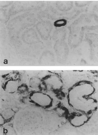
process was operative in our rats, the juxtaglomerular renin content at 11 weeks was assessed in both clipped and undipped kidneys using a monoclonal antibody to renin (Figure 1), and tissue renin levels were calculated for clipped, undipped, and control kidneys. These results are summarized in Table 1 and demonstrate that renin production was significantly
FIGURE 1. Representative juxtaglomerular renin staining in
undipped (a) and clipped (b) kidneys of rats with Goldblatt hy pertension. Renin staining in clipped kidneys occasionally ex tended from the afferent arteriole into the glomerulus (x630).
180 ENG ET AL AJH-FEBRUARY 1994-VOL. 7, NO. 2
TABLE 1. ASSESSMENT OF TISSUE RENIN Immunostaining for
Kidney Juxtaglomerular Renin*
Tissue Renin (ng AI/h/g tissue) Sham (n = 6) 26.8% (±11.3) 470.0 (±187.3) Clipped (n = 8) 47.3% (±6.1)t 1506.6 (±790.1)1 1 Undipped (n = 8) 8.9% (±7.9)t 86.4 (±76.7)** Al, angiotensin I.
*Reported as percent positive of total glomeruli counted per biopsy
SD.
fP < .001 compared with sham.
£P < .001 compared with sham and clipped.
fP < .05 compared with sham.
**P < .05 compared with sham and clipped.
elevated in clipped kidneys compared with sham, but significantly suppressed in undipped kidneys. Taken together, these data strongly suggest an All-driven state.
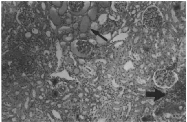
By light microscopy (periodic acid-Schiff stain), glo meruli of undipped hypertensive kidneys were often hyalinized and occasionally had areas of focal seg mental glomerulosclerosis (Figure 2 ) . Microaneu rysms and intraluminal thrombi were rarely present, although dilated glomerular capillary loops were common in most specimens. Bowman's capsule was thickened and frequently surrounded by mild peri-glomerular fibrosis in rats with higher mean SBPs. Synechiae were seen infrequently. Tubulointerstitial changes included tubular dilatation ranging in degree from mildly increased luminal diameter to massively dilated tubules with almost complete effacement of the tubular cells. Almost all biopsies of undipped kid neys contained dilated tubules, although biopsies
FIGURE 2. Periodic acid-Schiff stain of an undipped kidney of
a hypertensive (mean SBP >200 mm Hg) Goldblatt rat showing tubular dilatation and formation (narrow arrow), and dense focal interstitial infiltrates of mononuclear cells (wide arrow) (xlOO).
demonstrating the most dilated tubules and tubular casts were seen in rats with the most severe hyper tension (SBP > 2 0 0 mm Hg). In addition, dense focal interstitial m o n o n u c l e a r cellular infiltrates w e r e noted, especially in areas of tubular damage, in rats with severe hypertension. Tubular architecture in the regions of infiltrating cells was often dis rupted. In contrast, rats with less severe hyperten sion (SBP < 2 0 0 mg Hg) had only mild interstitial infiltrates.
Glomeruli from clipped kidneys appeared normal by light microscopy. Occasional low-grade mononu clear cellular infiltrates were seen in interstitial areas. With the exception of one kidney, no tubular dilation, disruption, or casts were seen.
Proliferation Cell proliferation was quantitated by immunostaining tissue sections for PCNA, a nuclear
protein that is increased from late G2 to the M phase
of the cell c y c l e .2 2 The number of glomerular PCNA +
cells did not significantly differ in sham animals (0.33 ± 0.08) compared with clipped rats (0.24 ± 0.16), although a mild, but significant, increase in the num ber of PCNA + glomerular cells was noted in the un dipped kidneys (0.81 ± 0.43, Ρ < .05 compared with sham and clipped kidneys). In contrast, a dramatic increase in PCNA 4- cells was seen in the tubuloin-terstitium of undipped hypertensive kidneys, com pared with both clipped and control (Figure 3 ) . Be cause both tubular and interstitial cell proliferation occurred focally, quantitation was difficult. Although occasional tubules had one or two proliferating cells, other primarily distal tubules were seen in which al most every cell was PCNA + . In the interstitium, mononuclear PCNA + cells were also clustered, fre quently in zones of injury. Although more tubular cell proliferation was seen in clipped kidneys com pared with sham-operated control kidneys, the pro liferation was relatively minor and never included en tire cross sections of PCNA-positive tubules. Foci of PCNA + interstitial mononuclear cells were also in creased in clipped kidneys, but never as large as the dense areas in the undipped kidneys.
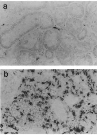
Leukocyte Infiltration To determine what popula tion of cells constituted the dense interstitial infil trates seen predominantly in the undipped kidneys, tissues were stained with specific antibodies to mono cyte-macrophages and dendritic cells (ED-1), neutro phils (RP-3), and lymphocytes (OX-22). Glomerular monocyte-macrophage counts were similar in sham (1.99 ± 0.69), undipped (1.42 ± 0.79), and clipped (1.06 ± 0.55) kidneys. However, undipped kidneys stained heavily and focally with ED-1 in damaged tubulointerstitial regions (Figure 4). All biopsies were uniformly negative for RP-3. Lymphocytes were
FIGURE 4. Interstitial monocyte-macrophages are occasionally
seen in clipped kidneys of Goldblatt rats (a) and sham controls, but dense foci are present only in undipped kidneys (b), especially in areas of tubular damage (x400).
FIGURE 3. Immunostaining for the proliferating nuclear cell
antigen (PCNA) (arrow) in sham control kidneys (a) shows only rare positive cells. Tubules and interstitium of clipped kidneys (b) have occasional PCNA + cells, whereas frequent areas of damage in undipped kidneys (c) have many PCNA+ tubular cells
(X400).
noted in rare dense focal groups in the undipped, but not clipped, kidneys in interstitial locations.
To determine w h e t h e r monocyte-macrophages were the proliferating interstitial cells, tissue was double immunostained for ED-1 and PCNA. Only 8.6% of PCNA + cells were double labeled. However, b e c a u s e E D 1 m a y n o t l a b e l all m o n o c y t e -macrophage populations, it is possible that some pro liferating monocyte-macrophages may not have been recognized.
Cytoskeletal Changes Desmin is expressed nor mally by mesangial cells and the staining in mesan gial regions in both clipped and undipped kidneys was similar to that seen in control animals. However, when evaluated using a semiquantitative score, desmin staining in glomerular visceral epithelial cells was mildly increased in clipped kidneys (0.86 ± 0.29) compared with sham controls (0.60 ± 0.45, Ρ < .05), but markedly increased in undipped, hypertensive kidneys (2.16 ± 0.49, Ρ < .05 compared with sham and clipped kidneys) (Figure 5). Desmin was also ex pressed by cells in the interstitium in areas of tubular injury within the undipped kidneys. In contrast, α-smooth muscle actin was not expressed to any sig nificant degree within glomeruli of clipped or un dipped kidneys (mean glomerular actin score 0.28 ± 0.23 in undipped ν 0.12 ± 0.09 in clipped kidneys, and 0.13 ± 0.03 in sham kidneys). However, staining for this actin isoform was markedly increased in a population of mesenchymal cells in areas of tubu lointerstitial damage in a manner similar to desmin (Figure 6).
Fibrosis Staining for types I and IV collagen was comparable in the kidneys of control animals and the
182 ENG ET AL AJH-FEBRUARY 1994-VOL. 7, NO. 2
FIGURE 5. Glomerular desmin staining in predominantly mes
angial regions in clipped kidneys (a) is similar to that seen in sham controls. Staining for desmin in undipped kidneys (b) is markedly increased, especially in epithelial cell locations (x630).
clipped kidneys of Goldblatt rats, and was seen in periglomerular, tubular, and vascular (types I and IV) and intraglomerular (type IV) areas. However, a dra matic increase of these collagens was noted in the interstitium in areas of tubular injury in undipped kidneys (Figure 7).
DISCUSSION
Recent studies have shown that short-term continu ous infusion of All rats results in renal cell prolifera
tion and phenotypic c h a n g e s .6 In doses causing hy
pertension, All induced tubular and interstitial cell proliferation, de novo mesangial cell and interstitial cell expression of the vascular smooth m u s c l e -associated actin isoform, α-smooth muscle actin, and upregulation of the intermediate filament protein, desmin, by glomerular visceral epithelial cells.
It could not be determined from these previous studies whether the proliferative and phenotypic changes resulted from hypertension caused by All or from nonhemodynamic effects of AIL Elevated blood
pressure has been reported to have trophic2 3 2 4 or mi
t o g e n i c1 1 , 2 5 effects on vascular smooth muscle cells.
Crane and D u t t a2 6 have demonstrated similar
mito-FIGURE 6. Staining for α-smooth muscle actin is positive only
in blood vessels of sham or clipped kidneys (a), but is dramatically increased in undipped kidneys (b). Expression of this actin iso form represents a phenotypic change and is seen in focal areas of undipped, but not clipped, kidneys. A similar tubulointerstitial staining pattern is seen for desmin (x400).
genie effects on glomerular and tubular cells in rats with All-independent deoxycorticosterone acetate (DOCA) salt-induced hypertension. In addition, in direct evidence for the proliferative effects of hyper tension has been provided by studies in both All-dependent and inAll-dependent models of hypertension in which administration of various classes of antihy pertensive drugs prevented rise in both systemic blood pressure and vascular smooth muscle cell DNA
synthesis.1 1 Indeed, it has been suggested that DNA
synthesis in these cells correlates with the rate of the
rise, rather than the peak, of blood p r e s s u r e .2 7
However, All itself has been reported to be mito genic for a variety of cells in culture under certain conditions. In vitro studies have demonstrated pro liferative effects of All on human neonate and juve
nile aortic smooth muscle c e l l s ,2 8 and human fetal1 0
an transformed m u r i n e2 9 mesangial cells. Evidence
for the mitogenic role of All in vivo has also been provided by studies showing inhibition of vascular smooth muscle cell proliferation after balloon catheter injury in rats treated with a specific All receptor
FIGURE 7. Immunostaining for collagen type IV (arrow) in
clipped kidneys (a) and sham controls is seen in the tubular base ment membrane, but is markedly increased in areas of tubuloint erstitial damage and fibrosis in undipped kidneys (b) (x400).
We, therefore, attempted to separate the hemody namic from nonhemodynamic effects of All on renal cell proliferation and phenotypic changes by study ing the two-kidney, one-clip Goldblatt model in rats in which circulating renin and All levels are known to be elevated and in which one kidney (ie, the un dipped kidney) is exposed to systemic pressures, whereas the other, clipped kidney is effectively pro tected. Although we did not measure All levels, it is known that renal tissue levels of All are elevated in both clipped and u n d i p p e d kidneys at 7 and 25
d a y s .3 1 Therefore, the evidence that our model was
All dependent is indirect, but it is suggested by the immunostaining for renin, as well as tissue renin levels, both of which are significantly increased in the clipped kidney, but suppressed in the undipped kidney.
In our study, dramatic tubular and interstitial cell proliferation was seen primarily in the undipped, hy pertensive kidney. A small increase in tubular cell proliferation was seen in the clipped, nonhyperten-sive kidneys, and is consistent with the mild stimu
latory effect of All on other cell t y p e s .1 0'2 8 , 2 9 This mi
togenic effect could be mediated by the All receptors
shown to reside on tubular c e l l s ,3 2 and is in contrast
to in vitro findings that have shown no mitogenic
effects of All on murine proximal tubular cells.8 Al
though our results do not rule out a potentiating role for All, as has been shown with epidermal growth
factor on cultured proximal tubular c e l l s ,9 , 3 3 the dra
matic increase in tubular cell proliferation seen only in unprotected kidneys suggests that it is mainly through its hemodynamic effects that All mediates proliferation.
These results are at variance with those of Cantin et
a l3 4 who also examined proliferative changes in an
All-mediated model of hypertension induced by par tial ligation of the aorta between the two renal arter ies. Examination of the left, nonhypertensive kidney revealed little glomerular cell proliferation, but marked proliferation of inner cortical (and to a lesser extent, medullary) tubules as detected by autoradiog raphy. However, their kidneys were ischemic and showed marked atrophy. In contrast, we excluded kidneys with apparent evidence of ischemia or atro phy, in the belief that this would be a confounding variable.
Similar to the tubular and interstitial cell prolifera tive changes, increased glomerular visceral epithelial cell desmin expression, the only dramatic glomerular finding in our study, was likewise upregulated pri marily in the unprotected kidney, whereas its expres sion in mesangial cells was unchanged in all three groups. Although normally a marker for muscle-derived cells, desmin has been shown to be variably
present in glomeruli visceral epithelial c e l l s ,1 2'3 5 , 3 6
and to be increased in these cells in several different
experimental models of glomerulonephritis.3 7 The
observation that increased desmin expression in glo merular epithelial cells occurred primarily in the un dipped, but not clipped, kidneys emphasizes the im portance of the hypertensive effects of AIL Indeed, one might postulate that desmin expression in the glomerular epithelial cell marks an adaptive response to the epithelial cells to increased glomerular pressure or stretch-mediated shape changes induced by glo merular hypertension. Although desmin expression in glomerular epithelial cells was increased in both
t h e p r e s e n t a n d p r e v i o u s s t u d y ,6 g l o m e r u l a r
α-smooth muscle actin expression was not increased in our model. It is possible, however, that such glo merular phenotypic modulation, unlike that seen in the tubulointerstitium, occurred early and resolved before biopsy at 11 weeks.
Traditionally, the majority of interstitial cells in the renal cortex and outer medulla have been thought to
be fibroblasts,3 8 but in at least two models of renal
injury they have been shown to express smooth mus
cle-associated p r o t e i n s .6 , 3 9 The finding of de novo
expression of the muscle-related proteins desmin and α-smooth muscle actin by interstitial cells in
un-184 ENG ET AL AJH-FEBRUARY 199^-VOL. 7, NO. 2
clipped kidneys in this study may represent transi tion of these cells to a myofibroblast state, as has been suggested for renal interstitial cells in animals infused with A I L6 Interstitial fibroblasts have been shown re
cently to express the ß-subunit of the platelet-derived growth factor r e c e p t o r .4 0 This suggests a mechanism
by which activated interstitial fibroblasts may be stim ulated to proliferate, which could lead to increased collagen production and eventual fibrosis.
Angiotensin II has been demonstrated to increase the expression of collagen type I in culture murine mesangial, and collagen type IV in murine proximal tubular, cells in v i t r o .9'2 9 However, in vivo, the im
portance of the pressor effects of All are further un derscored by the increased expression of types I and IV collagen in the renal interstitium in hypertensive, but not protected, kidneys. Such changes of fibrosis were also reported in rats made hypertensive by All infusion.6 In both models of All-mediated hyperten
sion, kidneys exposed to high pressures were also infiltrated by monocyte-macrophages, which is of in terest as macrophages are known to express a large number of vasoactive substances, cytokines, and growth factors,4 1 including platelet-derived growth
factor, basic fibroblast growth factor, and transform ing growth factor-ß. These cytokines might then act directly or in concert with All to stimulate tubular and interstitial cells to proliferate or produce extracel lular matrix.
These data are consistent with the hypothesis that the hemodynamic effects of All are necessary for the tubular and interstitial cell proliferative and pheno typic changes in this experimental model. We recog nize that it is possible that the hemodynamic manip ulations may also affect other regulatory or counter-r e g u l a t o counter-r y f a c t o counter-r s t h a t c o u l d c o n f o u n d t h e interpretation of the study. Unfortunately, there is no way to separate the hemodynamic from the nonhe-modynamic effects of All without affecting blood flow. However, two important findings emerge from this study. First, significant tubulointerstitial injury occurs in the undipped hypertensive kidneys, with tubular cell proliferation, interstitial cell proliferation and phenotype change, macrophage infiltration, and collagen deposition. This is important given previous observations that tubular and interstitial injury is crit ical in the progression to renal failure in a variety of renal diseases.4 2""*5 Second, despite very high levels
of tissue renin (and probably All), the clipped kid neys were morphologically normal and had minimal proliferative or phenotypic changes in the intersti tium. Therefore, this study suggests that in this model of injury, All in the absence of hypertension has only minimal effect in the initiation and progres sion of the tubulointerstitial changes characteristic of chronic renal injury.
A C K N O W L E D G M E N T S
The authors thank Dr. William Couser for his encourage ment and advice.
REFERENCES
1. Hall JE, Guyton AC, Saigado HC, et al: Renal hemo dynamics in acute and chronic angiotensin II hyper tension. Am J Physiol 1978;235:F174-F179.
2. Zimmerman BG, Sybertz EJ, Wong PC: Interaction be tween sympathetic and renin-angiotensin system. J Hypertens 1984;2:581-587.
3. Koletsky S, Rivera-Velez JM, Pritchard WH: Produc tion of hypertension and vascular disease by angioten sin. Arch Pathol 1966;82:99-106.
4. Wilson C, Byrom FB, Lond MD: Renal changes in ma lignant hypertension. Lancet 1939;1:136-139. 5. Wilson C, Byrom FB: The vicious circle in Bright's dis
ease. Experimental evidence from the hypertensive rat. Q J Med 1940;10:65-96.
6. Johnson RJ, Alpers CE, Yoshimura A, et al: Renal in jury from angiotensin II mediated hypertension. Hy pertension 1992;19:464-474.
7. Schelling P, Gantin D, Speck G, Fischer H: Effects of angiotensin II and angiotensin II antagonist saralasin on cell growth and renin in 3T3 and SV 3T3 cells. J Cell Physiol 1979;98:503-514.
8. Geisterfer AAT, Peach MJ, Owens GK: Angiotensin II induces hypertrophy, not hyperplasia, of cultured rat aortic smooth muscle cells. Circ Res 1988;62:749-756. 9. Wolf G, Neilson EG: Angiotensin II induces cellular
hypertrophy in cultured murine proximal tubular cells. Am J Physiol 1990;259:F768-F777.
10. Ray PE, Aguilera G, Kopp JB, et al: Angiotensin II receptor-mediated proliferation of cultured human fe tal mesangial cells. Kidney Int 1991;40:764-771. 11. Loeb AL, Bean BL: Antihypertensive drugs inhibit hy
pertension-associated aortic DNA synthesis in the rat. Hypertension 1986;8:1135-1142.
12. Johnson RJ, Iida H, Alpers CE, et al: Expression of smooth muscle cell phenotype by rat mesangial cells in immune complex nephritis. J Clin Invest 1991;87: 847-858.
13. Galen FX, Devausx C, Atlas S, et al: New monoclonal antibodies directed against human renin. J Clin Invest 1984;74:723-735.
14. Ogata K, Kurki P, Celis JE, et al: Monoclonal antibod ies to a nuclear protein (PCNA/cyclin) associated with DNA replication. Exp Cell Res 1987;168:475-486. 15. Dijkstra CD, Döpp EA, Joling P, Kraal G: The hetero
geneity of mononuclear phagocytes in lymphoid or gans: distinct macrophages subpopulations in the rat recognized by monoclonal antibodies EDI, ED2 and ED3. Immunology 1985;54:589-599.
16. Sekiya S, Gotoh S, Yamashita T, et al: Selective deple tion of rat neutrophils by in vivo administration of a monoclonal antibody. J Leukoc Biol 1989;49:96-102. 17. Skalli O, Ropraz P, Trzeciak A, et al: A monoclonal
for smooth muscle differentiation. J Cell Biol 1986;103: 2787-2796.
18. Iida H, Seifert R, Alpers CE, et al: Platelet-derived 32. growth factor (PDGF) and PDGF receptor are induced in mesangial proliferative nephritis in the rat. Proc
Natl Acad Sei 1991;88:6560-6564. 33
19. Floege J, Burns MW, Alpers CE, et al: Glomerular cell proliferation and PDGF expression precede glomeru losclerosis in the remnant kidney model. Kidney Int 0 4
1992;41:297-309.
20. DeRouffignac C, Bonvalet JP, Menard J: Renin content in superficial and deep glomeruli of normal and salt loaded rats. Am J Physiol 1974;226:150-154.
21. Koletsky S, Pavlicko KM, Rivera-Velez JM: Renin- 35. angiotensin activity in hypertensive rats with a single ischemic kidney. Lab Invest 1971;24:41-44.
22. Kurki P, Vanderlaan M, Dolbeare F, et al: Expression 36. of proliferating cell nuclear antigen (PCNA)/cyclin during the cell cycle. Exp Cell Res 1986;166:209-219.
23. Owens GK, Rabinovitch PS, Schwartz SM: Smooth 37. muscle cell hypertrophy versus hyperplasia in hyper
tension. Proc Natl Acad Sei 1981;78:7759-7763. 24. Owen GK, Schwartz SM: Vascular smooth muscle cell
hypertrophy and hyperploidy in the Goldblatt hyper
tensive rat. Circ Res 1983;53:491-501. 38. 25. Chobanian AV, Lichtenstein AH, Schwartz JH, et al:
Effects of deoxycorticosterone/salt hypertension on 39. cell ploidy in rat aortic smooth muscle cells. Circula
tion 1978;75(suppl 1):102-106.
26. Crane WAJ, Dutta LP: The utilization of tritiated thy midine for deoxyribonucleic acid synthesis by the le- 40. sions of experimental hypertension in rats. J Pathol Bacteriol 1963;86:83-97.
27. Carlier PG, Rorive G, Barbason H: Kinetics of prolif eration of rat aortic smooth muscle cells in Goldblatt one-kidney, one-clip hypertension. Clin Sei 1983;65: 351-357.
Campbell-Bos well M, Robertson AL: Effects of an giotensin II and vasopressin on human smooth mus cle cells in vitro. Exp Mol Pathol 1981;35:265-276. 28.
29.
41. 42.
43. Wolf G, Haberstroh U, Neilson EG: Angiotensin II stimulates the proliferation and biosynthesis of type I collagen in cultured murine mesangial cells. Am J
Pathol 1992;140:95-107. 4 4 30. Prescott MF, Webb RL, Reidy MA: Angiotensin
converting enzyme inhibitor versus angiotensin IIATX
receptor antagonist: effects on smooth muscle cell mi gration and proliferation after balloon catheter injury. 4c Am J Pathol 1991;139:1-6.
31. Guan S, Fox J, Mitchell KD, Navar LG: Angiotensin and angiotensin converting enzyme tissue levels in
two-kidney, one clip hypertensive rats. Hypertension 1992;20:763-767.
Mujais SK, Kauffman S, Katz AL: Angiotensin II bind ing sites in individual segments of the rat nephron. J Clin Invest 1986;77:315-318.
Norman J, Badie-Dezfooley B, Nord EP: EGF-induced mitogenesis in proximal tubular cells: potentiation by angiotensin II. Am J Physiol 1987;253:F299-F309. Cantin M, Araujo-Nascimento M, Benchimol S, Des-ormeaux Y: Metaplasia of smooth muscle cells into juxtaglomerular cells in the juxtaglomerular appara tus, arteries and arterioles of the ischemic (endocrine) kidney. Am J Pathol 1977;87:581-602.
Stamenkovic I, Skalli O, Gabbiani G: Distribution of intermediate filament proteins in normal and diseased human glomeruli. Am J Pathol 1986;125:465-475. Yaoita E, Kawasaki K, Yamamoto T, Kihara I: Variable expression of desmin in rat glomerular epithelial cells. Am J Pathol 1990;136:899-908.
Floege J, Alpers CE, Sage EH, et al: Marker of com plement-dependent and complement-independent glomerular visceral epithelial cell injury in vivo: ex pression of anti-adhesive proteins and cytoskeletal changes. Lab Invest 1992;67:486-497.
Lemley KV, Kriz W: Anatomy of the renal intersti-tium. Kidney Int 1991;39:370-381.
Nagle RB, Kneiser MR, Bulger RE, Benditt EP: Induc tion of smooth muscle characteristics in renal intersti tial fibroblasts during obstructive nephropathy. Lab Invest 1973;29:422-427.
Alpers CE, Seifert RA, Hudkins KL, et al: PDGF-receptor localizes to mesangial, parietal epithelial and interstitial cells in human and primate kidneys. Kid ney Int 1993;43:286-294.
Nathan CF: Secretory products of macrophages. J Clin Invest 1987;79:319-326.
Risdon RA, Sloper JC, de Wardener HE: Relationship between renal function and histological changes found in renal-biopsy specimens from patients with persistent glomerular nephritis. Lancet 1986;1:363-366.
Schainuck LI, Striker GE, Cuttler RE, Benditt EP: Structural-functional correlations in renal disease. Part II: the correlations. Hum Pathol 1970;1:631-641. Striker GE, Schainuck LI, Cutler RE, Benditt EP: Struc tural-functional correlations in renal disease. Part I: a method for assaying and classifying histopathologic changes in renal disease. Hum Pathol 1970;1:615-630. Bohle A, Mackensen-Haen S, von Gise H, et al: The consequences of tubulointerstitial changes for renal function in glomerulopathies: a morphometric and cy-tological analysis. Pathol Res Pract 1990;186;135-144.