Noggin null allele mice exhibit a microform
of holoprosencephaly
Eva Lana-Elola
1,2, Przemko Tylzanowski
3, Maarit Takatalo
4, Kirsi Alakurtti
4, Lotta Veistinen
4,
Thimios A. Mitsiadis
5, Daniel Graf
5, Ritva Rice
1,2, Frank P. Luyten
3and David P. Rice
1,2,4,6,∗1
Department of Craniofacial Development and2Department of Orthodontics, King’s College, London, UK,3Laboratory of Skeletal Development and Joint Disorders, Division of Rheumatology, Department of Musculoskeletal Sciences, Katholieke Universiteit Leuven, Leuven, Belgium,4Department of Orthodontics, Institute of Dentistry, University of Helsinki, Finland, 5Department of Orofacial Development and Structure, Institute of Oral Biology, ZZMK, Faculty of Medicine, University of Zurich, Zurich, Switzerland and6Oral and Maxillofacial Diseases, Helsinki University Central Hospital, Helsinki, Finland
Received July 6, 2011; Revised and Accepted July 25, 2011
Holoprosencephaly (HPE) is a heterogeneous craniofacial and neural developmental anomaly characterized in its most severe form by the failure of the forebrain to divide. In humans, HPE is associated with disruption of Sonic hedgehog and Nodal signaling pathways, but the role of other signaling pathways has not yet been determined. In this study, we analyzed mice which, due to the lack of the Bmp antagonist Noggin, exhibit elevated Bmp signaling. Noggin2/2 mice exhibited a solitary median maxillary incisor that developed from a single dental placode, early midfacial narrowing as well as abnormalities in the developing hyoid bone, pituitary gland and vomeronasal organ. In Noggin2/2 mice, the expression domains of Shh, as well as the Shh target genes Ptch1 and Gli1, were reduced in the frontonasal region at key stages of early facial devel-opment. Using E10.5 facial cultures, we show that excessive BMP4 results in reduced Fgf8 and Ptch1 expression. These data suggest that increased Bmp signaling in Noggin2/2 mice results in downregulation of the hedgehog pathway at a critical stage when the midline craniofacial structures are developing, which leads to a phenotype consistent with a microform of HPE.
INTRODUCTION
Holoprosencephaly (HPE) is a developmental anomaly that can present as a range of midline craniofacial and neural malfor-mations. HPE is the most common developmental defect of the forebrain and is characterized by the abnormal separation of the telencephalon into left and right hemispheres and the associated abnormal development of midline neural structures. Alobar HPE is the most severe form of this developmental defect, in which the prosencephalon does not separate along both its medial –lateral and anterior –posterior axes which result in a single uncleaved cerebral lobe, and the telencephalon and diencephalon remain unsegmented. There is a spectrum of clinical facial features including the presence of only one upper central incisor tooth with an absent maxillary midline frenum, cleft of the lip and or palate, nasal defects including single
nostril and nasal agenesis, central proboscis, hypotelorism and cyclopia (1,2). When these features appear without defects in the central nervous system, they are termed ‘microforms’. HPE occurs in 1 in 7600 live births (3). However, the incidence is much higher, 1 in 240, during early embryogenesis with most embryos undergoing spontaneous abortion (4).
The etiology of HPE is heterogeneous with both genetic and environmental factors. Environmental factors include poorly controlled maternal diabetes, alcohol, vitamin A analogues and alteration in cholesterol metabolism (5). Approximately 25% of HPE cases are syndromic; these syndromes include Pallister – Hall, Rubinstein – Taybi and Smith – Lemli – Opitz. In non-syndromic cases, 12 different chromosomal loci have been implicated in HPE and mutations in 7 genes have been found to cause HPE, and these are all members of the Hedge-hog and Nodal signaling pathways. Genes with mutations
∗To whom correspondence should be addressed at: Department of Orthodontics, Institute of Dentistry, Room C229a, Biomedicum Helsinki, PL 63
(Hartmaninkatu 8), 00014 University of Helsinki, Finland. Tel:+358 919127387; Fax: +358 919125371; Email: [email protected]
#The Author 2011. Published by Oxford University Press. All rights reserved. For Permissions, please email: [email protected]
Human Molecular Genetics, 2011, Vol. 20, No. 20 4005–4015 doi:10.1093/hmg/ddr329
known to cause HPE are SHH (HPE3), the Hedgehog receptor Patched 1 (PTCH1) (HPE7), the transcription factors GLI2, SIX3 (HPE2) and ZIC2 (HPE5), the Hedgehog regulator GAS1, as well as Transforming growth factor interacting factor (TGIF) and Teratocarcinoma-derived growth factor 1 (TDGF1/CRIPTO) (6–8).
Evidence from animal forebrain developmental studies and the characterization of some of the genes causing HPE in humans suggest that distinct mechanisms underlie the two major classes of HPE (9). First, ‘classic’ HPE in which the ventral telencephalon is most severely affected and where the ventralizing effects of Shh signaling are disrupted. Second, midline interhemispheric HPE or syntelencephaly in which the dorsal telencephalon fails to split while the ventral telence-phalon may be normal, and the dorsalizing effect of Bone mor-phogenetic protein (Bmp) signaling is disrupted. Evidence for the role of Bmp signaling in the pathogenesis of midline interhe-mispheric HPE comes from the analysis of Bmpr1a2/2; Bmpr1b2/2compound mutant mice which exhibit a loss of all dorsal midline cell types without affecting the specification of cortical and ventral precursors in the telencephalon (10).
Bmps regulate many aspects of craniofacial morphogenesis (11). Variations in Bmp signaling create morphological species-specific diversification of the middle and upper face (12,13) and Bmps together with retinoid acid specify the identity and regulate the growth of the frontonasal mass and maxillary prominences (14,15). Noggin is an extracellular Bmp antagonist which binds, with picomolar affinity, to Bmp2, 4, 6 and 7 (16). This high-binding affinity prevents Bmps from high-binding to their receptors and subsequent signal transduction. Noggin null allele mice exhibit elevated Bmp signaling which results in patterning and growth defects of the neural tube and somites. In the appendicular and axial skeleton cartilage is hyperplastic and there is a failure to initiate joint formation (17–20).
In this study, we show that Noggin is expressed in the midline of the developing upper face and that Noggin2/2 mouse embryos exhibit craniofacial abnormalities which equate to a microform HPE. More specifically, the mutant mice display a solitary median maxillary incisor, midfacial narrowing and abnormalities in the developing hyoid bone, pituitary gland and vomeronasal organ (VO). The Shh signal-ing pathway regulates midface development and has also been implicated in HPE. We found smaller expression domains of Shh, and the Shh target genes Ptch1 and Gli1 in the frontona-sal region of Noggin2/2mice compared with their wild-type (WT) littermates. Using bead implantation assays on E10.5 WT upper facial explants, we show that excessive BMP4 protein results in reduced expression of both Fgf8 and Ptch1. Taken together, the present data suggest that disrupted Bmp signaling can result in HPE by regulating the Fgf8-Shh pathway and we put forward the Noggin2/2 mouse as a model of microform HPE.
RESULTS
Noggin2/2mice exhibit a narrowing of the frontonasal process and a solitary median upper incisor
Noggin expression, revealed by LacZ staining, was analyzed in mice at keys stages when the midline of the upper face is
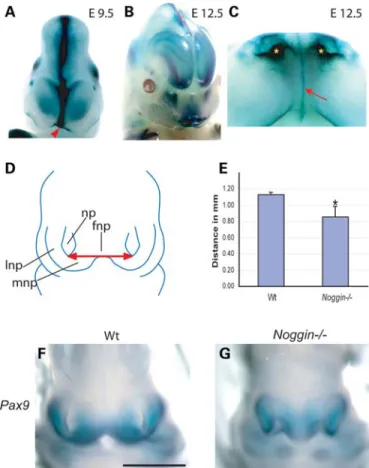
patterned and undergoing major morphogenesis. At E9.5 and E12.5, Noggin was detected in the midline of the developing frontonasal process and in the developing nasal pits (Fig. 1). E11.5 Noggin2/2mouse embryos exhibited a shorter distance between the lateral nasal processes compared with their WT lit-termates. Furthermore, the morphology of the frontonasal mass was abnormal, with Noggin mutant mice lacking the character-istic invagination of the anterior/oral surface (Fig.1). To quan-tify these changes, we have carried out measurements on tissue sections from WT and noggin null mice stained for Pax9 expression, a transcription factor used as a marker of the nasal processes. In the absence of Noggin, the distance between the nasal pits was found to be significantly reduced at E11.5 (P ¼ 0.02; WT ¼ 1.14 mm, n ¼ 12; Noggin2/2 0.86 mm, n ¼ 9, non-paired t-test) (Fig.1).
WT mice have two upper incisor teeth, each within separate premaxillary bones. Noggin2/2mice had a single upper incisor
Figure 1. Noggin is expressed in the developing face and Noggin2/2mice exhibit a narrow frontonasal process. (A – C) Whole-mount LacZ staining of E9.5 and E12.5 mice. At E9.5, Noggin is expressed in the midline of the devel-oping frontonasal process (arrowhead). Later Noggin expression is detected in the midline of the frontonasal process as fine line which extends into the oral cavity (arrow) and in the developing nasal pits (asterisks). (D and E) Schematic diagram illustrates the upper face of an E11.5 mouse embryo. The red arrow indi-cates the distance between the nasal pits. Noggin2/2mice have a shorter distance between the nasal pits. (F and G) Localization of Pax9 in the developing upper face of E11.5 WT and Noggin2/2mice. Using Pax9 as a marker of the lateral and medial nasal processes, we were able to record a reduction in the distance between the nasal pits in Noggin2/2heads compared with their WT littermates. fnp, frontonasal process; lnp, lateral nasal process; mnp, medial nasal process; np, nasal pit. Scale bar: 1 mm. (F and G, same magnification). Error bars: stan-dard deviation.
located in the midline in a single premaxillary bone (Fig.2). The morphology of the incisor was analyzed in histological sections at E16.5 (Fig. 2C and D). Instead of two separated tooth germs positioned adjacently to the nasal cavity and separated by the inter-premaxillary suture, Noggin2/2mice had a single midline incisor situated below the nasal septum.
In order to establish whether this tooth had developed from a single primordium or from the fusion of two well-defined primordia, we analyzed early tooth development in Noggin2/2mice and their WT littermates. During tooth devel-opment, Shh is expressed in dental placodes, which are thick-enings of the oral epithelium and represent the earliest stages of tooth development. We performed whole-mount in situ hybridization for Shh mRNA at E11.5 and E12.5 and found that Noggin2/2 mice exhibit a single Shh expression domain in the midline of the frontonasal process (9/10 mice)
(Fig. 2). This expression domain had not formed from two placodes, as occurs in WT mice. In WT mice, two maxillary incisor primordia were seen at E12.5; these had formed from a continuous dental lamina. Also, we noted that the Shh expression domain in the incisor placode was smaller in Noggin2/2 mice compared with WT littermates (Fig.2).
Consistent with previous reports, we found that the cranial neural tube of most Noggin2/2 mice (18/22 E17.5 and E18.5 mice) failed to close leaving the mutants with an exencephalic phenotype (19).
Shh signal transduction is decreased in the oral region in Noggin2/2 mice
As Shh expression domains in the incisor placode were smaller in Noggin2/2 mice compared with the WT and because Shh
Figure 2. Noggin2/2mice have a solitary median upper incisor tooth. (A and B) Premaxillary region of new born WT and Noggin2/2mice stained with alcian
blue (cartilage) and alizarin red (bone). WT mice have two upper incisor teeth (A, arrowheads), whereas Noggin2/2mice have only one (B, arrowhead). (C and D) Hematoxylin and eosin stained frontal sections from E16.5 WT and Noggin2/2heads confirming the presence of two upper incisors in WT mice and a single upper incisor in Noggin2/2mice (arrowheads). The developing upper incisor in Noggin2/2mice has an abnormal thick stalk attachment to the oral epithelium and abnormal epithelial convolutions (D). Associated with the dental epithelium is a condensed dental mesenchyme. (E – H) Shh whole-mount in situ hybrid-ization of E11.5 and E12.5 WT and Noggin2/2oral region. (E) In WT mice, at E11.5, Shh expression is seen in the two mandibular incisors (arrowheads) and in the continuous maxillary dental lamina (arrow). (F) In Noggin2/2mice, dental development is delayed in that Shh mRNA is not detected in the oral region.
(G and H) At E12.5, WT mice have two upper incisor primordia (G, arrowheads) and two lower incisor primordia which express Shh. Noggin2/2 exhibit two lower primordia which express Shh but have only one centrally positioned incisor primordium which is smaller than its WT counterparts (F, arrowhead). np, nasal pit; ns, nasal septum; t, tongue. Scale bars: 0.5 mm. Images on the left of the panel are the same magnification as the images on the right.
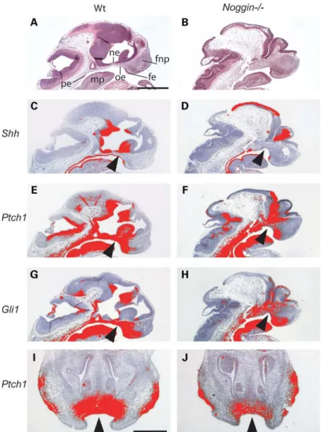
signaling has been shown to developmentally regulate midfa-cial width, we compared the expression of several genes involved in the Shh pathway in the midline facial regions of WT and Noggin2/2mice during early embryogenesis (E11.5 and E12.5) (Fig. 3). In WT mice, Shh was expressed in the oral epithelium and facial ectoderm, as well as the pharyngeal endoderm and diencephalic epithelium. In Noggin2/2 mice, Shh was absent from the oral epithelium (Fig.3D). The trans-membrane receptor Ptch1 and the transcription factor Gli1 are both targets of Shh. Consistent with previous reports that there is a gradient of Hh signaling activity across the craniofacial region (21), we found that in WT mice the expression of Ptch1 and Gli1 was strongest near to the expression domains
of Shh and that this mesenchymal expression reduced with increasing distance from the epithelium (Fig. 3C – J). In Noggin2/2 mice, the expression levels of Ptch1 and Gli1 in the mesenchyme adjacent to the oral epithelium were reduced, consistent with the reduction in Shh expression (Fig.3F, H, J).
As Bmp2, -4 and -7 are binding partners of Noggin and because the Noggin null phenotype is caused by the deregula-tion of Bmp signaling (17,22), we analyzed the expression of Bmp2, -4 and -7 during early facial development. Noggin is expressed in the epithelium of the developing frontonasal mass (Fig. 1A – C) (23). At E10.5, Bmp2 was expressed in the nasal epithelium, then at E12.5 in the pharyngeal
Figure 3. Decreased Shh signal transduction in the oral region of Noggin2/2mice. (A – H) Sagittal sections through the midline region of WT and Noggin2/2
mice. (A – H) E11.5 (A and B) hematoxylin and eosin stained sections. (C – J)35S-in situ hybridization for Shh signaling pathway members. (C and D) Shh is expressed in the oral epithelium (C, arrowhead), pharyngeal endoderm and neuro epithelium of the diencephalon. In Noggin2/2mutant mice, the expression of
Shh is missing from the oral epithelium (D, arrowhead). (E and G) Ptch1 and Gli1 are expressed in the oral epithelium and in the facial mesenchyme (arrow-heads). They show a gradient of transcriptional activity across these regions away from the epithelium, the source of hedgehog ligand. (E – H) In Noggin2/2
embryos, there is a reduction in the expression of Ptch1 and Gli1 in facial region compared with WT littermates (arrowheads). (I and J) E12.5 frontal sections. Ptch1 expression domain smaller in Noggin2/2mice compared with WT littermates (arrowheads). fe, facial ectoderm; fnp, frontonasal process; mp, mandibular
process; ne, neural ectoderm; oe, oral epithelium; pe, pharyngeal endoderm. Scale bars: (A) 200 mm; (I) 500 mm. (A – H, same magnification) (I and J, same magnification).
endoderm and in the mandibular process. Bmp4 and Bmp7 were expressed in the facial, oral and mandibular epithelia as well as in the mesenchyme of the frontonasal mass and the first branchial arch (mandible) at both E10.5 and E12.5 (Supplementary Material, Fig. S1).
It has been reported that Bmp4 expression is more intense and has an expanded domain in the first branchial arch of Noggin2/2 mice compared with WT littermates (24). Using in situ hybridization, we tested whether the expression of Bmp4 and Bmp7 were changed in the midline of the develop-ing face in Noggin2/2mice. Similar to WT littermates, Bmp4 and Bmp7 were expressed in the epithelium and mesenchyme of the frontonasal mass and in the first branchial arch. Levels of intensity were comparable to those found in WT littermates (Supplementary Material, Fig. S1).
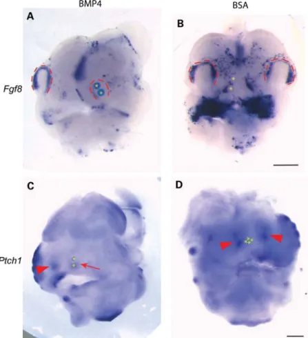
BMP4 inhibits Fgf8 and Ptch1 in the developing midface In the developing chick face, it has been shown that increasing levels of Bmp leads to a downregulation of Fgf8 and Shh (23). We postulated that increased Bmp sig-naling could, via Fgf8, reduce Shh sigsig-naling in the develop-ing frontonasal process and thereby result in the Noggin2/2
phenotype. Using bead assays, we tested the effects of adding exogenous BMP4 to E10.5 mouse facial organ cul-tures. BMP4 downregulated Fgf8 in the nasal pit [8/9 explants, compared with 2/9 with bovine serum albumin (BSA)] (Fig. 4). Fgf8 and Shh are expressed in the facial ectoderm; however, Shh signals to its receptor Ptch1 which is located in the facial mesenchyme to regulate facial mor-phogenesis. As ligand binding to Ptch1 initiates signal trans-duction and as Ptch1 is also upregulated by the binding of Hh, Ptch1 expression is regarded as a readout of Hh signal-ing. We therefore investigated the effects of BMP4 on Ptch1 expression in the developing mouse frontonasal process and found that BMP4 downregulated Ptch1 expression (9/11 explants, compared with 2/11 with BSA). Thus, in Noggin null allele mice, increased Bmp activity may inhibit Fgf 8/ Shh-dependent facial development.
Normal palate formation despite abnormal early shelf morphogenesis
Cleft lip and/or palate are part of the spectrum of features in human HPE. We examined the developing lips and palate of Noggin2/2 and WT mice from the initial stages of
Figure 4. BMP4 downregulates Fgf8 and Ptch1 in the developing mouse face. (A – D) WT mouse heads cultured overnight with beads impregnated with BMP4 or BSA. (A and B) E10.5 whole-mount in situ hybridization for Fgf8. Fgf8 is downregulated in the nasal pit in response to BMP4. (C and D) E11.5 whole-mount in situ hybridization for Ptch1. Ptch1 is downregulated in mesenchyme underlying the nasal pit in response to BMP4 (arrow), but Ptch1 expression is maintained in the contralateral side (C, arrowhead) and in response to BSA (negative control, D arrowheads). Nasal pits demarcated with red dotted lines. Asterisks indicate bead locations. Scale bars 500 mm. (A and B, same magnification) (C and D, same magnification).
development at E12.5 until after palatal shelf fusion is com-plete at E16.5 and E18.5. Normally, the palatal shelves develop as buds from maxillary processes and then start to grow downwards between the tongue and the floor of the mouth. The palatal shelves elevate rapidly between E14.0 and E14.5 to a position above the tongue, where after they approximate in the midline and fuse. In Noggin2/2 mice,
the palatal shelves appeared morphologically normal at E12.5; however at E13.5, they were thick and bulbous in shape and had failed to grow down between the tongue and floor of mouth instead remaining above the dorsum of the tongue (Fig. 5). Noggin2/2 palatal shelves met in the midline and fused normally so that by E16.5 a normal palate had formed (20/21 mice) (Fig.5G and H).
Figure 5. Palate and hyoid bone development in Noggin2/2mice. (A – H) Hematoxylin and eosin stained frontal sections of WT heads and Noggin2/2heads E12.5 – E16.5. In WT mice, the palatal shelf primordia bud from the maxillary processes (A), they extend into a position between the tongue and the floor of the mouth, (C) then elevate to a position between the tongue dorsum and the nasal capsule (E) and fuse in the midline. (D) In contrast, the palatal shelves of Noggin2/2mice were in a position above the tongue, already at E13.5. (D and F) Also, the shape of the Noggin2/2palatal shelves was not ‘finger-like’ but
more bulbous. (H) At E16.5, the palatal shelves of Noggin2/2mice appeared normal having fused together. (I – L) Alcian blue and alizarin red staining of hyoid bones from WT and Noggin2/2mice. (I and J) At E16.5, the Noggin2/2hyoid is bigger than its WT counterpart. In both WT and Noggin2/2mice,
the body of the hyoid has started to mineralize (arrowheads). (K and L) At E18.5, the body of the Noggin2/2hyoid bone is larger. It has four instead of two cornua which are thicker, shorter and prematurely mineralized compared with their WT littermates. b, body of the hyoid bone; gc, greater cornua of hyoid bone; m, upper molar tooth; mc, Meckel’s cartilage; nc, nasal cavity; np, nasal projection of the maxillary bone; ns, cartilage primordium of the nasal septum; oc, oral cavity; t, tongue; vc, vomeronasal cartilage;∗palatal shelves. Scale bars: (A) 500 mm, (I) 1 mm (A – H, same magnification) (I – L, same magnification).
It seems likely that this phenotype is, at least in part, sec-ondary to abnormalities causing the mandible and tongue to be held down so that the palatal shelves remain above the tongue. Meckel’s cartilages in Noggin2/2 mice were greatly enlarged holding the oral cavity open (Fig. 5). In addition, there were developmental abnormalities in the hyoid bone. The hyoid bone is a U-shaped bone positioned below the mandible in the anterior triangle of the neck. It gives attach-ment to muscles controlling the function of the tongue and mandible. The hyoid comprises a body, two greater cornua and two lesser cornua. It develops from cartilages from the pharyngeal arches (PA), the body from both PA II and III, the lesser cornua from PA II and the greater cornua from PA III. We examined the hyoid bones by alizarin red (bone) and alcian blue (cartilage) staining at E16.5, E17.5 and E18.5. Already at E16.5, the mutant hyoid bones/cartilages were much larger than in their WT littermates (Fig. 5I and J). In WT E18.5 mice, the hyoid was composed of a body and greater cornua with the lesser cornua developing later, while the hyoid of Noggin2/2 mice was abnormal in size, shape and number of cornua (Fig. 5K and L) (5/5 mice). The body of the hyoid bone was enlarged greatly and its shape changed with four instead of two cornua. These cornua were shorter, wider and mineralized distally compared with the cornua of WT littermates.
In WT mice, the palatal shelves develop in an oral cavity that is expanding in all directions. As Noggin2/2 mice exhibit a lack of midfacial transverse growth, the oral cavity is narrow and this may allow the palatal shelves to touch and fuse so that a palate could form. That said, 2 out of 31 Noggin2/2 mice exhibited facial clefts and 1 out of 21 mice exhibited a cleft palate (data not shown).
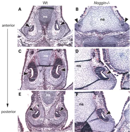
The vomeronasal capsule is absent in Noggin2/2 mice The VO (also known as Jacobson’s organ) is an auxiliary olfactory sense organ situated adjacent to the cartilage of the nasal septum, mainly used to detect pheromones. The VO is a c-shaped structure surrounded by a cartilagi-nous capsule which opens into the base of the nasal cavity. The vomeronasal capsule is a structure distinct from the nasal septum, which it neighbors. Histological analysis of the frontal section of Noggin2/2 E16.5 heads revealed that the epithelial section of the VO appeared normal (Fig. 6). However, the vomeronasal capsule was missing in the Noggin2/2 mutants and the mesenchyme around the VO was reduced in size (in all animals examined).
Figure 6. Abnormalities of the VO in Noggin2/2mice. (A – F) E16.5 hematoxylin and eosin stained frontal sections of WT and Noggin2/2heads showing the
developing nasal septum and VO. The VO is similar in WT and Noggin2/2mice (arrowheads), however the vomeronasal cartilage (vc) which cups the WT VO is missing in Noggin2/2mice, the mesenchyme surrounding the VO is reduced as is the developing nasal projection of the maxillary bone. The cartilage of the
nasal septum is greatly enlarged in Noggin2/2mice. np, nasal projection of the maxillary bone; ns; cartilage primordium of the nasal septum; vc, vomeronasal cartilage. Scale bar: (A) 200 mm (A – F, same magnification).
Pituitary gland abnormalities in Noggin2/2 mice
The developing pituitary gland was analyzed in sagittal and coronal sections at E13.5, E14.5 and E16.5 (Supplementary Material, Fig. S2, data not shown). The most notable feature in Noggin2/2 mice was that the developing pituitary was smaller when compared with WT mice, especially the pars anterior. The pars anterior (pars distalis) ultimately produces the most of the pituitary hormones. Other defects that have previously been reported in Noggin2/2 mice include a rostral displacement of Rathke’s pouch and the induction of secondary pituitary tissue (25).
DISCUSSION
Noggin null allele mice exhibit several craniofacial abnormal-ities including a narrowed frontonasal process, a solitary median maxillary incisor and defects in the hyoid bone, VO and pituitary gland. All these Noggin-related facial anomalies are characteristic of a microform of HPE and are associated with reduced expression of hedgehog target genes. Our find-ings are consistent with the hypothesis that Bmps have a role in midline patterning and the regulation of midface width by inhibiting Shh signaling.
Solitary median upper incisor
WT mice have two upper incisors. We found that Noggin2/2 mice have a solitary median upper incisor (100% penetrant), which develops from single expression domain in the midline of a single premaxillary bone. We cannot rule out that the single Shh expression domain is a consequence of a lack midline tissue intervening between two anlagen which have subsequently fused together, although this is relatively unlikely as the centrally located Shh domain in the Noggin2/2 mice is smaller than the individual domain of WT upper incisors. Alterations in the number of teeth can be caused by irregularities in the tooth initiation process (26). In Noggin2/2 mice, the single incisor is part of a HPE midfacial phenotype where the lack of tissue in the midline and a patterning defect are responsible for the tooth pheno-type. Overactivation of Noggin in the oral epithelium using transgenic tools (K14 promoter) does not result in a change in the number of incisors. However, the morphology and struc-ture of the K14-Noggin incisors are altered: incisors are large with abnormalities in enamel and dentine formation. In addition, all mandibular and maxillary third molars are lost due to developmental arrest at the early bud stage (27).
Humans normally have two deciduous and two permanent central incisors and the development of a solitary median maxillary central incisor (SMMCI) in the primary and/or secondary dentition is rare. SMMCI can occur as part of a holoprosencephalic phenotype, in association with abnormal-ities not related to HPE or as an apparently isolated finding. Missense mutations in SHH have been reported in patients with SMMCI that do not have other features of HPE (28,29). The I111F mutation has been found in eight members of the same family and it has been suggested that this mutation may be specific for SMMCI as it has not been found in either HPE patients or in a normal population (28).
Hypopituitarism
Hypopituitarism due to pituitary hypoplasia has been described in patients with SMMCI together with other HPE-like features and has been reported in patients with loss of function mutations in the HH signaling transcription factor GLI2 (30,31). In zebrafish, it has been shown that Gli1 and Gli2 regulate Hh signaling to induce and pattern the developing adenohypophysis (32). And in mice, 50% of Gli22/2 mutants and 100% of Gli22/2;Gli12/2 mutants fail to develop a pituitary gland (33). Whether Bmp and Hh signal-ing interact dursignal-ing pituitary development is not known. In Noggin2/2 mice, the pituitary abnormalities appear to be due to an expanded domain of Bmp4 activity that results in Fgf10 repression and rostral shift of the Bmp4 and Shh boundary (25).
Midfacial width is regulated by Noggin
Several studies have analyzed the role of Bmps and other growth factors in the control of facial proximal/distal out-growth, but very few have addressed what controls facial width. Here we show that Bmp, by modulating Shh signaling, plays a fundamental role in regulating facial width. Although the facial features of Noggin2/2 mice have not been pre-viously reported, there is evidence that Bmps control facial patterning and growth and that alteration of Bmp signaling can lead to HPE. For instance, compound mouse mutants for Chordin and Noggin (34,35) or chick embryos that have had BMP4-soaked beads implanted into their forebrains (36) exhibit holoprosencephalic facial features including cyclopia, a central proboscis and orofacial clefting. With regard to the proximal/distal outgrowth of the facial processes, Noggin-soaked beads implanted into the developing chick face at different developmental stages results in decreased cell proliferation and subsequently decreased outgrowth, smaller frontonasal and maxillary processes and deletion of the maxil-lary and palatine bones (14,15,23).
We show that in Noggin2/2 mice, Shh and the Shh target genes Ptch1 and Gli1 are misexpressed at E11.5 and E12.5 in the developing face. This suggests that disrupted Bmp signaling in Noggin2/2 mice results in downregulation of the hedgehog pathway at the critical time when the midline craniofacial structures are developing. This is supported by experiments in the developing chick that demonstrate that excessive exogenous BMP2 (micro bead implantation) down-regulates Shh expression (23), and transient loss of SHH sig-naling by either excision of the frontonasal epithelium or by the introduction of Shh neutralizing antibodies inhibits growth of the facial primordia and results in a narrowing of the mid and upper face and subsequent hypotelorism (37). Also, mice lacking the transmembrane protein Cdo, which positively regulates Shh signaling, exhibit HPE with a midface hypoplasia and hypotelorism (38).
Disruption in Shh signal transduction by exposing chick embryos, at Hamilton Hamburger stages 15 and 17, to the Smoothened inhibitor cyclopamine, results in a continuum of HPE-related defects including hypotelorism (39). The time when Shh signaling, and presumably Bmp signaling, is blocked is critical. Early blockade of Shh (before the division
of the eye field into two) (stage 4) causes cyclopia, Shh block-age later (stblock-ages 15 and 17) results hypotelorism, midfacial hypoplasia and orofacial clefting, while blockage later in development has no effect (39). Conversely, excessive SHH in the embryonic face leads to increased proliferation in the frontonasal process, increased width of mid and upper facial processes and consequently hypertelorism (37). When consid-ering facial development, it is important to differentiate between early abnormal Shh signaling from the developing brain and signaling later from the developing facial structures. In this study, we show that exogenous BMP4 inhibits Fgf8 and Ptch1 in the developing mouse frontonasal process. Fgf8 is a key regulator of facial development which is known to regulate Shh signaling in several embryonic locations, includ-ing the limb and genital tubercle. Blockinclud-ing Fgf signalinclud-ing in the developing chick face results in a narrowing of the upper face, approximation of the maxillary processes and a coalescence of the nasal pits into a single central pit (40). There is good evi-dence that during very early chick facial development Bmp signaling acts to restrict Fgf8 expression (41). Equally, block-ing endogenous Bmp signalblock-ing in the developblock-ing chick fronto-nasal prominence by overexpressing Noggin results in an expansion of the Fgf8 domain (40). Taken together, the down-regulation of Fgf8 and Hedgehog signaling by excessive Bmp signaling provides a mechanism underlying the HPE in the Noggin null allele mouse.
Mutations in SHH/Shh, as well as mutations in several hedgehog signaling pathway members, result in HPE in both humans and mice. Human and mouse mutations of GLI2/ Gli2 have been shown to cause HPE-like phenotypes, includ-ing midfacial hypoplasia with a sinclud-ingle maxillary incisor (31,42). A SHH missense mutation has been associated with HPE and shown to cause defective binding to the HH regulator GAS1 (43). Indeed, mutations in GAS1 alone or in addition to mutations in SHH can result in HPE in humans (8). Similar to Noggin2/2 mutants, 40 – 50% of Gas12/2 mice exhibit several features consistent with a microform of HPE including a single maxillary incisor, a narrow frontonasal process and abnormalities in the pituitary, palate and VO (21). The Gas12/2 phenotype appears to be due to a reduction in Shh signaling as loss of a single allele of Shh in the Gas12/2 back-ground worsened the craniofacial phenotype and Gas12/2 mice exhibit a reduced Ptch1 expression domain in the devel-oping frontonasal process (21,44).
Noggin2/2mice have a narrow facial width and malformed neural crest cell derivatives, namely the upper incisors, pre-maxillary bones, nasal septum and the palate. There is evidence that Bmp signaling is important in neural crest induc-tion, delamination and migration and it has been suggested that elevated Bmp signaling disrupts the development of post-migratory, differentiating skeletal neural crest cells (18,24,34). Thus, overexpression of Noggin in the second branchial arch (Hoxa2) results in reduced numbers of neural crest cells and consequently hypomorphic skeletal and neural elements (45). Also, Noggin may protect neural crest cells from apoptosis induced directly by elevated Bmp signaling, and indirectly by maintaining Shh signaling as Noggin and also Chordin promote the rostral expression of Shh (34). Noggin and Chordin are Bmp antagonists that have similar biochemical activity and expression domains and may well compensate
functionally for each other. Compound mutant mice for Noggin and Chordin display a variety of HPE craniofacial defects (24,34,35). Consistent with our data, disruption of Bmp and Follistatin signaling in the chick results in a holopro-sencephalic phenotype through a mechanism which involves modulating midline Shh signaling (46).
Despite multiple regulators controlling the Bmp pathway, disruption of Noggin alone is sufficient to result in the midfa-cial phenotype described in this paper. In conclusion, we demonstrate the importance of Noggin in the correct pattern-ing of the face and in the etiology of HPE.
MATERIALS AND METHODS Mice
The generation, breeding and genotyping of the Noggin mutant mice used in this study have been described previously (17,22). The initial inactivation of the Noggin gene was done in an inbred 129SvJ genetic background. Heterozygote males have been serially backcrossed with CD1 females for at least 10 generations. NogginLacZ reporter mice were backcrossed to C57BL/6 and used as heterozygotes.
Skeletal and LacZ staining
Alcian blue/alizarin red and LacZ staining were performed according to Rice et al. (2010) and Zouvelou et al. (2009) (47,48).
Organ culture
E10.5 and E11.5 WT mouse heads were dissected and placed on Nuclepore filters in a Trowell-type organ culture system as described previously (49). Affi-gel agarose beads (Biorad) were incubated with recombinant human BMP4 (100 ng/ml, R&D Systems) or BSA at 378C for 1 h and stored at 48C before being placed on the explants. Bead assays were cultured overnight.
Preparation of probes and in situ hybridization
35
S in situ hybridization on paraffin sections was performed as previously described (50). Both bright and dark field images were taken of hybridized sections. Silver grains were selected from the dark field images, colored red and then superimposed onto the identical bright field image using Adobe Photoshop 6.0 software. Whole-mount in situ hybridization was per-formed using digoxigenin-UTP-labelled riboprobes as pre-viously described (51). The preparation of the Bmp, Fgf8, Gli1, Pax9, Ptch1 and Shh RNA probes has been described previously (52–54).
Statistical analysis
An independent samples t-test was used for the statistical analysis of normally distributed samples. A P-value of ,0.05 was considered statistically significant. SPSS 15.0 was used for the statistical analysis of the data.
SUPPLEMENTARY MATERIAL
Supplementary Material is available at HMG online.
ACKNOWLEDGEMENTS
We thank Airi Sinkko for her technical assistance. The Pax9 cDNA was a kind gift from Heiko Peters.
Conflict of Interest statement. None declared
FUNDING
This work was supported by the Academy of Finland, Sigrid Juse´lius Foundation, Helsinki University Research Foun-dation, Biocentrum Helsinki, Medical Research Council UK and the Flemish Science Foundation (FWO) grant number G2.214.07. T.A.M. was supported by grants from the Univer-sity of Zurich.
REFERENCES
1. Gorlin, R., Cohen, M.J. and Hennekam, R. (2001) Syndromes of the Head and Neck, 4th edn. Oxford University Press, New York.
2. Muenke, M. (1994) Holoprosencephaly as a genetic model for normal craniofacial development. Dev. Biol., 5, 293 – 301.
3. Leoncini, E., Baranello, G., Orioli, I.M., Anneren, G., Bakker, M., Bianchi, F., Bower, C., Canfield, M.A., Castilla, E.E., Cocchi, G. et al. (2008) Frequency of holoprosencephaly in the International
Clearinghouse Birth Defects Surveillance Systems: searching for population variations. Birth Defects Res. A Clin. Mol. Teratol., 82, 585 – 591.
4. Matsunaga, E. and Shiota, K. (1977) Holoprosencephaly in human embryos: epidemiologic studies of 150 cases. Teratology, 16, 261 – 272. 5. Cohen, M.M. Jr and Shiota, K. (2002) Teratogenesis of
holoprosencephaly. Am. J. Med. Genet., 109, 1 – 15.
6. Traiffort, E., Dubourg, C., Faure, H., Rognan, D., Odent, S., Durou, M.R., David, V. and Ruat, M. (2004) Functional characterization of sonic hedgehog mutations associated with holoprosencephaly. J. Biol. Chem., 279, 42889 – 42897.
7. Wallis, D. and Muenke, M. (2000) Mutations in holoprosencephaly. Hum. Mutat., 16, 99 – 108.
8. Ribeiro, L.A., Quiezi, R.G., Nascimento, A., Bertolacini, C.P. and Richieri-Costa, A. (2010) Holoprosencephaly and holoprosencephaly-like phenotype and GAS1 DNA sequence changes: report of four Brazilian patients. Am. J. Med. Genet. A, 152A, 1688– 1694.
9. Fernandes, M. and Hebert, J.M. (2008) The ups and downs of holoprosencephaly: dorsal versus ventral patterning forces. Clin. Genet., 73, 413 – 423.
10. Fernandes, M., Gutin, G., Alcorn, H., McConnell, S.K. and Hebert, J.M. (2007) Mutations in the BMP pathway in mice support the existence of two molecular classes of holoprosencephaly. Development, 134, 3789– 3794.
11. Nie, X., Luukko, K. and Kettunen, P. (2006) BMP signalling in craniofacial development. Int. J. Dev. Biol., 50, 511 – 521.
12. Abzhanov, A., Protas, M., Grant, B.R., Grant, P.R. and Tabin, C.J. (2004) Bmp4 and morphological variation of beaks in Darwin’s finches. Science, 305, 1462 – 1465.
13. Wu, P., Jiang, T.X., Suksaweang, S., Widelitz, R.B. and Chuong, C.M. (2004) Molecular shaping of the beak. Science, 305, 1465 – 1466. 14. Lee, S.H., Fu, K.K., Hui, J.N. and Richman, J.M. (2001) Noggin and
retinoic acid transform the identity of avian facial prominences. Nature, 414, 909 – 912.
15. Barlow, A.J. and Francis-West, P.H. (1997) Ectopic application of recombinant BMP-2 and BMP-4 can change patterning of developing chick facial primordia. Development, 124, 391 – 398.
16. Zimmerman, L.B., De Jesus-Escobar, J.M. and Harland, R.M. (1996) The Spemann organizer signal noggin binds and inactivates bone
morphogenetic protein 4. Cell, 86, 599 – 606.
17. Brunet, L.J., McMahon, J.A., McMahon, A.P. and Harland, R.M. (1998) Noggin, cartilage morphogenesis, and joint formation in the mammalian skeleton. Science, 280, 1455– 1457.
18. Anderson, R.M., Lawrence, A.R., Stottmann, R.W., Bachiller, D. and Klingensmith, J. (2002) Chordin and noggin promote organizing centers of forebrain development in the mouse. Development, 129, 4975– 4987. 19. McMahon, J.A., Takada, S., Zimmerman, L.B., Fan, C.M., Harland, R.M.
and McMahon, A.P. (1998) Noggin-mediated antagonism of BMP signaling is required for growth and patterning of the neural tube and somite. Genes Dev., 12, 1438 – 1452.
20. Wijgerde, M., Karp, S., McMahon, J. and McMahon, A.P. (2005) Noggin antagonism of BMP4 signaling controls development of the axial skeleton in the mouse. Dev. Biol., 286, 149 – 157.
21. Seppala, M., Depew, M.J., Martinelli, D.C., Fan, C.M., Sharpe, P.T. and Cobourne, M.T. (2007) Gas1 is a modifier for holoprosencephaly and genetically interacts with sonic hedgehog. J. Clin. Invest., 117, 1575– 1584.
22. Tylzanowski, P., Mebis, L. and Luyten, F.P. (2006) The Noggin null mouse phenotype is strain dependent and haploinsufficiency leads to skeletal defects. Dev. Dyn., 235, 1599 – 1607.
23. Ashique, A.M., Fu, K. and Richman, J.M. (2002) Endogenous bone morphogenetic proteins regulate outgrowth and epithelial survival during avian lip fusion. Development, 129, 4647– 4660.
24. Stottmann, R.W., Anderson, R.M. and Klingensmith, J. (2001) The BMP antagonists Chordin and Noggin have essential but redundant roles in mouse mandibular outgrowth. Dev. Biol., 240, 457 – 473.
25. Davis, S.W. and Camper, S.A. (2007) Noggin regulates Bmp4 activity during pituitary induction. Dev. Biol., 305, 145 – 160.
26. Mitsiadis, T.A. and Graf, D. (2009) Cell fate determination during tooth development and regeneration. Birth Defects Res. C Embryo Today, 87, 199 – 211.
27. Plikus, M.V., Zeichner-David, M., Mayer, J.A., Reyna, J., Bringas, P., Thewissen, J.G., Snead, M.L., Chai, Y. and Chuong, C.M. (2005) Morphoregulation of teeth: modulating the number, size, shape and differentiation by tuning Bmp activity. Evol. Dev., 7, 440 – 457. 28. Nanni, L., Ming, J.E., Du, Y., Hall, R.K., Aldred, M., Bankier, A. and
Muenke, M. (2001) SHH mutation is associated with solitary median maxillary central incisor: a study of 13 patients and review of the literature. Am. J. Med. Genet., 102, 1 – 10.
29. Garavelli, L., Zanacca, C., Caselli, G., Banchini, G., Dubourg, C., David, V., Odent, S., Gurrieri, F. and Neri, G. (2004) Solitary median maxillary central incisor syndrome: clinical case with a novel mutation of sonic hedgehog. Am. J. Med. Genet. A, 127A, 93 – 95.
30. Artman, H.G. and Boyden, E. (1990) Microphthalmia with single central incisor and hypopituitarism. J. Med. Genet., 27, 192 – 193.
31. Roessler, E., Du, Y.Z., Mullor, J.L., Casas, E., Allen, W.P.,
Gillessen-Kaesbach, G., Roeder, E.R., Ming, J.E., Ruiz i Altaba, A. and Muenke, M. (2003) Loss-of-function mutations in the human GLI2 gene are associated with pituitary anomalies and holoprosencephaly-like features. Proc. Natl Acad. Sci. USA, 100, 13424 – 13429.
32. Devine, C.A., Sbrogna, J.L., Guner, B., Osgood, M., Shen, M.C. and Karlstrom, R.O. (2009) A dynamic Gli code interprets Hh signals to regulate induction, patterning, and endocrine cell specification in the zebrafish pituitary. Dev. Biol., 326, 143 – 154.
33. Park, H.L., Bai, C., Platt, K.A., Matise, M.P., Beeghly, A., Hui, C.C., Nakashima, M. and Joyner, A.L. (2000) Mouse Gli1 mutants are viable but have defects in SHH signaling in combination with a Gli2 mutation. Development, 127, 1593 – 1605.
34. Anderson, R.M., Stottmann, R.W., Choi, M. and Klingensmith, J. (2006) Endogenous bone morphogenetic protein antagonists regulate mammalian neural crest generation and survival. Dev. Dyn., 235, 2507 – 2520. 35. Bachiller, D., Klingensmith, J., Kemp, C., Belo, J.A., Anderson, R.M.,
May, S.R., McMahon, J.A., McMahon, A.P., Harland, R.M., Rossant, J. et al. (2000) The organizer factors Chordin and Noggin are required for mouse forebrain development. Nature, 403, 658 – 661.
36. Golden, J.A., Bracilovic, A., McFadden, K.A., Beesley, J.S., Rubenstein, J.L. and Grinspan, J.B. (1999) Ectopic bone morphogenetic proteins 5 and 4 in the chicken forebrain lead to cyclopia and holoprosencephaly. Proc. Natl Acad. Sci. USA, 96, 2439 – 2444.
37. Hu, D. and Helms, J.A. (1999) The role of sonic hedgehog in normal and abnormal craniofacial morphogenesis. Development, 126, 4873– 4884. 38. Zhang, W., Kang, J.S., Cole, F., Yi, M.J. and Krauss, R.S. (2006) Cdo
functions at multiple points in the Sonic Hedgehog pathway, and Cdo-deficient mice accurately model human holoprosencephaly. Dev. Cell, 10, 657 – 665.
39. Cordero, D., Marcucio, R., Hu, D., Gaffield, W., Tapadia, M. and Helms, J.A. (2004) Temporal perturbations in sonic hedgehog signaling elicit the spectrum of holoprosencephaly phenotypes. J. Clin. Invest., 114, 485 – 494.
40. Brugmann, S.A., Kim, J. and Helms, J.A. (2006) Looking different: understanding diversity in facial form. Am. J. Med. Genet. A, 140, 2521– 2529.
41. Shigetani, Y., Nobusada, Y. and Kuratani, S. (2000) Ectodermally derived FGF8 defines the maxillomandibular region in the early chick embryo: epithelial-mesenchymal interactions in the specification of the craniofacial ectomesenchyme. Dev. Biol., 228, 73 – 85.
42. Hardcastle, Z., Mo, R., Hui, C.C. and Sharpe, P.T. (1998) The Shh signalling pathway in tooth development: defects in Gli2 and Gli3 mutants. Development, 125, 2803– 2811.
43. Martinelli, D.C. and Fan, C.M. (2009) A sonic hedgehog missense mutation associated with holoprosencephaly causes defective binding to GAS1. J. Biol. Chem., 284, 19169 – 19172.
44. Martinelli, D.C. and Fan, C.M. (2007) Gas1 extends the range of Hedgehog action by facilitating its signaling. Genes Dev., 21, 1231 – 1243. 45. Kanzler, B., Foreman, R.K., Labosky, P.A. and Mallo, M. (2000) BMP
signaling is essential for development of skeletogenic and neurogenic cranial neural crest. Development, 127, 1095– 1104.
46. Towers, P., Patel, K., Withington, S., Isaac, A. and Cooke, J. (1999) Flik, a chick follistatin-related gene, functions in gastrular dorsalisation/neural
induction and in subsequent maintenance of midline Sonic hedgehog signalling. Dev. Biol., 214, 298 – 317.
47. Zouvelou, V., Luder, H.U., Mitsiadis, T.A. and Graf, D. (2009) Deletion of BMP7 affects the development of bones, teeth, and other ectodermal appendages of the orofacial complex. J. Exp. Zool. B Mol. Dev. Evol., 312B, 361 – 374.
48. Rice, D.P., Connor, E.C., Veltmaat, J.M., Lana-Elola, E., Veistinen, L., Tanimoto, Y., Bellusci, S. and Rice, R. (2010) Gli3Xt-J/Xt-J mice exhibit lambdoid suture craniosynostosis which results from altered
osteoprogenitor proliferation and differentiation. Hum. Mol. Genet., 19, 3457 – 3467.
49. Kim, H.J., Rice, D.P., Kettunen, P.J. and Thesleff, I. (1998) FGF-, BMP-and Shh-mediated signalling pathways in the regulation of cranial suture morphogenesis and calvarial bone development. Development, 125, 1241 – 1251.
50. Vainio, S., Karavanova, I., Jowett, A. and Thesleff, I. (1993) Identification of BMP-4 as a signal mediating secondary induction between epithelial and mesenchymal tissues during early tooth development. Cell, 75, 45 – 58.
51. Rice, D.P., Kim, H.J. and Thesleff, I. (1997) Detection of gelatinase B expression reveals osteoclastic bone resorption as a feature of early calvarial bone development. Bone, 21, 479 – 486.
52. Rice, R., Connor, E. and Rice, D.P. (2006) Expression patterns of Hedgehog signalling pathway members during mouse palate development. Gene Expr. Patterns, 6, 206 – 212.
53. Neubuser, A., Koseki, H. and Balling, R. (1995) Characterization and developmental expression of Pax9, a paired-box-containing gene related to Pax1. Dev. Biol., 170, 701 – 716.
54. Aberg, T., Wozney, J. and Thesleff, I. (1997) Expression patterns of bone morphogenetic proteins (Bmps) in the developing mouse tooth suggest roles in morphogenesis and cell differentiation. Dev. Dyn., 210, 383 – 396.