Female Sex Hormones, Salt,
and Blood Pressure Regulation
Antoinette Pechère-Bertschi and Michel Burnier
There are gender-associated differences in blood pressure(BP) in humans, with men having higher BP than age-matched pre-menopausal women and being at greater risk for cardiovascular and renal diseases. The mechanisms responsible for the gender differences in BP control and regulation are not clear, although there is some evidence that interactions between sex hormones and the kidneys could play a role. However, the response to salt in pre- and post-menopausal women, and in particular the influence of exogenous and endogenous female sex hormones on renal hemodynamics and tubular segmental sodium handling, have been poorly investigated. Recently we have shown that both endogenous and exogenous female sex hormones markedly influence the systemic and renal hemodynamic response to salt. We have found that BP in young normo-tensive women, regardless of oral contraceptive use, is rather insensitive to salt. However, the renal hemodynamic and the tubular responses to salt vary significantly during
the normal menstrual cycle and with the administration of oral contraceptives. Furthermore, after the menopause, BP tends to become salt sensitive, a pattern that could be due to aging as well as to the modification of the sex hormone profile. These observations provide new insights pertain-ing to potential mechanisms explainpertain-ing the lower inci-dence of cardiovascular disease and progression of renal disease in pre-menopausal women (which tend to disap-pear with the menopause); these observations also empha-size the importance of considering more carefully the phase of the menstrual cycle whenever conducting phys-iologic studies in women and enrolling women in clinical studies. Finally, increased salt sensitivity in menopausal women strongly encourages the use of diuretics. Am J Hypertens 2004;17:994 –1001 © 2004 American Journal of Hypertension, Ltd.
Key Words: Female hormones, blood pressure, sodium, endogenous lithium, renal hemodynamics.
G
ender-associated differences in blood pressure (BP) have been observed in animals as well as in hu-mans.1–3 Thus, in humans, men have higher BP levels than women until the age of 60 to 70 years, when BP becomes progressively more comparable in both men and women. Hence, men are also at greater risk for developing cardiovascular and renal complications.1,2The mechanisms responsible for these gender differences in BP are not well understood, but several hypotheses have been proposed sug-gesting a role of androgens and female sex hormones.3The kidneys play a major role in the regulation of BP. According to the hypothesis of Guyton et al, a decrease in renal sodium excretion or a rightward shift of the pres-sure–natriuresis relationship can lead to a long-term in-crease in BP and the development of hypertension.4 In accordance with this hypothesis, Reckelhoff et al have reported that the pressure–natriuresis curve is blunted in
male spontaneously hypertensive rats (SHR) and that cas-tration of male SHR restores the pressure–natriuresis re-lationship, suggesting that androgens do contribute to the higher BP measured in males.5In addition, androgen
re-ceptor blockade lowers BP in male SHR to the level of female SHR and appears to protect against hypertension and end-organ damage.3,6 Reckelhoff et al also reported that the administration of testosterone to ovariectomized female SHR increases BP and modifies the pressure– natriuresis relationship.5This latter finding would indicate that androgens could also play a role in the rise in BP observed in menopausal women.3Yet, the role of
andro-gens in mediating the increase in BP in post-menopausal women remains a topic of discussion. Several studies have also suggested that sex steroid hormones have direct vas-cular effects that may contribute to the gender differences in BP regulation.7–9
Received February 2, 2004. First decision March 22, 2004. Accepted June 4, 2004.
From the Medical Policlinic and Service of Endocrinology (AP-B), University Hospital, Geneva, Switzerland; and Division of Hyper-tension and Vascular Medicine (MB), University Hospital, Lausanne, Switzerland.
Supported by the Fonds National Suisse de la Recherche Scientifique
(grants 32-52471.97 and 31-63947.00, to MB) and by a Marie-Heim Vögtlin grant (3234-45005.95, to AP-B).
Address correspondence and reprint requests to Dr. Antoinette Pechère-Bertschi, Policlinique of Medicine and Service of Endocrinol-ogy, University Hospital, 1211 Geneva 4, Switzerland; e-mail: Antoinette.Pechere@hcuge.ch
0895-7061/04/$30.00 © 2004 by the American Journal of Hypertension, Ltd.
Whether female sex hormones also modulate the renal handling of sodium and thereby contribute to the gender-associated differences observed in animal models and in humans is also a subject of controversy. In this review, we discuss the potential links among female sex hormone status, salt balance, and renal function and their possible influence on BP regulation. We also present recent results indicating that both endogenous and exogenous female sex hormones have important effects on the systemic and renal responses to changes in sodium intake in young, healthy, normotensive women, regardless of whether they are using oral contraceptives (OC), and in menopausal women who are not receiving hormonal replacement therapy.
Gender Difference in BP
Regulation and Pressure–
Natriuresis in Animal Models
As mentioned earlier here, there is good evidence to sup-port the role of androgens in mediating the increased BP in male rats and perhaps in elderly female rats.3 However,
there is also some evidence suggesting that female sex hormones may actually protect against a salt-induced in-crease in BP, possibly by augmenting the renal excretion of sodium. Thus, when Dahl salt-sensitive (DS) rats re-ceive a high-sodium diet, female rats become less hyper-tensive than male rats.10 In this animal model,
gonadectomy results in an accelerated development of salt-sensitive hypertension in females.10 Whether
gonad-ectomy affects BP in the male DS rats is more controver-sial. Indeed, results obtained so far are inconsistent. Rowland and Fregly found that gonadectomy reduces BP in male DS rats,11but this observation was not reported by
others.10 The increase in salt sensitivity observed after
ovariectomy is actually associated with a blunted pres-sure–natriuresis relationship. Interestingly, reversal of the diet to a low salt intake reverses the hypertension in intact male and female DS rats, but this was not the case in ovariectomized female DS rats, suggesting that female sex hormones act to suppress sodium dependent as well as sodium independent increases in BP.12 A greater rise in
BP has also been reported in spontaneously hypertensive female rats after ovariectomy.13,14Dietary phytoestrogens
have been found to protect ovariectomized female SHR from dietary salt-sensitive hypertension.15 The
sympa-thetic nervous system seems to play an important role in mediating this effect, as ganglionic blockade reduces the sodium-induced rises in arterial pressure.15
Of note, female sex hormones may affect not only renal sodium excretion but perhaps also sodium intake per se. Indeed, in many mammals, the spontaneous free sodium intake appears to be greater in females than in males of the same species. In this respect, estradiol has recently been found to modulate the sodium intake of hypertensive (ie, SHR) and normotensive (ie, Wistar-Kyoto) female rats, and is positively correlated with sodium intake in both
strains, with the hypertensive rats consuming more sodium than the normotensive rats.16 This remarkable
gender-related difference in sodium ingestion may originate phy-logenetically in the need to preserve sodium losses during pregnancy. Taken together, these experimental findings suggest that female sex hormones indeed modulate sodium balance in animals and may therefore contribute to the regulation of BP.
Gender Differences in the Activity
of the Renin-Angiotensin System
The renin-angiotensin system is one of the key hormonal systems regulating BP and modulating the pressure–natriure-sis relationship. Several studies have reported gender differ-ences in various components of the renin-angiotensin cascade that could partially explain the gender differences in BP.17Thus, in a normotensive population, plasma renin activity (PRA) has been reported to be higher in men than in women regardless of age and ethnic heritage.18Exogenous female sex hormones administered for oral contraception have also been shown to stimulate angiotensinogen production, which may lead to a increase in BP in some women.19Other studies have found that PRA is higher in post-menopausal than in pre-menopausal women, although PRA remains higher in men than in women of the same age.17In animals significant differences have also been observed between males and fe-males. The administration of testosterone to ovariectomized female rats increases PRA, and PRA is lower in males after castration.20,21Finally, in Sprague-Dawley rats, a linear cor-relation between the levels of testosterone and plasma renin activity levels has been reported,3,17 suggesting that testos-terone stimulates the renin-angiotensin system. In accordance with this observation, several studies have found that andro-gens like oestrogens enhance renal angiotensinogen mRNA.20,22
There is also some evidence that the response to a stimulation of the renin-angiotensin system differs in men and women. Thus, Miller et al have compared the renal hemodynamic response to the infusion of exogenous an-giotensin II in young normotensive pre-menopausal women and in age-matched men and found striking dif-ferences.23Both groups exhibited an increase in BP and a decrease in effective renal plasma flow (ERPF) with an-giotensin II, but only men maintained their glomerular filtration rate (GFR) resulting in an increased filtration fraction. In women, the administration of angiotensin II decreased GFR, leading to a reduction in filtration frac-tion. This has been interpreted as a lesser increase in intraglomerular pressure in women, an observation which may play a role in renal disease progression. The incidence of end-stage renal disease is indeed lower in women than in men even after adjustment for age and race.24
Further-more, the rate of progression to end-stage renal disease is often slower in women than in men, independently of BP or serum cholesterol levels.25
Female Sex Hormones
and the BP Response to Salt
Few clinical studies have assessed specifically the effect of gender on the BP response to salt. There are some sugges-tions that the hormonal status modulates this response and that hypertensive females are more salt sensitive than males, but this has not been confirmed by all investigators.1,26The
relationship between salt intake and BP tends to become more marked in post-menopausal than in pre-menopausal women, but this observation may be attributed to aging. Little attention has also been given to the changes in BP occurring in young women during the normal menstrual cycle, and the few investigations performed so far have led to rather incon-sistent results. Indeed, the BP has been shown to be higher in the luteal phase or at the onset of menstruation in some studies27–29or significantly decreased in the mid-luteal phase
of the cycle both in normotensive30,31 and hypertensive
women32or even unchanged across the entire cycle.33Most
of these studies were based on office or home BP and with an uncontrolled sodium intake. More recently a prospective trial suggested that the ambulatory systolic BP is higher through-out the menstrual cycle in women taking OC than in women who do not, but no change in ambulatory pressure was found across the menstrual cycle.34Because little is known about
the effects of female sex hormones on the BP response to salt, we have recently performed a series of studies to assess prospectively the influence of exogenous and endogenous female sex hormones on the systemic hemodynamic response to changes in sodium intake in young healthy normotensive women and in menopausal women.35–38
In brief, 35 young normotensive white females who had regular menstruation and were not using OC were ran-domly assigned to be studied during the follicular (n⫽ 17) and the luteal (n⫽ 18) phases of the menstrual cycle. In addition, 27 young white healthy women who were using OC (monophasic combination of 30 g ethinylestradiol and 150 g desogestrel for ⬎6 months) and 12 meno-pausal women not receiving hormonal replacement ther-apy were investigated using a comparable protocol.
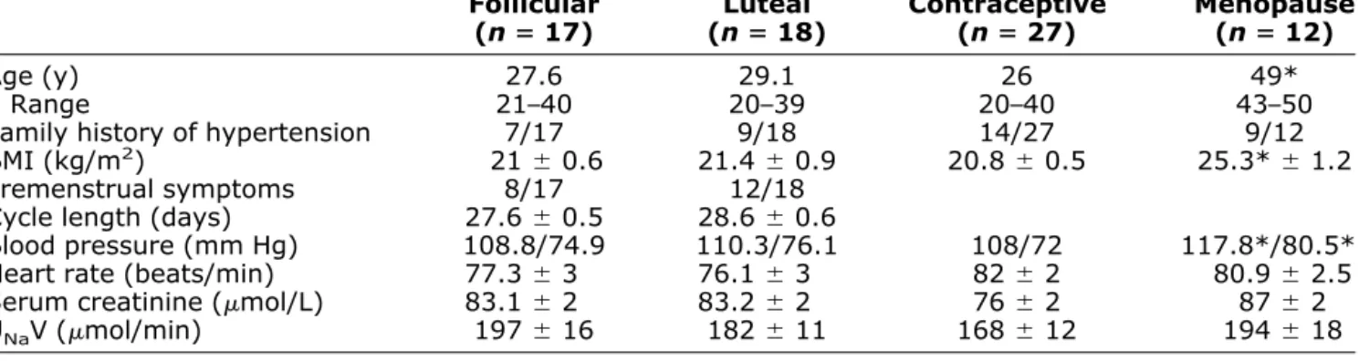
Characteristics of these subjects are presented in Table 1. All subjects were randomized to receive a diet low in sodium (40 mmol Na/day) and a diet high in sodium (250 mmol Na/day) for a 7-day period for 2 consecutive months. At the end of each 7-day diet period, 24-h ambu-latory BP as well as renal hemodynamics and renal sodium handling and an hormonal profile were measured.35–39
As shown in Fig. 1, we found that the BP and heart rate response to salt is comparable in the luteal and in the follicular phases of the normal menstrual cycle. In both phases of the cycle, the pressure–natriuresis relationship is steep, suggesting that these women are essentially insen-sitive to salt. The administration of OC does not modify the pressure–natriuresis relationship, suggesting little if any effect of exogenous female sex hormones on the BP response to salt. In contrast, the pressure–natriuresis curve of menopausal women is significantly shifted to the right, indicating that BP becomes salt sensitive after the meno-pause as suggested previously by Weinberger et al.40
Unfortunately, our study does not allow any conclusion Table 1. Characteristics of the study groups
Follicular
(n ⴝ 17) (n ⴝ 18)Luteal Contraceptive(n ⴝ 27) Menopause(n ⴝ 12)
Age (y) 27.6 29.126 49*
Range 21–40 20–39 20–40 43–50
Family history of hypertension 7/17 9/18 14/27 9/12
BMI (kg/m2) 21 ⫾ 0.6 21.4 ⫾ 0.9 20.8 ⫾ 0.5 25.3* ⫾ 1.2
Premenstrual symptoms 8/17 12/18
Cycle length (days) 27.6 ⫾ 0.5 28.6 ⫾ 0.6
Blood pressure (mm Hg) 108.8/74.9 110.3/76.1 108/72 117.8*/80.5*
Heart rate (beats/min) 77.3 ⫾ 3 76.1 ⫾ 3 82 ⫾ 2 80.9 ⫾ 2.5
Serum creatinine (mol/L) 83.1 ⫾ 2 83.2 ⫾ 2 76 ⫾ 2 87 ⫾ 2
UNaV (mol/min) 197 ⫾ 16 182 ⫾ 11 168 ⫾ 12 194 ⫾ 18
BMI ⫽ body mass index; UNaV ⫽ urinary sodium excretion.
Data are mean ⫾ SEM. * P ⬍ .05 v other phases.
FIG. 1. Pressure–natriuresis relationship in normotensive women
during the normal menstrual cycle, during use of oral contracep-tives, and after menopause. All women received randomly a diet low in sodium (40 mmol Na/day) and high in sodium (250 mmol Na/day) for 1week. Blood pressure was measured over 24 h using ambula-tory blood pressure monitoring.
about the main mechanism of this increase in salt sensi-tivity that develops after the menopause and, particularly, whether it is due to aging only or to the modification of the hormonal profile. In humans, some studies have suggested that the BP increase after the onset of menopause is linked to a reduced synthesis of estradiol or to an imbalance between androgens and female sex hormones, but this remains to be demonstrated.3 Nevertheless, our results
may provide new clues to explain why the incidence of hypertension clearly increases in post-menopausal fe-males, exceeding that in age-matched men.41 Indeed, if
sodium intake is not reduced, the increased responsiveness to sodium at the menopause may contribute to increase BP and to favour the development of hypertension. Of note, the menopause is often associated with a weight gain, and the prevalence of obesity is higher in post-menopausal women than in age-matched men.42 This would indicate
that sodium intake is certainly not reduced in menopausal women. We have also reported previously that the increase in body weight occurring at the time of the menopause is actually associated with a significant increase in ambula-tory BP.43
Female Sex Hormones
and the Renal Hemodynamic
Response to Salt
The literature is also conflicting concerning the possible changes in renal hemodynamics occurring during the normal menstrual cycle.30,31,44 – 48Indeed, using either endogenous
creatinine clearance44or [51Cr] EDTA clearance,45– 47some
investigators have reported small increases in GFR together with either no change or an increase in renal plasma flow during the luteal phase, whereas others found no change.30,31,44 – 48 In another study, investigators have
as-sessed the renal and peripheral hemodynamic responses to angiotensin II receptor blockade with losartan during the two phases of the menstrual cycle in subjects receiving a fixed sodium diet containing ⬍150 mmol/day.49 In this study,
despite an apparent activation of the renin-angiotensin system during the luteal phase, a comparable renal vasodilatation during angiotensin II blockade was observed during the two phases of the menstrual cycle.49
In our normotensive subjects studied either during the follicular or during the luteal phase of their menstrual cycle, GFR and renal plasma flow were similar in the two phases of the cycle when subjects were studied while receiving a low sodium intake.35 However, whereas salt
loading had no effect on renal hemodynamics in the fol-licular phase, the administration of salt during the luteal phase induced a marked renal vasodilation with no change in GFR (Fig. 2). Hence, a significant decrease in filtration fraction was observed while receiving high salt during the luteal but not during the follicular phase.35 Plasma renin
activity (PRA) and aldosterone levels decrease signifi-cantly after sodium loading in the two phases of the
menstrual cycle. Yet, the salt-induced suppression of PRA is partly blunted in the luteal compared with the follicular phase.35
Thus, our data suggest that in contrast to arterial pres-sure, the control of the renal circulation is salt sensitive during the luteal phase of the menstrual cycle and that endogenous female sex hormones act in a direct or indirect way on glomerular hemodynamics with a possible effect on the efferent arteriole, inasmuch as the filtration fraction decreases upon salt loading. By comparison, the renal hemodynamic response to salt in young normotensive male subjects is characterized by an increase in renal plasma flow and a concomitant rise in GFR, which was not observed in women studied in the luteal phase.50To
ex-plain the salt-induced renal vasodilatation and fall in fil-tration fraction in women studied during the luteal phase, we propose the hypothesis that estrogens modulate the renal hemodynamics indirectly via the nitric oxide (NO) pathway and perhaps prostaglandin formation, thereby inhibiting the effects of angiotensin II on glomerular he-modynamics. Indeed, estrogen levels are high in the luteal phase, and estradiol has been shown to increase local prostaglandin E2(PGE2) and I2(PGI2) production and to
activate NO synthase.51,52Because NO has been proposed
as a physiologic antagonist of angiotensin II53and because
blockade of endothelium-derived NO synthesis has been shown to increase intraglomerular BP,54,55 female sex
hormones could modulate indirectly, via the NO pathway, and perhaps prostaglandin synthesis, the effect of angio-tensin II on glomerular hemodynamics and particularly on efferent arteriolar tone. Through this mechanism, estro-gens could cause renal vasodilation and a decrease in filtration fraction.
There are few data on the renal hemodynamic effects of OC.19,55–57An early study by Hollenberg et al has shown
that OC in healthy young women reduces the effective
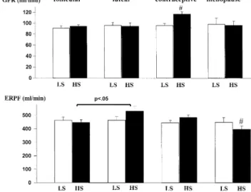
FIG. 2. Salt-induced variations in glomerular filtration rate (GFR)
and effective renal plasma flow (ERPF) in normotensive women during the normal menstrual cycle, with the use of oral contracep-tives, and after the menopause. HS ⫽ high-sodium diet; LS ⫽ low-sodium diet. #P ⬍ .05 LS versus HS.
renal plasma flow, an effect that appears to be due to an activation of the renin-angiotensin system by OC.19 The
administration of OC has also been found to increase creatinine clearance in young women studied while receiv-ing a free sodium intake.57Kang et al reported significant
increases in systolic BP, renal vascular resistance, and filtration fraction in OC users compared with nonusers and showed that these differences were at least partially abol-ished by angiotensin II blockade.58Based on their data, the
authors have suggested that despite humoral evidence of renin angiotensin system activation by OC, the tissue response may actually be down-regulated, perhaps at a receptor level. In a more recent study, Ribstein et al failed to confirm that the renin-angiotensin system plays a prom-inent role in maintaining a high BP in women with OC-associated hypertension.56
In our normotensive women, OC had a marked influ-ence on the renal hemodynamic response to salt, although systemic BP was not affected34(Fig. 2). Indeed, upon salt
loading, GFR increased significantly in women taking OC, whereas renal plasma flow was unchanged; consequently, filtration fraction was significantly increased, suggesting an increased intraglomerular pressure. Importantly, plasma renin activity in our OC users was several-fold higher than the activity measured in OC nonusers studied at comparable levels of salt intake. Therefore, the mech-anism resulting in the salt-induced increase in GFR in OC users may actually be the same as the one that we hypoth-esize to occur during the luteal phase of the menstrual cycle but with one important difference: namely, the base-line activity of the renin-angiotensin system. Thus, in OC users, the vasodilatory effect of estrogens may be more prominent at the level of the afferent arteriole, thereby leading to an increase in GFR and filtration fraction. An alternative explanation for the apparent difference between endogenous and exogenous female sex hormones, as re-flected by the more pronounced effect of salt loading on renal hemodynamic in OC users than in nonusers, is per-haps a difference in potency between endogenous and exogenous female sex hormones. Indeed, combined OC agents deliver pharmacologic levels of estrogens that ex-hibit six to 10 times the estrogenic activity provided by endogenous estrogens.59
Several investigators have found that sodium induces a renal vasoconstriction in salt-sensitive patients.60 We
found a similar pattern, with a salt-induced reduction in renal plasma flow and an increase filtration fraction, in our group of salt-sensitive normotensive menopausal women.38 This maladaptive response to a sodium load
may be attributed to a lack of estrogens leading again to a reduced capacity of the renal circulation to vasodilate. An endothelial dysfunction has frequently been observed after the menopause, with an impaired systemic endothelium-dependent relaxation. In addition, it is also known that PRA is higher in post-menopasual than in pre-menopausal women. In our evaluation of normotensive menopausal women, suppression of PRA is blunted after salt loading.
At the time of menopause, in addition to aging and to-gether with the loss of estradiol (which is known to de-crease renin release), this poor suppression of renin by salt could contribute to the development of salt-sensitive hy-pertension in elderly women and may reflect an interaction between the renin system and menopausal status.
Altogether, our results demonstrate that endogenous as well as exogenous female sex hormones strongly affect the renal hemodynamic response to salt in young normoten-sive women, although these hormones do not affect the BP response to sodium.
Female Sex Hormones
and Renal Sodium Handling
The menstrual cycle as well as the menopause are char-acterized by great variations in plasma progesterone, es-trogen, aldosterone, and plasma renin activity— hormonal systems that are known to modulate the renal tubular handling of sodium either in the proximal or in the distal nephrons. In addition, androgen receptors have been lo-calized in the proximal tubule segment of the nephron, suggesting that testosterone could also affect renal sodium reabsorption.61 So far, several studies have examined the
effects of female sex hormones on the overall 24-h sodium excretion.62– 67 The results of these studies are often
con-tradictory, perhaps because most studies were conducted in subjects ingesting uncontrolled amounts of sodium or because subjects were receiving large amounts of water to measure renal function, which is an important confound-ing factor interactconfound-ing with renal sodium handlconfound-ing.50None
of these studies have really attempted to analyze in more detail the respective changes in proximal and distal so-dium reabsorption occurring either during the normal menstrual cycle or with the administration of OC.
In the past few years, we have used the endogenous lithium clearance technique to investigate the respective changes in proximal and post-proximal tubular sodium reab-sorption to investigate the tubular adaptation occurring with large changes in sodium intake in normotensive as well as hypertensive individuals.50,68,69 This technique is currently
considered to be the most reliable approach to investigate proximal segmental renal sodium handling in humans. When young normotensive men are shifted from a low-salt to a high-salt diet, the lithium clearance increases significantly and the fractional distal reabsorption of sodium decreases, indicating that both the proximal and the distal reabsorptions of sodium are reduced to maintain sodium balance.50
Similar investigations were conducted in our normo-tensive women, and significant differences in tubular re-sponses were observed between the follicular and the luteal phases.36 In the follicular phase, the renal tubular
response to a sustained increase in sodium intake is com-parable to that in men. In contrast, during the luteal phase, increasing sodium intake leads to a marked sodium escape from the distal nephron and no change in proximal sodium reabsorption, suggesting that while maintaining a diet high
in sodium, the sodium balance is controlled mainly by the distal segments of the nephron with a paradoxical increase in sodium reabsorption in the proximal nephron. Thus, the relationship between the proximal reabsorption of sodium as measured by the fractional excretion of lithium (FELi)
and the overall fractional excretion of sodium (FENa)is flat
and significantly different from that in women studied in the follicular phase (Fig. 3). The finding of significantly reduced sodium reabsorption in the distal nephron in the luteal phase when consuming a high-salt diet may reflect the pivotal role of progesterone to act as an anti-aldoste-rone compound and to participate in the maintenance of sodium balance during the pre-menstrual phase. This ef-fect of progesterone is particularly demonstrable with a high-salt diet when circulating aldosterone levels are low.36The increased reabsorption of sodium in the
prox-imal tubule with a high-salt diet is more surprising. Al-though one cannot exclude a direct tubular effect of progesterone to stimulate proximal sodium reabsorption, possibly in relation with the renal vasodilation, we believe that the low lithium clearance represents a compensatory mechanism of the proximal tubule to avoid an excessive loss of sodium by the distal nephron, thereby preventing sodium wasting and maintaining sodium balance. The increase in proximal sodium reabsorption may actually provide a clue for the blunted salt-induced decrease in plasma renin activity observed in the luteal phase com-pared with the follicular phase as discussed earlier here. Indeed, as sodium reabsorption is enhanced in the proxi-mal segments of the nephron, the delivery of sodium to the macula densa is reduced. A decreased sodium delivery at the juxta-glomerular apparatus will stimulate renin secre-tion. In turn, the increased PRA and angiotensin II further enhance the sodium reabsorption by the proximal nephron. Endogenous progesterone is known to be natriuretic, but synthetic derivatives of progesterone may lack
natri-uretic properties, for they have decreased affinity for renal mineralocorticoid receptors.70In our young patients using
OC, the renal tubular handling of sodium was similar to that observed in normal women in the follicular phase. However, the FELi /FENarelationship is shifted to lower
levels of FELi(Fig. 3). This shift may be attributed to the
activation of the renin-angiotensin system by OC, leading to an increased proximal reabsorption of sodium. A high reabsorption of sodium was also found to occur in the distal tubule as reflected by the very high distal reabsorp-tion of sodium. Thus, combined OC do not appear to affect the balance between the proximal and the distal nephron segments. However, because of the activation of the renin-angiotensin system, women using OC may have a greater tubular responsiveness.
Effect of Sodium Loading
on Body Weight in Women
Despite the common belief that women gain weight pre-menstrually because of sodium retention, there is little convincing evidence that sodium and water retention in-deed occur during the luteal phase of a normal menstrual cycle.62– 67 We found that under carefully controlled so-dium diets, a high soso-dium intake induces a weight gain of the same magnitude in the follicular and the luteal phase of the cycle (⬃1.0 kg), showing that no systematic sodium retention occurs in the luteal phase of the normal men-strual cycle (Fig. 4). This observation was based on weight determination, which of course provides no information on water redistribution within the body. The results suggest that the symptoms of edema and bloating reported by some women in the second phase of the cycle may be due to either a redistribution of fluid from the intravascular into the interstitial compartment rather than to sodium retention or to increased sodium appetite during the luteal phase of the menstrual cycle.71,72 When the diet is changed from a low-salt to a high-salt diet, the weight gain is comparable in women using OC. However, the weight gain is significantly more marked in menopausal women (⫹2.5 kg for comparable changes in sodium intake).
Mul-FIG. 4. Changes in body weight measured in normotensive women
during the normal menstrual cycle, under oral contraceptives and after the menopause when going from a low to a high salt intake. *P ⬍ .05 menopause versus other groups.
FIG. 3. Relationship between the fractional excretion of
endoge-nous lithium (FELi), an index of the amount of sodium coming out of
the proximal tubule, and the fractional excretion of sodium (FENa),
the expression of the overall sodium excretion. In the luteal phase, we observed a marked sodium escape from the distal nephron com-pared with the follicular phase and with OC users on a high-salt diet, indicating that sodium balance is controlled mainly by the distal segments of the tubule with a paradoxical proximal increase in sodium reabsorption.
tivariate analysis of the four groups disclosed that salt-induced change in body weight was a strong determinant of the change in BP (P ⬍ .03; 95% confidence interval 0.10 to 2.28).
Conclusion
In conclusion, endogenous and exogenous sex female hor-mones profoundly influence systemic and renal response to salt in women. Our data show that the BP of young normo-tensive women, regardless of whether they are using contra-ceptives, is rather insensitive to salt, with a normal pattern of adaptation of renal proximal and distal reabsorption to chang-ing salt intake. In contrast, women become salt sensitive after menopause. In contrast to the systemic pressure, the renal hemodynamic response to salt is modulated by female sex hormones. These hormones also affect the regulation of so-dium excretion. These recent observations provide new in-sights into potential mechanisms explaining the lower incidence of cardiovascular disease and the lower rate of progression of renal disease in pre-menopausal women, which tend to disappear with menopause. They also empha-size the importance of considering more carefully the phase of the menstrual cycle whenever conducting physiologic and pharmacologic studies in pre-menopausal women and when enrolling women in clinical studies, to prevent the strong influence of progesterone and high levels of estrogens on renal and systemic parameters. The increased salt sensitivity in menopausal women strongly encourages the use of diuret-ics in hypertensive women, which is in agreement with recent hypertension guidelines.
References
1. Stamler J, Stamler R, Riedlinger WF, Algera G, Roberts RH: Hypertension screening Evaluation Clinic (CHEC) Program, 1973– 1975. J Am Med Assoc 1976;235:2206 –2299.
2. August P, Oparil S: Hypertension in women. J Clin Endocrinol Metab 1999;4:1862–1866.
3. Reckelhoff JF: Gender differences in the regulation of blood pres-sure. Hypertension 2001;37:1199 –1208.
4. Guyton AC, Coleman TG, Cowley AW Jr, Scheel KW, Manning RD, Norman RA: Arterial pressure regulation: overriding domi-nance of the kidneys in long-term regulation and in hypertension. Am J Med 1972;52:584 –594.
5. Reckelhoff JF, Zhang H, Granger JP: Testosterone exacerbates hypertension and reduces pressure–natriuresis in male spontane-ously hypertensive rats. Hypertension 1998;31:435– 439. 6. Baltatu O, Cayla C, Iliescu R, Andreev D, Bader M: Abolition of
end-organ damage by antiandrogen treatment in female hyperten-sive transgenic rats. Hypertension 2003;41:830 – 833.
7. Shan J, Resnick LM, Liu QY, Wu XC, Barbagallo M, Pang PK: Vascular effects of 17 beta-estradiol in male Sprague-Dawley rats. Am J Physiol 1994;266:H967–H973.
8. Barbagallo M, Dominguez LJ, Licata G, Shan J, Bing L, Karpinski E, Pang PK, Resnick LM: Vascular effects of progesterone: role of cellular calcium regulation. Hypertension 2001;37:142–147. 9. Barbagallo M, Doninbues LJ, Licata G, Ruggero R, Lewanczuk RZ,
Pang PK, Resnick LM: Effect of testosterone on intracellular Ca⫹⫹ in vascular smooth muscle cells. Am J Hypertens 2001;14:1273– 1275.
10. Dahl K, Knudson D, Ohanien EV, Muirhead M, Tuthil R: Role of gonads in hypertension-prone rats. J Exp Med 1975;142:748 – 759.
11. Rowland NE, Fregly MJ: Role of gonadal hormones in hypertension in the Dahl salt sensitive rat. Clin Exp Hypertens A 1992;14:367– 375.
12. Hinojosa-Laborde C, Lange DL, Haywood JR: Role of female sex hormones in the development and reversal of Dahl hypertension. Hypertension 2000;35:484 – 489.
13. Masubuchi Y, Kumai T, Uematsu A, Komoriyama, Hirai M: Gon-adectomy-induced reduction in blood pressure in adult spontane-ously hypertensive rats. Acta Endocrinol 1982;101:154 –160. 14. Chen YF, Meng QM: Sexual dimorphism of blood pressure in
spontaneously hypertensive rats is androgen dependent. Life Sci 1991;48:85–96.
15. Fang Z, Carlson SH, Chen YF, Oparil S, Wyss JM: Estrogen depletion induces NaCl sensitive hypertension in female spontane-ously hypertensive rats. Am J Physiol Regul Integr Comp Physiol 2001;281:R1934 –R1939.
16. Kensicki E, Dunphy G, Ely D: Estradiol increases salt intake in female normotensive and hypertensive rats. J Appl Physiol 2002; 93:479 – 483.
17. Kang AK, Miller JA: Impact of gender on renal disease: the role of the renin angiotensin system. Clin Invest Med 2003;26:38 – 44. 18. James GD, Sealey JE, Muller F, Alderman M, Madhavan S, Laragh
JH: Renin relationship to sex, race and age in normotensive popu-lation. J Hypertens 1986;4(Suppl 5):S387–S389.
19. Hollenberg NK, Williams GH, Burger B, Chenitz W, Hoosmand I, Adams DF: Renal blood flow and its response to angiotensin II. An interaction between oral contraceptive agents, sodium intake, and the renin-angiotensin system in healthy young women. Circ Res 1976;38:35– 40.
20. Ellison KE, Inglfinger JR, Pivor M, Dzau VJ: Androgen regulation of rat renal angiotensinogen messenger RNA expression. J Clin Invest 1989;83:1941–1945.
21. Katz FH, Roper EF: Testosterone effect on renin system in rats. Proc Soc Exp Biol Med 1977;155:330 –333.
22. Chen YF, Naftilan Aj, Oparil S: Androgen-dependent angiotensino-gen and renin messenger RNA expression in hypertensive rats. Hypertension 1992;19:456 – 463.
23. Miller JA, Anacta LA, Cattran DC: Impact of gender on the renal response to angiotensin II. Kidney Int 1999;55:278 –285. 24. Neugarten J: Gender and the progression of renal disease. J Am Soc
Nephrol 2002;13:2807–2809.
25. Rosman JB, Langer K, Brandl M, Piers-Becht TP, van der Hem GK, ter Wee PM, Donker AJ: Protein-restricted diets in chronic renal failure: a four year follow-up shows limited indications. Kidney Int 1989;27(Suppl):S96 –S102.
26. Kojima S, Murakami K, Kimura G, Sanai T, Yoshida K, Imanishi M, Abe H, Kawamura M, Kawano Y, Ashida T: A gender difference in the association between salt sensitivity and family history of hypertension. Am J Hypertens 1992;5:1–7.
27. Greenberg G, Imeson J.D, Thompson S.G, Meade TW: Blood pressure and the menstrual cycle. Br J Obstet Gynaecol 1984;91: 1254 –1260.
28. Kelleher C, Joyce C, Kelly G, Ferriss JB: Blood pressure alters during the normal menstrual cycle. Br J Obstet Gynaecol 1986;93: 523–526.
29. Dunne FP, Barry DG, Ferriss JB, Grealy G, Murphy D: Changes in blood pressure during the normal menstrual cycle. Clin Sci 1991; 81:515–518.
30. Chapman AB, Zamudio S, Woodmansee W, Merouani A, Osorio F, Johnson A, Moore LG, Dahms T, Coffin C, Abraham WT, Schrier RW: Systemic and renal hemodynamic changes in the luteal phase of the menstrual cycle mimic early pregnancy. Am J Physiol Renal Physiol 1997;273:F777–F782.
31. Hassan AA, Carter G, Tooke JE: Postural vasoconstriction in women during the normal menstrual cycle. Clin Sci 1990;78:39 – 47.
32. Karpanou EA, Vyssoulis GP, Georgoudi DG, Toutouza MG, Tout-ouzas PK: Ambulatory blood pressure changes in the menstrual cycle of hypertensive women. Significance of plasma renin activity values. Am J Hypertens 1993;6:654 – 659.
33. Hirshoren N, Tzoran I, Makrienko I, Edoute Y, Plawner MM, Itskovitz-Eldor J, Jacob G: Menstrual cycle effects on the neurohu-moral and autonomic nervous systems regulating the cardiovascular system. J Clin Endocrinol Metab 2002;87:1569 –1575.
34. Williamson PM, Buddle ML, Brown MA, Whitworth JA: Ambulatory blood pressure monitoring (ABPM) in the normal menstrual cycle and in women using oral contraceptives. Comparison with conventional blood pressure measurement. Am J Hypertens 1996;9:953–958. 35. Pechère-Bertschi A, Maillard M, Stalder H, Brunner HR, Burnier
M: Blood pressure and renal hemodynamic response to salt during the normal menstrual cycle. Clin Sci 2000;98:697–702.
36. Pechère-Bertschi A, Maillard M, Stalder H, Brunner HR, Burnier M: Renal segmental tubular response to salt during the normal menstrual cycle. Kidney Int 2002;61:425– 431.
37. Pechère-Bertschi A, Maillard M, Stalder H, Bischof P, Fathi M, Brunner HR, Burnier M: Renal hemodynamic and tubular responses to salt in women using oral contraceptives. Kidney Int 2003:64: 1374 –1380.
38. Pechère-Bertschi A, Maillard M, Stalder H, Burnier M: Contrasting blood pressure (BP) and renal hemodynamic effects of sodium in pre- and peri-menopausal women (abstract). J Am Soc Nephrol 1999;10:369.
39. Pechère-Bertschi A, Nussberger J, Decosterd L, Armagnac C, Siss-mann J, Bouroudian M, Brunner HR, Burnier M: Renal response to the angiotensin II receptor subtype 1 antagonist irbesartan versus enalapril in hypertensive patients. J Hypertens 1998;16:385–393. 40. Weinberger MH, Miller JZ, Luft FC, Grim CE, Fineberg NS:
Definitions and characteristics of sodium sensitivity and blood pres-sure resistance. Hypertension 1986;8(Suppl II): II-127–II-134. 41. Burl VL, Whelton P, Roccella EJ, Brown C, Cutler JA, Higgins
M, Horan MJ, Labarthe D: Prevalence of hypertension in the US adult population: results from the Third National Health and Nutrition Examination Survey, 1988 –1991. Hypertension 1995: 25:305–313.
42. Rappelli A: Hypertension and obesity after the menopause. J Hy-pertens 2002;20(Suppl 2):S26 –S28.
43. Bidlingmeyer I, Burnier M, Bidlingmeyer M, Waeber B, Brunner HR: Isolated office hypertension: a pre-hypertensive state? J Hyper-tens 1996;14:327–332.
44. Davison JM, Noble MCB: Serial changes in 24 hour creatinine clearance during normal menstrual cycles and in the first trimester of pregnancy. Br J Obstet Gynaecol 1980;88:10 –17.
45. Brochner-Mortensen J, Paaby G, Fjelborg P, Raffn K, Larsen CE, Moller-Petersen J: Renal hemodynamics and extracellular ho-meostasis during the menstrual cycle. Scand J Clin Lab Invest 1987;47:829 – 835.
46. Gault MH, Chafe L, Prabkaran V: Mid-menstrual cycle decline in creatinine and urea clearances. Nephron 1994;67:158 –166. 47. Van Beek E, Houben AJHM, van Es PN, Willekes C, Korten
ECCM, de Leeuw PW, Peeters LLH: Peripheral hemodynamics and renal function in relation to the menstrual cycle. Clin Sci 1996;91: 163–168.
48. Nafziger AN, Schwartzman MS, Bertino JS: Absence of tobramycin pharmacokinetic and creatinine variation during the menstrual cy-cle: implied absence of variation on glomerular filtration rate. J Clin Pharmacol 1989;29:757–763.
49. Chidambaram M, Duncan JA, Lai VS, Cattran DC, Floras JS, Scholey JW, Miller JA: Variation in the renin angiotensin system throughout the normal menstrual cycle. J Am Soc Nephrol 2002; 13:446 – 452.
50. Burnier M, Monod M-L, Chioléro A, Maillard M, Nussberger J, Brunner HR: Renal sodium handling in acute and chronic salt loading/depletion protocol: the confounding influence of acute wa-ter loading. J Hypertens 2000;18:1657–1664.
51. Terragno NA, Terragno DA, Pacholczy D: Prostaglandins and the regulation of uterine blood flow in pregnancy. Nature 1974;249:57– 58.
52. Weiner CP, Soain IL, Baykis SA, Knowtes RG, Charles IG, Moncada S: Induction of calcium-dependent nitric oxide synthase by sex hormones. Proc Natl Acad Sci USA 1994;91;5212–5216. 53. De Nicola L, Blantz RC, Gabbai FB: Nitric oxide and angiotensin II.
Glomerular and tubular interaction in the rat. J Clin Invest 1992; 89:1248 –1256.
54. Zatz R, Nucci G: Effects of acute nitric oxide inhibition on rat glomerular microcirculation. Am J Physiol 1991;261:F360 –F363. 55. Baylis C, Mitruka B, Deng A: Chronic blockade of nitric oxide
synthesis in the rat produces systemic hypertension and glomerular damage. J Clin Invest 1992;90:278 –281.
56. Ribstein J, Halimi JM, Du Cailar G, Mimran A: Renal characteris-tics and effect of angiotensin suppression in oral contraceptive users. Hypertension 1999;33:90 –95.
57. Brandle E, Gottwald E, Melzer H, Sieberth HG: Influence of oral contraceptive agents on kidney function and protein metabolism. Eur J Clin Pharmacol 1992;43:643– 646.
58. Kang A, Duncan J, Cattran D, Floras JS, Lai V, Scholey JW, Miller JA: Effect of oral contraceptives on the renin angiotensin system and renal function. Am J Physiol Regul Integr Comp Physiol 2001;280: R807–R8013.
59. Stachenfeld NS, Silva C, Keefe DL, Kokoszka CA, Nadel ER: Effects of oral contraceptives on body fluid regulation. J Appl Physiol 1999;87:1016 –1025.
60. Schmidlin O, Forman A, Tanaka M, Sebastian A, Morris RC Jr: NaCl-induced renal vasoconstriction in salt sensitive African Amer-icans: antipressor and hemodynamic effects of potassium bicarbon-ate. Hypertension 1999;33:633– 639.
61. Pelletier G: Localization of androgen and estrogen receptors in rat and primate tissues. Histol Histopathol 2000;15:1261–1270. 62. Thorn GW, Nelson KR, Thorn DW: A study of the mechanism of
edema associated with menstruation. Endocrinology 1938;22: 155–163.
63. Watson PE, Robinson MF: Variations in body-weight of young women during the menstrual cycle. Br J Nutr 1965;19:237–248. 64. Michelakis AM, Stant EG, Brill AB: Sodium space and electrolyte
excretion during the menstrual cycle. Am J Obstet Gynecol 1971; 109:150 –154.
65. Bisson DL, Dunster GD, O’Hare JP, Hampton D, Penney MD: Renal sodium retention does not occur during the luteal phase of the men-strual cycle in normal women. Br J Obstet Gynaecol 1992;99:247–252. 66. Olson BR, Forman MR, Lanza E, McAdam PA, Beecher G, Kimzey LM, Campbell WS, Raymond EG, Brentzel SL, Guttsches-Ebeling B: Relation between sodium balance and menstrual cycle symptoms in normal women. Ann Intern Med 1996;125:564 –567.
67. Edwards OM, Bayliss RIS: Urinary excretion of water and electrolytes in normal females during the follicular and luteal phases of the men-strual cycle: the effect of posture. Clin Sci Mol Med 1973;45:495–504. 68. Steinhauslin F, Burnier M, Magnin JL, Munafo A, Buclin T, Diezi J, Biollaz J: Fractional excretion of trace lithium and uric acid in acute renal failure. J Am Soc Nephrol 1994;4:1429 –1437. 69. Chiolero A, Maillard M, Nussberger J, Brunner HR, Burnier M:
Proximal sodium reabsorption: an independant determinant of blood pressure response to salt. Hypertension 2000;36:631– 637. 70. Wambach G, Higgins JR, Kenn DC, Kaufmann W: Interaction of
synthetic progestagens with renal mineralocorticoids receptors. Acta Endocrinol 1979;92:560 –567.
71. Jones EM, Fox RH, Verow PW: Variations in capillary permeability to plasma proteins during the menstrual cycle. J Obstet Gynaecol Br Commonwealth 1966;73:666 – 669.
72. Van Beek E, Houben AJHM, van Es PN, Willekes C, Korten EC, de Leeuw PW, Peeters LL, Higgins JR, Kem DC, Kaufmann W: Peripheral haemodynamics and renal function in relation to the menstrual cycle. Clin Sci 1996;91:163–168.