BRIEF COMMUNICATION
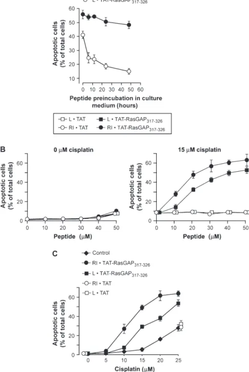
osteosarcoma U2OS cells ( 12 ) to undergo apoptosis [assessed by scoring the percent-age of cells displaying pycnotic nuclei ( 9 )] decreased as a function of increasing pep-tide preincubation time in serum-contain-ing medium, suggestserum-contain-ing that this peptide is sensitive to degradation by serum proteases ( Figure 1, A ). This sensitivity to degrada-tion is likely to adversely affect the antitu-mor activity of the peptide in vivo. One way to render a peptide more resistant to proteolytic degradation is to convert its amino acids from the natural l -form to the protease-resistant d -form. To best mimic the structure of the natural peptide, the sequence of the d -peptide is generally inverted, generating the so-called retro-inverso form ( 13 , 14 ). After preincubation in serum-containing medium for up to 2 days, the retro-inverso form of TAT-RasGAP 317 – 326 (RI·TAT-RasGAP 317 – 326 ) showed almost no decrease in its ability to sensitize U2OS cells to cisplatin-induced apoptosis ( Figure 1, A ). This fi nding suggests that RI·TAT-RasGAP 317 – 326 is a more stable compound than L·TAT-RasGAP 317 – 326 . Dose – response analyses indicated that RI·TAT-RasGAP 317 – 326 was more effective than L·TAT-RasGAP 317 – 326 in sensitizing U2OS cells to the apoptosis-inducing action of cisplatin ( Figure 1, B and C ), a likely refl ection of its higher stability com-pared with L·TAT-RasGAP 317 – 326 .
Another parameter that can affect the effi cacy of an anticancer compound in vivo is its rate of clearance from the circulation. We used liquid chromatography coupled with mass spectrometry to examine the persistence of RI·TAT-RasGAP 317 – 326 in blood samples taken at various times from Chemotherapeutic drugs kill tumor cells by
activating apoptosis. If this activation is com-promised (eg, through additional gene muta-tions), a tumor may develop resistance to the anticancer drugs ( 1 , 2 ). Consequently, strate-gies to restore tumor sensitivity to apoptosis are promising approaches for treating can-cer. Much is now known about the mode of action of proteins that regulate cell death in cancer cells. This knowledge has led to the design of peptides that, in vitro, can perturb the resistance of cancer cells to anticancer agents ( 3 , 4 ). However, few studies have examined the effi cacy of those peptides as anticancer compounds in vivo ( 3 ).
We have previously reported that frag-ment N2, a caspase-generated polypeptide from RasGAP, a regulator of Ras- and Rho-dependent pathways, strongly sensi-tizes tumor cells, but not normal cells, to genotoxin-induced apoptosis ( 5 , 6 ). The tumor sensitization property of the N2 frag-ment is contained within a 10 – amino acid sequence (amino acids 317 – 326) ( 7 ). A syn-thetic peptide (called TAT-RasGAP 317 – 326 ) corresponding to this sequence fused to a
cell-permeable peptide derived from the human immunodefi ciency virus TAT pro-tein was found to effi ciently increase the extent of apoptosis induced by a variety of genotoxins ( 7 ) and other anticancer treat-ments ( 8 ) in several tumor cell lines. Although it is now known that tumor cells must be able to activate the p53 and p53-upregulated modulator of apoptosis (PUMA) pathway for them to undergo TAT-RasGAP 317 – 326 peptide – mediated sensitization to genotoxin-induced apoptosis ( 9 ), the molecular mechanisms underlying the apoptosis sen-sitization property of TAT-RasGAP 317 – 326 remain to be characterized in detail. Here, we assessed the ability of TAT-RasGAP 317 – 326 to increase the effi cacy of genotoxins in an in vivo context.
We fi rst examined the functional stabil-ity of the peptide in biological fl uids because it has been shown that peptides that are susceptible to proteolytic degradation can rapidly lose their function ( 10 , 11 ). Indeed, the ability of the natural l -form of TAT-RasGAP 317 – 326 (L·TAT-RasGAP 317 – 326 ) to sen-sitize cisplatin-treated human p53-positive
Effect of RasGAP N2 Fragment – Derived
Peptide on Tumor Growth in Mice
David Michod , Alessandro Annibaldi , Stephan Schaefer , Christine Dapples , Bertrand Rochat , Christian Widmann
Peptides that interfere with the natural resistance of cancer cells to genotoxin-induced apoptosis may improve the efficacy of anticancer regimens. We have previ-ously reported that a cell-permeable RasGAP-derived peptide (TAT-RasGAP 317 – 326 ) specifically sensitizes tumor cells to genotoxin-induced apoptosis in vitro. Here, we examined the in vivo stability of a protease-resistant d -form of the peptide,
RI·TAT-RasGAP 317 – 326 , and its effect on tumor growth in nude mice bearing subcutaneous human colon cancer HCT116 xenograft tumors. After intraperitoneal injection, RI·TAT-RasGAP 317 – 326 persisted in the blood of nude mice for more than 1 hour and was detectable in various tissues and subcutaneous tumors. Tumor-bearing mice treated daily for 7 days with RI·TAT-RasGAP 317 – 326 (1.65 mg/kg body weight) and cisplatin (0.5 mg/kg body weight) or doxorubicin (0.25 mg/kg body weight) dis-played reduced tumor growth compared with those treated with either genotoxin alone (n = 5 – 7 mice per group; P = .004 and P = .005, respectively; repeated measures analysis of variance [ANOVA, two-sided]). This ability of the RI·TAT-RasGAP 317 – 326 peptide to enhance the tumor growth inhibitory effect of cisplatin was still observed at peptide doses that were at least 150-fold lower than the dose lethal to 50% of mice. These findings provide the proof of principle that RI·TAT-RasGAP 317 – 326 may be useful for improving the efficacy of chemotherapy in patients.
J Natl Cancer Inst 2009;101: 828 – 832
Affiliations of authors: Department of Physiology
(DM, AA, CW), Department of Pathology (SS, CD), and Quantitative Mass Spectrometry Facility, University Hospital Center (BR), University of Lausanne, Lausanne, Switzerland .
Correspondence to: Christian Widmann, PhD,
Department of Physiology, University of Lausanne, Rue du Bugnon 7/9, 1005 Lausanne, Switzerland (e-mail: christian.widmann@unil.ch ).
See “Funding” and “Notes” following “References.”
DOI: 10.1093/jnci/djp100
© The Author 2009. Published by Oxford University Press. All rights reserved. For Permissions, please e-mail: journals.permissions@oxfordjournals.org.
a 7-week-old female NMRI Nu/Nu (nude) mouse (Janvier, Le Genest-St-Isle, France) that received a single intraperitoneal injec-tion with the peptide at 3.3 mg/kg body weight. All experiments involving mice were approved by the State of Vaud Veterinary Offi ce (Lausanne, Switzerland). RI·TAT-RasGAP 317 – 326 was detectable in the circulation as early as 15 minutes after injection and for at least 90 minutes after injection; peak concentrations were observed 1 hour after the injection of the peptide (Supplementary Figure 1, A and B, available online). By contrast, we could not detect the peptide in blood samples obtained from two mice that were similarly injected with L·TAT-RasGAP 317 – 326 (data not shown), consistent with the notion that the l -peptide is less stable, or more rapidly cleared, than the retro-inverso form. The observation that RI·TAT-RasGAP 317 – 326 could be detected in the circulation for relatively long periods of time suggested that it might be able to reach distant tis-sues, including tumors, after it is injected into mice. To test this possibility, a nude mouse bearing a visible tumor derived from subcutaneous injection of human colon cancer HCT116 cells was injected intrap-eritoneally with RI·TAT-RasGAP 317 – 326 (4.8 mg/kg body weight). The mouse was killed approximately 1 hour after peptide injection, and its liver, lungs, brain, and the tumor were removed and processed for peptide detection by mass spectrometry. RI·TAT-RasGAP 317 – 326 was detectable in the liver and lungs of this mouse but was not found in the brain (Supplemental Figure 1, C, available online), suggesting that it may not cross the blood – brain bar-rier. Importantly, RI·TAT-RasGAP 317 – 326 was also detected in the subcutaneous tumor.
We next evaluated the toxicity of RI·TAT-RasGAP 317 – 326 in nude mice that were injected intraperitoneally twice per week for 4 weeks with RI·TAT-RasGAP 317 – 326 or RI·TAT (a control peptide consisting of the retro-inverso form of the TAT pep-tide) (dose range = 0.8 – 7.2 mg peptide/kg body weight; n = 3 – 4 mice per group). Intraperitoneal injection of RI·TAT-RasGAP 317 – 326 at a dose of 4.8 mg/kg of mouse body weight was lethal (three of the three injected mice died, all within 45 – 60 minutes after the fi rst injection), whereas a dose of 2.4 mg/kg body weight was not,
even after repeated injections (three of the three injected mice were alive after the eighth injection) (Supplementary Table 1, available online). These results indicate that the dose of RI·TAT-RasGAP 317 – 326 that is lethal to 50% of mice (ie, the LD 50 ) is between 2.4 and 4.8 mg/kg body weight. The lethality induced by high doses of RI·TAT-RasGAP 317 – 326 did not seem to be due to the presence of the cell-permeable TAT peptide because none of the three mice injected intraperitoneally with RI·TAT at a dose of 4.8 mg/kg body weight died. Necropsy revealed that the cause of death in mice injected with lethal doses of RI·TAT-RasGAP 317 – 326 appeared to be mas-sive hemorrhages in the lungs and kidneys (Supplementary Figure 1, D, available online). No damage to the liver, pancreas, or spleen was observed (data not shown).
We next conducted two types of in vivo experiments to examine the effect of RI·TAT-RasGAP 317 – 326 on the effi cacy of cisplatin against tumors, in particular under conditions in which the doses of cisplatin had poor or marginal effects. In the fi rst experiment, 7-week-old female nude mice were injected intraperitoneally with 1.5 × 10 6 HCT116 cells. Beginning the
next day, the mice were injected intraperi-toneally twice per week for 4 weeks with phosphate-buffered saline (PBS), cisplatin (1 mg/kg body weight; catalog no. P4394, Sigma-Aldrich, St Louis, MO), RI·TAT-RasGAP 317 – 326 (2.4 mg/kg body weight), or cisplatin plus RI·TAT-RasGAP 317 – 326 (n = 15 – 24 mice per group). At the end of the treatment period, the mice were killed by cervical dislocation, and their tumors were removed, weighed (when possible), and scored empirically as follows: tumors weighing more than 1 mg (score 5), tumors weighing more than 0.5 mg up to 1 mg (score 4), tumors weighing more than 0.1 mg up to 0.5 mg (score 3), vascularized tumors grouped in clumps but too small to be weighed (score 2), or nonvascularized dispersed tumors too small to be weighed (score 1). Mice treated with RI·TAT-RasGAP 317 – 326 plus cisplatin developed sta-tistically signifi cantly fewer tumors with higher scores than mice treated with cispla-tin alone ( P = .032, two-sample location
exact two-sided Wilcoxon test; all statistical analyses were performed using SAS/STAT software v9.1.3, SAS Institute, Inc. (Cary, NC) ( Figure 2, A ). This fi nding indicates
that cisplatin in combination with the pep-tide hampers tumor growth better than cisplatin alone.
In the second type of experiment, HCT116 cells were injected subcutane-ously into the left and right upper fl anks of 7-week-old female nude mice (2.5 × 10 5
cells injected per fl ank). One week later, when visible tumors had developed, the mice were injected intraperitoneally once per day for 1 week with PBS, RI·TAT-RasGAP 317 – 326 (1.65 mg/kg body weight), or cisplatin (0.5 mg/kg body weight) or doxo-rubicin (0.25 mg/kg body weight; catalog no. D1515, Sigma-Aldrich), alone or in combination with RI·TAT-RasGAP 317 – 326 (n = 4 – 7 mice per group). We tested doxo-rubicin because it is structurally different from cisplatin and has a different mode of action ( 15 ). The orthogonal dimensions of C O N T E X T A N D C A V E A T S
Prior knowledge
Peptides that interfere with the natural resistance of cancer cells to genotoxin-induced apoptosis in vitro, such as
TAT-RasGAP 317 – 326 , a cell-permeable
RasGAP-derived peptide, may improve the efficacy of anticancer regimens in vivo.
Study design
Examination of the in vitro and in vivo
sta-bility of a protease-resistant d -form of the
peptide, RI·TAT-RasGAP 317 – 326 , and its effect
on tumor growth in nude mice bearing sub-cutaneous human colon cancer HCT116 xenograft tumors.
Contribution
RI·TAT-RasGAP 317 – 326 was stable in
biologi-cal fluids, and after injection into mice, it persisted in the bloodstream for more than 1 hour, reached distant tissues and subcu-taneous tumors, was effective at doses at least 150-fold below the dose lethal to 50% of mice, and improved the efficacy of genotoxins.
Implications
RI·TAT-RasGAP 317 – 326 may be useful for
improving the efficacy of chemotherapy in patients.
Limitations
Tumors in nude (ie, immunocompromised) mice may not behave the same as synge-neic tumors in immunocompetent mice.
Figure 1 . Effects of the L and the RI forms of TAT-RasGAP 317 – 326 on cisplatin-induced apoptosis in U2OS cells. Sequences of the peptide used in the fi gure: RI·TAT-RasGAP 317 – 326 , DTRLNTVWM-WGGRRRQRRKKRG ( d -amino acid); L·TAT-RasGAP 317 – 326 , GRKKRRQRRRGGWMWVTNLRTD ( l -amino acid); RI·TAT, RRRQRRKKRG ( d -amino acid); and L·TAT, GRKKRRQRRR ( l -amino acid). Apoptosis was assessed by counting the cells that displayed a pycnotic nucleus. The results presented in each panel correspond to the mean percentage of apoptotic cells for three independent experiments; error bars correspond to 95% confi dence intervals. A ) Peptide functional stability assay. U2OS cells were incubated for 20 hours with 15 µ M cisplatin plus the indicated peptides, which were previously preincubated for the indicated times in Dulbecco ’ s modifi ed Eagle medium containing 10% newborn calf serum. Apoptosis was then assessed. B ) Tumor cell sensitization in response to increasing peptide concentrations. U2OS cells were treated for 20 hours with increasing concentrations of the indi-cated peptides in the absence ( left panel ) or pres-ence ( right panel ) of 15 µ M cisplatin. Apoptosis was then assessed. C ) Tumor cell sensitization induced by the peptides in response to increasing cisplatin concentrations. U2OS cells were treated for 20 hours without (Control) or with (20 µ M fi nal concen-tration) the indicated peptides in the presence of increasing concentrations of cisplatin. Apoptosis was then assessed. The values for L·TAT and RI·TAT are slightly offset for better visualization. L = natural; RI = retro-inverso.
the tumors were measured daily with the use of a caliper on isofl urane-anesthetized mice, beginning on the fi rst day of treat-ment injection (day 0, ie, when tumors were just visible), for up to 13 days. Tumor volume was calculated as [(largest orthogo-nal dimension) 2 × (smallest orthogonal
dimension) × ( /6)]. When a mouse devel-oped tumors on both fl anks, the volumes of the two tumors were averaged to get a sin-gle value for that mouse to allow statistical comparisons with mice that developed only one tumor. All mice in a given experiment
were killed by cervical dislocation while still anesthetized when the largest tumors exceeded a specifi c threshold volume (500 – 1000 mm 3 , depending on the experiment).
Mice treated with RI·TAT-RasGAP 317 – 326 plus either cisplatin or doxorubicin devel-oped statistically signifi cantly smaller tumors than mice treated with RI·TAT-RasGAP 317 – 326 ( P = .005 and P < .001, respec-tively) or with the respective genotoxin alone ( P = .004 and P = .005, respectively;
repeated measures analysis of variance [ANOVA, two-sided]) ( Figure 2, B and C ).
Compared with PBS, RI·TAT-RasGAP 317 –
326 by itself had no effect on tumor growth
( P = .89 and P = .41 for the cisplatin and
doxorubicin experiments, respectively; repeated measures ANOVA [two-sided]). These data demonstrate that RI·TAT-RasGAP 317 – 326 can inhibit the growth of already formed and detectable tumors when used in combination with a genotoxin (cis-platin or doxorubicin).
Given our fi nding that one injection of a lethal dose of TAT-RasGAP 317 – 326 led to hemorrhages in lungs and kidneys
Figure 2 . In vivo chemosensitizing effi cacy of RI·TAT-RasGAP
317 – 326 . A ) Effect of RI·TAT-RasGAP 317 – 326 on
intraperi-toneal tumors. Nude mice (7-week-old females) were injected with 1.5 million HCT116 cells intraperitoneally. The following day and thereafter twice a week, the mice were injected intraperitoneally with PBS, 2.4 mg/kg of RI·TAT-RasGAP 317 – 326 , 1 mg/kg cisplatin, or a combination of cisplatin and RI·TAT-RasGAP
317 – 326 . After 28 days, the
mice were killed and the development of the tumors scored (score 5, tumors weighing more than 1 mg; score 4, tumors weighing between 0.5 and 1 mg; score 3, tumors weighing between 0.1 and 0.5 mg; score 2, vascu-larized tumors grouped in clumps but too small to be weighed; and score 1, nonvascularized dispersed tumors too small to be weighed); examples of tumors with scores 1 and 2 are shown on the right in ( A ). Mice that did not show any presence of tumors (ie, mice in which the tumors did not “take”) were excluded from the analysis (PBS: four mice excluded of 15 injected; RI·TAT-RasGAP
317 – 326 : three mice excluded of 15 injected; cisplatin:
one mouse excluded of 21 injected; cisplatin + RI·TAT-RasGAP
317 – 326 : four mice excluded of 24 injected); the
pro-portion of excluded mice did not differ statistically signifi cantly among the groups (Fisher exact test, P = .225). The dots represent individual mice. Statistical analysis for the difference between the cisplatin and the cisplatin + RI·TAT-RasGAP 317 – 326 groups was performed with a two-sample location exact two-sided Wilcoxon test. B and C ) Cisplatin- and doxorubicin-sensitizing effect of RI·TAT-RasGAP
317 – 326 on subcutaneous tumors. Nude mice
(7-week-old females) were injected subcutaneously with 250 000 HCT116 cells on the left and right upper fl anks. Seven days later, the mice that developed visible tumors were injected each day for 7 consecutive days (in red in the fi gure) with PBS (300 µ L), RI·TAT-RasGAP 317 – 326 (1.65 mg/kg in 300 µ L PBS), cisplatin (0.5 mg/kg in 300 µ L PBS), or RI·TAT-RasGAP 317 – 326 plus cisplatin ( B ). In the experi-ment described in ( C ), cisplatin was replaced with 0.25 mg/kg doxorubicin. Tumor volume was plotted as a func-tion of time (for mice that developed tumors on both fl anks, the two tumor volumes were averaged). The num-ber of mice analyzed is indicated in the fi gure. Mean val-ues are plotted, and error bars correspond to 95% confi dence intervals. D ) Dose – response analysis of the in vivo sensitizing effect of the peptide. Nude mice were treated and analyzed as in ( B ) but with the conditions indicated in the fi gure. E ) Peptide injection frequency. Nude mice were injected subcutaneously with 500 000 HCT116 cells and treated as described in ( D ), except that 0.16 mg/kg body weight RI·TAT-RasGAP
317 – 326 was injected
on the indicated days [ open diamonds , triangles , and squares ; cisplatin was injected every day as in ( D )]. The P values in ( B – E ) are from repeated measures ANOVA (two-sided). ANOVA = analysis of variance; PBS = phosphate-buffered saline; RI = retro-inverso.
(Supplementary Figure 1, D, available online ), we assessed whether the nonlethal dose of RI·TAT-RasGAP 317 – 326 that enhanced the sensitivity of tumors to cis-platin and doxorubicin in the previous experiment had any effect on these organs. Non – tumor-bearing mice were injected with PBS, RI·TAT-RasGAP 317 – 326 , cispla-tin, or RI·TAT-RasGAP 317 – 326 plus cisplatin as described above (n = 2 mice per group); the mice were killed by cervical dislocation after the last injection (ie, 8 days after the fi rst injection), and histological sections of
their lungs and kidneys were prepared. We observed no damage to these organs in any of the mice (Supplementary Figure 1, E, available online). Thus, a nonlethal dose of RI·TAT-RasGAP 317 – 326 (1.65 mg/kg body weight), which was only approximately two- to three fold lower than the lethal dose ( ~ 5 mg/kg body weight), did not appear to cause visible alterations in the lungs or kidneys, even when it was admin-istered in the presence of cisplatin.
To examine if RI·TAT-RasGAP 317 – 326 doses lower than 1.65 mg/kg body weight
could exert a genotoxin-sensitizing effect on tumor growth in mice, we used the experi-mental design described above but with decreasing doses of injected RI·TAT-RasGAP 317 – 326 and with a dose of cisplatin that is at the threshold of inducing an inhibi-tory effect on tumor growth by itself. This dose of cisplatin was determined by inject-ing mice with various doses of cisplatin only (range = 0.005 – 1 mg/kg body weight) using the injection protocol shown in Figure 2, B . The highest dose of cisplatin tested that did not reduce tumor growth was found to be
0.1 mg/kg body weight (data not shown). HCT116 tumor – bearing mice were treated with PBS or RI·TAT-RasGAP 317 – 326 (at doses of 0, 0.16, 0.048, or 0.160 mg/kg body weight) combined with cisplatin at 0.1 mg/ kg body weight (n = 4 – 8 mice per group). Cisplatin alone (0.1 mg/kg body weight) had no effect on the growth of HCT116 tumors compared with PBS ( P = .77; repeated mea-sures ANOVA [two-sided]) ( Figure 2, D ). However, cisplatin plus RI·TAT-RasGAP 317 –
326 at 0.16 mg/kg body weight effi ciently
inhibited tumor growth compared with cis-platin alone ( P = .03; repeated measures
ANOVA [two-sided]). Importantly, RI·TAT-RasGAP 317 – 326 at 0.016 mg/kg body weight, a dose more than 150-fold lower than the estimated LD 50 (ie, between 2.4 and 4.8 mg/kg body weight), also improved the effi cacy of cisplatin to inhibit the growth of HCT116 tumors compared with cisplatin alone ( P = .004; repeated measures ANOVA [two-sided]).
We also examined the effect of varying the frequency of RI·TAT-RasGAP 317 – 326 peptide injection on tumor growth. HCT116 tumor – bearing mice were injected for 1 week with RI·TAT-RasGAP 317 – 326 (0.16 mg/ kg body weight), daily, every other day, or every 3.5 days, in combination with a daily injection of cisplatin (0.1 mg/kg body weight) (n = 3 – 7 mice per group). Control mice were injected daily with PBS or cispla-tin alone. Tumor volumes were measured every 3 – 4 days. Injection of the peptide every other day sensitized tumor cells to cisplatin (every-other-day peptide injection + cisplatin vs cisplatin only: P = .02; repeated measures ANOVA [two-sided]) as effi ciently as daily injection of the peptide (every-oth-er-day peptide injection + cisplatin vs daily peptide injection + cisplatin: P = .94; repeated measures ANOVA [two-sided]). Injection of the peptide every 3.5 days also sensitized the tumors somewhat to cisplatin (every 3.5 days peptide injection + cisplatin vs cisplatin only: P = .15; repeated measures ANOVA
[two-sided]) but with reduced effi ciency compared with daily or every-other-day injections of the peptide. These results indi-cate that RI·TAT-RasGAP 317 – 326 does not need to be injected every day along with cisplatin to exert its sensitization property, probably because of its increased resistance to degradation in biological fl uids.
A limitation of this study is that tumors in immunocompromised mouse models
(eg, nude mice) may not behave as synge-neic tumors in immunocompetent mice. Indeed, there is now clear evidence that the immune system positively modulates some anticancer therapies ( 16 ). For example, doxorubicin-induced death of colon carci-noma cells implanted in syngeneic mice elicits an immune response that favors the elimination of the tumor cells ( 17 ). The effi cacy of RI·TAT-RasGAP 317 – 326 against human tumors implanted in nude mice might therefore be attenuated by the lack of an intact immune system in nude mice.
The properties of RI·TAT-RasGAP 317 – 326 described here in mouse tumor models indi-cate that this peptide has the potential to be used in humans to sensitize tumor cells to genotoxin treatments (ie, to enhance the antitumor effect of genotoxins): it is stable in biological fl uids, it persists in the blood-stream for more than 1 hour after intraperi-toneal injection, it reaches distant tissues and organs (including subcutaneous tumors), its effi cacious doses are at least 100-fold below the LD 50 , and it greatly improves the effi cacy of genotoxins. To our knowledge, this peptide is the only compound that has been shown to improve the effi cacy of geno-toxins and that behaves strictly as a chemo-sensitizer, that is, it has no effect on tumors by itself ( 4 ). This compound would therefore have the potential to improve the effi -cacy of chemotherapeutic agents that are currently used in the clinic, particularly in situations in which doses of genotoxin have to be lowered to reduce side effects.
References
1. Debatin K-M . Apoptosis pathways in cancer and cancer therapy . Cancer Immunol Immunother . 2004 ; 53 ( 3 ): 153 – 159 .
2. Schmitt CA , Lowe SW . Apoptosis and ther-apy . J Pathol . 1999 ; 187 ( 1 ): 127 – 137 . 3. Mendoza FJ , Espino PS , Cann KL , Bristow
N , McCrea K , Los M . Anti-tumor chemo-therapy utilizing peptide-based approaches — apoptotic pathways, kinases, and proteasome as targets . Arch Immunol Ther Exp. (Warsz) . 2005 ; 53 ( 1 ): 47 – 60 .
4. Michod D , Widmann C . DNA-damage sensi-tizers: potential new therapeutical tools to improve chemotherapy . Crit Rev Oncol
Hematol . 2007 ; 63 ( 2 ): 160 – 171 .
5. Yang J-Y , Widmann C . Antiapoptotic signaling generated by caspase-induced cleavage of RasGAP . Mol Cell Biol . 2001 ; 21 ( 23 ): 5346 – 5358 . 6. Yang J-Y , Walicki J , Michod D , Dubuis G , Widmann C . Impaired Akt activity down-modulation, caspase-3 activation, and apopto-sis in cells expressing a caspase-reapopto-sistant mutant
of RasGAP at position 157 . Mol Biol Cell .
2005 ; 16 ( 8 ) : 3511 – 3520 .
7. Michod D , Yang JY , Chen J , Bonny C , Widmann C . A RasGAP-derived cell perme-able peptide potently enhances genotoxin-induced cytotoxicity in tumor cells . Oncogene . 2004 ; 23 ( 55 ): 8971 – 8978 .
8. Rehm M , Huber HJ , Dussmann H , Prehn JH . Systems analysis of effector caspase activation and its control by X-linked inhibitor of apopto-sis protein . EMBO J . 2006 ; 25 ( 18 ): 4338 – 4349 . 9. Michod D , Widmann C . TAT-RasGAP 317-326
requires p53 and PUMA to sensitize tumor cells to genotoxins . Mol Cancer Res . 2007 ; 5 ( 5 ): 497 – 507 .
10. Widmann C , Maryanski JL , Romero P , Corradin G . Differential stability of antigenic MHC class I-restricted synthetic peptides . J Immunol . 1991 ; 147 ( 11 ) : 3745 – 3751 . 11. Blanchet JS , Valmori D , Dufau I , et al . A
new generation of Melan-A/MART-1 peptides that fulfi ll both increased immunogenicity and high resistance to biodegradation: implication for molecular anti-melanoma immunotherapy . J Immunol . 2001 ; 167 ( 10 ): 5852 – 5861 . 12. Ponten J , Saksela E . Two established in vitro
cell lines from human mesenchymal tumours . Int J Cancer . 1967 ; 2 ( 5 ): 434 – 447 .
13. Chorev M , Goodman M . Recent develop-ments in retro peptides and proteins — an ongoing topochemical exploration . Trends
Biotechnol . 1995 ; 13 ( 10 ): 438 – 445 .
14. Fischer PM . The design, synthesis and appli-cation of stereochemical and directional pep-tide isomers: a critical review . Curr Protein
Pept Sci . 2003 ; 4 ( 5 ): 339 – 356 .
15. Momparler RL , Karon M , Siegel SE , Avila F . Effect of adriamycin on DNA, RNA, and pro-tein synthesis in cell-free systems and intact cells . Cancer Res . 1976 ; 36 ( 8 ): 2891 – 2895 . 16. de la Cruz-Merino L , Grande-Pulido E ,
Alberto-Tamarit A , Codes-Manuel de Villena ME . Cancer and immune response: old and new evidence for future challenges . Oncologist . 2008 ; 13 ( 12 ): 1246 – 1254 .
17. Casares N , Pequignot MO , Tesniere A , et al . Caspase-dependent immunogenicity of doxo-rubicin-induced tumor cell death . J Exp Med . 2005 ; 202 ( 12 ): 1691 – 1701 .
Funding
Swiss National Science Foundation ( 31003A_119876 to C.W.). D. Michod and C. Widmann are coinven-tors of the TAT-RasGAP 317 – 326 compound as a geno-toxin sensitizer (patent owned by the University of Lausanne) and may receive royalties from patent licensing if the compound is commercialized. The authors did not receive any specifi c support from private or institutional bodies that might have com-mercial interests in the patented product .
Notes
D. Michod and A. Annibaldi contributed equally to this work.
We thank Dr Rudolf Kraftsik for the statistical analyses of the data.
Manuscript received April 30 , 2008 ; revised February 19 , 2009 ; accepted March 27 , 2009 .