Insular volume abnormalities associated with
different transition probabilities to psychosis
R. Smieskova1,2, P. Fusar-Poli3, J. Aston1, A. Simon4,5, K. Bendfeldt2, C. Lenz6, R.-D. Stieglitz1, P. McGuire3, A. Riecher-Ro¨ssler1and S. J. Borgwardt1,2,3*1Department of Psychiatry, University of Basel, Switzerland 2Medical Image Analysis Centre, University of Basel, Switzerland
3Department of Psychosis Studies, King’s College London, Institute of Psychiatry, London, UK 4Specialized Out-patient Service for Early Psychosis, Department of Psychiatry, Bruderholz, Switzerland 5University Hospital of Psychiatry, University of Bern, Switzerland
6Radiological Physics, University Hospital Basel, Switzerland
Background. Although individuals vulnerable to psychosis show brain volumetric abnormalities, structural alterations underlying different probabilities for later transition are unknown. The present study addresses this issue by means of voxel-based morphometry (VBM).
Method. We investigated grey matter volume (GMV) abnormalities by comparing four neuroleptic-free groups : individuals with first episode of psychosis (FEP) and with at-risk mental state (ARMS), with either long-term (ARMS-LT) or short-term ARMS (ARMS-ST), compared to the healthy control (HC) group. Using three-dimensional (3D) magnetic resonance imaging (MRI), we examined 16 FEP, 31 ARMS, clinically followed up for on average 3 months (ARMS-ST, n=18) and 4.5 years (ARMS-LT, n=13), and 19 HC.
Results. The ARMS-ST group showed less GMV in the right and left insula compared to the ARMS-LT (Cohen’s d 1.67) and FEP groups (Cohen’s d 1.81) respectively. These GMV differences were correlated positively with global functioning in the whole ARMS group. Insular alterations were associated with negative symptomatology in the whole ARMS group, and also with hallucinations in the ARMS-ST and ARMS-LT subgroups. We found a significant effect of previous antipsychotic medication use on GMV abnormalities in the FEP group.
Conclusions. GMV abnormalities in subjects at high clinical risk for psychosis are associated with negative and positive psychotic symptoms, and global functioning. Alterations in the right insula are associated with a higher risk for transition to psychosis, and thus may be related to different transition probabilities.
Received 2 August 2011 ; Revised 25 October 2011 ; Accepted 26 October 2011 ; First published online 30 November 2011 Key words: At-risk mental state (ARMS), insula, magnetic resonance imaging (MRI), psychosis, transition.
Introduction
A growing body of magnetic resonance imaging (MRI) evidence suggests that subjects at high clinical risk for psychosis show structural abnormalities in the frontal, insular and temporal regions (Pantelis et al. 2003 ; Borgwardt et al. 2007a, b, 2008 ; Meisenzahl et al. 2008 ; Koutsouleris et al. 2009 ; Witthaus et al. 2009). Several MRI studies have examined whether there are specific neuroanatomical differences between high-risk sub-jects who subsequently develop psychosis and those who do not (Pantelis et al. 2003 ; Borgwardt et al. 2007a, b, 2008 ; Koutsouleris et al. 2009). Structural deficits associated with transition to psychosis can be
seen as vulnerability markers of very early stages of psychosis (Smieskova et al. 2010) and are of crucial relevance to the field of preventive interventions in psychosis.
Early clinical intervention in psychosis has recently become a major objective of mental health services. Research at this stage is a potential way of investigat-ing the mechanisms of underlyinvestigat-ing psychosis, as the same individuals can be studied before and after the onset of psychosis, often with minimal confounding effects of previous antipsychotic treatment. The identification of a clinical risk syndrome, an at-risk mental state (ARMS), that reflects an ultra-high clinical risk predisposition to psychosis is fundamental to both clinical and research work in this area. Most transitions to psychosis in ARMS individuals have been found in the first 2 years after baseline assess-ment and are much less probable afterwards (Yung et al. 2007 ; Riecher-Ro¨ssler et al. 2009), suggesting The online version of this article is published within an Open Access environment subject to the conditions of the Creative Commons
* Address for correspondence : Dr S. J. Borgwardt, Professor of Neuropsychiatry, Department of Psychiatry, University of Basel, c/o University Hospital Basel, Petersgraben 4, 4031 Basel, Switzerland.
rapid dynamic anatomical and neurophysiological changes in the same patients during the pre-psychotic phases. Independent studies have confirmed complex neurophysiological changes underlying the prodro-mal psychotic phases involving not only brain structure but also disrupted dopaminergic and gluta-matergic neurotransmission (Jessen et al. 2006 ; Hurlemann et al. 2008). However, to the best of our knowledge no study has explicitly addressed potential neurobiological markers of different levels of risk across the prodromal phase. We therefore investigated separately ARMS individuals with a short or long duration of the ARMS. All these individuals fulfilled the ARMS criteria [similar to the Personal Assessment and Crisis Evaluation Clinic (PACE) criteria] at the time of scanning. In the first group [short-term dur-ation ARMS (ARMS-ST)] the scan was obtained di-rectly after referral to our specialized early detection clinic, that is at the time of ascertainment of the ARMS (within 3 months on average). According to published data, the probability of developing psychosis in this group is 20–40 % over 2 years (Yung et al. 1998 ; Riecher-Ro¨ssler et al. 2007, 2009). In the second group [long-term duration ARMS (ARMS-LT)] the scan was obtained on average after 4.5 years of follow-up. Although at the time of the scan this group was still on the risk continuum to develop psychosis, according to the published data (Yung et al. 2007 ; Cannon et al. 2008 ; Riecher-Ro¨ssler et al. 2009), they had a lower probability of developing subsequent psychosis when compared to ARMS-ST. Importantly, most of the ARMS individuals who encountered transition (90.5 %) did so in the first 2 years after their ARMS was ascertained (Riecher-Ro¨ssler et al. 2009). After these 2 years, only 3 % of included ARMS individuals developed psychosis (Riecher-Ro¨ssler et al. 2009). Some studies have reported that the transition rate of newly referred individuals is declining in recent years (Haroun et al. 2006 ; Yung et al. 2007 ; Frommann et al. 2010). Thus, the transition rate in high-risk popu-lations declined from 31 % during the first 2 years as published in 2003 (Pantelis et al. 2003) to 16 % as published 5 years later (Yung et al. 2008). There may be some protective mechanism in the high-risk in-dividuals who compensate for risk factors and remain without subsequent transition to psychosis.
Our aim was to examine the neuroanatomical brain abnormalities associated with higher or lower tran-sition probability and the reduced risk of developing psychosis. In a previous multimodal analysis we found an association between functional and struc-tural abnormalities in groups with different transition probabilities to psychosis (Smieskova et al. 2011). Psy-chopathological symptoms are often associated with negative symptoms (Lencz et al. 2004 ; Riecher-Ro¨ssler
et al. 2009) and cognitive deficits (Brewer et al. 2006 ; Simon et al. 2007 ; Riecher-Ro¨ssler et al. 2009). Conse-quently, we aimed to clarify the correlation between structural alterations and clinical outcomes during the prodromal phases of psychosis.
Expanding on previous voxel-based morphometry (VBM) studies in ARMS (Pantelis et al. 2003 ; Borgwardt et al. 2007a, 2008 ; Koutsouleris et al. 2009 ; Witthaus et al. 2009), in the current study we in-vestigated an ARMS-LT group with a lower prob-ability of developing psychosis compared to an ARMS-ST group (Yung et al. 2007) with a higher probability of developing psychosis.
We tested the following hypotheses :
(1) The magnitude of volumetric abnormalities would be in parallel with the clinical status (ARMS-LT<ARMS-ST<FEP) compared to the healthy control (HC) group.
(2) Significant correlations between GMV and psy-chotic symptoms or global functioning were expected in the regions showing volumetric differ-ences in ARMS.
Method
Study population
Since 1999, the FEPSY (Fru¨herkennung von Psychosen; early detection of psychosis) Clinic at the Psychiatric Outpatient Department, Psychiatric University Clinics in Basel recruited and followed up ARMS individuals and patients experiencing a first episode of psychosis (FEP) over up to 7 years (Riecher-Ro¨ssler et al. 2009). The entire group of individuals with an ARMS (ARMS-ST and ARMS-LT ; n=31) corresponds to the criteria by Yung et al. (1998) used in previous MRI studies (Pantelis et al. 2003 ; Borgwardt et al. 2007a, b, 2008 ; Sun et al. 2009). The FEP patients (n=16) met operational criteria for FEP according to Breitborde et al. (2009). All included individuals were assessed at baseline and at the time of the MRI scan.
Inclusion to the ARMS group required one or more of the following : (a) ‘ attenuated ’ psychotic symptoms, (b) brief limited intermittent psychotic symptoms (BLIPS) or (c) a first-degree relative with a psychotic disorder plus a marked decline in social or occu-pational functioning (Yung et al. 2004 ; Riecher-Ro¨ssler et al. 2007, 2009).
We divided the ARMS group into two subgroups depending on the duration of the ARMS status since the baseline assessment. Individuals with short-term ARMS (ARMS-ST) fulfilled ARMS criteria for a time period shorter than 2 years and individuals with long-term ARMS (ARMS-LT) were in an ARMS over a follow-up period longer than 2 years. ARSM-ST 1614 R. Smieskova et al.
https:/www.cambridge.org/core/terms. https://doi.org/10.1017/S0033291711002716
individuals had an MRI scan as soon as practicable, on average within 2.64 (S.D.=4.8) months [i.e. 0.22 (S.D.=0.4) years]. ARMS-LT individuals were scanned on average 4.62 (S.D.=2.06) years after first ascertain-ment and made no transition until the point of data analysis (March 2011). At the time of scanning, all the ARMS-ST and ARMS-LT individuals fulfilled the criteria of Yung et al. for the ARMS (Yung et al. 1998 ; Riecher-Ro¨ssler et al. 2008) but had different prob-abilities of developing psychosis (Cannon et al. 2008 ; Yung et al. 2008 ; Riecher-Ro¨ssler et al. 2009).
From the baseline assessment, the ARMS-ST and ARMS-LT subjects were followed up by the clinical service and received standard psychiatric case man-agement. At the time of scanning, all of the ARMS in-dividuals (from both groups) were antipsychotic naive, except for one antipsychotic-free ARMS-ST subject (olanzapine 2.5 mg/day during the period of 9 months, stopped 4 months ago). Furthermore, four of the FEP, six of the ARMS-LT and six of the ARMS-ST were re-ceiving antidepressants at the time of the MRI scan.
The FEP patients (n=16) were defined as subjects who met the operational criteria for ‘ first-episode psychosis ’ (Breitborde et al. 2009). Inclusion required scores ofo4 on the hallucination item or o5 on the unusual thought content, suspiciousness or concep-tual disorganization items of the Brief Psychiatric Rating Scale (BPRS ; Yung et al. 1998). The symptoms must have occurred at least several times a week and persisted for more than 1 week. None of the FEP group were receiving antipsychotic medication at time of scanning. Ten of our FEP patients were antipsychotic naive and six were antipsychotic free ; antipsychotic medication (one risperidone, five aripiprazole, two olanzapine) was stopped 1, 2, 4, 19 and 24 months ago, one subject with unknown withdrawal period.
We recruited healthy volunteers (HC, n=19) from the same geographical area as the other groups. All subjects were representative of the local population of individuals presenting with an ARMS in terms of age, gender, handedness, and alcohol and cannabis con-sumption. These individuals had no current psychi-atric disorder, no history of psychipsychi-atric illness, head trauma, neurological illness, serious medical or surgi-cal illness, or substance abuse, and no family history of any psychiatric disorder as assessed by an experi-enced psychiatrist in a detailed clinical assessment.
We applied the following exclusion criteria to our groups : history of previous psychotic disorder ; psy-chotic symptomatology secondary to an ‘ organic ’ disorder ; substance abuse according to ICD-10 re-search criteria ; psychotic symptomatology associated with an affective psychosis or a borderline personality disorder ; age<18 years; inadequate knowledge of the German language ; and IQ<70.
Instruments
We assessed subjects using the Basel Screening Instrument for Psychosis (BSIP ; Riecher-Ro¨ssler et al. 2007, 2008), the BPRS, the Scale for the Assessment of Negative Symptoms (SANS) and the Global Assessment of Functioning (GAF) at the time of scan-ning. The BSIP evaluates ‘ prodromal ’ symptoms (according to DSM-III-R) occurring in the past 5 years ; non-specific ‘ prodromal ’ signs (Riecher-Ro¨ssler et al. 2007) in the past 2 years ; previous or current psychotic symptoms, psychosocial functioning over the past 5 years, substance dependency ; and psychotic dis-orders among first- and second-degree relatives (Riecher-Ro¨ssler et al. 2008). We obtained current and previous psychotropic medication, alcohol, nicotine, cannabis and other illegal drug consumption using a semi-structured interview adapted from the Early Psychosis Prevention and Intervention Centre (EPPIC) Drug and Alcohol Assessment Schedule (www.eppic. org.au).
Magnetic resonance image acquisition Structural MRI
A three-dimensional (3D) T1-weighted magnetization prepared rapid gradient echo (MPRAGE) sequence was acquired on a 3-T MRI system (Magnetom Verio, Siemens Healthcare, Germany) with sagittal orien-tation based on a 256r256r176 matrix, with 1 mm isotropic spatial resolution, inversion time (TI) of 1000 ms, repetition time (TR) of 2 s and echo time (TE) of 3.4 ms. All of the scans were screened for gross radiological abnormalities by an experienced neuro-radiologist.
Image analysis
We examined group-related differences in grey matter volume (GMV) using SPM8 software (www.fil.ion. ucl.ac.uk/spm ; Wellcome Department of Cognitive Neurology, UK) running under Matlab 7.1 (MathWorks, USA). T1-weighted MPRAGE images were preprocessed using the VBM8 toolbox (http:// dbm.neuro.uni-jena.de/vbm8/). This approach in-volves the creation of a study-specific template and the segmentation of each individual image using the template, with the aim of maximizing accuracy and sensitivity (Yassa & Stark, 2009). We provided the following steps : (1) checking for scanner artefacts and gross anatomical abnormalities for each subject ; (2) using the New Segmentation approach with differ-ent treatmdiffer-ent of the mixing proportions ; (3) using the DARTEL toolbox to produce a high-dimensional nor-malization protocol (Ashburner, 2007) ; (4) checking
Table 1.Demographic and clinical characteristics
Characteristic FEP (n=16) ARMS-ST (n=18) ARMS-LT (n=13) HC (n=19)
Statistics
F/x2 df p
Gender male, n ( %) 12 (75) 14 (77.8) 8 (72.7) 10 (52.6) x2=4.790 3 0.091
Mean age (years), mean (S.D.) 25.13 (4.6) 25.11 (6.2) 24.62 (2.2) 26.58 (4.2) F=0.579 65 0.637
Handedness (left), n ( %) 1 (6.3) 0 0 1 (5.3) x2=1.856 3 0.603
MWT-B IQ, mean (S.D.) 103 (10.7) 112 (14.2) 107 (13.3) 114 (9.7) F=2.710 56 0.054
Years since presentation, mean (S.D.) 1.56 (2.7) 0.22 (0.4) 4.23 (2.0) 0 F=16.914 46 <0.0001
Alcohol currently, n ( %) x2=15.303 6 0.018 None 4 (25) 2 (11.1) 0 0 Moderate 11 (68.8) 7 (38.9) 8 (61.5) 14 (73.7) Drunkenness 11 (6.3) 9 (50.0) 5 (38.5) 5 (26.3) Cannabis, n ( %) 6 (37.5) 7 (38.89) 4 (30.8) 4 (21.1) x2=2.107 3 0.550 BPRS total, mean (S.D.) 54.0 (13.4) 40.1 (8.1) 32.8 (6.8) 24.6 (1.2) F=37.200 61 <0.0001 58.56 (15.0)a 39.8 (8.3)a Post-hoc >HC : p<0.0001 >HC : p=0.0001 >HC : p=0.050 <FEP : p=0.001 <FEP : p=0.001 BPRS 9, mean (S.D.) 3.6 (1.4) 2.5 (1.2) 1.6 (0.8) 1 (0) F=20.105 60 <0.0001 3.9 (1.6)a 2.5 (1.3)a Post-hoc >HC : p=0.0001 >HC : p=0.0001 >ARMS-ST : p=0.0001 >ARMS-LT : p=0.019 BPRS 10, mean (S.D.) 3.5 (2.0) 1.94 (1.2) 1.4 (1.0) 1.0 (0) F=12.245 60 <0.0001 4.0 (2.1)a 1.87 (1.2)a Post-hoc >HC : p=0.0001 >HC : p=0.018 >ARMS-ST : p=0.004 >ARMS-LT : p=0.0001 BPRS 11, mean (S.D.) 4.0 (1.7) 2.3 (1.4) 1.4 (0.8) 1.0 (0) F=20.172 60 <0.0001 4.4 (1.9)a 2.2 (1.4)a Post-hoc >HC : p=0.0001 >HC : p=0.008 >ARMS-ST : p=0.001 >ARMS-LT : p=0.019 BPRS 15, mean (S.D.) 2.2 (1.3) 1.7 (0.9) 1.3 (0.9) 1.0 (0) F=4.777 60 <0.005 2.2 (1.6)a 1.6 (0.9)a Post-hoc >HC : p=0.003 APS, mean (S.D.) 13.2 (4.1) 8.4 (3.3) 5.8 (2.5) 4.0 (0) F=32.304 60 <0.0001 Post-hoc >HC : p=0.0001 >HC : p=0.0001 <FEP : p=0.0001 <FEP : p=0.0001 1616 R. Smieskova et al. https:/www.cambridge.org/core/terms . https://doi.org/10.1017/S0033291711002716 Downloaded from https:/www.cambridge.org/core
. University of Basel Library
, on
30 May 2017 at 14:45:43
for homogeneity across the sample ; and (5) using 8-mm standard smoothing. We identified two subjects with a mean covariance below two standard deviations and screened their volumes carefully, but found no artefacts and the quality of images was reasonable. We repeated the analyses without these two subjects, with the same results, and therefore decided not to exclude them from the analysis. After this pre-processing, we obtained segmented, normalized and smoothed data that were used for the statistical analysis.
We performed an analysis of covariance (ANCOVA) to compare grey matter images from all four groups (FEP, ARMS-ST, ARMS-LT, HC). We modelled age, gender and total GMV as covariates of no interest to reduce the potential impact of these variables on the findings. Statistical significance was assessed at cluster level using the non-stationary ran-dom field theory (Hayasaka et al. 2004). The first step of this cluster-level inference strategy consisted of identifying spatially contiguous voxels at a threshold of p<0.001, uncorrected (cluster-forming threshold) (Petersson et al. 1999). Statistical inferences were then made at p<0.05 after family-wise error (FWE) correc-tion. Additionally, we calculated effect size (Cohen’s d) for volumetric differences in bilateral insular volumes.
To label the regions of brain activation, Montreal Neurological Institute (MNI) coordinates were trans-formed into Talairach space (www.ebire.org/hcnlab/ cortical-mapping ; Talairach Daemon software).
Correlation of GMV and clinical data
In addition to the whole-brain VBM analysis, corre-lation analyses were used to examine associations be-tween GMV and scales measuring clinical symptoms (positive, negative and global functioning). We chose two areas with between-group volumetric differences that stayed stable even after exclusion of seven anti-psychotic-free individuals.
To examine the association between positive and negative symptoms and global functioning, we extracted grey matter values from the peak voxels [(39,x19, 7) and (x33, –21, 10)] with sphere 2 mm and performed a series of two-tailed Pearson’s corre-lation analyses using SPSS version 19.0 (SPSS Inc., USA) ; see online Supplementary Table S2. The stat-istical threshold was set at p<0.05.
Statistical analysis of demographic data
We examined clinical and sociodemographic differ-ences between groups using one-way analysis of variance (ANOVA), the F test or the x2
test (Table 1). For post-hoc analyses we used multiple two-sided
SANS total, mean ( S . D .) 28.4 (15.8) 21.0 (13.1) 11.3 (16.6) 0 F = 16.130 61 < 0.0001 Post-hoc > HC : p< 0.0001 > HC : p< 0.0001 < FEP : p= 0.006 > HC : p= 0.100 GAF, mean ( S . D .) 49.6 (17.8) 59.9 (12.9) 76.0 (13.5) 88.6 (4.5) F = 30.990 62 < 0.0001 Post-hoc < HC : p= 0.0001 < HC : p< 0.0001 < HC : p= 0.051 < ARMS-LT : p= 0.0001 < ARMS-LT : p= 0.006 FEP, First episode of psychosis ; ARMS, at-risk mental state ; ARMS-LT, long-term ARMS ; ARMS-ST, short-term ARMS ; M WT-B, multiple choice vocabulary IQ test (Mehrfachwahl-Wortschatz-Intelligenztest, Form B) ; BPRS, Brief Psychiatric Rating Scale (item 9 suspiciousness, 10 hallucinations, 11 unusual thought content , 15 conceptual disorganization) ; APS, attenuated psychotic symptoms ; SANS, Scale for the Assessment of Negative Symptoms ; GAF, Global Assessment of Functioning ; HC, healthy controls ; d f, degrees of freedom. For post-hoc analyses, the Bonferroni correction (at p= 0.05) in SPSS 19.0 was calculated. aValues obtained on repeating the demographic analysis after exclusion of all individuals with a history of antipsychotic medication. Antipsychoti c-naive FEP showed slightly higher BPRS and better GAF scores than FEP with antipsychotic history.
t tests with Bonferroni’s correction. Statistical analyses were performed with SPSS version 19.0 and the level of significance was set at p<0.05.
Results
Clinical and demographic characteristics
There were no significant differences between the FEP, ARMS-ST, ARMS-LT and HC groups in age (p=0.637), gender (p=0.091), handedness (p=0.603), IQ (p=0.054) and current cannabis use (p=0.550; Table 1). The groups of patients did not differ in terms of antidepressant medication : 25 % in the FEP group, 33.3 % in the ARMS-ST group and 46.2 % in ARMS-LT group were receiving antidepressants (p=0.488). We found a significant difference in cigarette smoking (F=3.187, p=0.030). Post-hoc analysis showed that the FEP patients smoked more cigarettes compared to the HC (p=0.012). We repeated demographic analysis after exclusion of all individuals with a history of antipsychotic medication (seven patients : six FEP and one ARMS-ST). The difference in cigarette smoking disappeared when we excluded these seven patients (F=1.614, p=0.197).
There were significant between-group differences in positive and negative symptoms and in global func-tioning. These differences corresponded to the clinical status (FEP>ARMS-ST>ARMS-LT) compared to the HC group. The ARMS-ST group showed lower GAF scores compared to the ARMS-LT group (p=0.004). Both ARMS groups differed significantly in BPRS, SANS and GAF scores compared to the HC group (Table 1). Antipsychotic-naive FEP (FEP after ex-clusion of individuals with a history of antipsychotic medication) showed slightly higher BPRS and lower SANS and GAF compared to the FEP with anti-psychotic history.
GMV differences (VBM results)
GMV differences between groups are presented in Table 2 and group-specific deviations from the overall mean in online Supplementary Table S1.
Psychosis-associated volumetric abnormalities
The FEP individuals compared to the whole group of ARMS showed less GMV in the left uncus, para-hippocampal gyrus and right middle temporal gyrus (Table 2, p<0.05, FWE corrected). Both ARMS-ST and ARMS-LT had more GMV in the left parahippocampal gyrus when tested separately as compared to the FEP group.
In the FEP group we found more GMV in the right thalamus and superior frontal gyrus, and in the left
caudate, insula, superior frontal and cingulate gyrus compared to the entire ARMS group. Compared to the ARMS-ST group, the FEP had more GMV in the bilat-eral insula and superior frontal gyrus. Compared to the ARMS-LT, the FEP showed more GMV in the left caudate and superior frontal gyrus.
Volumetric abnormalities associated with antipsychotic medication
At the time of the study, none of the included in-dividuals were receiving any antipsychotic medi-cation. However, there were seven antipsychotic-free patients (six in the FEP and one in the ARMS-ST group), who had a history of antipsychotic medi-cation, receiving low doses of atypical antipsychotics (five of them aripiprazole, two olanzapine and one risperidone ; the duration of atypical antipsychotic medication use was 1–60 months ; withdrawal 1–24 months ago). We repeated our analysis after exclusion of these seven individuals and found that the majority of volumetric differences had lost their significance. The 10 antipsychotic-naive FEP patients did not show any volumetric abnormalities compared to either the entire ARMS group or the ARMS-LT group or the HC group. Only one cluster in the left insula (x33x21 10) remained significant and showed more GMV on comparing the FEP to the ARMS-ST group (Table 2, p<0.05, FWE corrected) with large effect size (Cohen’s d 1.81).
Consequently, we compared antipsychotic-naive FEP (n=10) and FEP subjects with a previous history of antipsychotic use (n=15: n=6 antipsychotic-free and n=9 currently receiving atypical antipsychotics ; these are not included in this study). We found less GMV in medicated FEP compared to the anti-psychotic-naive FEP group in the right thalamus [(3, x21, x2), p=0.032, FWE corrected] and right parahippocampal gyrus [(33, x1, x15), p=0.041, FWE corrected].
Volumetric abnormalities in the ARMS-LT v. ARMS-ST Compared to the ARMS-LT group, the ARMS-ST group had less GMV in the right insula (Table 2, p<0.05, FWE corrected), with a Cohen’s d of 1.67 (large effect). Conversely, the ARMS-LT had more GMV in the right insula compared to the HC (Table 2, p<0.05, FWE corrected), with an effect size (Cohen’s d) of 1.54 (large effect).
Correlation analyses of GMV and clinical outcomes Within the whole ARMS group (ARMS-LT+ARMS-ST) there was a negative correlation (p<0.05) between negative symptoms (SANS total score) and GMV in 1618 R. Smieskova et al.
https:/www.cambridge.org/core/terms. https://doi.org/10.1017/S0033291711002716
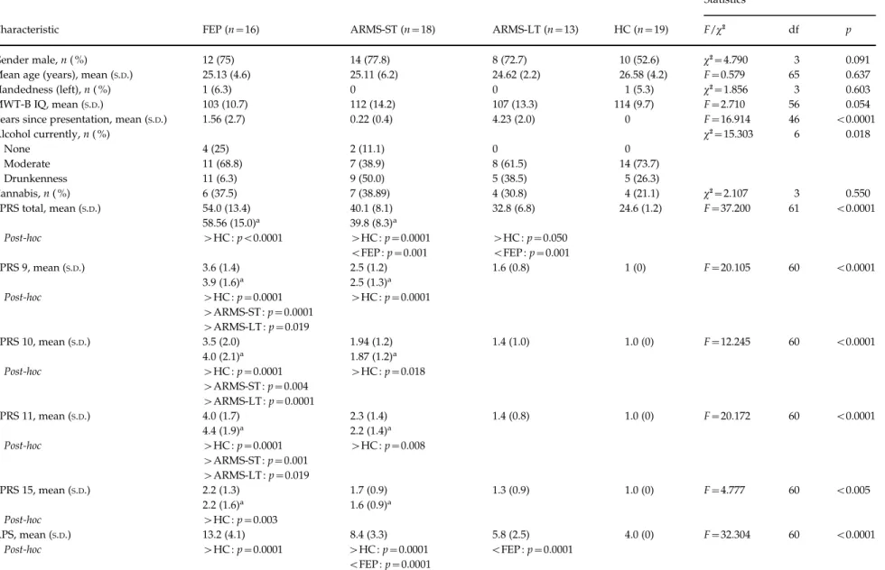
the right insula [(39, x19, 7), Pearson’s correlation coefficientx0.411, Fig. 1] and in the left insula [(x33, x21, 10), Pearson’s correlation coefficient x0.460, Fig. 2, online Supplementary Table S2]. Global func-tioning (GAF total score) correlated positively (p<0.01) with GMV in the right insula [(39, x19, 7), Pearson’s correlation coefficient 0.473, Fig. 1] and in the left insula [(x33, x21, 10), Pearson’s correlation coefficient 0.502, Fig. 2] within the whole ARMS group. We found a positive correlation (p<0.05) with global functional decline in the ARMS-LT group in the right insula [(39, x19, 7), Pearson’s correlation coefficient 0.633 ; p<0.05]. There were no significant correlations between positive symptoms (BPRS total score) and GMV in the whole ARMS group. When the above correlations were repeated within the
ARMS-ST, we found a significant relationship be-tween GMV and hallucinations (p<0.05) in the left insula [(x33, x21, 10), Pearson’s correlation coef-ficient 0.502 and 0.564 respectively, Fig. 2, Sup-plementary Table S2] and conceptual disorganization in the right insula [(39,x19, 7), Pearson’s correlation coefficient x0.533, Fig. 1, Supplementary Table S2]. The ARMS-LT group showed a positive correlation between GMV and hallucinations in the right insula [(39, x19, 7), Pearson’s correlation coefficient 0.720; p<0.01, Fig. 1, Supplementary Table S2].
Discussion
We used structural MRI to examine individuals with FEP, individuals at high clinical risk of psychosis Table 2.Group differences in brain structure
Contrast pFWEcorr. Cluster T MNI (x, y, z) Region
FEP<ARMSa 0.002 6.16 x18, x9, x39 L Uncus 0.007 5.86 x15, x16, x33 L Parahippocampal G (BA 28) 0.047 5.25 36,x1, x35 R MTG (BA 21) FEP>ARMS 0.014 631 5.42 14,x27, x6 R Thalamus 0.005 790 4.68 x18, x19, 21 L Caudate 0.042 483 4.74 18, 21, 58 R SFG 0.049 463 4.92 x44, x15, 16 L Insula (BA 13) 0.005 784 4.49 x6, 20, 61 L SFG 0.027 541 4.11 x9, x12, 42 L CG
FEP<ARMS-STa 0.014 5.64 x18, x9, x31 L Parahippocampal G (BA 35)
FEP>ARMS-ST 0.005 793 5.12 x33, x21, 10 L Insula (BA 13)b
0.015 626 4.84 x34, 44, 27 L SFG 0.020 584 4.81 18, 20, 58 R SFG 0.012 650 4.80 36,x9, 10 R Insula (BA 13) 0.013 640 4.39 8, 15,x23 R medial FG FEP<ARMS-LTa 0.023 5.48 x15, x16, x33 L Parahippocampal G FEP>ARMS-LT 0.003 871 4.81 x6, 23, 60 L SFG 0.013 647 4.39 x20, x21, 21 L Caudate
ARMS-ST<ARMS-LT 0.002 921 4.44 39,x19, 7 R Insula (BA 13)b
ARMS-LT>HC 0.005 790 4.15 34,x27, 22 R Insula (BA 13)b
FEP<HCa 0.019 5.55 14, 15, 43 R CG (BA 32)
FEP>HC 0.040 489 4.58 x42, x15, 16 L Insula (BA 13)
FEP, First episode of psychosis ; ARMS, at-risk mental state ; ARMS-LT, long-term ARMS ; ARMS-ST, short-term ARMS ; FEW, family-wise error ; MNI, Montreal Neurological Institute ; L, left ; R, right ; G, gyrus ; HC, healthy controls ; BA, Brodmann area ; MTG, middle temporal gyrus ; SFG, superior frontal gyrus ; CG, cingulate gyrus.
Group differences in grey matter volume (GMV) calculated from full factorial ANCOVA using SPM8 with the VBM8 toolbox with covariates age, gender, and VBM-GMV with cluster forming p value uncorrected p<0.001.
There were no significant differences in contrasts : ARMS-ST>ARMS-LT, ARMS-LT<HC, (ARMS-ST+ARMS-LT)<HC, (ARMS-ST+ARMS-LT)>HC, ARMS-ST<HC, ARMS-ST>HC.
aDifferences were not significant at cluster level only at voxel level.
We repeated the analysis after exclusion of patients with a history of previous antipsychotic medication (currently anti-psychotic-free subjects : six FEP and one ARMS-ST) to exclude the confounding effect of previous antipsychotic medication. The differences lost their significance in contrasts : FEP<ARMS, FEP>ARMS, FEP<ARMS-ST, FEP<ARMS-LT, FEP>ARMS-LT, and FEP<HC, FEP>HC. The only volumetric difference in the left insula (x33x21 10) stayed significantly different after exclusion of seven antipsychotic-free subjects in contrast FEP>ARMS-ST (p=0.028).
(ARMS) and healthy controls. The ARMS subjects were subcategorized according to different duration of ARMS and probability for transition to psychosis and then scanned with MRI. We compared short- and long-term ARMS individuals who differed in the duration of the ARMS status and consecutively in the probability to transit. The ARMS-ST group showed less right insular GMV than the ARMS-LT group.
Alterations in the right insula related to negative symptomatology and global functioning in the whole ARMS group, and to hallucinations and conceptual disorganization in the ARMS-LT and ARMS-ST sub-groups respectively. Moreover, we found a significant
100 80 60 40 20
SANS and GAF
0 40 50 60 R Insula GMV (×10–2) 70 80 90 100 6 5 4 3 2 hallucinations (BPRS 10) 0 1 40 50 60 R Insula GMV in (×10–2) 70 80 90 100 ARMS-ST ARMS-LT 110 (a) (b) (c)
Fig. 1.Correlation of psychopathology and grey matter volume (GMV) in the right (R) insula. (a) The cluster in the right insula (39,x19, 7) reflects reduced GMV in the short-term at-risk mental state (ARMS-ST) compared to the long-term ARMS (ARMS-LT) group (39,x19, 7) [p<0.05 family-wise error (FEW) corrected]. (b) Correlation of psychopathology and GMV in this cluster across the whole ARMS sample (ARMS-LT+ARMS-ST) [Pearson’s correlation coefficient isx0.411 (p=0.05 two-tailed) for Scale for the Assessment of Negative Symptoms (SANS) and 0.473 (p=0.01 two-tailed) for Global Assessment of Functioning (GAF)]. (c) Positive correlation with hallucinations [Pearson’s correlation coefficient for Brief Psychiatric Rating Scale (BPRS) item 10 (hallucinations) is 0.564 (p=0.05 two-tailed) for ARMS-ST and 0.632 (p=0.05 two-tailed) for ARMS-LT]. The left side of the brain is shown on the left side of the image. 100 80 60 40 20
SANS and GAF
0 40 45 50 55 60 65 70 75 80 L insula GMV (×10–2) 6 5 4 3 2 1 hallucinations (BRPS 10) 0 40 45 50 55 60 65 70 75 ARMS-ST ARMS-LT 80 L insula GMV (×10–2) 10 14 16 (a) (b) (c)
Fig. 2.Correlation of psychopathology and grey matter volume (GMV) in the left (L) insula. (a) The cluster in the left insula (x31x21 10) reflects more GMV in the first episode of psychosis (FEP) compared to the short-term at-risk mental state (ARMS-ST) group [p<0.05 family-wise error (FEW) corrected]. (b) Correlation of psychopathology and GMV in this cluster across the whole ARMS sample [long-term ARMS (ARMS-LT)+ARMS-ST ; Pearson’s correlation coefficient is x0.460 (p=0.05 two-tailed) for Scale for the Assessment of Negative Symptoms (SANS) and 0.502 (p=0.01 two-tailed) for Global Assessment of Functioning (GAF)]. (c) Positive correlation of hallucinations with GMV in the ARMS-ST group [Pearson’s correlation coefficient for Brief Psychiatric Rating Scale (BPRS) item 10 (hallucinations) is 0.564 (p=0.05 two-tailed) for ARMS-ST and 0.632 (p=0.05 two-tailed) for ARMS-LT]. The left side of the brain is shown on the left side of the image.
1620 R. Smieskova et al.
https:/www.cambridge.org/core/terms. https://doi.org/10.1017/S0033291711002716
effect of previous antipsychotic medication use on brain volumetric changes.
Volumetric differences associated with psychosis In line with previous MRI studies we found that FEP patients showed volumetric reductions in the cingulate gyrus relative to controls and in the para-hippocampal gyrus compared to the ARMS groups and HC (for meta analyses, see : Glahn et al. 2008 ; Ellison-Wright & Bullmore, 2010). These alterations can be interpreted as state-marker risk factors for the disease and are qualitatively similar to those seen in patients with chronic schizophrenia (Ellison-Wright et al. 2008). However, we also found more GMV in subcortical regions (thalamus and caudate) in the FEP group compared to the whole ARMS group and to the ARMS-LT subgroup. These differences lost statistical significance after exclusion of individuals with any history of antipsychotic medication. However, not only subcortical regions showed more GMV in the FEP group compared to the ARMS group. The FEP group showed more GMV in bilateral superior frontal and insular regions compared to the ARMS and ARMS-ST groups. The changes in these regions are probably associated with long-lasting effects of previous treat-ment with antipsychotics (Ho et al. 2011). After ex-clusion of all patients who were ever receiving antipsychotics, only one cluster in the left insula re-mained significant. Contrary to our findings, insular atrophy associated with auditory hallucinations, with delusions and with the duration of schizophrenia (Kurth et al. 2010 ; Cascella et al. 2011 ; Nickl-Jockschat et al. 2011) was reported recently. Thus, intriguingly, more GMV in the left insula may be associated with the onset of psychosis, which is in contrast to evidence of reduced GMV in the prodromal phase and the chronic phase of psychosis (Smieskova et al. 2010). However, the increased insula volume in FEP com-pared to ARMS is associated specifically with the transition phase, which may account for this discrep-ancy. This result corresponds to brain swelling de-scribed in fronto-temporal brain regions during exacerbation of psychosis (Garver et al. 2000) and to blood–brain barrier dysfunction in schizophrenia (Uranova et al. 2010). A higher water diffusion co-efficient in the fronto-temporal regions has been re-ported in schizophrenia (Shin et al. 2006). Astrocytes, altered in psychosis, are involved in the permeability of the blood–brain barrier and regulation of water homeostasis (del Zoppo & Hallenbeck, 2000). Thus, altered function in cerebral microvasculature at the ultrastructural level may play an important role in psychosis (Uranova et al. 2010). Additionally, it may be speculated that increases in regional brain volume in
high-risk populations may reflect timely delayed and spatially expanded synaptic pruning as a compensa-tory mechanism for earlier pre/postnatal disruptions (Ettinger et al. 2011).
Volumetric differences associated with previous antipsychotic medication
The effect of antipsychotic medication on brain struc-ture and function has been underestimated for a long period of time. The influence of antipsychotics on brain structure is not entirely clear ; however, anti-psychotics may affect GMV (Dazzan et al. 2005 ; Navari & Dazzan, 2009 ; Ho et al. 2011), as reported specifically for the basal ganglia (Smieskova et al. 2009). Anti-psychotics may contribute to volumetric brain reduc-tions involving multiple subregions, and to specific volumetric increase in subcortical regions, as de-scribed in a longitudinal study with 211 patients (Ho et al. 2011). The patients included in that study were medicated and followed up for an average of 7 years. Our FEP patients received lower doses of anti-psychotics for a shorter time and were not medicated at least 1 month ago. The differences we detected might reflect long-lasting changes in brain volume caused by previous antipsychotic medication but also reparatory/compensatory changes started after the withdrawal of the treatment. The changes associated with antipsychotic medication may be reversible, as shown in a study with healthy volunteers who re-ceived haloperidol injection (Tost et al. 2010). The temporarily diminished GMVs in the striatum re-turned to almost their original size within a day (Tost et al. 2010). However, we are aware that the influence of medication and the process of the disease itself are changing over time. Such complex processes cannot be entirely explained using a cross-sectional design and need to be studied longitudinally.
Volumetric differences associated with vulnerability to transition probability to psychosis
We found no significant differences between HC and the whole ARMS group or ARMS-ST subgroup. Comparing ARMS-LT and HC revealed more GMV in the right insular region. This finding is in line with previous published volumetric abnormalities found in ARMS (Borgwardt et al. 2007b, 2008 ; Meisenzahl et al. 2008 ; Koutsouleris et al. 2009 ; for a review of VBM studies in high-risk subjects see Fusar-Poli et al. 2011a). Moreover, there was a difference in the right insular volume between ARMS-ST and ARMS-LT associated with different transition probabilities. Compared to ARMS-LT individuals, ARMS-ST showed reduced GMV, corresponding to the reductions seen in ARMS
subjects who later transit to psychosis (Borgwardt et al. 2007b ; Takahashi et al. 2009b). Such alterations may represent the neurobiological substrate of dif-ferent transition probabilities within the ARMS. Furthermore, GVM alteration in the insula may be as-sociated directly with the consistent neurofunctional alterations observed in the ARMS during cognitive tasks (Fusar-Poli et al. 2011b).
By comparing the ARMS-LT and HC groups, we revealed more right insular volume than volume re-ductions. This might reflect associations with protect-ing factors in the transition process to psychosis in addition to a different dynamics of underlying pro-tective and/or psychosis-associated changes in the brains of ARMS and FEP individuals. At a symptoms level, we showed that negative symptoms correlated negatively and global functioning positively with volumetric differences within the ARMS subjects. Positive symptoms correlated positively with GMV in ARMS-ST and hallucinations in both ARMS-ST and ARMS-LT groups.
Comparing the ARMS-LT to the HC and to the ARMS-ST subjects revealed more volume in the right insula. Thus the insular GMV increase may represent activity-related hypertrophy (Draganski et al. 2006) secondary to neurofunctional activation of insular areas when experiencing auditory hallucinations (O’Daly et al. 2007).
Repeating the analysis with antipsychotic-naive FEP only, we found more GMV in the left insula compared to the ARMS-ST. However, we cannot dis-tinguish those antipsychotic-naive FEP who had better internal resources from those FEP who were diag-nosed recently (with a higher hallucination score) and had not yet received their antipsychotic medication.
Clinical implications and correlation with clinical outcomes
The mean duration of the ARMS was 4.5 years in the ARMS-LT group ; the probability that any of these subjects will develop psychosis in the future is low (Cannon et al. 2008 ; Riecher-Ro¨ssler et al. 2009). In the ARMS-ST subjects, we expect a transition rate of ap-proximately 30 % (Riecher-Ro¨ssler et al. 2009) in the next 1 to 2 years. The ARMS-ST group with 30 % probability of subsequently developing psychosis allows the investigation of vulnerability and higher transition probability-associated changes in brain ac-tivation compared to the HC. Of note, our ARMS-LT did not differ from the ARMS-ST with respect to age ; this might be because of the small sample sizes and needs further investigation (Whitwell, 2009). However, the differences between ARMS-ST and ARMS-LT did not refer with regard to the effect of
ageing in one of those groups. The structural abnor-malities we observed were not attributable to anti-psychotic treatment, as all of the ARMS individuals were antipsychotic naive except for one ARMS-ST subject who was antipsychotic free at the time of scanning. Consequently, we suggest that the volu-metric difference between these two groups reflects both disrupted brain structure and protective pro-cesses. It remains to be determined whether the structural abnormalities we found are reversible or compensatory in nature. Some recent studies suggest reversibility of prodromal symptoms, especially in an adolescent high-risk group (Simon & Umbricht, 2010). As hypothesized, we found a correlation between volumetric differences and positive and negative psychotic symptoms. Negative symptoms correlated negatively with the GMV in the insula within our whole ARMS population. There was a positive corre-lation of GMV changes with positive symptoms in the ARMS-ST group and with hallucinations in both ARMS-ST and ARMS-LT groups. Of note, there was a negative correlation with conceptual disorganization in the ARMS-LT group. Previously studies have re-ported negative correlations (Crespo-Facorro et al. 2000 ; Pressler et al. 2005), no correlation (Takahashi et al. 2005 ; Crespo-Facorro et al. 2010) and a positive correlation (Takahashi et al. 2009a) between positive symptoms and GMV in schizophrenia patients.
GMV in the bilateral insula correlated positively with global functioning within the whole ARMS group. This is in line with previous behavioural re-ports indicating that the duration of prodromal symptoms predicts the longitudinal GAF scores in subjects with an ARMS (Fusar-Poli et al. 2009).
Limitations
Some limitations of this study should be considered. First, although the ARMS-ST group had a 20–40 % probability of transition to psychosis, there was a high false-positive probability of approximately 60–80 %. Our two groups had a different inherent risk for the development of illness : 20–40 % in the ARMS-ST group and 2–4 % in the ARMS-LT group. A compari-son of those who had converted (that is had 100 % risk of transition) could give us a much more concrete differentiation in risk. However, our ARMS-ST group showed similar volumetric deficits to ARMS in-dividuals who subsequently transited. Thus, the ARMS-ST with volumetric deficits in the right insula at the very beginning of their risk state may be asso-ciated with a higher transition probability subse-quently. If this finding is confirmed in longitudinal studies, insular abnormalities may be an important neuroimaging marker of higher probability to 1622 R. Smieskova et al.
https:/www.cambridge.org/core/terms. https://doi.org/10.1017/S0033291711002716
psychosis. Second, most of the ARMS-ST individuals could become ARMS-LT in several months, and with the aim of revealing the longitudinal differences in the same individuals, we should scan them again in 4.5 years. Third, our healthy volunteers had no family history of any psychiatric disorder among first- and second-degree relatives, as assessed in a detailed questionnaire. Thus, given the very high prevalence rates of mental illness in almost every family, our group could be seen as an exceptional group of heal-thy controls. Fourth, the exact meaning of brain tissue loss or increase in the period of life when the young adult brain is maturating and eliminating superfluous synapses is very complex. In addition, confounding factors including antidepressant medication, smoking, consumption of alcohol and cannabis, and socio-economic status may have influenced the results (Cardenas et al. 2007). However, although alcohol use differed significantly between groups, there was no difference with respect to antidepressant or cannabis use. Fifth, the use of VBM methodology implicates problems of brain registration and the size of the smoothing kernel, especially when relatively small differences are expected. The exact meaning of the volumetric abnormalities (exaggerated dendritic pruning, impaired myelination, apoptosis of neuropil) is not entirely clear. Nevertheless, this technique al-lows comparisons of the entire brain volume at the single voxel level and we used the improved seg-mentation and normalization SPM8 protocols in our analyses.
Conclusions
Structural alterations in subjects at high clinical risk for psychosis are associated with negative and positive psychotic symptoms, and impairment in global func-tioning. Specific GMV abnormalities within the ARMS may distinguish different transition probabilities. In particular, volume loss in the right insula is associated with a higher risk for transition to psychosis.
Note
Supplementary material accompanies this paper on the Journal’s website (http://journals.cambridge.org/ psm).
Acknowledgements
We thank the subjects who took part in this study and also the FEPSY study group for recruitment and management of participants. R.S. and S.J.B. were sup-ported by the Swiss National Science Foundation (No. 3232BO_119382).
Declaration of Interest None.
References
Ashburner J(2007). A fast diffeomorphic image registration algorithm. NeuroImage 38, 95–113.
Borgwardt SJ, McGuire PK, Aston J, Berger G, Dazzan P, Gschwandtner U, Pflu¨ger M, D’Souza M, Radue EW, Riecher-Ro¨ssler A(2007a). Structural brain abnormalities in individuals with an at-risk mental state who later develop psychosis. British Journal of Psychiatry. Supplement 51, s69–s75.
Borgwardt SJ, McGuire PK, Aston J, Gschwandtner U, Pflu¨ger MO, Stieglitz RD, Radue EW, Riecher-Ro¨ssler A (2008). Reductions in frontal, temporal and parietal volume associated with the onset of psychosis. Schizophrenia Research 106, 108–114.
Borgwardt SJ, Riecher-Ro¨ssler A, Dazzan P, Chitnis X, Aston J, Drewe M, Gschwandtner U, Haller S, Pfluger M, Rechsteiner E, D’Souza M, Stieglitz RD, Radue EW, McGuire PK(2007b). Regional gray matter volume abnormalities in the at risk mental state. Biological Psychiatry 61, 1148–1156.
Breitborde NJ, Srihari VH, Woods SW(2009). Review of the operational definition for first-episode psychosis. Early Intervention in Psychiatry 2009, 259–265.
Brewer WJ, Wood SJ, Phillips LJ, Francey SM, Pantelis C, Yung AR, Cornblatt B, McGorry PD(2006). Generalized and specific cognitive performance in clinical high-risk cohorts : a review highlighting potential vulnerability markers for psychosis. Schizophrenia Bulletin 32, 538–555. Cannon TD, Cadenhead K, Cornblatt B, Woods SW,
Addington J, Walker E, Seidman LJ, Perkins D, Tsuang M, McGlashan T, Heinssen R(2008). Prediction of psychosis in youth at high clinical risk : a multisite longitudinal study in North America. Archives of General Psychiatry 65, 28–37.
Cardenas VA, Studholme C, Gazdzinski S, Durazzo TC, Meyerhoff DJ(2007). Deformation-based morphometry of brain changes in alcohol dependence and abstinence. NeuroImage 34, 879–887.
Cascella NG, Gerner GJ, Fieldstone SC, Sawa A, Schretlen DJ(2011). The insula-claustrum region and delusions in schizophrenia. Schizophrenia Research. Published online : 27 August 2011. doi :10.1016/ j.schres.2011.08.004.
Crespo-Facorro B, Kim J, Andreasen NC, O’Leary DS, Bockholt HJ, Magnotta V(2000). Insular cortex abnormalities in schizophrenia : a structural magnetic resonance imaging study of first-episode patients. Schizophrenia Research 46, 35–43.
Crespo-Facorro B, Roiz-Santianez R, Quintero C, Perez-Iglesias R, Tordesillas-Gutierrez D, Mata I, Rodriguez-Sanchez JM, Gutierrez A,
Vazquez-Barquero JL(2010). Insular cortex morphometry in first-episode schizophrenia-spectrum patients : diagnostic specificity and clinical correlations. Journal of Psychiatric Research 44, 314–320.
Dazzan P, Morgan KD, Orr K, Hutchinson G, Chitnis X, Suckling J, Fearon P, McGuire PK, Mallett RM, Jones PB, Leff J, Murray RM(2005). Different effects of typical and atypical antipsychotics on grey matter in first episode psychosis : the AESOP study. Neuropsychopharmacology 30, 765–774.
del Zoppo GJ, Hallenbeck JM(2000). Advances in the vascular pathophysiology of ischemic stroke. Thrombosis Research 98, 73–81.
Draganski B, Gaser C, Kempermann G, Kuhn HG, Winkler J, Buchel C, May A(2006). Temporal and spatial dynamics of brain structure changes during extensive learning. Journal of Neuroscience 26, 6314–6317. Ellison-Wright I, Bullmore E(2010). Anatomy of bipolar
disorder and schizophrenia : a meta-analysis. Schizophrenia Research 117, 1–12.
Ellison-Wright I, Glahn DC, Laird AR, Thelen SM, Bullmore E(2008). The anatomy of first-episode and chronic schizophrenia : an anatomical likelihood estimation meta-analysis. American Journal of Psychiatry 165, 1015–1023.
Ettinger U, Williams SC, Fannon D, Premkumar P, Kuipers E, Moller HJ, Kumari V(2011). Functional magnetic resonance imaging of a parametric working memory task in schizophrenia : relationship with performance and effects of antipsychotic treatment. Psychopharmacology 216, 17–27.
Frommann I, Pukrop R, Brinkmeyer J, Bechdolf A, Ruhrmann S, Berning J, Decker P, Riedel M, Moller HJ, Wolwer W, Gaebel W, Klosterko¨tter J, Maier W, Wagner M(2010). Neuropsychological profiles in different at-risk states of psychosis : executive control impairment in the early – and additional memory dysfunction in the late – prodromal state. Schizophrenia Bulletin 37, 861–873.
Fusar-Poli P, Borgwardt S, Crescini A, Deste G,
Kempton MJ, Lawrie S, McGuire P, Sacchetti E(2011a). Neuroanatomy of vulnerability to psychosis : a voxel-based meta-analysis. Neuroscience and Biobehavioral Reviews 35, 1175–1185.
Fusar-Poli P, Broome MR, Woolley JB, Johns LC, Tabraham P, Bramon E, Valmaggia L, Williams SC, McGuire P(2011b). Altered brain function directly related to structural abnormalities in people at ultra high risk of psychosis : longitudinal VBM-fMRI study. Journal of Psychiatric Research 45, 190–198.
Fusar-Poli P, Meneghelli A, Valmaggia L, Allen P, Galvan F, McGuire P, Cocchi A(2009). Duration of untreated prodromal symptoms and 12-month functional outcome of individuals at risk of psychosis. British Journal of Psychiatry 194, 181–182.
Garver DL, Nair TR, Christensen JD, Holcomb JA, Kingsbury SJ(2000). Brain and ventricle instability during psychotic episodes of the schizophrenias. Schizophrenia Research 44, 11–23.
Glahn DC, Laird AR, Ellison-Wright I, Thelen SM, Robinson JL, Lancaster JL, Bullmore E, Fox PT(2008). Meta-analysis of gray matter anomalies in schizophrenia : application of anatomic likelihood estimation and network analysis. Biological Psychiatry 64, 774–781.
Haroun N, Dunn L, Haroun A, Cadenhead KS(2006). Risk and protection in prodromal schizophrenia : ethical implications for clinical practice and future research. Schizophrenia Bulletin 32, 166–178.
Hayasaka S, Phan KL, Liberzon I, Worsley KJ, Nichols TE (2004). Nonstationary cluster-size inference with random field and permutation methods. NeuroImage 22, 676–687. Ho BC, Andreasen NC, Ziebell S, Pierson R, Magnotta V
(2011). Long-term antipsychotic treatment and brain volumes : a longitudinal study of first-episode
schizophrenia. Archives of General Psychiatry 68, 128–137. Hurlemann R, Matusch A, Kuhn KU, Berning J,
Elmenhorst D, Winz O, Kolsch H, Zilles K, Wagner M, Maier W, Bauer A(2008). 5-HT2A receptor density is decreased in the at-risk mental state. Psychopharmacology 195, 579–590.
Jessen F, Scherk H, Traber F, Theyson S, Berning J, Tepest R, Falkai P, Schild HH, Maier W, Wagner M, Block W(2006). Proton magnetic resonance spectroscopy in subjects at risk for schizophrenia. Schizophrenia Research 87, 81–88.
Koutsouleris N, Schmitt GJ, Gaser C, Bottlender R, Scheuerecker J, McGuire P, Burgermeister B, Born C, Reiser M, Mo¨ller HJ, Meisenzahl EM(2009).
Neuroanatomical correlates of different vulnerability states for psychosis and their clinical outcomes. British Journal of Psychiatry 195, 218–226.
Kurth F, Zilles K, Fox PT, Laird AR, Eickhoff SB(2010). A link between the systems : functional differentiation and integration within the human insula revealed by meta-analysis. Brain Structure and Function 214, 519–534. Lencz T, Smith CW, Auther A, Correll CU, Cornblatt B
(2004). Nonspecific and attenuated negative symptoms in patients at clinical high-risk for schizophrenia.
Schizophrenia Research 68, 37–48.
Meisenzahl EM, Koutsouleris N, Gaser C, Bottlender R, Schmitt GJ, McGuire P, Decker P, Burgermeister B, Born C, Reiser M, Mo¨ller HJ(2008). Structural brain alterations in subjects at high-risk of psychosis : a voxel-based morphometric study. Schizophrenia Research 102, 150–162.
Navari S, Dazzan P(2009). Do antipsychotic drugs affect brain structure ? A systematic and critical review of MRI findings. Psychological Medicine 39, 1763–1777.
Nickl-Jockschat T, Schneider F, Pagel AD, Laird AR, Fox PT, Eickhoff SB(2011). Progressive pathology is functionally linked to the domains of language and emotion : meta-analysis of brain structure changes in schizophrenia patients. European Archives of Psychiatry and Clinical Neuroscience. Published online : 10 September 2011. doi :10.1007/s00406-011-0249-8.
O’Daly OG, Frangou S, Chitnis X, Shergill SS(2007). Brain structural changes in schizophrenia patients with persistent hallucinations. Psychiatry Research 156, 15–21. Pantelis C, Velakoulis D, McGorry PD, Wood SJ,
Suckling J, Phillips LJ, Yung AR, Bullmore ET, Brewer W, Soulsby B, Desmond P, McGuire PK(2003). Neuroanatomical abnormalities before and after onset of psychosis : a cross-sectional and longitudinal MRI comparison. Lancet 361, 281–288.
1624 R. Smieskova et al.
https:/www.cambridge.org/core/terms. https://doi.org/10.1017/S0033291711002716
Petersson KM, Nichols TE, Poline JB, Holmes AP(1999). Statistical limitations in functional neuroimaging. II. Signal detection and statistical inference. Philosophical Transactions of the Royal Society of London. Series B, Biological Sciences 354, 1261–1281.
Pressler M, Nopoulos P, Ho BC, Andreasen NC(2005). Insular cortex abnormalities in schizophrenia : relationship to symptoms and typical neuroleptic exposure. Biological Psychiatry 57, 394–398.
Riecher-Ro¨ssler A, Aston J, Ventura J, Merlo M, Borgwardt S, Gschwandtner U, Stieglitz RD(2008). The Basel Screening Instrument for Psychosis (BSIP) : development, structure, reliability and validity [in German]. Fortschritte der Neurologie-Psychiatrie 76, 207–216. Riecher-Ro¨ssler A, Gschwandtner U, Aston J, Borgwardt S,
Drewe M, Fuhr P, Pflu¨ger M, Radu¨ W, Schindler C, Stieglitz RD(2007). The Basel early-detection-of-psychosis (FEPSY)-study – design and preliminary results. Acta Psychiatrica Scandinavica 115, 114–125.
Riecher-Ro¨ssler A, Pflueger MO, Aston J, Borgwardt SJ, Brewer WJ, Gschwandtner U, Stieglitz RD(2009). Efficacy of using cognitive status in predicting psychosis : a 7-year follow-up. Biological Psychiatry 66, 1023–1030.
Shin YW, Kwon JS, Ha TH, Park HJ, Kim DJ, Hong SB, Moon WJ, Lee JM, Kim IY, Kim SI, Chung EC(2006). Increased water diffusivity in the frontal and temporal cortices of schizophrenic patients. NeuroImage 30, 1285–1291.
Simon AE, Cattapan-Ludewig K, Zmilacher S, Arbach D, Gruber K, Dvorsky DN, Roth B, Isler E, Zimmer A, Umbricht D(2007). Cognitive functioning in the
schizophrenia prodrome. Schizophrenia Bulletin 33, 761–771. Simon AE, Umbricht D(2010). High remission rates from
an initial ultra-high risk state for psychosis. Schizophrenia Research 116, 168–172.
Smieskova R, Allen P, Simon A, Aston J, Bendfeldt K, Drewe J, Gruber K, Gschwandtner U, Klarhoefer M, Lenz C, Scheffler K, Stieglitz RD, Radue EW, McGuire P, Riecher-Ro¨ssler A, Borgwardt SJ(2011). Different duration of at-risk mental state associated with neurofunctional abnormalities. A multimodal imaging study. Human Brain Mapping. Published online : 16 September 2011. doi :10.1002/hbm.21360. Smieskova R, Fusar-Poli P, Allen P, Bendfeldt K,
Stieglitz RD, Drewe J, Radue EW, McGuire PK, Riecher-Ro¨ssler A, Borgwardt SJ(2009). The effects of antipsychotics on the brain : what have we learnt from structural imaging of schizophrenia ? A systematic review. Current Pharmaceutical Design 15, 2535–2549.
Smieskova R, Fusar-Poli P, Allen P, Bendfeldt K, Stieglitz RD, Drewe J, Radue EW, McGuire PK, Riecher-Ro¨ssler A, Borgwardt SJ(2010). Neuroimaging predictors of transition to psychosis – a systematic review and meta-analysis. Neuroscience and Biobehavioral Reviews 34, 1207–1222.
Sun D, Phillips L, Velakoulis D, Yung A, McGorry PD, Wood SJ, van Erp TG, Thompson PM, Toga AW, Cannon TD, Pantelis C(2009). Progressive brain structural
changes mapped as psychosis develops in ‘ at risk ’ individuals. Schizophrenia Research 108, 85–92.
Takahashi T, Suzuki M, Zhou SY, Hagino H, Tanino R, Kawasaki Y, Nohara S, Yamashita I, Seto H, Kurachi M (2005). Volumetric MRI study of the short and long insular cortices in schizophrenia spectrum disorders. Psychiatry Research 138, 209–220.
Takahashi T, Wood SJ, Soulsby B, McGorry PD, Tanino R, Suzuki M, Velakoulis D, Pantelis C(2009a). Follow-up MRI study of the insular cortex in first-episode psychosis and chronic schizophrenia. Schizophrenia Research 108, 49–56.
Takahashi T, Wood SJ, Yung AR, Phillips LJ, Soulsby B, McGorry PD, Tanino R, Zhou SY, Suzuki M,
Velakoulis D, Pantelis C(2009b). Insular cortex gray matter changes in individuals at ultra-high-risk of developing psychosis. Schizophrenia Research 111, 94–102. Tost H, Braus DF, Hakimi S, Ruf M, Vollmert C, Hohn F, Meyer-Lindenberg A(2010). Acute D2 receptor blockade induces rapid, reversible remodeling in human cortical-striatal circuits. Nature Neuroscience 13, 920–922. Uranova NA, Zimina IS, Vikhreva OV, Krukov NO,
Rachmanova VI, Orlovskaya DD(2010). Ultrastructural damage of capillaries in the neocortex in schizophrenia. World Journal of Biological Psychiatry 11, 567–578. Whitwell JL(2009). Voxel-based morphometry : an
automated technique for assessing structural changes in the brain. Journal of Neuroscience 29, 9661–9664. Witthaus H, Kaufmann C, Bohner G, Ozgurdal S,
Gudlowski Y, Gallinat J, Ruhrmann S, Brune M, Heinz A, Klingebiel R, Juckel G(2009). Gray matter abnormalities in subjects at ultra-high risk for schizophrenia and first-episode schizophrenic patients compared to healthy controls. Psychiatry Research 173, 163–169.
Yassa MA, Stark CE(2009). A quantitative evaluation of cross-participant registration techniques for MRI studies of the medial temporal lobe. NeuroImage 44, 319–327.
Yung AR, Nelson B, Stanford C, Simmons MB, Cosgrave EM, Killackey E, Phillips LJ, Bechdolf A, Buckby J, McGorry PD(2008). Validation of ‘ prodromal ’ criteria to detect individuals at ultra high risk of psychosis : 2 year follow-up. Schizophrenia Research 105, 10–17. Yung AR, Phillips LJ, McGorry PD, McFarlane CA,
Francey S, Harrigan S, Patton GC, Jackson HJ(1998). Prediction of psychosis. A step towards indicated prevention of schizophrenia. British Journal of Psychiatry. Supplement 172, 14–20.
Yung AR, Phillips LJ, Yuen HP, McGorry PD(2004). Risk factors for psychosis in an ultra high-risk group : psychopathology and clinical features. Schizophrenia Research 67, 131–142.
Yung AR, Yuen HP, Berger G, Francey S, Hung TC, Nelson B, Phillips L, McGorry P(2007). Declining transition rate in ultra high risk (prodromal) services : dilution or reduction of risk ? Schizophrenia Bulletin 33, 673–681.