Publisher’s version / Version de l'éditeur:
Biomaterials, 32, 4, pp. 1167-1176, 2010-10-28
READ THESE TERMS AND CONDITIONS CAREFULLY BEFORE USING THIS WEBSITE. https://nrc-publications.canada.ca/eng/copyright
Vous avez des questions? Nous pouvons vous aider. Pour communiquer directement avec un auteur, consultez la
première page de la revue dans laquelle son article a été publié afin de trouver ses coordonnées. Si vous n’arrivez pas à les repérer, communiquez avec nous à PublicationsArchive-ArchivesPublications@nrc-cnrc.gc.ca.
Questions? Contact the NRC Publications Archive team at
PublicationsArchive-ArchivesPublications@nrc-cnrc.gc.ca. If you wish to email the authors directly, please see the first page of the publication for their contact information.
NRC Publications Archive
Archives des publications du CNRC
This publication could be one of several versions: author’s original, accepted manuscript or the publisher’s version. / La version de cette publication peut être l’une des suivantes : la version prépublication de l’auteur, la version acceptée du manuscrit ou la version de l’éditeur.
For the publisher’s version, please access the DOI link below./ Pour consulter la version de l’éditeur, utilisez le lien DOI ci-dessous.
https://doi.org/10.1016/j.biomaterials.2010.10.013
Access and use of this website and the material on it are subject to the Terms and Conditions set forth at
The cell labeling efficacy, cytotoxicity and relaxivity of copper-activated MRI/PET imaging contrast agents
Patel, Daksha; Kell, Arnold; Simard, Benoit; Xiang, Bo; Lin, Hung Yu; Tian, Ganghong
https://publications-cnrc.canada.ca/fra/droits
L’accès à ce site Web et l’utilisation de son contenu sont assujettis aux conditions présentées dans le site LISEZ CES CONDITIONS ATTENTIVEMENT AVANT D’UTILISER CE SITE WEB.
NRC Publications Record / Notice d'Archives des publications de CNRC: https://nrc-publications.canada.ca/eng/view/object/?id=8995fb89-1e99-4d06-804c-4d1a13635d75 https://publications-cnrc.canada.ca/fra/voir/objet/?id=8995fb89-1e99-4d06-804c-4d1a13635d75
The cell labeling efficacy, cytotoxicity and relaxivity of
copper-activated MRI/PET imaging contrast agents
Daksha Patela, b, Arnold Kella, Benoit Simarda, Bo Xiangc, 1, Hung Yu Linc, 1, Ganghong Tianb, c,
a Steacie Institute for Molecular Science, National Research Council of Canada, 100
Sussex Drive, Ottawa, Ontario, Canada K1A 0R6
b Department of Physiology, University of Manitoba, 745 Bannatyne Avenue,
432BMSB, Winnipeg, Manitoba Canada R3E 0J9
c Institute for Biodiagnostics, National Research Council Canada, Winnipeg, MB,
Canada R3B 1Y6
Abstract
A new class of nanoparticle-based dual-modality positron emission
tomography/magnetic resonance imaging (PET/MRI) contrast agents has been developed. The probe consists of a superparamagnetic iron oxide (SPIO) or
manganese oxide core coated with 3,4-dihydroxy-d,l-phenylalanine (dl-DOPA). The chelator 1,4,7,10-tetraazacyclododecane-1,4,7,10-tetraacetic acid (DOTA) was conjugated to DOPA termini. The DOTA modified nanoparticles allow chelation of copper for PET imaging. These surface functionalized nanoparticle-based probes have been characterized by various analytical techniques. The cell-labeling efficacy,
cytotoxicity and relaxivity of these nanoparticles have been evaluated and compared with the same properties of one of the most commonly utilized MRI contrast agents, Feridex®. Evidently, this new nanoparticle has a great potential for use in cell tracking with MRI and PET in the absence of transfecting agent. It is noteworthy that there is a sharp increase in r2 relaxivity of these nanoparticles on coordination with Cu2+ ions.
Thus these iron oxide nanoparticles can also be explored as the smart magnetic
resonance (MR) sensor for the detection of micromolar changes in copper concentration for neurodegenerative diseases such as Alzheimer’s disease, Menkes and Wilson’s diseases, amyotrophic lateral sclerosis and prion diseases.
Keywords
Nanoparticle; Magnetic resonance imaging; Positron emission tomography; Multimodal imaging probe; Relaxivity; Stem cell labeling
1. Introduction
Molecular imaging plays a very important role in molecular or personalized medicine, which has been described as the future of patient management and healthcare [1]. Molecular imaging enables visualization of the biological targets and understanding its complexities for diagnosis and treatment of the disease. The molecular abnormalities are traced by different imaging techniques for the understanding of the disease process. An accurate and real-time imaging of biological targets provides a thorough
understanding of the fundamental biological processes and helps to diagnose various diseases successfully [2] and [3]. It is difficult to obtain all the necessary information about the biological structure and function of an organ by any single imaging modality among all the existing imaging techniques. Therefore attempts are being made to fuse the advantages of different imaging techniques by combining two or more imaging modalities while eliminating or reducing their disadvantages. Thus dual or multimodal imaging methods are used to enhance the quality of the images to achieve proper visualization of the organs and a better reliability of the collected data. Multimodal contrast agents are becoming increasingly important for biomedical applications. A variety of combinations of different modalities including MRI–optical, PET–near-infrared optical fluorescence (NIRF) and PET–CT have been reported [1], [4], [5], [6], [7] and [8]. The fusion of PET and MRI in a single contrast agent has proved to be beneficial as it gives images of high sensitivity and high resolution [1]. For example, the low sensitivity of magnetic resonance imaging (MRI) is often found insufficient for detection of tissue injury, assessment of tissue/organ function and more recently for tracking of implanted stem cells. To overcome this limitation of low sensitivity, a number of contrast agents have been developed, however, these contrast agents often have to be administered in a high concentration in order to obtain clinically useful images [9], [10], [11] and [12]. On the contrary positron emission tomography (PET) has the advantage of high sensitivity and isotropism i.e., ability to detect and quantify the exogenous radioactive isotopes accurately [13]. However, the low spatial resolution of PET in comparison to MRI often makes PET imaging insufficient to obtain reliable biological information [14],
[15] and [16].
Nanoparticles are the ideal scaffolds that allow the integration of several different imaging modalities onto a single platform. For example, a superparamagnetic nanoparticle can act as a magnetic resonance imaging modality, higher effective
concentrations of a second imaging probe such as a fluorophore or radionuclide can be incorporated into the nanoparticle shell and the introduction of a targeting moiety on the surface of the nanoparticle allows the specific labeling of the region of interest in vivo. The usefulness of the multi-component nanoparticle probes is not limited to diagnostic purposes only. These hybrid nanoparticles can be used as probes for biological
processes such as cell trafficking and gene delivery under in vitro and in vivo conditions when conjugated with the biologically active species such as stem cells and viruses [17] and [18]. When target specificity and tissue regeneration capabilities are imparted by endocytosis of the magnetic nanoparticles into the stem cells, the composite
trafficking and stem cell functions [19]. However, if the nanoparticles are to be exploited in these important research areas, their surfaces must be modified such that they can be readily taken up by the cells.
Metal oxide nanoparticles are valuable probes for multimodal detection because their diameters and surface functional groups can be easily fine tuned and may act as contrast enhancement probes in MRI (e.g. Fe3O4 and MnO). Recently,
nanoparticle-based PET agents have been developed for multimodal imaging, however, only a few combined PET/MRI probes have been reported to date, while the majority of the bi-modal probes have been developed for use in optical imaging in combination with either PET or MRI. [1], [20], [21], [22], [23] and [24]. Chen and coworkers reported
nanoparticle-based probe for dual-modality PET/MRI, by using MnMEIO as MRI agent and 124I for PET [25]. There is increasing interest in the development of nontraditional
positron-emitting radionuclides [26] and [27]. With respect to nanoparticle-based PET probes, copper-based radionuclides are particularly interesting and offer a varying range of half-lives and positron energies. In addition, there is a multitude of well-established ligands that can strongly coordinate with copper ions and can be
incorporated onto the surface of a nanoprobes and provide in vivo stability [3]. There have been reports of attempts to build a scanner with the capabilities to simultaneously image MRI and PET [14], [29], [30] and [31].
We have recently reported potential MRI/PET dual contrast agents by suitable surface modification of iron oxide nanoparticles by silica shell and a ligand to bind Cu2+ ions.
These nanoparticles can be used for cell labeling and interestingly upon binding with Cu2+ ions to the surface anchored ligands, these nanoparticles exhibited significant lowering of the T2 relaxivity of the iron oxide nanoparticles. This lowering of the T2
relaxivity upon Cu2+ ion binding onto the nanoparticles offers a potential mean for the
detection of Wilkinson’s disease, which is caused by excess Cu2+ ion storage in the liver
[32]. Though we were able to control the T2-weighted MRI contrast through coordination
with Cu2+, the T
2 values were lower than anticipated, presumably due to dampening of
the water relaxation within the silica shell. To remedy this deleterious effect we have eliminated the silica shell and have bound the ligands directly to the nanoparticle surface. Specifically, to utilize the potential benefits of multimodal functionality in
biomedical applications, we have designed and fabricated multifunctional T1 (MnO) and T2 (Fe3O4) based nanoparticles (Scheme 1). The magnetic nanoparticles have been
derivitized with 3,4-dihydroxy-d,l-phenylalanine and subsequently conjugated with 1,4,7,10-tetraazacyclododecane-1,4,7,10-tetraacetic acid (DOTA) to bind with a nuclear tracer, 64Cu, yielding the bi-modal imaging probes for PET and MRI. As demonstrated in Scheme 1, the free carboxylate group of 3,4-dihydroxy-d,l-phenylalanine molecule remains available for further modifications such as attachment of a target specific moiety or fluorescent tag. Herein we report the development of a new class of MRI/PET agents and the preliminary result of their MRI and cell labeling studies. It is important to highlight that the strategies leading to the effective endocytosis of nanoparticles into cells in the absence of transfecting agents is critical for any real applications. In this regard we have also introduced ligands that are capable of fine-tuning the surface charge of the nanoparticles in order to facilitate endocytosis without the aid of a
transfecting agent. In addition the relaxivities of these nanoparticle-based contrast agents increase sharply upon binding of Cu2+ ions with the DOTA moieties conjugated
with dl-DOPA on the surface of the nanoparticles providing the opportunity to explore them as a new class of chemosensors for molecular imaging of copper biology in living systems.
Scheme 1 2. Experimental procedures 2.1. Materials
The reagents and solvents were obtained from the commercial sources: iron (II) chloride tetrahydrate (99%), iron (III) chloride hexahydrate (97%), diethylene glycol (99%),
sodium hydroxide (97%; 20–40 mesh beads), oleic acid (OA, 90%), 3,4-dihydroxy-dl-phenylalanine (dl-DOPA), (3-carboxypropyl)trimethylammonium chloride (CTAC), 1-octadecene, 1-hexadecene, were purchased from Aldrich and used without further purification. 1,4,7,10-tetraazacyclododecane-1,4,7,10-tetraacetic acid mono(N-hydroxysuccinimide ester) (DOTA–NHS-ester) was purchase form Macrocyclics. X-ray diffractometry (XRD) measurements were performed with a Bruker AXS X-ray system equipped with a graphite monochromator (Cu Kα radiation, λ 1.54056 Å). A transmission electron microscope (TEM) Philips CM20 FEG operating at 200 kV was used to determine particle size and morphology. The dynamic Light scattering (DLS) measurements of all nanoparticles were performed using the NanoSight NTA. NMR studies were performed on a Varian Gemini-300 spectrometer. FT-IR spectra were obtained on a Perkin–Elmer 1600 spectrometer using KBr pellets. XPS spectrum for Cu was recorded on a Physical Electronics 5600 multi-technique system. UV–visible
spectra were recorded on a Cary 5000 instrument. To determine the metal content of the nanoparticles, ICP analyses were carried out using Varian Vista-Pro ICP-ES CCD spectrometer at the chemistry department of the University of Ottawa. The magnetic susceptibility measurements for the nanoparticles were obtained using a Quantum Design SQUID magnetometer MPMS-XL housed at the chemistry department of the University of Ottawa. This magnetometer works between 1.8 and 400 K for dc applied fields ranging from −7 to 7 T. Magnetic susceptibility measurements were performed at 300 K and 2 K. The magnetic data were corrected for the sample holder and the
diamagnetic contributions. The surface modifications by ligand exchange are shown in Scheme 2.
Scheme 2. Schematics of the ligand exchange procedure.
2.2. Synthesis of the nanoparticles and ligand exchange
Magnetite nanoparticles were prepared as describe previously by hydrolysis of chelated metal alkoxide complexes at an elevated temperature dissolved in diethylene glycol as the chelating alcohol [33]. Organic soluble two different MnO nanoparticles with uniform diameters of 7 and 20 nm were synthesized by the thermal decomposition of Mn-oleate complex using 1-hexadecene and 1-octadecene as the solvents, respectively [28]. Water-dispersible and biocompatible nanoparticles were prepared through ligand exchange processes. Specifically, a dispersion of the as synthesized nanoparticles (Fe3O4 or MnO) in hexane (10.0 mL, 0.5 mg/mL) was mixed with a solution of dl-DOPA
(40.0 mg) in buffer (5.0 mL, pH 5.5): EtOH (10:1 mL, v/v) and a mixture of dl-DOPA (40.0 mg) and (3-carboxypropyl)trimethylammonium chloride (10.0 mg) in
buffer (5.0 mL, pH 5.5): EtOH (10:1 mL, v/v) in separate experiments. These different reaction conditions lead to nanoparticles with increasingly more positive charge on their surfaces as the (3-carboxypropyl)trimethylammonium chloride is introduced. The
biphasic mixture was stirred vigorously at room temperature for 12 h. The water-soluble nanoparticles were obtained by separating the aqueous phase from the hexane layer. The resultant aqua dispersion of the nanoparticles was dialyzed for 24 h in dialysis tubing (MWCO 3500) against water to remove excess ligands. These nanoparticles are
denoted as Fe3O4-DOPA, Fe3O4-DOPA(+), (MnO)7-DOPA, (MnO)7-DOPA(+),
(MnO)20-DOPA, (MnO)20-DOPA(+) and used for further conjugation with DOTA. Water was removed from the aqua dispersion of the nanoparticles by centrifugation and
decantation. To remove trace amounts of water from nanoparticles they were
redispersed in DMF, centrifuged and the supernatant was decanted. This process was repeated thrice. Finally the dl-DOPA modified nanoparticles were dispersed in DMF and used for further DOTA conjugation.
2.3. Conjugation of DOTA with the dl-DOPA modified nanoparticles
The reactive NHS ester of DOTA (15 mg) was added to a dispersion of the dl-DOPA modified nanoparticles (20 mg) in DMF (10.0 mL). The resulting mixture was stirred over vortex for 12 h. The DOTA conjugated nanoparticles were separated by
centrifugation washed thrice with water (5.0 mL) and dialyzed the resultant
nanoparticles overnight in dialysis tubing against water to remove excess NHS ester of DOTA. These nanoparticles are denoted as Fe3O4-L, Fe3O4-L(+), L,
(MnO)7-L(+), (MnO)20-L, (MnO)20-L(+) (L = dl-DOPA conjugated DOTA).
2.4. Binding of Cu2+ with the nanoparticles
A stock solution of 0.01 mm cupric acetate was prepared in 0.1m sodium acetate (pH 7.2). This cupric acetate solution (100 μL) was mixed with a dispersion of Fe3O4-L,
Fe3O4-L(+) (1.15 mg) in sodium acetate buffer (1.0 mL) and stirred for 1 h. The resulting
Fe3O4-L, Fe3O4-L(+) nanoparticles coordinated with Cu2+ ions were magnetically
confined from the solution by placing an external magnet next to the reaction vessel. The (MnO)7-L, (MnO)7-L(+), (MnO)20-L, (MnO)20-L(+) nanoparticles were coordinated to Cu2+ under identical conditions, though the nanoparticles were purified through centrifugation. After the supernatant solution was discarded, the nanoparticles were washed five times with 10 mL portions of deionized water to remove any
non-coordinated Cu2+ ions.
2.5. Measurement of relaxivity
Relaxation times of the in vitro phantoms with different concentrations of nanoparticles were determined using a 3.0 T MR system (MAGNETOM Trio, Siemens Healthcare, Erlangen, Germany). For measurements of T1 relaxation times of the phantoms, a
spin-echo sequence with an spin-echo time of 7 ms and 14 repetition-times (TR) was used. The 14 TRs were 100, 300, 500, 700, 900, 1100, 1300, 1500, 1700, 2000, 23000, 2600, 2900, and 3200 ms. For measurements of their T2 relaxation times, a multiple spin-echo
sequence with a TR of 2200 ms and viable TEs (TE = 7, 17, 27, 40, 60, 80, and 100 ms) was used. For T2* measurements, a spoiled gradient-echo pulse sequences with TE of
5, 7, 9, 11, 13, 15, 17, 19, 21, 23 and 25 ms were performed. Common acquisition parameters were: field of view = 151 × 230 mm, matrix = 168 × 256, spatial
resolution = 0.9 × 0.9 mm2. The flip angle of the spoiled gradient-echo sequence was 15°.
2.6. Cell culture and stem cells labeling with the contrast agents
Rat adipose-derived stem cells (ASC) were isolated from subcutaneous fat tissue in abdominal and inguinal regions of Lewis rats. Briefly, excised adipose tissue was washed extensively with sterile phosphate-buffered saline (PBS) to remove
contaminating debris and blood cells, and then it was finely minced and digested with 2 mg/mL collagenase in 37 °C water bath for 20 min. After filtration through 70 μm filters and centrifugation at 1000 g for 10 min, the cell pellet was re-suspended in DMEM (Dulbecco’s Modified Eagle’s Medium) supplemented with 15% fetal bovine serum (FBS) (Thermo Scientific Inc. USA), 100 I.U./mL penicillin (Thermo Scientific Inc. USA), 100 μL/mL Streptomycin (Thermo Scientific Inc. USA), 2 mmol l-glutamine (Thermo Scientific Inc. USA), 1 mmol sodium pyruvate (Thermo Scientific Inc. USA) and 1% MEN/NEAA (100X) (Thermo Scientific Inc. USA). The purified ASCs were seeded in 24-well plate at 3 × 10⁴/24-well. New nanoparticles were then added to the cell cultures at a concentration of 10, 20 or 30 μg/mL with 1.5 μg/mL poly-l-lysine (Sigma–Aldrich Co, USA) as a transfecting agent. ASCs were cultured in the conditions for 48 h. The labeled cells were assessed using Prussian blue staining technique.
3. Results and discussion
3.1. Synthesis of the nanoparticles
The iron oxide nanoparticles were synthesized by utilizing a green synthesis approach developed by Caruntu et al., in which iron (II) and iron (III) salts are chelated to
diethylene glycol and reduced at high temperature to obtain highly crystalline, non-aggregated nanoparticles [33]. Oleic acid capped iron oxide nanopartilces were
obtained by adding oleic acid at the last step of the synthesis. Manganese oxide (MnO) nanoparticles with two different particle sizes 7 nm and 20 nm, dispersed in non-polar organic solvent (n-hexane and chloroform), were synthesized by the thermal
decomposition of Mn-oleate complex in 1-hexadecene and 1-octadecene as the solvent, respectively [28].
3.2. Crystal structure and morphology of the nanoparticles
The powder X-ray diffractometry (XRD) analysis of the as synthesized SPIO shows that the nanoparticles are highly crystalline and the XRD pattern matches well with the literature values of cubic crystalline bulk magnetite (Fe3O4) (JCPDS file No. 19-629). No
other secondary iron oxide phases could be traced in the XRD pattern. The PXRD data are presented in the supporting information (Fig. S-1). The refined lattice parameter derived from this PXRD data is 8.38 Å, which is essentially identical to that of the lattice parameter reported for the bulk Fe3O4 (8.396 Å) [34]. The size of the Fe3O4
nanocrystals calculated using Scherrer’s formula from the most intense peak of the XRD pattern was 5.5 ± 1 nm [35], which also agrees well with the average SPIO
diameter calculated from the transmission electron microscopy (TEM) images (Fig. S-2). We used TEM images to compare the morphologies of the as prepared magnetite
TEM images of the magnetite nanoparticles made by following the reported procedures of Caruntu et al. [33] indicated a narrow size distribution and the monodispersed nature. The TEM images of surface modified Fe3O4-L nanoparticles indicated that the
morphology and crystallinity of these nanoparticles remain unchanged (Fig. S-2). The powder X-ray diffractometry (XRD) of the manganese oxide nanoparticles (7 nm and 20 nm) (Fig. S-3) showed that the nanoparticles are highly crystalline and the XRD pattern corresponds to the literature values of cubic MnO (JCPDS file No. 07-0230). The presence of no other secondary oxide phases could be traced in the XRD pattern [28] and [36]. The sizes of the MnO nanocrystals were calculated using Scherrer’s formula [35] from the most intense peak of the XRD pattern. The sizes of the two types of MnO nanoparticles were found to be 7 ± 1 nm and 20 ± 1 nm and these values agree well with the average diameter of the MnO nanoparticles calculated from the TEM images. The TEM images (Fig. S-4, 5) of both the types of the MnO nanoparticles indicated that these nanoparticles were uniform, well dispersed and highly crystalline in nature. The aqua dispersion of both the types of the nanoparticles did not show any indication of degradation or aggregation when stored over several months. These nanoparticles are antiferromagnetic in nature (see Supporting Information) thus they do not exert the susceptibility artifacts in MRI as observed with the SPIO-based T2 contrast
agents.
Both the Fe3O4 and MnO nanocrystals were coated with hydrophobic ligands (i.e., oleic
acid) and are only soluble in organic solvents n-hexane, Chloroform etc. However, for their use as intracellular imaging probes, it was necessary to appropriately modify the nanocrystal surface with hydrophilic ligands to promote solubility in aqueous media and simultaneously to provide a moiety for radioactive metal ion binding. Water-dispersible and biocompatible nanoparticles were prepared according to a reported method with some modifications [36], [37], [38], [39] and [40]. The ligand exchanges of these nanoparticles were carried out as shown in Scheme 2. The dispersion of different nanoparticles in n-hexane were agitated, in separate experiments, with an aqueous solution of dl-DOPA or a mixture of dl-DOPA and (3-carboxypropyl)trimethylammonium chloride to result dl-DOPA modified nanocrystals with neutral zwitterionic surface and mixed dl-DOPA: (3-carboxypropyl)trimethylammonium modified nanocrystals with positively charged surface, respectively.
In aqueous medium, the (3-carboxypropyl)trimethylammonium chloride ligands are attached to the nanoparticle surface (Scheme 2) with the carboxylate end, while the appended ammonium cation introduces a positive charge and hydrophilicity. Thus manifestation of two different surfaces, neutral and positive could be achieved by simple manipulation of the addition of surface modifying agents. Excess of the surface
modifying ligands were removed from the isolated nanoparticles by repeated dialysis. dl-DOPA is a derivative of phenylalanine and abundantly present in the mussel
adhesive protein Mytilus edulis, [41] and considered to have high affinity binding group to stabilize iron oxide nanoparticles in water [42] and physiologic buffers via M–O (M = Fe, Mn) bonding [43], [44], [45] and [46]. In addition dl-DOPA can provide the opportunity of conjugating DOTA on the surface of the nanoparticles through amide bond formation. The spectroscopic study by Rajh [46] suggested that bidentate enediol
ligands such as DOPA convert the under-coordinated Fe surface sites back to a bulk-like lattice structure with an octahedral geometry for oxygen-coordinated iron, which is expected to result in tight binding of DOPA to iron oxide. DOTA is a well-established ligand to form stable complex with metal ions like Cu(II), Gd(III) and In(II) [47]. DOTA derived ligands have widely been utilized to coordinate radioactive metal ions to use as PET imaging agent [48]. For the controlled incorporation of 64Cu, highly efficient Cu(II) binding ligand, DOTA was chosen. The amide linkage formation via active ester group of mono (N-hydroxysuccinimide ester) of DOTA was selected for the coupling reaction with the free amine group of the DOPA on the surface of the nanoparticles. The dl-DOPA modified nanoparticles were reacted with mono-(N-hydroxysuccinimide ester) of DOTA, where one of the carboxylic acid groups of DOTA is activated as succinimidyl ester. The reaction led to conjugation of DOTA with the amine group of dl-DOPA, attached to the surface of the nanoparticles. The excess un-reacted DOTA molecules were removed by dialysis.
The DOTA conjugation with the dl-DOPA, anchored on the surface of the nanoparticles was proved indirectly by 1H NMR analysis (Fig. S-6). The absence of free DOTA in the solution phase of a dispersion of the nanoparticles in D2O was indicated by the diffusion
ordered spectroscopy (DOSY) analyses. In the DOSY measurements the resonances of the free and conjugated surface-bound ligands can be easily identified based on the respective diffusion dimensions [49]. The DOTA conjugated nanoparticles (MnO)7-L and (MnO)20-L were dispersed in D2O and the 1H NMR spectrum was recorded. The
peak due to the methylene protons of DOTA conjugated with the surface-bound dl-DOPA appears as a broad singlet at δ 1.80 ppm based on the correlation of the resonance chemical shifts and the diffusion coefficients.Further, the rapid diffusion of this signal also indicates that the DOTA is attached to the paramagnetic nanoparticle surface through conjugation with the amine group of the surface anchored dl-DOPA molecules.
DOTA is able to coordinate with Cu2+ to form thermodynamically stable complex.
Coordination between the surface anchored DOTA and Cu2+ was also confirmed by UV–vis and X-ray photoelectron spectroscopy (XPS) (Figs. S-7–9). The DOTA
anchored nanoparticles were reacted with 0.01 mm aqueous cupric acetate solution and the UV–visible spectra of the supernatants were checked after the removal of the Cu2+
bound nanoparticles by magnetic separation or centrifuge technique. The lowering of the intensity of the d–d transition band of the supernatants in comparison to the Cu2+
control solution prior to binding with DOTA modified nanoparticles, allows the
quantification of Cu2+ binding to the surface of the nanoparticles. XPS measurements were also employed to characterize the Cu2+ binding to the nanoparticles. As
highlighted in Figs. S 7 and S 8 the peaks at 951 ev and 931 ev correspond to copper bound to the ligand on the nanoparticles surface.
The dynamic light scattering (DLS) measurements of the nanopartilces revealed the hydrodynamic diameter of the nanoparticles in aqueous dispersion. The hydrodynamic diameter data of the nanoparticles before and after Cu2+ coordination are collected in
higher compared to the diameter of the nanoparticles obtained from the TEM
measurements. Such a phenomenon of difference in the mean diameters obtained by DLS and TEM has already been observed with other nanomaterials by Bootz et al. [50]. The larger colloidal particle size in aqueous dispersion than in the dry state or in the non-polar organic dispersion is essentially due to the highly hydrated ligand and surface molecule layer on the nanoparticles. The molecules at the surface of the nanoparticles, described here, bear large numbers of polar groups viz carboxylate, amide, tertiary amine and quaternary ammonium ions. All these functional groups are capable of forming hydrogen bonding with the solvent water molecules or being solvated by water molecules through polar interaction causing an extensive hydration of the nanoparticles. Thus the z-average diameters of the nanoparticles obtained by the DLS measurements are higher. As the TEM only visualizes the iron oxide cores, the particle size obtained from the TEM measurements in a dried state or in a non-polar organic dispersion is expected to be lesser than the values obtained from the DLS measurements [51], [52], [53] and [54]. Our interest for acquiring the DLS data for these nanoparticles was to understand the interaction among the nanoparticles after Cu2+ coordination. The DLS data of the diameter of the nanoparticles before and after Cu2+ coordination do not alter substantially indicating that after the Cu2+ coordination there is no inter-nanoparticle
associative interaction or conjugation.
3.3. Magnetic measurement
Magnetic measurements of the Fe3O4 nanoparticles on an SQUID magnetometer
indicate that the nanoparticles are superparamagnetic at room temperature (Fig. 1). The Fe3O4 nanoparticles, both before and after surface modification, showed
superparamagnetic behavior at room temperature. This indicates that the thermal energy can overcome the anisotropy energy barrier of a single particle and the net magnetization of the particle assemblies in the absence of an external field is zero [55]. It can be seen that these nanoparticles are superparamagnetic at 300 K and
ferromagnetic at 2 K, with a coercivity of 225 Oe, 60 Oe, and 150 Oe, for the as synthesized Fe3O4 and Fe3O4-L and Fe3O4-L-Cu, respectively, while the saturation
magnetizations (Ms) for these nanoparticles are 51.3 emu g−1, 38.3 emu g−1and
33.3 emu g−1, respectively (Fig. 1). Due to the surface modification of the nanoparticles with the ligands which are bound directly to the iron ions at the surface of the
nanoparticles, a surface spin disorder is caused, which induces significant changes in the magnetic properties of the nanocrystalline materials [56], [57], [58], [59],
[60] and [61]. Hence Fe3O4-L and Fe3O4-L-Cu has lower saturation magnetization
values than as the as prepared Fe3O4 nanoparticles. The saturation magnetization
values at 300 K are systematically lower than those measured at 2 K, presumably, due to the thermal fluctuations of the magnetic moments of the nanoparticles at higher temperatures [61].
Fig. 1. The magnetic hysteresis loops of the as synthesis iron oxide nanoparticles, surface modified Fe3O4 (L) and Fe3O4 (L)-Cu.
The Fe3O4-L-Cu NPs show slightly reduced magnetization compared to the Fe3O4 and
Fe3O4-L NPs. This may be caused by the presence of Cu2+ ions bound to the DOTA on
the surface of the Fe3O4-l-Cu NPs. Apparently no virtual change in the magnetic
properties due to the presence of the paramagnetic Cu2+ ions was expected, as observed earlier [56]. The paramagnetic moment from the Cu2+ ion is evidently
outweighed by the strong magnetization induced by the iron oxide core [62]. The blocking temperature TB shifted from 24 K to 20 K after the surface modification (Fig. S
10). The lowering of TB was observed in the literature when the nanocrystals are
chemically coated with a thin layer of nonmagnetic material. The TB of the nanoparticles
was suppressed to a lower temperature compared to that of the approximately similar sized uncoated nanoparticles [63]. The (MnO)7-L samples showed the
antiferromagnetic behavior at 2 K [64] and [36]. While the (MnO)7-l-Cu shows ferromagnetic behavior with the coercivity of 1795 Oe. The observed blocking temperature TB was 34.5 K for both (MnO)7-L and (MnO)7-l-Cu NPs (Fig. S 11, 12). 3.4. Relaxivity
The relaxation properties of the superparamagnetic nanoparticles were verified by measuring the spin–spin relaxation times (T2) of the water protons in the dispersion of
the nanoparticles in 1% agarose gel. The superparamagnetic Fe3O4 nanocrystals
shortened the water proton spin–spin relaxation times, by perturbing the magnetic relaxation processes of the protons in the surrounding water molecules resulting in a decrease in the MRI signal intensity and consequent darkening of the MR images [65] and [66]. Relaxivities were calculated using the relaxation times and the
concentrations obtained from inductively coupled plasma atomic emission spectroscopy (ICP-AES) measurements with each type of nanoparticles. For the as synthesized iron oxide nanoparticles the r2 and r2∗ relaxivities were determined to be 54 mm−1 s−1 and
148 mm−1 s−1, respectively. Compared to the Feridex (r2 = 148 mm−1 s−1), a
commercially available iron-oxide based MR contrast agent used in the clinics, the surface modified Fe3O4-L and F3O4-L(+) nanoparticles exhibited much less r2 relaxivities
(52 and 66 mm−1 s−1, respectively). Thus the r2 relaxivities of Fe3O4-L remain essentially
identical to that of the unmodified Fe3O4 NPs, while the r2 relaxivities of Fe3O4-L(+)
increases slightly (from 55 to 66 mm−1 s−1). However, the r2∗ relaxivity decreased
substantially upon surface modification. The r2∗ relaxivity is related to the T2 relaxation
time as 1/T2∗ = 1/T2 + 1/T2′, with T2′ being a time constant arising from magnetic field
inhomogeneity [67]. The r2∗ relaxivities of the neutral surface Fe3O4-L (115 mm−1 s−1)
and the positively charged surface Fe3O4-L(+) (136 mm−1 s−1) are smaller than the same
for the as synthesized iron oxide nanoparticles (148 mm−1 s−1) and Feridex
(215 mm−1 s−1) indicating that the surface modification and DOTA conjugation decrease the magnetic field inhomogeneity contribution towards the r2∗ relaxivity. This decrease in
the magnetic field inhomogeneity contribution towards the r2∗ relaxivity may be attributed
to the nature of the charge of the newly added surface on the original nanoparticles, however, the exact nature of the interaction causing this effect is still unknown. It is noteworthy that both the r2 and r2∗ relaxivities for Fe3O4-L(+) is higher than Fe3O4-L. The
surface of the Fe3O4-L(+) nanoparticles are modified with
(3-carboxypropyl)trimethylammonium chloride in addition to DOTA conjugated dl-DOPA resulting in a positively charged surface. Thus it appears that the positively charged iron oxide nanoparticles are having higher relaxivities compared to their neutral
counterparts. The similar behavior is consistent with the observations from previous work we had carried out with positively charged and neutral silica coated iron oxide nanoparticles, though it is unclear exactly why this effect occurs [32]Fig.2.
Fig. 2. r1 and r2 values of the iron oxide nanopartilces with and without copper
The relaxation properties after binding of Cu2+ with the ligand on the surface modified
Fe3O4 nanoparticles was also studied to evaluate the potential of these nanoparticles as
MRI/PET dual agents. The Fe3O4-L and Fe3O4-L(+) nanoparticles were mixed with
increasing known concentrations of aqueous Cu2+ in order to obtain the Fe3O4-l-Cu2+
and Fe3O4-L(+)-Cu2+. As described earlier, excess Cu2+ ions were removed through
repeated centrifugation and decantation washing cycles with deionized water. The relaxivities of these Cu2+ coordinated iron oxide nanoparticles were measured and clear trend was found. The first observation demonstrated that as Cu2+ is anchored to the
nanoparticles, the r2 relaxivity increased from 52 mm−1 s−1 to 167 mm−1 s−1 for Fe3O4-L vs. Fe3O4-l-Cu2+ while from 66 mm−1 s−1 to 198 mm−1 s−1 for Fe3O4-L(+) vs. Fe3O4
-L(+)-Cu2+. These relaxivity data are presented in Table S-2. The shortening of the T 2
relaxation time of the water proton is known to originate from the induction by the paramagnetic moment of the superparamagnetic iron oxide nanoparticles in the
presence of an external magnetic field. The effect is eventually distributed to the bulk by the diffusion of the water molecules from the closer proximity of the nanoparticles to the outer sphere environment of the nanoparticles, i.e., the bulk water [68].
The enhanced r2 relaxivities upon Cu2+ coordination may be attributed to the geometry
around the Cu2+ ion on the surface of the nanoparticles and the modified surface
obtained after Cu2+ binding. DOTA binds with the Cu2+ ion through the nitrogen atoms of the tetra-aza macrocyclic ring. The acetate coordination is labile in aqueous media and the water molecules can establish an association–dissociation equilibrium with the Cu2+
ions coordinated by the DOTA moieties on the surface of the nanoparticles. This
dynamic association–dissociation equilibrium facilitates the propagation of the magnetic induction of the nanoparticles to the bulk water molecules. Further, the interface
between the surface of the superparamagnetic nanoparticles and the aqueous phase is the closest possible approach of the water molecules towards the iron oxide surface. When the surfaces of both the Fe3O4-L and Fe3O4-L(+)are anchored by Cu2+ ions
through binding with the DOTA moieties on the modified surface, the molecular weight and the surface area are higher than the same for the bare iron oxide nanoparticles. The larger interface between the copper anchored magnetic nanoparticles and the bulk water lead to a favorable interaction of the Cu2+ anchored iron-oxide nanoparticles with
the bulk water resulting in higher relaxivity.
Further, the binding of the Cu2+ by DOTA on the surface of nanoparticle will relieve steric congestion around the iron oxide surface. This will affect the dynamic
association–dissociation equilibrium between the Cu2+ ions on the nanoparticles surface
and the water molecules in the intimate vicinity resulting in a shorter residence lifetime,
τm, of coordinated water. The high degree of cooperative interaction facilitates the
induction of the paramagnetic moment of the superparamagnetic iron oxide
nanoparticles to the bulk water and consequently a larger r2 relaxivity. The enhanced r2
relaxivities obtained after Cu2+ coordination to the ligands of the nanoparticles is essentially not due to clustering of the nanoparticles. Form DLS data the difference in the hydrodynamic diameter of the nanoparticles before and after Cu2+ coordination is
not too high (approximately ± 20–30 nm). This observation proves that there is no cluster formation among the nanoparticles or conjugation between nanoparticles. The
increased relaxivity values after the Cu2+ coordination is purely due to the coordination
of the Cu2+ ions to the DOTA moiety of the ligand on the nanoparticle surface. The
increase in r2 relaxivity is also reflected in the T2-weighted phantom images as shown in
Fig. 3. The negative contrast of the MR images was increased for copper coordinated nanoparticles compared to the nanoparticles with no coordinated Cu2+ ions. The
enhanced r2 and r2∗ relaxivities were observed upon Cu2+ binding for both the types of
the iron oxide nanoparticles and the relaxivity values are comparable to the same for Feridex. The relaxivity data are summarized in Table S-2.
Fig. 3. T2 and T2∗ images of nanoparticles, 10 μg/mL in 1% agarose gel, acquired
with a 3T Siemens MR system using a gradient-echo sequence with echo time of 60 ms and 10 ms respectively.
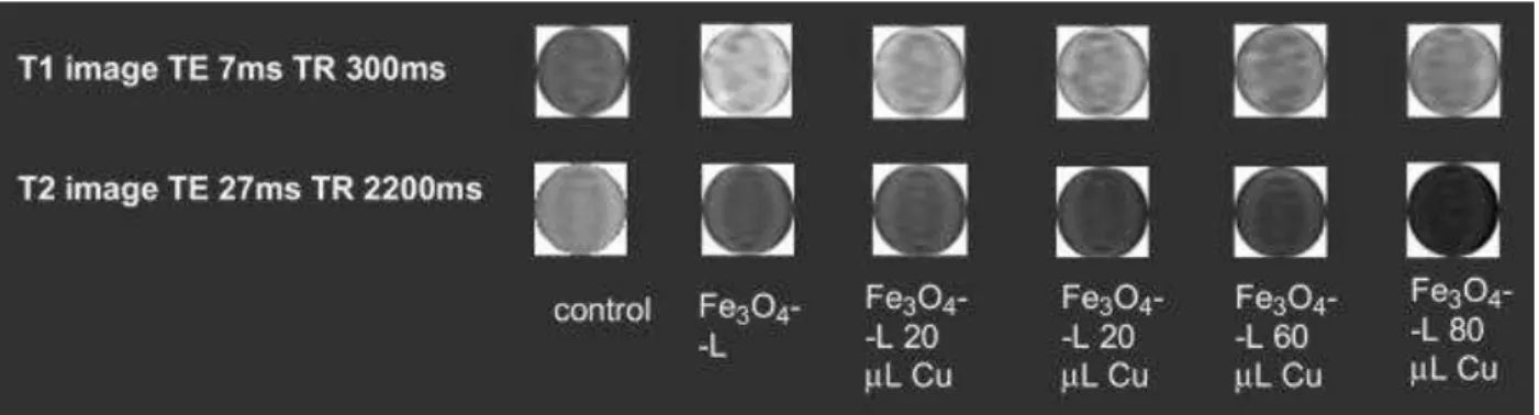
Further, the variation of the relaxivities as a function of the amount of Cu2+ bound onto the surface of the Fe3O4-L nanoparticles were monitored to understand the trends of the
signal enhancement in the MR images. In separate experiments, 20 μL, 40 μL, 60 μL and 80 μL, 0.01 mm Cu2+ solutions were allowed to bind with 1.15 mg Fe
3O4-L
nanoparticles in 1.0 mL 0.01 m sodium acetate buffer at pH 7.2. It was observed that Fe3O4-L nanoparticles treated with 20 μL 0.01 mm Cu2+ solution has more colloidal
stability. The colloidal stability of the nanoparticles decreased with increased amount of added 0.01 mm Cu2+ solution. The r2 relaxivity increased when 20 μL 0.01 mm Cu2+
was bound with 1.15 mg of Fe3O4-L compared to the r2 relaxivity of the bare Fe3O4-L
nanoparticles. This higher r2 relaxivity practically remained unchanged when 40 μL and
60 μL 0.01 mm Cu2+ was bound with 1.15 mg of Fe
3O4-L. A further increase in the r2
relaxivity was observed when 80 μL 0.01 mm Cu2+ was bound with 1.15 mg of Fe 3O4-L
nanoparticles (Table S-3). The enhanced r2 relaxivities were reflected in the phantom
MR images obtained with Fe3O4-L bound with different amount of Cu2+ ions (Fig. 4). As
these contrast agents lead to change in r2 values in response to Cu2+ ions, they can
also be utilized as the chemosensors. It will also be useful to understand the
system and for the diseases like Wilson’s disease [69] by using MR-based molecular imaging.
Fig. 4. T1 and T2 weighted phantom images of iron oxide nanoparticles with
increased concentrations of Cu2+ and without Cu2+.
When the radio isotope of Cu2+ will be made to bind with the Fe3O4-L or Fe3O4-L(+), the
combined effect of the presence of radio isotope of Cu2+ and the higher r
2 relaxivities of
Fe3O4-l-Cu2+ and Fe3O4-L(+)-Cu2+ are expected to enable them as the suitable
candidates for MRI/PET dual agents. This is evident from the contrast in the phantom images obtained with the Fe3O4-L before and after Cu2+ binding as shown in Fig. 4.
In contrast to the iron oxide based nanoparticles, manganese oxide (MnO) nanoparticles were shown to give brighter signal in the T1 weighted MR images
indicating that MnO nanoparticles are potential T1 contrast agents [2]. To explore the
ability of the MnO nanoparticles of different diameters to be used as MRI contrast agents, relaxivities of water protons in the dispersion of the MnO nanoparticles in 1% agarose gel were determined. In general MnO-L exhibit higher r1 relaxivities, but the
MnO nanoparticles of 7 nm particle size exhibit higher r1 relaxivity (16.4 and
17.8 mm−1 s−1 for (MnO)7-L and (MnO)7-L(+), respectively) compared to the MnO nanoparticles of 20 nm particle size (10.8 and 11.5 mm−1 s−1 for (MnO)20-L and
(MnO)20-L(+), respectively). The relaxivity data have been collected in Table S-4. The relaxivity data are also reflected in the brightness of the phantom images obtained with the MnO nanoparticles, as shown in Fig. 5. The higher relaxivities of the smaller
nanoparticles may be attributed to the larger surface area/volume ratios of these nanoparticles. That is, the larger surface area of the MnO nanoparticles of smaller size exposes large number of MnO units to the water molecules in the immediate vicinity of the nanoparticles and thereby facilitating a more efficient shortening of the T1 relaxation
time of water protons and hence the higher r1 relaxivity. Consequently the relatively
Fig. 5. T1 and T2 weighted phantom images of the MnO nanoparticles with and
without copper.
To explore the MnO nanoparticles as MRI/PET dual agent copper coordination with to MnO-L was carried out. Upon Cu2+ coordination, the r
1 relaxivities actually decrease.
The decreased r1 relaxivities upon Cu2+ ion binding suggests that there is a lowering of
the net paramagnetic moment of the coordinated nanoparticles. This may be caused by the anti-parallel alignment of the magnetic spin of the paramagnetic Cu2+ ions on the
surface of the nanoparticles and the magnetic spin of the MnO units at the surface of the nanoparticles. The changes in the relaxivities are also reflected in the brightness of the phantom images obtained with these nanoparticles as shown in Fig. 5.
3.5. Cell labeling
The Fe3O4 nanoparticles, reported in this study, show promising MR signal
enhancement as contrast agents, particularly those based on Fe3O4-L. Cell labeling
studies with superparamagnetic iron oxide particles (SPIO) has emerged as a powerful tool to monitor trafficking of transplanted cells by magnetic resonance imaging. Such studies are valuable for tracking cells used in regenerative medicine in vivo. Generally, intracellular labeling is carried out with the use of a transfecting agent such as
Lipofectamine, Super-Fect, or polylysine (PLL) [69]. These transfecting agents are not approved for clinical use as they cannot be used in humans without the threat of potentially dangerous side effects. As such, it is important to develop the surface chemistry that will facilitate effective endocytosis into the cells without the use of transfection agents. Herein, we evaluate how well our nanoparticles can be
endocytosised by adipose-derived stem cells in the presence as well as in absence of the transfecting agent, PLL and elucidate how the presence of a positively charged dilutant ligand affects the nanoparticle uptake. A similar study was also performed with the commercially available SPIO (Feridex) for comparison. Cell viability experiments were conducted on the rat adipose-derived stem cells (ASC). The labeled cells were assessed using prussian blue staining technique for the detection of intracellular iron. This investigation included Fe3O4-L, Fe3O4-l-Cu2+, Fe3O4-L(+) and Fe3O4-L(+)-Cu2+ and
Feridex over the dosages ranges of 20–80 μg mL−1 of the nanoparticles, with the 0 μg mL−1 dosage being the control.
Surface modification of the nanoparticles is a general strategy utilized to enhance the cellular uptake of the nanoparticles. The differential behavior concerning the uptake into
the stem cells between the two surface modified nanoparticles and Feridex was established. The cell labeling efficiencies were evaluated by following the standard method [70], [71] and [72]. A strong Prussian blue staining was observed when the cells were incubated with the Feridex plus transfecting agent. When Feridex was used for the cell labeling in the absence of transecting agent, there was very little nanoparticle
uptake as evidenced by the lack of blue staining within the cells, indicating little
intracellular uptake. The presence of many blue particles in the cells can be observed when labeling was carried out with the Fe3O4-L and Fe3O4-L(+) both in the presence as
well as in the absence of the transfecting agent (Fig. 6). Thus the efficiency of the surface modified nanoparticles for the cell labeling is higher compared to Feridex in absence of the transfecting agent.
Fig. 6. Stem cells were incubated with nanoparticles with and without PLL as transfecting agent and cellular labeling were visualized histochemically by the Prussian blue reaction.
Feridex being a dextran modified SPIO does not bear any charge on its surface, while Fe3O4-L and Fe3O4-L(+) bear carboxylic acid groups and carboxylic acid groups in
combination with positively charged quaternary ammonium chloride groups,
respectively. Presumably the presence of the ionic surface on the surface modified nanoparticles render them a better cell labeling agents compared to Feridex in absence of the transfecting agents. The presence of PLL as the transfecting agent does not exhibit any beneficial effect on the cell labeling efficiency of either Fe3O4-L or Fe3O4
-L(+). This observation may be attributed to the fact that under the physiological
conditions (pH 7.4), the carboxylic acid group mostly remain in the deprotonated form,
i.e., negatively charged and as reported earlier the negatively charged surface of the
nanoparticles facilitate the phagocytosis of the nanoparticles and hence the cell labeling [72]. When Cu2+ ions are coordinated to the DOTA on the surface of the nanoparticles,
the carboxylate groups of DOTA moiety would no longer remain free resulting in a significant change in surface charges. Upon Cu2+ binding, the surface charges on the Fe3O4-L and Fe3O4-L(+) nanopartiles become less negative and essentially positive,
respectively. The result of changes in surface charge affects the cellular uptakes of Fe3O4-l-Cu, Fe3O4-L(+)-Cu by the stem cell. Fe3O4-l-Cu has less cell labeling compared
to Fe3O4-L(+)-Cu in absence of transfecting agent. The decreased number of free
carboxylic groups and the absence of positively charged quaternary ammonium chloride groups on the surface of Fe3O4-l-Cu, lower its cell labeling efficiency. The presence of
Cu2+ on the surface of Fe3O4-l-Cu nanoparticles helps the endocytosis of the
nanoparticles in the ASC cells to take place however, the extent of cell labeling is definitely lowered compared to the same for Fe3O4-L. The reactive carboxylic acid
groups (-COOH) present on the nanoparticle surface are expected to provide the opportunity for further functionalization of the surface. It is also expected that a variety of other bio-molecules may be immobilized onto the nanoparticle surface to enhance specific cell recognition for use in targeting studies.
The contrast agents showed an efficient cellular uptake and high cell labeling efficiency for the ACS stem cells. All the surface modified nanoparticles both under Cu2+ bound
and Cu2+ unbound state are suitable agents for labeling at a low concentration without using a transfecting agent. In contrast, Feridex is not so effective for cell labeling in the absence of a transfecting agent. This offers the opportunity to omit transfecting agents from the labeling protocol for the surface modified nanoparticles [73].
4. Conclusions
In summary, the hydrophilic and biocompatible multimodal probes, consisted of a monocrystalline superparamagnetic iron oxide (SPIO) or managanse oxide core and stable in water or physiological environment, have been developed. Surface
modifications and subsequent DOTA conjugation enable the fine-tuning of the surface charge of these nanoparticles. The relaxivity studies indicate a relaxivity enhancement upon Cu2+ binding. These properties enable the nanoprobes as potent for dual
PET/MRI, intracellular labeling without transfecting agent and in vivo MR/PET tracking. We are currently investigating the targeting abilities of these nanoprobes, the scope of these nanoparticles as T2 CEST agents for dual PET/MRI, application to disease
detection and stem cell treatment.
Acknowledgement
This study was supported by the Canadian Institutes of Health Research.
Appendix.
Figures with essential color discrimination. Fig. 1 and Fig. 6 in this article is difficult to interpret in black and white. The full color image can be found in the online version, at doi:10.1016/j.biomaterials.2010.10.013.
Appendix. Supplementary material
Supplementary data related to this article can be found online
References
1. L.E. Jennings, N.J. Long Two is better than one—probes for dual-modality molecular imaging Chem Commun (2009), pp. 3511–3524
2. R. Weissleder Molecular imaging in cancer Science, 312 (2006), pp. 1168–1171 3. J. Cheon, J.H. Lee Synergistically integrated nanoparticles as multimodal probes
for nanobiotechnology Acc Chem Res, 41 (12) (2008), pp. 1630–1640
4. V. Ntziachristos, A.G. Yodh, M. Schnall, B. Chance Concurrent MRI and diffuse optical tomography of breast after indocyanine green enhancement Proc Natl Acad Sci U S A, 97 (2000), pp. 2767–2772
5. T. Beyer, D.W. Townsend, T. Brun, P.E. Kinahan, M. Charron, R. Roddy et al. Combined PET/CT scanner for clinical oncology J Nucl Med, 41 (2000), pp. 1369–1379
6. C.B. Murray, D.J. Norris, M.G. Bawendi Synthesis and characterization of nearly monodisperse CdE (E = sulfur, selenium, tellurium) semiconductor
nanocrystallites J Am Chem Soc, 115 (1993), pp. 8706–8715
7. S. Link, M.A. El-Sayed Spectral properties and relaxation dynamics of surface plasmon electronic oscillations in gold and silver nanodots and nanorods J Phys Chem B, 103 (1999), pp. 8410–8426
8. A.P. Alivisatos Semiconductor clusters, nanocrystals, and quantum dots Science, 271 (1996), pp. 933–937
9. K. Toth, L. Helm, A.E. Merbach The chemistry of contrast agents in medical magnetic resonance imaging Wiley, Chichester (2001)
10. P. Caravan, J.J. Ellison, T.J. McMurry, R.B. Lauffer Gadolinium(III) chelates as MRI contrast agents: structure, dynamics and applications Chem Rev, 99 (1999), pp. 2293–2352
11. K.N. Raymond, V.C. Pierre Next generation, high relaxivity gadolinium MRI agents Bioconjug Chem, 16 (2005), pp. 3–8
12. W.S. Seo, J.H. Lee, X. Sun, Y. Suzuki, D. Mann, Z. Liu et al. FeCo/graphitic-shell nanocrystals as advanced magnetic-resonance-imaging and near-infrared agents Nat Mater, 5 (2005), pp. 971–976
13. G. Sun, J. Xu, A. Hagooly, R.D. Rossin, A. Moore, C.J. Hawker et al. Strategies for optimized radiolabeling of nanoparticles for in vivo PET imaging Adv Mater, 19 (2007), pp. 3157–3162
14. C. Catana, Y.B. Wu, M.S. Judenhofer, J.Y. Qi, B.J. Pichler, S.R. Cherry
Simultaneous acquisition of multislice PET and MR images: initial results with a MR-compatible PET scanner J Nucl Med, 47 (2006), pp. 1968–1976
15. S.R. Cherry The 2006 Henry N. wagner lecture: of mice and men (and positrons) advances in PET imaging technology J Nucl Med, 47 (2006), pp. 1735–1745 16. S.R. Cherry Fundamentals of positron emission tomography and applications in
preclinical drug development J Clin Pharmacol, 41 (2001), pp. 482–491 17. H.T. Song, J.S. Choi, Y.M. Huh, S. Kim, Y.W. Jun, J.S. Suh et al. Surface
modulation of magnetic nanocrystals in the development of highly efficient magnetic resonance probes for intracellular labeling J Am Chem Soc, 127 (2005), pp. 9992–9993
18. Y.M. Huh, E.S. Lee, J.H. Lee, Y.W. Jun, P.H. Kim, C.O. Yun et al. Hybrid nanoparticles for magnetic resonance imaging of target-specific viral gene delivery Adv Mater, 19 (2007), pp. 3109–3112
19. J.W.M. Bulte, D.L. Kraitchman Monitoring cell therapy using iron oxide MR contrast agents Curr Pharm Biotechnol, 5 (2004), pp. 567–584
20. M.M. Huber, A.B. Staubli, K. Kustedjo, M.H.B. Gray, J. Shih, S.E. Fraser et al. Fluorescently detectable magnetic resonance imaging agents Bioconjug Chem, 9 (1998), pp. 242–249
21. L. Josephson, C.H. Tung, A. Moore, R. Weissleder High-efficiency intracellular magnetic labeling with novel superparamagnetic-tat peptide conjugates
Bioconjug Chem, 10 (1999), pp. 186–191
22. E.A. Schellenberger, D. Sosnovik, R. Weissleder, L. Josephson Magneto/optical annexin V, a multimodal protein Bioconjug Chem, 15 (2004), pp. 1062–1067 23. Tsourkas, V.R. Shinde-Patil, K.A. Kelly, P. Patel, A. Wolley, J.R. Allport et al. In
vivo imaging of activated endothelium using an anti-vcam-1 magnetooptical probeBioconjug Chem, 16 (2005), pp. 576–581
24. W. Wang, S. Ke, S. Kwon, S. Yallampalli, A.G. Cameron, K.E. Adams et al. New optical and nuclear dual-labeled imaging agent targeting interleukin 11 receptor alpha-chain Bioconjug Chem, 18 (2007), pp. 397–402
25. J.S. Choi, J.C. Park, H. Nah, S. Woo, J. Oh, M. Kim et al. A hybrid nanoparticle probe for dual-Modality positron emission tomography and magnetic resonance imaging Angew Chem Int Ed, 47 (2008), pp. 6259–6262
26. G. Charles, R. Raffaella, J.W. Michael, B. Gang In vivo evaluation of 64Cu-labeled magnetic nanoparticles as a dual-modality PET/MR imaging agent Bioconjug Chem, 21 (2010), pp. 715–722
27. M. Lewin, N. Carlesso, C.H. Tung, X.W. Tang, D. Cory, D.T. Scadden et al. Tat peptide-derivatized magnetic nanoparticles allow in vivo tracking and recovery of progenitor cells Nat Biotechnol, 18 (2000), pp. 410–414
28. H.B. Na, J.H. Lee, K. An, Y. Park, M. Park, I. Lee et al. Development of a T1 contrast agent for magnetic resonance imaging using MnO nanoparticles Angew Chem Int Ed, 46 (2007), pp. 5397–5401
29. Y. Shao, Y. Shaoyk, S.R. Cherry, K. Farahaniz, K. Meadorsy, S. Siegely et al. Simultaneous MRI and PET imaging of a rat brain Phys Med Biol, 42 (1997), pp. 1965–1970
30. R.R. Raylman, S. Majewski, S.S. Velan, S. Lemieux, B. Kross, V. Popov et al. Simultaneous acquisition of magnetic resonance spectroscopy (MRS) data and positron emission tomography (PET) images with a prototype MR-compatible, small animal PET imager J Magn Reson, 186 (2007), pp. 305–310
31. C. Woody, D. Schlyera, P. Vaskaa, D. Tomasia, S.S. Najeraa, W. Rooneyc et al. Preliminary studies of a simultaneous PET/MRI scanner based on the RatCAP small animal tomography Nucl Instrum Methods Phys Res Sect A, 571 (2007), pp. 102–105
32. D. Patel, A. Kell, B. Simard, J. Deng, B. Xiang, Lin HYu et al. Cu2+-labeled, SPION loaded porous silica nanoparticles for cell labeling and multifunctional imaging probes Biomaterials, 31 (2010), pp. 2866–2873
33. D. Caruntu, G. Caruntu, Y. Chen, C.J. O’Connor, G. Goloverda, V.L.
Kolesnichenko Synthesis of variable-sized nanocrystals of Fe3O4 with High
surface reactivity Chem Mater, 16 (2004), pp. 5527–5534
34. R.M. Cornell, U. Schwertmann The iron oxides: structure, properties, reactions, occurrence and uses VCH, New York (1996) p. 28
35. A.R. West Solid state chemistry and its applications John Wiley & Sons, London (1984) p 174
36. W.S. Seo, H.H. Jo, K. Lee, B. Kim, S.J. Oh, J.T. Park Size-dependent magnetic properties of colloidal Mn3O4 and MnO nanoparticles Angew Chem Int Ed, 43
(2004), pp. 1115–1117
37. J. Park, K. An, Y. Hwang, J.G. Park, H.J. Noh, J.Y. Kim et al. Ultra-large-scale syntheses of monodisperse nanocrystals Nat Mater, 3 (2004), pp. 891–895 38. M. Yin, S. O’Brien Synthesis of monodisperse nanocrystals of manganese
oxides J Am Chem Soc, 125 (2003), pp. 10180–10181
39. J. Park, E. Kang, C.J. Bae, J.G. Park, H.J. Noh, J.Y. Kim et al. Synthesis, characterization, and magnetic properties of uniform-sized MnO nanospheres and nanorods J Phys Chem B, 108 (2004), pp. 13594–13598
40. B. Dubertret, P. Skourides, D.J. Norris, V. Noireaux, A.H. Brivanlou, A. Libchaber In vivo imaging of quantum dots encapsulated in phospholipid micelles Science, 298 (2002), pp. 1759–1762
41. J.H. Waite, M.L. Tanzer Polyphenolic substance of mytilus edulis: novel
adhesive containing l-Dopa and hydroxyproline Science, 212 (1981), pp. 1038– 1040
42. H.W. Gu, Z.M. Yang, J.H. Gao, C.K. Chang, B. Xu Heterodimers of
nanoparticles: formation at a liquid-liquid interface and particle-specific surface modification by functional molecules J Am Chem Soc, 127 (2005), pp. 34–35 43. C.J. Xu, K.M. Xu, H.W. Gu, R.K. Zheng, H. Liu, X.X. Zhang et al. Dopamine as a
robust anchor to immobilize functional molecules on the iron oxide shell of magnetic nanoparticles J Am Chem Soc, 126 (32) (2004), pp. 9938–9939
44. J. Xie, C. Xu, N. Kohler, Y. Hou, S. Sun Controlled PEGylation of monodisperse Fe3O4 nanoparticles for reduced non-specific uptake by macrophage cells Adv
Mater, 19 (20) (2007), pp. 3163–3166
45. J. Xie, C.J. Xu, Z.C. Xu, Y.L. Hou, K.L. Young, S.X. Wang et al. Linking
hydrophilic macromolecules to monodisperse magnetite (Fe3O4) nanoparticles
via trichloro-s-triazine Chem Mater, 18 (23) (2006), pp. 5401–5403
46. T. Rajh, L.X. Chen, K. Lukas, T. Liu, M.C. Thurnauer, D.M. Tiede Surface
restructuring of nanoparticles: an efficient route for ligand-metal oxide crosstalk J Phys Chem B, 106 (2002), pp. 10543–10552
47. M. Shokeen, C.J. Andesron Molecular imaging of cancer with copper-64
radiopharmaceuticals and positron emission tomography (PET) Acc Chem Res, 42 (2009), pp. 832–841
48. E.D. Pressly, R. Rossin, A. Hagooly, K. Messmore, M.J. Welch, K.L. Wooley et
al. Structural effects on the biodistribution and positron emission tomography
(PET) imaging of well-defined 64Cu-labeled nanoparticles comprised of
49. F. Bernd, M. Iwan, L. Petra, K. Rolf, H. Zeger, C.M. Jose In situ observation of rapid ligand exchange in colloidal nanocrystal suspensions using transfer NOE nuclear magnetic resonance spectroscopy J Am Chem Soc, 131 (2009), pp. 3024–3032
50. Bootz, V. Vogel, D. Schubert, J. Kreuter Comparison of scanning electron microscopy, dynamic light scattering and analytical ultracentrifugation for the sizing of poly(butyl cyanoacrylate) nanoparticles Eur J Pharm Biopharm, 57 (2004), pp. 369–375
51. C.W. Jung, P. Jacobs Physical and chemical properties of super
paramagneticiron oxide MR contrast agents: ferumoxides, ferumoxtran, ferumoxsil Magn Reson Imaging, 13 (1995), pp. 661–674
52. Esther, G. Torben, B. Idalia, T. Marcus, R. Erik Ultrastable iron oxide nanoparticle colloidal suspensions using dispersants with catechol-derived anchor groups Nano Lett, 9 (2009), pp. 4042–4048
53. S.M. Hussain, G.P. Krestin Superparamagnetic iron oxide contrast agents: physicochemical characteristics and application in MR imaging Eur J Radiol, 11 (2001), pp. 2319–2331
54. P. Wunderbaldinger, L. Josephson, R. Weissleder Crosslinked iron oxides
(CLIO): a new platform for the development of targeted MR contrast agents Acad Radiol, 9 (2002), pp. S304–S306
55. S. Sun, H. Zeng, D.B. Robinson, S. Raoux, P.M. Rice, S.X. Wang et al. Monodisperse MFe2O4 (M = Fe, Co, Mn) nanoparticles J Am Chem Soc, 126 (2004), pp. 273–279
56. R.H. Kodama, A.E. Berkowitz, E.J. McNiff, S. Foner Surface spin disorder in ferrite nanoparticles (invited) J Appl Phys, 81 (1997), pp. 5552–5557
57. Y. Lee, J. Lee, C.J. Bae, J.G. Park, H.J. Noh, J.H. Park et al. Large-scale synthesis of uniform and crystalline magnetite nanoparticles using reverse
micelles as nanoreactors under reflux conditions Adv Funct Mater, 15 (2005), pp. 503–509
58. M. Grigorova, H.J. Blythe, V. Blaskov, V. Rusanov, V. Petkov, V. Masheva et al. Magnetic properties and Mossbauer spectra of nanosized CoFe2O4 powders
J Magn Magn Mater, 183 (1998), pp. 163–172
59. C.R. Vestal, Z.J. Zhang Magnetic spinel ferrite nanoparticles from microemulsions Int J Nanotechnol, 1 (2004), pp. 240–263
60. L.D. Tung, V. Kolesnichenko, D. Caruntu, N.H. Chou, C. O’Connor, J.L. Spinu Magnetic properties of ultrafine cobalt ferrite particles J Appl Phys, 93 (2003), pp. 7486–7488
61. D. Caruntu, G. Caruntu, C.J. O’Connor Magnetic properties of variable-sized Fe3O4 nanoparticles synthesized from non-aqueous homogeneous solutions of
polyols J Phys D Appl Phys, 40 (2007), pp. 5801–5809
62. J. Choi, J.C. Kim, Y.B. Lee, I.S. Kim, Y.K. Parka, N.H. Hur Fabrication of silica-coated magnetic nanoparticles with highly photoluminescent lanthanide probes Chem Commun (2007), pp. 1644–1646
63. Y.P. He, S.Q. Wang, C.R. Li, Y.M. Miao, Z.Y. Wu, B.S. Zou Synthesis and characterization of functionalized silica-coated Fe3O4 superparamagnetic
nanocrystals for biological applications J Phys D Appl Phys, 38 (2005), pp. 1342– 1350
64. G.H. Lee, S.H. Huh, J.W. Jeong, B.J. Choi, S.H. Kim, H.C. Ri Anomalous magnetic properties of MnO nanoclusters J Am Chem Soc, 124 (41) (2002), pp. 12094–12095
65. R. Weissleder, A. Moore, U. Mahmood, R. Bhorade, H. Benveniste, E.A. Chiocca
et al. In vivo magnetic resonance imaging of transgene expression Nat Med, 6
(2000), pp. 351–354
66. Y.M. Huh, Y.W. Jun, H.T. Song, S. Kim, J.S. Choi, J.H. Lee et al. In vivo magnetic resonance detection of cancer by using multifunctional magnetic nanocrystals J Am Chem Soc, 127 (2005), pp. 12387–12391
67. Leslie La, N. Nitin, B. Gang Magnetic nanoparticle probes Nano Today (2005), pp. 32–38
68. S.H. Koenig, K.E. Kellar Theory of proton relaxation in solutions of magnetic nanoparticles, including the superparamagnetic size range Acad Radiol, 3 (1996), pp. S273–S276
69. E.L. Que, C.J. Chang A smart magnetic resonance contrast agent for selective copper sensing J Am Chem Soc, 128 (2006), pp. 15942–15943
70. A.S. Arbab, G.T. Yocum, L.B. Wilson, A. Parwana, E.K. Jordan, H. Kalish et al. Comparison of transfection agents in forming complexes with ferumoxides, cell labeling efficiency, and cellular viability Mol Imaging, 3 (2004), pp. 24–32 71. J.A. Frank, H. Zywicke, E.K. Jordan, J. Mitchell, B.K. Lewis, B. Miller et al.
Carboxylated Superparamagnetic iron oxide particles label cells intracellularly without transfection agentsAcad Radiol, 9 (2002), pp. S484–S487
72. D.L. Kraitchman, A.W. Heldman, E. Atalar, L.C. Amado, B.J. Martin, M.F.
Pittenger et al. In vivo magnetic resonance imaging of mesenchymal stem cells in myocardial infarction Circulation, 107 (2003), pp. 2290–2293
73. V. Mailänder, M.R. Lorenz, V. Holzapfel, A. Musyanovych, K. Fuchs, M.
Wiesneth et al. Carboxylated superparamagnetic iron oxide particles label cells intracellularly without transfection agents Mol Imaging Biol, 10 (2008), pp. 138– 146


![[PDF] Programmation C pdf support de cours et travaux pratiques - Cours langage c](data:image/gif;base64,R0lGODlhAQABAIAAAP///wAAACH5BAEAAAAALAAAAAABAAEAAAICRAEAOw==)