Biological Scaffolds for the Peptide-Directed Assembly of
Nanoscale Materials and Devices
by
Daniel Joseph Solis
B.S. Chemistry B.A. Physics
California Polytechnic State University, San Luis Obispo, CA 93407. 2001
SUBMITTED TO THE DEPARTMENT OF CHEMISTRY IN PARTIAL FULFILMENT OF
THE REQUIREMENTS FOR THE DEGREE OF
DOCTORATE OF PHILOSOPHY IN PHYSICAL CHEMISTRY
AT THE
MASSACHUSETTS INSTITUTE OF TECHNOLOGY, CAMBRIDGE, MA 02139
,jW[ 5 February 2006.
© 2006 Daniel J. Solis
All rights reserved.
The author hereby grants to MIT permission to reproduce and to distribute publicly paper and
electronic copies of this thesis document in whole or part in any medium now known
or hereafter created.
S itre- -o - - D ai eJ. Soi
Daniel J. Solis
November
10th, 2005
('artififdy-'
-Angela M. Belcher
Professor of Materials Science & Engineering
A
_
*Thesis
Supervisor
__~~
.1" Is
~~~~
r
Accepted by I Robert W. Field
MASWCHUSETSMIE OF TECHNOLOGY
APR 0
20|6
LIBRARIES
THESIS COMMITTEE CERTIFICATION OF APPROVAL OF
Biological Scaffolds for the Peptide-Directed Assembly of
Nanoscale Materials and Devices
by
Daniel Joseph Solis
GRADUATE THESIS CQMMITTEE
A
Angela M. Belcher
Professor of Materials Science
& Bioengineering
VL I
Moungi G. Bawendi
Professor of Chemistry Thesis Committee Chair
Keith A. Nelson
Professor of Chemistry
I
Biological Scaffolds for the Peptide-Directed Assembly of
Nanoscale Materials and Devices
by
Daniel Joseph Solis
Submitted to the Department of Chemistry on February 1, 2006, in partial fulfillment of the requirements for the
Degree of Doctorate of Philosophy in Physical Chemistry.
Abstract
The utilization of biological factors in the design, synthesis and fabrication of nano-scaled materials and devices presents novel, large scale solutions for the realization of future technologies. In particular, we have genetically modified the M13 Filamentous Bacteriophage for its use as a biological scaffold in the peptide-controlled nucleation and patterning of nanoscale semiconducting and magnetic materials. Through evolutionary phage display screening of inorganic substrates, ftunctional peptides that influence material properties such as size, phase and composition during nucleation have been identified. The incorporation of these specific, nucleating peptides into the generic scaffold of the M13 coat structure provides a viable linear template for the directed synthesis of semiconducting and magnetic nanowires. Through further modification of the remaining proteins on the virus scaffold, other functionalities can be incorporated such as the directed patterning of the virus/nanowires assemblies into nanoscaled devices with tunable properties as determined by the genetic information carried within the virus scaffold. Multi-functional viruses provide a truly self assembled system for the design and execution of a myriad of nanoscaled devices in a green, scalable and cost effective manner.
Thesis Supervisor: Angela M. Belcher
ACKNOWLEDGEMENTS
Conlige suspectos semper habitos
I would like to thank everyone who, in many different ways, has brought me to this place in life. I would like to thank my advisor, Angela Belcher for helping me to explore uncharted areas of research with great freedoms. I was fortunate to have more than one great mentor, and for this I will forever be grateful for the friendship of Brian Reiss. I would not have made it through some of the rougher patches with out my brother in arms, Stephen Kottmann. I was fortunate in many ways to have begun my graduate studies at the University of Texas, Austin, and met many incredible scientists and friends. I would especially like to thank the boys in the band (guillaume et les cogneurs de couilles). At MIT, I would like to thank Glenn McCloud, with out whom I would never have really known Boston. I would also like to thank Sandra and Craig Breen who helped me in finishing up my time at MIT. I also thank
Ioannis
Kimissis for his help in the final stages of my research.I would also like to acknowledge those people who contributed to my life during graduate school, although in a non-academic way. First and foremost, I will forever be in the debt of Rebecca Golden-Harrell, who has always been, and will continue to be, the best part of my life. One can never overstate the support of ones parents, so thank you mom and pop. And finally, I would like to thank everyone whom I met and enriched my life during the past four years.
TABLE OF CONTENTS
List of Tables ... 007 List of Figures ... 008 Chapter 1: Introduction 1.0 Introduction ... 010 1.1: Biomineralization ... 012 1.2: Phage Display ... 014 1.3: M113 Bacteriophage ... 016 1.4: Materials ... 019 1.4.1: Noble Metals ... 020 1.4.2: Semiconducting Materials ... 021 1.4.3: Magnetic Materials ... 022 1.5: Scope of Work ... 025 References Chapter 2: Selection of Functional Peptides2.1: ntroduction ...
035
2.2: Phage Display Methods ... 035
2.3: Substrates
...
038
2.3.1: Magnetic Materials ... 039 2.3.1.1: Cobalt ... 039 2.3.1.2: Cobalt Platinum ... 0412.3.1.3: Iron Platinum ...
044
2.3.2: Noble Metals
...
046
2.3.2.1: Gold ...
046
2.3.2.2: Platinum ...
049
2.3.2.3: Copper ... 051 2.4: Discussion ... 0522.5: Characterization of Functional Peptides ... 054
2.5.1 :Binding Affinities ... 054
2.5.2: Computational Structural Analysis ... 058
References Chapter 3: Genetic Incorporation of Functional Peptides into the M13 Bacteriophage
3.1: Introduction ...
062
3.2: Display of Peptides ... 062
3.2.1: Proximal Tip Display ... 062
3.2.2: Capsid Display ... 063
3.2.2: Distal Tip Display ... 068
3.3: Modeling of Displayed Peptide ... 069
3.4: Discussion ... 073
References Chapter 4: Peptide Directed Synthesis
4.1: Introduction ...
074
4.2: Methods and Materials ... 074
4.3: Nucleation
...
077
4.3.1: Proximal Tip Nucleation ... 078
4.3.2: Free Peptide Nucleation ... 080
4.3.3: Capsid Nucleation ... 082
4.4: D)iscussion
...
092
References Chapter 5: Device Assembly
5.1: Introduction ...
096
5.2: Methods and Materials ... 097
5.3: Specific attachment of Bacteriophage ... 099
5.4: Nucleation...
101
5.5 Device characteristics... .102
5.6: Discussion ... 106
References Biographical Note ... 108
LIST OF TABLES
2.1 Cobalt binding Sequences determined from the Ph.D. 12 library 41 2.2 Cobalt Platinum binding Sequences determined from the Ph.D. 12 library 43 2.3 Cobalt Platinum binding Sequences determined from the Ph.D. 7c library 43 2.4 Iron Platinum binding Sequences determined from the Ph.D. 12 library 45 2.5 iron Platinum binding Sequences determined from the Ph.D. 7c library 46 2.6 Gold binding Sequences determined from the Ph.D. 12 library 48 2.7 Gold binding Sequences determined from the Ph.D. 7c library 49 2.8 Platinum binding Sequences determined from the Ph.D. 12 library 51
LIST OF FIGURES
1.1 Phage based device assembly 11
1.2 Examples of Biomineralization 12
1.3 SEM images of haliotis rufescens Nacre 13
1.4 Diagram of Phage Display Process 14
1.5 Wild Type M13 Bacteriophage Genome 15
1.6 Diagram of the M1 3 Bacteriophage 16
1.7 L 10crystal structure 25
2.1 XRD of chemically prepared Cobalt Nanoparticles 40
2.2 XRD of chemically prepared Iron Platinum Nanoparticles 45
2.3 XRD of Single crystal (111) Gold Ingot 47
2.4 XRD of Platinum Film 50
2.5 UV/Vis spectrum of Copper Selection Eluate 52
2.6 Adsorption Isotherm of Cobalt binding peptide 53
2.7 SPR sensogram of a Gold binding bacteriophage 56
2.8 Adsorption Isotherm of Gold binding bacteriophage 57
2.9 Adsorption Isotherm of Gold binding peptide 58
2. 10 Molecular Mechanics Model of the Gold Binding Peptide 59
3., 1 pMoPac33 vector for phagmid systems 64
3.2 Nearest Neighbor distance between capsid displayed peptides 71
3.3 Visualization of gPVllI modified capsid down the c-axis 71
3.4 Visualization of the gPVIII modified capsid along the c-axis 72
3.5 Representations of the average dihedral angles of the A7 peptide 73
4.1 FePt and CoPt Nanoparticles synthesized by phage 79
4.2 SQUID of phage synthesized FePt nanoparticles 81
4.3 Synthetic Peptide nucleated FePt nanoparticles 83
4.4 Phage synthesized ZnS nanowires 85
4.5 Single crystal CdS nanowires 86
4.6 Phage Synthesized CoPt nanowires 88
4.7 Phage Synthesized CoPt nanowires 88
4.8 Annealed phage synthesized CoPt nanowire 89
4.9 Phage synthesized FePt nanowires 90
4.10 SQUID of CoPt nanowires 91
4.11 TGA of CoPt nanoparticle/phage assemblies 92
4.12 TEM Thermal analysis of CoPt/phage system 92
5.1 Phage directed assemble of nanoscale electronics 97
5.2 Electrode Mask Design 99
5.3 CdS aggregation 100
5.4 AFM of gold specific phage binding events 101
5.5 Controlled electrode bridging phage densities 101
5.6 Phage directed CdS nucleation 102
5.7 CdS nucleated phage matt 103
5.8 AFM of pre- and post- annealed CdS nucleated phage 103
5.9 IV characteristics of pure and CdS nucleated phage 104
5.10 SEM of annealed CdS structures 105
CHAPTER 1
1.0 Introduction
The reliance of future technologies on developing scalable and economic methods for the fabrication of one-dimensional systems has spurred intense and rapid progress in the interdisciplinary field of materials science. In particular, one-dimensional materials have been enthusiastically pursued for their applications in the study of electrical transport (1), optical phenomena (2), and as functional units in nanoscaled circuitry (3). Pursuit of "bottom up" methods for the synthesis of semiconducting, metallic and magnetic nanowires has yielded strategies including, but not limited to, vapor liquid solid (VLS) (4), chemical (5), solvothermal, vapor phase, and template-directed fabrication (6). Although each method developed for the production of nanowires has had success in achieving high quality materials, no distinct strategy to date has yielded monodisperse, crystalline nanowires of radically different compositions. The realization of such a system would require the combination of substrate specific ligands with the predictability of self-assembly commonly found in nature. Recently, biological factors have been exploited as synthesis directors for nanofibers (7, 8), virus-based particle cages (9), virus-particle assemblies (10., 11), and non-specific peptide templates (12). This is due to the high degree of organization, ease of chemical modification and naturally occurring self-assembly motifs inherent in these systems.
The development of the virus based scaffold for synthesizing and assembling nanoscale materials into function architectures is presented (figure 1.1). The ability to store information about a material, including composition, phase, and crystallographic detail, within the genetic code of the M13 bacteriophage virus DNA has proven to be a viable means of synthesizing and organizing materials on the nanometer scale (13,14). The use of phage display techniques
(utilizing peptide libraries consisting of ~ 109 random sequences) has led to the discovery of material specific peptides having preferential binding (13), control over nanoparticle nucleation (14), and the ability to order based on the inherent shape anisotropy of the filamentous M 13 virus (11).
Figure 1.1 Proposed device assembly using a substrate binding peptide on the proximal tip of the virus to anchor it specifically to pre-patterned gold electrodes. Pep tides expressed along the length of the phage can then induce nucleation of technologically relevant materials between the electrode gap. Thermal removal of the organic template results in continuous inorganic nanowires connected to the electrodes creating a functional device.
Because the protein sequences responsible for these attributes are gene linked and contained within the capsid of the virus, exact genetic copies of the virus scaffold are easily reproduced by infection into its bacterial host. The exploitation of the self-assembly motifs employed by the M13 bacteriophage to produce a biological scaffold provides a means of generating a complex, highly ordered, and economical template for the general synthesis of single crystal nanowires. By introducing programmable genetic control over the composition, phase and assembly of nanoparticles, a generic template for the universal synthesis of a variety of materials can be realized. Further advances in the fabrication of nanoscale materials and
devices can be achieved through modification of the remaining four proteins in the virus to incorporate device-assembly directors. Overall, modification of biological systems by the introduction of substrate specific peptides presents a means of achieving well ordered nanomaterials in a cost-effective and scalable manner (15). The following chapters will discuss the selection of functional peptides exhibiting a binding affmity for specific materials, the genetic manipulation of these functional peptides into different areas of the M 13 bacteriophage scaffold, and the subsequent ability to control materials SYnthesisand assembly.
1.1 Biomineralization
Nature's ability to form inorganic structures with controlled structure and properties, developed over millions of years, provides a unique chemistry for developing inorganic-organic materials. The field of biomineralization seeks to understand the mechanisms by which biological systems can uptake elements from its surroundings, and organize them into complex, highly ordered structures of defmed functionality (16). There are many types of organisms that utilize biomineralization, ranging from single-celled coccolithophorids (figure 1.2) that assemble calcite cages to mammals who depend on the biologically controlled mineralization of hydroxyapetite for bones.
Figure 1.2Examples of biomineralization. Left: Emi/iana Huxley coccoliths Right silica diatom. Images were taken fromwww.bigelow.org/images/ bulletin_coccolith.jpg and http://academics.hamilton.edu/ biology/ kbart/image/ diatom.j pg respectively.
Although these materials provide essential life functions for many organisms (17), it is their unique physical properties and inherently green synthesis of materials with precise control that has garnered attention from the materials community as a facile route to nanoscaled components for next generation technologies. Biomineralized materials are ordered over multiple length scales (18) beginning at the atomistic level with control over crystallographic phase and orientation and composition, to nanoscaled building blocks of controlled shape and size, to organized micro and macroscopic heterostructures( 19). The materials also exhibit desirable material characteristics such as fracture toughness, self-correction or "healing" and single crystal growth. Furthermore, biomineralization reactions proceed under aqueous conditions at or below ambient temperatures, yet often produce polymorphs typically SYnthesized at elevated temperatures and pressures. As an example, the shell of the red abalone (haliotis rufescens) has a "brick and mortar" like construction of aragonite tablets (a metastable phase of calcium carbonate) separated by layers of acidic glycoprotein's (figure 1.3), which gives the shell a fracture resistance 3000 times greater than geological aragonite (20).
Figure 1.3Scanning Electron Micrographs of the nacre component of the ha/iotis rufescens shell. The brick and mortar structure is evident and exhibits long range ordering.
Other examples of biomineralization systems include magnetotactic bacteria that produce 35-120nm diameter, single-domain, ferromagnetic Fe304 particles, allowing for the bacteria to migrate along the earth's magnetic field (21). Another common biomineralization product is marine silica (SiO2) as found in diatoms and sea sponge spicules. The successful isolation of biomineralization proteins, and their ability to maintain functionality in vitro, has provided many successes in understanding the mechanisms behind biomineralization. However, the complexity of natural systems has impeded the complete understanding of the biomineralization process, which had hindered the progress in extending the classes of materials that can be processed by this chemistry. In order to exploit the tremendous advantages of biomineralization, a rapid method for developing functional, material-specific peptides needed to be developed in order to extend the materials available from common minerals, to technologically relevant materials including semiconductors, conductors, and highly anisotropic magnetic materials.
1.2 Phage Display
Almost all biological processes in living organisms rely on specific, protein-ligand interactions. Biomineralization is no different in that the inorganic substrate serves as the ligand, and a highly specific biomolecule dictates the organization and construction of the inorganic structure. There are two common techniques for isolating biomolecules that exhibit the necessary affinity for specific targets, both routinely used in the pharmaceutical industry, Rational design and Combinatorial screening. Both of these provide pathways for developing new biomineralization chemistries for synthesizing materials not found in nature. However, because the exact mechanisms for biomineralization are still not fully understood, and the complexity of biological systems, there is usually poor correlation between computer predictions
of biomolecule functionality and that observed in vitro. Also, the computational costs of analyzing the vast number of relevant mutants precludes this method from being a practical and rapid method for developing bioinorganic synthesis routes for the multiple classes of materials needed to produce the complex architectures found in today's technology. The second approach of using a combinatorial library to screen a target provides a rapid and economical means of identifying biomolecules that exhibit the required specificity for a given target, in this case an inorganic substrate. Combinatorial libraries comprised of biological systems can also employ the same evolutionary processes found in nature of mutation and selection. The main limitation of combinatorial libraries then lies in their low complexity and sample size. There are multiple types of libraries available including combinatorial chemistry, yeast two way systems, ribosomal display and cell surface display however, phage display was chosen for its combinatorial size, the complexity of its banks, the diversity of applications, and its ease of use.
Phage display libraries are systems in which a peptide or protein is expressed (displayed) on the surface of a filamentous bacteriophage virus (22). These libraries are commercially available with 10^9 molecules, but have been synthesized with banks as high as 10"12 (23). In essence, phage display is performed by incubating the target substrate with the phage library, followed by washing of the unbound phage and elution of the specifically bound phage. The eluted phage are then amplified, creating an enriched pool for subsequent screenings. The affinity of the selected phage for the target can be tailored either through employing more stringent washings, method of elution, or by the desired binding constants as determined via kinetic screening of substrates (figure 1.4).
Figure 1.4Introduction of the library (top left) to the substrate, is followed by washing and acid elution of the bound phage. Bacterial amplification enriches the phage pool, which is then screened against the same target. This process is repeated until a dominant binding sequence can be determined. (image from IEEE spectrum, Germs that build circuits, online.)
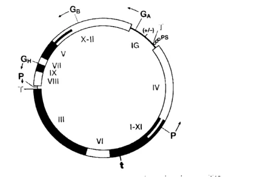
1.3 MI3 Bacteriophage
The M 13 class of bacteriophage, used in this work, is approximately 880nm long by 6nm wide, and is comprised of five capsid proteins that encapsulate a single stranded DNA. The M 13 phage DNA has 9 genes that encode for II proteins grouped on the single stranded, covalently closed DNA in order of their functionality during the life cycle of the virus (24). They are classified into DNA replication proteins (gene products (gP) Il,V,X), Capsid proteins (gP III, VI, VII, VIII, IX) and assembly proteins (gP I, IV, XI). The wild type (or naturally occurring, unmodified M13 phage) genome is given in figure 1.5. Addition of a randomized peptide insert on the gene III and a gene giving antibiotic resistance is added to the genome to create the gPIll library (25).
(
P¥ -r
Figure 1.5 The wild type genome of the Ff class of filamentous bacteriophage, which includes the M1 3 phage.
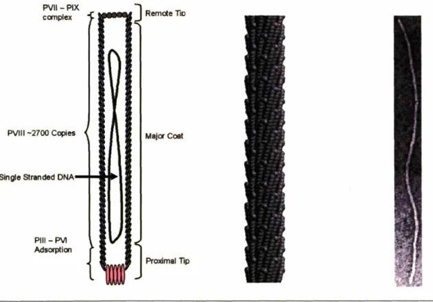
The five capsid proteins create a vesicle for DNA delivery and is comprised of: approximately five copies of the gPIII and gPVI as a complex at the proximal tip of the bacteriophage (as it is the first part of the virus to enter the bacterial host, and the first to exit); approximately five copies of a similar complex of the gP VII and IX and the distal tip of the bacteriophage; and 2700 copies of the gPVIII in the form of an uninterrupted alpha helix, in an overlapping shingle-type array having a five fold symmetry rotational axis with a twofold screw axis pitch of 3.2nm (figure 1.6). The pVIII monomer is then tilted with respect to the c-axis of the virus, allowing for it to wrap around the axis of the virus with a right handed twist (26). By incorporating standard combinatorial genetics to the M13 bacteriophage, chimeric proteins can be synthesized and incorporated into the phage during assembly. There seems to be no limit as to the size and type of peptide or protein that can be fused to the gPIII (27), however a limit of approximately six amino acids is imposed of fusions to the gPVIII (28). Peptides larger than six amino acids, or that are sterically bulky, and have large overall charge have deleterious effects on phage
assembly and will not be present in the final phage assembly (there are of course exceptions to this, but they require specialized DNA vectors, 29.)
PVII-PIX ccmplex
PVIII -2700 Copies
Single stranded DNA
PIII- PVI Adsorptioo
Major Coat
Proximal Tip
Figure 1.6. A diagram of the M 13 bacteriophage showing the location of the 5 capsid proteins with respect to the single stranded DNA (left.) Recreation of the virus capsid from fiber x-ray studies as given in the pdb file # I ifj (center.) Transmission Electron micrograph of a negatively stained wild type M13 bacteriophage (stained with uranyl acetate.) the virus is approximately 880nm long and 6nm wide (right.)
The ability to genetically modify the M 13 bateriophage has increased the usability of these libraries through incorporation of antibiotic resistance to reduce contamination and through the implementation of the tittering method as a reliable assay for quantifying the number of phage present in a system and as a simple means of harvesting single phage for DNA sequencmg.
Tittering exploits thelacZ gene that has been incorporated into the genome of the library phage. After infection of the phage into its bacterial host, lacZ transcription is promoted in the
presence of isopropyl-beta-D-thiogalactopyranoside (IPTG), producing the enzyme galactosidase. The galactosidase enzyme specifically cleaves X-gal, producing a blue color in the bacterial colonies grown on nutrient rich agarose plates. Titering is then the process of dilution of the phage solution and immobilization of the infected bacterial hosts onto IPTG/X-gal containing agarose plates. This process ensures the reporting of single infection events as individual blue plaques on the plate, providing a quantifiable assay of isolated phage. Therefore, all concentrations of phage as determined via tittering are given in plaque forming units (PFU), and are reported with a single digit accuracy (i.e. xlO^x) (22).
1.4 Materials Used
The ability of phage display to extend the repertoire of materials that can be manipulated with biomolecules has been exploited to advance the synthesis of metallic, magnetic and semiconducting materials on the nanometer scale. The materials explored in the following body of work where chosen for their importance in current technologies including data storage and components in advanced circuitry (magnetic and semiconducting), and to address the issue of wiring these components into nanoscaled devices (metals). They also provide a basis set with enough complexity to assemble rudimentary components in electronic circuitry. The development of each of these systems tests the boundaries of the types of materials that can be processed utilizing biology.
1.4.1 Noble Metals (Au, Cu, Pt)
Noble metals serve as an integral part of today's technologies and bridge the gap between device components and electrical input (30). Nanoscale gold systems also exhibit plasmon properties that make them useful for detection of a variety of compounds, including DNA
(31,32). Screening of these materials using phage display will provide a biological route for
developing strategies for fabricating bio-inorganic device architectures (33). Noble metals where also selected because of the numerous techniques that would be available for studying these systems such as Surface Plasmon Resonance (SPR, 34) and Surfaced Enhanced Raman Spectroscopy (SERS, 35). Other projects aimed at understanding the interaction of peptides with the gold surface have included molecular modeling techniques (36) and Nuclear Magnetic Resonance (NMR) studies (37).
Surface Plasmon Resonance
-Since the discovery of the optical surface plasmon resonance effect by Otto in 1968 (38), it has found many applications in the detection of surface species. Most recently it has been widely used for the in situ monitoring of biological molecules, providing real time information on the binding kinetics and thermodynamics (39). The excitation of surface plasmons in a thin metal film by a polarized optical source in a total internal reflectance (TIR) geometry shows a dramatic dip in the reflected intensity at the resonance angle (40). This resonance angle is highly dependant on the dielectric layer directly opposite the metal glass interface, i.e. the index of refraction, and molecules binding to the metal surface on the length scale dictated by the evanescent wave created by the TIR geometry (250nm) cause a dramatic shift in the resonance angle. This angle dependant dip in intensity, which is directly proportional to the amount of
analyte adsorbed, is detected via a photodiode or more recently, imaged on a Charged Coupled Device (41). SPR has proven to be an effect means of determining the time scales and thermodynamics of surface interactions (42), but does not provide data on specific interactions with the substrate.
NMR has produced significant results toward the understanding of ligation of gold nanoparticles in the recent literature (43), and is a useful avenue for understanding both peptides themselves and peptide-metal interactions.
1.4.2 Semiconducting Materials (CdS, ZnS)
The unique electrical properties of semiconducting materials on the nanometer scale, including GaN, Si, Cd(S,Se.Te), has spurred intense research in the synthesis of the materials with lowered dimensions including chemical (44) and biological routes (45). Most of the organo-metallic chemistry based synthetic strategies employ high temperatures and toxic chemical precursors. Using biological strategies has the potential for creating green synthetic routes that can also address current thermodynamic and chemical limitations (46). Evolution of substrate specific peptides through phage display technologies for the directed nucleation of materials on the nanometer scale has been previously reported and serves as the basis for the material specificity in the virus template (13). Screening of the ZnS and CdS (14, 47) systems using commercially available bacteriophage libraries (New England Biolabs) expressing either a disulphide constrained (Cys-Cys) heptapeptide or a linear dodecapeptide as a fusion to the gPIII protein located at the proximal tip of the virus has yielded nucleating peptides with the sequences: CNNPMHQNC (termed A7; ZnS), SLTPLTTSHLRS (termed J140; CdS). These peptides were incorporated into the phage scaffold described in this work to both show the
generality of the synthetic scheme and the unique electrical properties needed for designing nanoscale device elements.
It is believed that the adhesion characteristics of peptides with semiconductor surfaces stem from both the semiconductor specific electronegativity and the acidity of the amino-acid side groups within the peptide (48). These properties can be effected through changes in solution, including pH and ionic strength (49), adding an element of control over peptide-semiconductor binding events.
1.4.3 Magnetic Materials (Co, CoPt, FePt)
Biological organisms have evolved the ability to control the synthesis and assembly of inorganic materials through proteins under environmentally benign conditions. Several examples exist in nature of protein-mediated inorganic synthesis, and researchers have begun manipulating these organisms and proteins to synthesize inorganic materials with controlled composition and crystallinity (50). Most of these efforts have focused on preparing materials composed of sulfides (51,52,53), calcium carbonate (54,55), silicon oxide (56), iron oxides (57,58), and noble metals (59,60,61,62), but these materials are often similar to naturally-abundant, biologically prepared inorganic materials. Here we use biological interactions to control the nucleation of materials that are not isomorphous to materials found in nature.
In Stoner's 1936 treatise on the internal energy of ferromagnetics, it was predicted that a crystal domain on the order of 104 atoms (10Onm diameter spheres) could only support a single magnetic domain (63). Murray and co-workers at IBM's Watson Research Center have since demonstrated this unique property for 2-5nm diameter FePt and Cobalt nanoparticles (64). This 1:1 correlation between NP and magnetic moment (i.e. readable bit) makes NPs ideal candidates
for developing denser recording media. However, it has also been shown by Murray et.al. (65) that inconsistencies in NP size, shape, surface defects, and magnetocrystalline defects lead to magnetic anisotropies that render them useless for their implementation in the manufacturing of recording media. Current synthesis of FePt, CoPt, Co, FeCo and other known magnetic nanoparticles rely on the air sensitive, high temperature, and expensive polyol reduction of organometallic salts (66). Platinum alloy particles in particular require post synthesis annealing in order to under go a phase transition from superparamagnetic to ferromagnetic (67). The size and flocculation of these NPs are mediated by multi-surfactant systems and precipitation processes that lack the desired control over NP characteristics. Synthesizing NPs under peptide control provides an inexpensive route to highly ordered, defect free particles under ambient conditions (68).
Cobalt
-Synthesis and characterization of cobalt nanoparticles has garnered much attention in the literature over the past six years for it many size dependant properties. Although magnetic nanoparticles hold promise for medical applications, there has been significant research performed for their use in magnetic storage media (68,69). There has been many proposed synthetic routes for creating single domain magnetic cobalt, most notable has been the routes of Bawendi (70), Murray (71), and Alivisatos (72) and are based on the polyol process. The assembly of cobalt nanoparticles into well ordered structures is a promising route toward ultra-high density recording media (73). However, the magnetic anisotropy of cobalt is not large enough to overcome the superparamagnetic limit (74); the limit at which the magnetic anisotropy energy of the particles is on par with the thermal energy. At this limit, thermal fluctuations cause
random flipping of the magnetic moment, prohibiting any long term data storage capacities (75). To overcome the effect of diminishing magnetic anisotropy as particle sizes shrink, alloyed systems have been pursued due to their large magnetic moments (76).
Development of peptides that can control the nucleation of cobalt nanoparticles has been pursued as a model system for developing the methods needed to synthesize alloyed nanoparticles that have a more complex synthesis and chemical structure.
Magnetic Platinum Alloys
-The metal alloys FePt and CoPt are particularly interesting for ultra high density magnetic recording because they exhibit high magnetic anisotropy (77,78) and resist chemical oxidation. Future progress in ultra-high density magnetic data storage will depend on the development of metal thin film media with smaller particles, tighter size distributions and optimized compositions (79). This has lead several researchers to begin developing solution-based synthesis techniques for ferromagnetic nanoparticles (80,81,82) as an alternative to the sputtering techniques used for conventional media (83). These solution-based methods have proven to be excellent tools for preparing monodisperse metal nanoparticles of FePt and CoPt
(84,85,86) . These particles have also been shown to crystallize into ordered face-centered cubic
(FCC) and hexagonally close-packed (HCP) arrays, which can function as high density memory devices (87,88).
Although these synthetic strategies have had success in generating monodisperse, ordered arrays of CoPt and FePt nanoparticles, they are of the chemically unordered phase. In order to achieve the magnetic anisotropy needed for recording devices, these alloys must be in the chemically ordered L crystal phase. Because this phase is thermodynamically stable only
above 400°C and 500°C for FePt and CoPt respectively (67), post synthesis annealing is required. This annealing removes the protective organic layer used to stabilize the particles, and thus causes aggregation of the particles. Therefore a biological route that aims at exploiting nature's ability to nucleate metastable crystal phases at room temperature to develop a direct synthesis of Llo phase FePt and CoPt nanoparticles (figure 1.7).
ActS""
Figure 1.7 Diagmm of the Ll 0 crystal structure. Obtained from http://cst-www.nrl.navy.mil/lattice/struk.pictslll_ O.s.png
1.5 Scope of Work
The basis of the work presented is the ability of nature to control the synthesis of highly ordered bioinorganic structures such as bone, shells, teeth, as well as pure inorganic structures such as metallic and magnetic nanoparticles. This work seeks to utilize the knowledge gained in the field of biomineralization to expand upon the types and forms of materials that we can control using biological factors. The overall goal is to discover peptides that have similar
capabilities as naturally occurring biomineralization systems, but for materials that are not found in nature. Creation of biomineralization organisms for technologically relevant materials can then be achieved through incorporation of these peptides into biological systems. These biological scaffolds can then be developed to control the synthesis and organization of nanoscaled materials for their facile integration into next generation technologies including integrated circuitry and chemical sensors. This text describes the process of peptide selection, incorporation and function as laid out bellow.
Chapter 2 - discusses the experimental details in preparing and characterizing the substrates
used during the phage display screening. It also outlines the procedures used for screening the substrates, and the results of the screening experiments. Some of the peptides discovered through the phage display process where analyzed for their binding affinities to better understand the level of specificity that can be achieved from the library used. Both tittering and surface plasmon resonance where employed on two separate systems (Co and Au respectively). Lastly, computer modeling of the peptides was performed in order to elucidate any obvious substrate interactions that could be used to better understand the mechanism behind the peptide-substrate interaction, and how it influences particle nucleation.
Chapter 3 - discusses the Ml 3 bacteriophage scaffold in further detail, and how manipulation of
its genome and perturbations to its life cycle can yield multifunctional scaffolds for materials synthesis and fi:)r programmable assembly.
Chapter 4 - discusses in detail the development of the biomineralization process used to
synthesize magnetic, metallic and semiconducting nanoparticles and wires under ambient, aqueous conditions. It goes on to explore the effect of the scaffold on both the synthesis of nanoparticle and their assembly into 1 dimensional nanoparticle arrays. It also discusses the techniques developed to further process these arrays in order to produce highly crystalline free
standing nanowires.
Chapter
S
- explores the development of multi functional scaffolds for synthesizing, organizing and specific placement of these structures into functional devices, testing the proof of concept that a biological system can be designed to assemble nano-architectures for future technologies.Chapter 6 - provides an overview of the progress made in each area of the research; peptide
selection; bioscaffold development; peptide driven nanoparticle synthesis; biologically organized nanoparticle arrays; and genetic coding of a nanoscale material architecture into the M13 bacteriophage.
References
1. R. de Picciotto; H.L. Stormer; L.N. Pfeiffer; K.W. Baldwin; K.W. West, FourOterminal resistance of a ballistic quantum wire. Nature 2001, 411, pp. 51-54.
2. Y. Wang; L. Zhang; C. Liang; G. Wang; X. Peng, Catalytic growth and photoluminescence properties of semiconductor single-crystal ZnS nanowires. Chem.
Phys. Lett.
2002,
357,pp. 314-318.
3. Y. Huang et. al., Logic gates and computation from assembled nanowire building blocks.
Science 2001, 294, pp. 1313-1317.
4. A. M. Morales; C.M. Lieber, A laser ablation method for the synthesis of crystalline semiconductor nanowires. Science 1998, 279, pp. 208-211.
5. L. Manna; E. C. Scher; A. P. Alivisatos, Synthesis of soluble and processable rod-, arrow-, teardrop-, and tetrapod-shaped CdSe nanocrystals. J. Am. Chem. Soc. 2000, 122,
pp. 12700-12706.
6. Y. Xia et. al., One-dimensional nanostructures: synthesis, characterization, and applications. Adv. Mat. 2003, 15, pp. 353-389.
7. J. N. Cha; G. D. Stucky; D. E. Morse; T. J. Deming, Biomimetic synthesis of ordered
silica structures mediated by block copolypeptides. Nature 2000, 403, pp. 289-292. 8. J. D. Hartgerink; E. Beniash; S. I, Stupp, Self-assembly and mineralization of
peptide-amphiphile nanofibers. Science 2001, 294, pp. 1684-1688.
9. T. Douglas; M. Young, Host-guest encapsulation of materials by assembled virus protein cages. Nature 1998, 393, pp. 152-155.
10. E. Dujardin et. al., Organization of metallic nanoparticles using tobacco mosaic virus templates. Nano Lett. 2003, 3, pp. 413-417.
11. S. Lee; C. Mao; C. E. Flynn; A. M. Belcher, Ordering of quantum dots using genetically engineered viruses. Science 2002, 296, pp.8 9 2-8 9 5.
12. M. Reches; E. Gazit, Casting metal nanowires within discrete self-assembled peptide nanotubes. Science 2003, 300, pp. 625-627.
13. S. R. Whaley; D. S. English; E. L. Hu; P. F. Barbara; A. M. Belcher, Selection of
peptides with semiconductor binding specificity for directed nanocrystal assembly.
Nature 2000, 405, pp. 665-668.
14. C. Mao et. al., Viral assembly of oriented quantum dot nanowires. Proc. Natl. Acad Sci.
15. Naik, R.R. et. al. Peptide templates for nanoparticle synthesis derived from polymerase chain reaction-driven phage display. Adv. Funct. Mater., 2004, 14(1), pp. 25-30.
16. On Biomineralization, ed. H.A. Lowenstam and S. Weiner. 1989, Oxford: Oxford
University Press.
17. Kroger, N. and M. Sumper, The Biochemistry of Silica Formation in Diatoms, in Biomineralization, E. Baeuerlein, Editor. 2000, Wiley-VCH: Weinheim. pp. 151-170. 18. Belcher, A.M., et al., Control of crystal phase switching and orientation by soluble
mollusk-shell proteins. Nature 1996. 381(6577), pp. 56-58.
19. Weiner, S.; Traub, W. Bone-Structure - from Angstroms to Microns. Faseb Journal 1992, 6(3), pp. 879-885.
20. Jackson, A.P., J.F.V. Vincent, and R.M. Turner, Comparison of nacre with Other ceramic
composites. Journal of Materials Science 1990. 25(7), pp. 3173-8.
21. Bazylinski, D.A. and R.B. Frankel, Magnetic Iron Oxide and Iron Sulfide Minerals
within Microorganisms, in Biomineralization: From Biology to Biotechnology and
Medical Application, E. Baeuerlein, Editor. 2000, Wiley-VCH: Weinheim. pp. 25-46.
22. Ph.D.- 12TM, Ph.D.-7M, Ph.D.-C7CTM Phage Display Peptide Library Kit Instruction Manuals, New England Biolabs.
23. Sidhu, S.S. Engineering M13 for phage display, Biomolecular Engin. 2001, 18, pp.
57-63.
24. Gailus, V.; Rasched, I. The adsorption protein of bacteriophage fd and its neighbor minor coat protein build a structural entity. Euro. J. Biochem. 1994, 222, pp. 927-931.
25. Sidhu, S.S.; Weiss, G.A.; Wells, J.A. High copy display of large proteins on phage for functional selections. J. Mol. Bio. 2000, 296, pp. 487-495.
26. Martin, D.A. Filamentous phage structure, infection and assembly. Curr. Opin. Struct.
Bio. 1998, 8, pp. 150-158.
27. Huse, W. et. al. Generation of a large combinatorial library of immunoglobulin repertoire in phage lambda. Science 1989, 246, pp. 1275-1281.
28. Smith, G. Filamentous fusion phage: novel expression vectors that display cloned antigens on the viron surface. Science 1985, 228, 1315-1317.
29. Petrenko, V.; Smith, G.; Gong, X.; Quinn, T. A library of organic landscapes on filamenlous phage. Prot. Eng. 1996, 9(9), 797-801.
30. Yanson, A.; Bollinger, R.; van der Brom, H.; Agarait, N.; van Ruitenbeek, J. Formation and manipulation of a metallic wire of single gold atoms. Nature 1998, 395, pp.7 8 3-7 8 5. 31. Park, S.; Taton, A.; Mirkin, C. Array-based electrical detection of DNA with nanoparticle
probes. Science 2002, 295, pp. 1503-1506.
32. Woodbury, R.G. et. al. Construction of biosensors using a gold-binding polypeptide and a minerature intergrated surface plasmon resonance sensor. Biosernsors and Bioelectronics, 1998, 13, pp. 1117-1126.
33. Jeuken, L.J.C. et. al. Direct electrochemical interaction between a modified gold electrode and a bacterial membrane extract. Langmuir 2005, 21(4), pp. 1481-1488. 34. Malmborg, A.; Borrebaeck, C. BAcore as a tool in antibody engineering. J. Immun.
Methods 1995, 183, pp. 7-13.
35. Ooka, A.; Garrell, R. Surface enhanced raman spectroscopy of DOPA-containing peptides related to adhesive protein of marine mussel, Mytilus edulis. Biopoly.(Biospec.) 2000, 57, pp. 92-102.
36. Grater, F.; Schwarzl, S. M.; Dejaegere, A.; Fischer, S.; Smith, J. C. Protein/Ligand Binding Free Energies Calculated with Quantum Mechanics/Molecular Mechanics. J. Phys. Chem. B. 2005, 109(20), pp. 10474-10483.
37. Kohlmann, O.; Steinmetz, W.; Mao, X.; Wuelfing, W.; Templeton, A.; Murray, R.; Johnson Jr., C. NMR diffusion, relaxation, and spectroscopic studies of water soluble, monolayer-protected gold nanoclusters.
J.
Phys. Chem. B 2001, 105, p. 8801-8809.38. Otto, A. Z. Phys. 1968, 216, 398.
39. Malmborg, A.; Ohlin, M. Characterization of bacteriophages by the use of BIAcore and Origen analyzer. Int. J. Bio-Chrom. 1999, 4(3), pp. 163-173.
40. Webber, W. Modulated Surface-Plasmon Resonance for in situ metal-film surface studies. Phys. Rev. Lett. 1977, 39(3), pp. 153-156.
41. Stenberg, E.; Persson, B.; Roos, H.; Urbaniczky, C. Quantitative determination of surface concentration of protein with surface plasmon resonance using radiolabeled proteins. J.
Coil. Int. Sci. 1991, 143(2), pp. 513-526.
42. Malmborg, A.; Duenas, M.; Ohlin, M.; Soderlind, E.; Borrebaeck, C. Selection of binders from phage displayed antibody libraries using BIAcore biosensor.
J.
Immun. Methods,1996, 198, pp. 51-57.
43. Thomas, K.; Zajicek, J.; Kamat, P. Surface binding properties of tetraoctylammonium bromide-capped gold nanoparticles. Langmuir 2002, 18, pp. 3722-3727.
44. Cui, Y.; Wei, Q.; Park, H.; Lieber, C. Nanowire nanosensors for highly sensitive and selective detection of biological and chemical species. Science 2001, 293, pp. 1289-1292. 45. Matoussi, H.; Mauro, M.; Goldman, E.; Anderson, G,; Sundar, V.; Mikulec, F.; Bawendi, M. Self assempbly of CdSe-ZnS quantum dot bioconjugates using an engineered recombinant protein. J. Am. Chem. Soc. 2000, 122, pp. 12142-12150.
46. Sone, E.D.; Stupp, S.I. Semiconductor-encapsulated peptide-amphiphile nanofibers. J.
Am. Chem. Soc., 2004, 126, pp. 12756-12757.
47. Flynn, C.E. et. al. Synthesis and organization of nanoscale II-VI semiconductor materials using ecolved peptide specificity and viral capsid assembly. J. Mater. Chem., 2003, 13, pp. 2414-2421.
48. Goede, K.; Busch, P.; Grundmann, M. Binding specificity of a peptide on semiconductor surfaces. Nanoletters, 2004, 4(11), pp. 2115-2120.
49. Luey, J.; McGuire, J.; Sproull, R.D. The effect of pH and NaCl concentration on adsorption of beta-lactoglobulin at hydrophilic and hydrophobic silicon surfaces. J. Coll.
Inter. Sci., 1991, 143(2), pp. 489-500.
50. Wong, K.; Douglas, T.; Gider, S.; Awschalom, D.; Mann, S. Biomimetic synthesis and characterization of magnetic proteins (magnetoferritin.) Chem. Mater. 1998, 10, pp. 279-285.
51. Wong, K. K. W. & Mann, S. Biomimetric Synthesis of Cadmium Sulfide -Ferritin Nanocomposites. Adv. Mater. 8, 928-933 (1996).
52. Dameron, C. T. et al. Biosynthesis of Cadmium Sulphide Quantum Semiconductor Crystallites. Nature 338, 596-597 (1989).
53. Kowshik, M., Vogel, W., Urban, J., Kulkamrni, S. K. & Paknikar, K. M. Microbial
Synthesis of Semiconductor PbS Nanocrystallites. Adv. Mater. 14, 815-818 (2002). 54. Zaremba, C. M. et al. Critical Transformations in the Biofabrication of Abalone Shells
and Flat Pearls. Chem. Mater. 8 (1996).
55. Falini, G., Albeck, S., Weiner, S. & Addadi, L. Control of Aragonite or Calcite Polymorphism by Mollusk Shell Macromolecules. Science 271, 67-69 (1996).
56. Fowler, C. E., Shenton, W., Stubbs, G. & Mann, S. Tobacco Mosaic Virus Liquid Crystals as Templates for the Interior Design of Silica Mesophases and Nanoparticles.
57. Douglas, T. & Stark, V. T. Nanophase Cobalt Oxyhydroxide Mineral Synthesized with the Protein Cage of Ferritin. Inorg. Chem. 39, 1828-1830 (2000).
58. Shenton, W., Mann, S., Colfen, H., Bacher, A. & Fischer, M. Synthesis of Nanophase Iron Oxide in Lumazine Synthase Capsids. Adv. Mater. 40, 442-445 (2001).
59. Brown, S., Sarikaya, M. & Johnson, E. A Genetic Analysis of Crystal Growth. J. Mol.
Biol. 299, 725-735 (2000).
60. Dujardin, E., Peet, C., Stubbs, G., Culver, J. N. & Mann, S. Organization of Metallic Nanoparticles Using Tobacco Mosaic Virus Templates. Nanoletters (2002).
61. Naik, R. R., Stringer, S. J., Agarwal, G., Jones, S. E. & Stone, M. O. Biomimetric
Synthesis and Patterning of Silver Nanoparticles. Nature Mater. 1, 169-172 (2002). 62. Mukherjee, P. et al. Fungus-Mediated Synthesis of Ag Nanoparticles and their
Immobilization in the Mycelial Matrix: A Novel Biological Approach to
63. Stoner E.C. The internal energy of Ferromagnetics, Phil. Trans. Royal Soc. London
Series A', Math. And Phys. Sci. 1936, 235(750), pp. 165-193.
64. Sun, S.; Murray, C.; Weller, D.; Folks, L.; Moser, A. Monodisperse FePt nanoparticles and ferromagnetic FePt nanocrystal superlattices. Science 2000, 287, pp. 1989-1992.
65. Diehl, M.R.; Yu, J.Y.; Heath, J.R.; Held, G.A.; Doyle, H.; Sun, S.; Murray, C.B. Crystalline, Shape and Surface Anisotropy in Two Crystal Morphologies of Superparamagnetic Cobalt Nanoparticles by Ferromagnetic Resonance. J. Phys. Chem. B 2001, 105, p. 7913-7919. 66. Fivet, F.; Lagier, J.P.; Figlarz, M. Preparing monodisperse metal powders in micrometer
ad submicrometer sizes by the polyol process. MRS Bulletin 1989, December, pp. 29-34. 67. Barmak, K. et. al. Calorimetric studies of the A1 to L1i0 transformation in FePt and CoPt
thin films. App. Phys. Lett. 2002, 80(22), pp. 4268-4270.
68. Held, G.A.; Grinstein, G.; Doyle, H.; Sun, S.; Murray, C.B. Competing interactions in dispersions of superparamagnetic nanoparticles, Phys. Rev. B 2001, 64, 12408 (4 pages). 69. Kumbhar, A. Magnetic Properties of Cobalt and Cobalt-platinum alloy nanoparticles
synthesized via microemulsion technique. IEEE Trans. Mag. 2001, 37(4), pp. 2216-2218. 70. Jamet, M. et. al. Magnetic Anisotropy of a single cobalt nanocluster. Phys. Rev. Lett.
2001, 86(20), pp. 4676-4679.
71. Dinega, D.; Bawendi, M. A solution phase chemical approach to a new crystal structure of cobalt. Angew. Chem. Int. Ed. 1999, 38(12), 1788-1791.
72. Sun, S... Murray, C. B. & Doyle, H. Controlled Assembly of Monodisperse e-Cobalt-Based Nanocrystals. Mat. Res. Soc. Symp. Proc. 577, 385-398 (1999).
73. Puntes, V. F., Krishnan, K. M. & Alivisatos, A. P. Colloidal Nanocrystal Shape and Size Control: The Case of Cobalt. Science 291, 2115-7 (2001).
74. Sun, S.; Murray, C.B. Synthesis of monodisperse cobalt nanocrystals and their assembly into magnetic superlattices. J. App. Phys. 1999, 85(8), pp. 4325-4330.
75. Skumryev, V.; Stoyanov, S.; Zhang, Y.; Hadjipanayis, G.; Givord, D.; Nogues, J. Beating the superparamagnetic limit with exchange bias. Nature 2003, 423, pp. 850-853.
76. Weller, D. & Moser, A. Thermal Effect Limits in Ultrahigh-Density Magnetic Recording.
IEEE Trans. Mag. 35, 4423-4439 (1999).
77. Jeong, S.; Hsu, Y.; Laughlin, D.; McHenry, M.E. Magnetic properties of nanostructured CoPt and FePt thin films. IEEE Trans. Mag. 2000, 36(5), pp. 2336-2338.
78. Sakuma, A. First principle calculation of the magnetocrystalline anisotropy energy of FePt and CoPt ordered alloys. J. Phys. Soc. Japan 1994, 63(8), pp. 3053-3058.
79. Chang, G.S.; Whang, C.N.; Rhee, J.Y.; Lee, Y.P. Electronic and structural properties of equiatomnic Co-Pt alloy films at low temperatures. J. App. Phys., 2000, 87(4), pp.
1775-1779.
80. Yu, C.C.A.; Mizuno, M.; Sasaki, Y.; Kondo, H. Structural characteristics and magnetic properties of chemically synthesized CoPt nanoparticles. App. Phys. Lett. 2002, 8(20), pp. 3768-3770.
81. Chinnasamy, C.N.; B. Jeryadevan, B.; Shinoda, K.; Tohji, K. Polyol-process-derived CoPt nanoparticles: Structural and magnetic properties. J. App. Phys., 2003, 93(10), pp. 7583-7585.
82. Chen, M.; Nikles, D. Synthesis of spherical FePd and CoPt nanoparticles, J. App. Phys. 2002, 91(10), pp. 8477-8479.
83. Park, S.; Jung, P.; Kim, K. Magnetic properties and microstructural analysis of sputter-deposited and annealed CoPt alloys. J. App. Phys. 1995, 77(6), pp.2 6 4 1-2 6 4 7.
84. Uba, L. et. al. Influence of the crystal structure and chemical order on the magnetic and magneto-optical properties of equiatomic CoPt alloy. J. App. Phys. 2002, 91(2), pp.
775-779.
85. Huang, Y.; Zhang, Y.; Hadjipanayis, G.C.; Simopoulos, A.; Weller, D. Hysteresis behavior of CoPt nanoparticles. IEEE Trans. Mag., 2002, 38(5), pp. 2604-2606.
86. Dai, Z.R.; Sun, S.; Wang, Z.L. Phase transformation, coalescence and twinning of monodisperse FePt nanocrystals. Nanoletters 2001, 1(8), pp. 443-447.
87. Sun, S.;, Weller, D. Self assembling magnetic nanomaterials. J. Mag. Soc. Japan 2001,
25(8). pp. 1434-1440.
88. Zeng, H. et. al. Exchange-coupled FePt nanoparticle assembly, App. Phys. Lett. 2002, 80(14), pp. 2583-2585.
CHAPTER 2
2.1 Introduction
Previously it has been shown that polyanionic proteins isolated from abalone shells that possess a high affinity for CaCO3 can be used to control the crystallization of CaCO3 crystals grown in vitro (1,2). The peptides selected in these experiments which bind specifically to the screened materials may be able to exhibit similar control over the nucleation and growth of nanostructures. This approach would be comparable to the arrested precipitation techniques traditionally used to prepare inorganic nanoparticles (3). The key differences being: the substitution of genetically engineered phage for organic ligands, aqueous solvents, room temperature reaction conditions, and direct templating of the ordered ferromagnetic phase of FePt.
The use of the rapid peptide selection method of phage display has been used to determine materials specific amino acid sequences (4). Because the sequences of the peptides displayed on the surface of the bacteriophage are encoded in its DNA, the materials properties that can then be controlled by that peptide are gene-linked and therefore can be manipulated using standard biological techniques (5). It is therefore necesarry to develop a database of known materials binders in order to provide a toolkit from which researchers can design biological scaffolds for the synthesis and organization of multiple classes of materials.
2.2 Phage Display Methods
Three M13 bacteriophage libraries displaying 10^9 random dodeca- and constrained hepta- peptides., named Ph.D. 12 and Ph.D. 7c respectively, were obtained from New England Biolabs (NEB) and used without further modification. All solutions used for the screening of
materials, known as biopanning, are given in the library protocol (6). The substrates where prepared as described in the following text and screened against both libraries to determine high surface affinity peptide sequences. Dominant sequences, discovered after multiple rounds of biopanning, were tested for their functionality as materials binders and materials synthesizers as discussed in the following chapter.
Substrates where incubated with
1 OuL
of the original library (10OA ^12 pfu) in Tris Buffered Saline (TBS, pH 7.5) for one hour under orbital rocking at room temperature. Tween-20 (C5 8H1 402 6, M.W. 1227.54,CAS
9005-64-5), a non-ionic surfactant, was added to the solutionbuffer in increasing concentrations (from 0.1-0.5%) during subsequent rounds of screening to interrupt non specific interactions; effectively increasing the stringency of the phage selections. After the incubation period, the substrates where removed and washed ten times with TBS containing 0.1-0.5% Tween 20 (0.1-0.5% TBST) to remove non specific binding phage. After thorough washing, the bound phage were removed from the substrate using lmL of a general elution buffer, 0.2M Glycine-HCl (pH 2.2), known to nonspecifically disrupt phage binding interactions, for 5min. (6). Rapid neutralization of the phage containing elution buffer with 150uL of 1M Tris-HCl (pH 9.1) prevented any deleterious effects of the acidic environment on the phage. Ten fold dilutions of the neutralized eluate were prepared in TBS (10A1-10^4) using aerosol-resistant tips to prevent cross contamination. 1OuL of each dilution was then added to 200uL of an e. coli culture having an optical density at 600nm (O.D. 600 ) of 0.5, known as the
mid-log phase. The culture was prepared by inoculating 5-lOmL of Langmuir Broth (LB), having the appropriate antibiotic (in this case, tetracycline), with a single colony of the ER2738 strain of the bacteria Escherichia coli. The infected cells where then tittered on agarose plates containing IPTG/x-gal. The original elution was then amplified using a one-hundred fold
dilution of an overnight ER2738 culture in LB, and was titerd in a similar fashion (with dilutions of 10^8-10^11) to prepare a solution of 10^12 PFU's. This enriched library was then incubated with a fresh substrate, with the biopanning process being repeated through five rounds of selection. In order to increase the stringency of the selection process in subsequent rounds, the concentration of tween used during the incubation and wash steps was gradually increased. Tween-20 is commonly used to disrupt non-specific phage interactions and phage-phage interactions. After the third and subsequent rounds of selection, individual blue plaques from the eluate titer where isolated and prepared for DNA sequencing of the phage genome in order to determine the amino acid sequence of the displayed peptide. Ten Blue plaques were removed from the agarose plate using a sterile lance (either a toothpick of pipette tip) and amplified in a one-hundred fold dilution of an overnight culture of ER2738 in fresh LB for 4.5 hours. After amplification, the bacterial host was separated from the phage through centrifugation. The isolated, amplified, phage where then precipitated using the process of pegylation (7). Pegylation is the attachment of Poly(ethylene glycol) (M.W. 8000, CAS 25322-68-3) to a biological factor, in this case it serves to add additional drag and weight needed for the phage to be pulled down from solution using centrifugation. Specifically addition of an aqueous solution of 20% w/v Poly(ethylene glycol) and 2.5M NaCl at a ratio of 6:1 of the original volume, and incubated at 4"C overnight to allow for full precipitation of the phage. The phage precipitate was then isolated from solution by centrifugation. The resulting pellet was then resuspended in a sodium iodide buffer (10mM Tris-HCL, mM EDTA, 4M NaI) to extract the DNA from the phage. Ethanol precipitation of the DNA, followed by centrifugation was used to isolate the DNA which was then resuspended in sterile, type one water (having a resistivity of at least 18MOhms.) The DNA was then sequenced by the Institute for cellular and micro biology core facilities at University of
Texas, Austin, using a -96gIII primer (6). DNA sequences of the displayed peptide where then translated using the standard genetic code to determine the amino acid structure. The translated sequences where analyzed to determine dominant motifs in the sequenced peptides.
2.3 Substrates
The substrates screened were chosen as to increase the number and types of materials for which there were known, functional peptides. Previous studies had already determined binding sequences for the semiconducting materials GaAs; GaN; ZnS; CdS; the insulating materials CaCo3; and the magnetic material Fe304 (4,5,8,9). Any research presented in which a biological-materials interaction is used for any of the aforementioned materials relies on the peptides previously discovered. The materials screened in this work had the disadvantage that they were not isomorphous with any known naturally occurring biomineralization product. In order to test the range of materials for which the phage display method was applicable, the ferromagnetic metal Co, and the ferromagnetic metal alloys CoPt and FePt where chosen. As the research progressed, it became our goal to use the phage both as a screening vehicle and as a biological scaffold for programmable self assembly of biologically synthesized materials. To this end it was necessary to screen contact materials used in planar technologies as a means of wiring in the phage. The screening of the Noble metals gold, copper, and platinum provided a materials selection for the future design of multi-component devices. The selection of a gold binding peptide also allowed for the use of spectroscopic techniques to be used to study the binding strengths of the peptide-substrate interactions, as a means of understanding the limits of selectivity obtainable using phage display.
2.3.1 Magnetic Materials
2.3.1.1 Cobalt (Co)
Cobalt substrates where prepared by drop coating silicon wafers with Co nanoparticles under inert atmosphere, followed by thermal annealing (300°C, under 5%H2(g)) to achieve thin films of the ferromagnetic HCP phase. Synthesis of Cobalt nanoparticles was achieved through a modified version of the polyol based strategy developed by Alivisatos (10). In short, this method involves the rapid thermal decomposition of an organometallic precursor containing a zero-valent metal center in the presence of a cooperative surfactant system. Specifically, using standard airless techniques, a solution of Octacarbonyldicobalt (0.6g, C808Co2, M.W. 341.9, CAS 10210-68-1) and dichlorobenzene (3mL, C6H4C12, M.W. 245.5, CAS 106-46-7), and
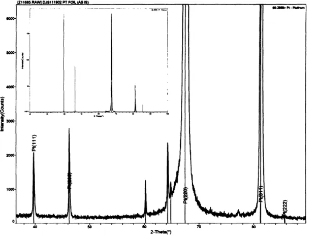
rapidly injected into a surfactant mixture of oleic acid (0.2 mL, CH3(CH2)7CHCH(CH2)7COOH, M.W. 282.58, CAS 112-80-1) and Trioctylphosphine Oxide (TOPO, 0.4g, [CH3(CH2)7]3PO, M.W. 386.65, CAS 78-50-2), dissolved in dichlorbenzene (12mL), at 182°C followed by refluxing for 3 minutes. The solution was allowed to cool to room temperature by removal of the heating mantle. Post synthesis processing involved the ethanol induced precipitation and centrifugation of the reaction product, followed by resuspension in hexane. This process was repeated thrice in order to further focus the size distribution of the cobalt nanoparticles. Verification of the synthetic process was achieved by transmission electron microscopy (TEM) and X-ray diffraction (XRD, figure 2.1). TEM samples were prepared by direct deposition of the cobalt particle solution onto carbon coated copper TEM grids (ted pella) and analyzed using a JEOL 200 cx microscope. XRD samples were prepared by drop coating aliquots of the particle solution onto 2cm2 pieces of silicon 110, followed by drying and then repeating the process
multiple times in order to achieve the particle density necessary for achieving an accepted signal count. Analysis was performed on a Phillips XRD using the Copper K-alpha line.
-,
n
_I 1 ~}-~.
I I I I 01 «I • IIIDoll,-.c.•
• • --., --'---. -. io---
...
---Figure 2.1 XRD of chemically synthesized Cobalt nanoparticles.
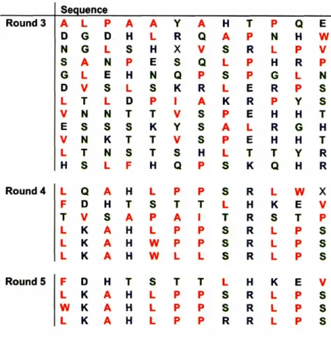
Substrates used in the screening of the phage display library were used immediately after removal from the furnace to prevent the onset of oxidation. All solutions used during the selection process were deairated under house vacuum to prevent oxidation. biopanning was performed with both the Ph.D. 12 library and yielded the sequences given in table 2.1, amino acids are color coded according to the reactivity of their side group: Hydrophobic (red); Hydrophilic (green); Negative (black); Positive (blue). All sequences are reported in order from N-terminus to C-terminus.