HAL Id: tel-02939373
https://tel.archives-ouvertes.fr/tel-02939373
Submitted on 15 Sep 2020HAL is a multi-disciplinary open access archive for the deposit and dissemination of sci-entific research documents, whether they are pub-lished or not. The documents may come from teaching and research institutions in France or abroad, or from public or private research centers.
L’archive ouverte pluridisciplinaire HAL, est destinée au dépôt et à la diffusion de documents scientifiques de niveau recherche, publiés ou non, émanant des établissements d’enseignement et de recherche français ou étrangers, des laboratoires publics ou privés.
orodental anomalies associated with rare diseases
Supawich Morkmued
To cite this version:
Supawich Morkmued. Clinical, preclinical and translational approaches of orodental anomalies asso-ciated with rare diseases. Development Biology. Université de Strasbourg, 2017. English. �NNT : 2017STRAJ040�. �tel-02939373�
UNIVERSITÉ DE STRASBOURG
ÉCOLE DOCTORALE DES SCIENCES DE LA VIE ET DE LA SANTÉ
Institut de génétique et de biologie moléculaire et cellulaire IGBMC,
CNRS-UdS UMR7104, Inserm U964
THÈSE
présentée par :
Supawich MORKMUED
soutenue le : 8 septembre 2017
pour obtenir le grade de : Docteur de l’Université de Strasbourg
Discipline/ Spécialité
: Biologie des organismes : développement et physiologie
Approches cliniques, précliniques et
translationnelles des anomalies
bucco-dentaires associées aux maladies rares
THÈSE dirigée par :
[Mme BLOCH-ZUPAN Agnès] Professeur, Université de Strasbourg RAPPORTEURS :
[M KLEIN Ophir] Professeur, University of California San Francisco, USA [Mme BABAJKO Sylvie] Docteure, Centre de Recherche des Cordeliers, Paris
AUTRES MEMBRES DU JURY :
[M MARK Manuel] Professeur, IGBMC, Illkirch
[M PIPPENGER Benjamin] Docteur, Institut Straumann AG, SWITZERLAND [Mme PITIPHAT Waranuch] Professeur, Khon Kaen University, THAILAND [Mme NIEDERREITHER Karen] Docteur, IGBMC, Illkirch
UNIVERSITY OF STRASBOURG
DOCTORAL SCHOOL OF LIFE AND HEALTH SCIENCES
The Institute of Genetics and Molecular and Cellular Biology IGBMC,
CNRS-UdS UMR7104, Inserm U964
THESIS
presented by :
Supawich MORKMUED
Defended on : 8 september 2017
For obtained the grade of: Doctor of University of Strasbourg Discipline/ Specialty
: Biology of organisms: development and physiology
Clinical, preclinical and translational
approaches of orodental anomalies
associated with rare diseases
THESIS directed by:
[Mme BLOCH-ZUPAN Agnès] Professor, University of Strasbourg REPORTERS :
[M KLEIN Ophir] Professor, University of California San Francisco, USA [Mme BABAJKO Sylvie] Doctor, the Cordeliers Research Centre (CRC), Paris
OTHER JURY MEMBERS :
[M MARK Manuel] Professor, IGBMC, Illkirch
[M PIPPENGER Benjamin] Doctor, Institut Straumann AG, SWITZERLAND [Mme PITIPHAT Waranuch] Professor, Khon Kaen University, THAILAND [Mme NIEDERREITHER Karen] Doctor, IGBMC, Illkirch
REMERCIEMENTS
Comme vous le savez, je suis venu en France il y a cinq ans pour obtenir ma thèse. En septembre s'achèvera cette étape qui sera pour moi une expérience inoubliable et j'aimerais remercier toutes les personnes qui ont rendu cela possible.
Tout d'abord, je veux exprimer ma plus profonde gratitude au Pr Madame Agnès BLOCH-ZUPAN pour avoir été mon principal superviseur. Elle m'a donné de nombreuses occasions de m'exprimer et de progresser en tant que chercheur, dentiste, conférencier et m'a même accueilli en France au début de mon séjour. Elle m'a fourni des ressources scientifiques exceptionnelles, un soutien continu ainsi qu'un mentorat et un excellent environnement de travail. Son engagement envers la recherche et le traitement des patients atteints de maladies rares est exceptionnel. Le Pr. BLOCH-ZUPAN m'a également donné des conseils personnels et des clés pour comprendre la culture française et le système académique français. Je suis venu vous dire, chère Agnès, que je m'en vais, comme dit la chanson, avec un immense sentiment de reconnaissance pour vous.
Je suis aussi extrêmement reconnaissant envers la Docteure Karen NIEDERREITHER, un co-superviseur qui possède une expertise considérable dans la recherche sur les rétinoïdes et la biologie du développement de la souris. Elle a toujours partagé ses expériences et aidé à diriger mon projet pour faire avancer efficacement mes travaux et mon développement intellectuel. Son engagement personnel pour l'excellence dans la science m'a beaucoup inspiré.
Je tiens à exprimer tous mes remerciements aux membres du Comité consultatif de Mi-thèse:
Le Pr. Ekkehart LAUSCH, Université de Fribourg, pour ses contributions en tant que membre externe du comité de thèse. C'est un merveilleux clinicien et chercheur qui m'a fourni des connaissances en biologie du développement et pour le traitement des patients. Sa carrière est très certainement un exemple pour mon avenir professionnel.
Le Dr Stéphane VINCENT, pour avoir éclairé mes recherches de son expertise en biologie et pour être un excellent chercheur.
Le Dr François CLAUSS, pour son soutien dans mes travaux et pour avoir partagé son expertise dans les expériences chirurgicales sur les souris, à la fois l'implant dentaire et la guérison (modèle d'extraction dentaire) à la faculté de médecine. Il m'a énormément aidé techniquement et intellectuellement à faire avancer mes recherches. J'apprécie particulièrement nos expériences conjointes et nos discussions approfondies.
Je tiens à remercier le Professeur Ophir KLEIN, de l'Université de Californie à San Francisco, aux États-Unis, et la Docteure Sylvie BABAJKO, du Centre de Recherche des Cordeliers de Paris pour avoir fait partie du jury de thèse de doctorat en tant que rapporteurs de ce travail.
Je tiens aussi à exprimer ma gratitude au Professeur Manuel MARK, de l'IGBMC, de l'Université de Strasbourg, au Dr Benjamin PIPPENGER de l'Institut Straumann AG et au Dr Waranuch PITIPHAT de l'Université de Khon Kaen pour avoir accepté d'être membres de ce jury de thèse. Je leur en suis très reconnaissant et suis très honoré de leur participation.
Je tiens également à remercier toutes les personnes qui m'ont encouragé et soutenu tout au long de la formation doctorale. J'ai reçu un soutien et une formation considérables et diversifiés pour des expériences et des discussions scientifiques et je remercie les personnes suivantes pour leur générosité dans le partage des connaissances et du matériel expérimental :
Tout d'abord, j'aimerais remercier deux ingénieurs talentueux : Brigitte SCHUHBAUR et Valérie FRAULOB qui m'ont aidé avec leur assistance de laboratoire et leurs conseils techniques. J'ai appris beaucoup de leurs expériences scientifiques et générales. Surtout, leur amitié m'a été précieuse tout au long des événements sociaux et de ma vie au laboratoire. Je ne trouve pas de mots suffisants pour les remercier toutes les deux.
Le Pr Dr Pascal DOLLE pour ses conseils, son soutien et son expertise en préparation de manuscrits. Cela a également favorisé mon avancement professionnel.
Je remercie tous les membres de l'équipe DOLLE, les membres actuels et passés du laboratoire :
Le Dr. Virginie LAUGEL-HAUSHALTER, pour m'avoir fait bénéficier de son engagement personnel et de sa technique du profilage biologique ;
Le Dr. Marie PASCHAKI, pour le soutien en biologie moléculaire et en génétique de souris ;
Le Dr. Anna NIEWIADOMSKA-CIMICKA, pour le soutien en biochimie des protéines, l'analyse des protéines, l'analyse des données de séquençage et pour sa gentillesse ;
Le Dr Carole HAUSHALTER, pour son soutien en biologie moléculaire, génétique de souris et pour sa belle amitié ;
Le Dr Muriel RHINN, pour m'avoir fourni un enseignement fondamental dans la manipulation de la souris et la biochimie ;
Le Dr Wojciech KREZEL, Ania PODLESNY, Marion CIACIA, le Dr Samir LAOUINA, Claire HUBER, Thomas HEID, le Dr Elyette BROLY et le Dr. Rose MBEDE NGA MVONDO pour le soutien en sciences de la biologie et avec qui j'ai partagé de beaux moments pendant ma vie de doctorat.
Je remercie les autres membres de l'IGBMC, de l’Institut clinique de la souris ICS, de la plate-forme d'histopathologie, de la plate-forme de microarray et de la génomique, de la phénotypisation et de la plate-forme d'imagerie μCT. Je tiens à remercier également les membres de la Faculté de médecine et de chirurgie dentaire de l’Université de Strasbourg:
Le Dr. Megana PRASAD, pour son enseignement fondamental dans les analyses de génétique humaine. Son expertise dans la recherche m'inspirera et elle sera un modèle pour moi ;
Le Dr Mathilde HUCKERT, pour le soutien et l'amitié en clinique d’odontologie pédiatrique, de la Faculté de chirurgie dentaire et du Pôle de médecine et chirurgie bucco-dentaire des Hôpitaux Universitaires de Strasbourg.;
Le Mr. Joseph HEMMERLE et le Mr. Eric MATHIEU, pour avoir donné son soutien lors des expériences et des résultats de microscopie électronique ;
Marzena KAWCZYNSKI, pour l'assistance dans la base de données, les échantillons salivaires et pour avoir su créer un environnement de travail joyeux lors de nombreux événements ;
Le Dr. Alexandru PARLOG, pour avoir donné son enseignement fondamental et pour sa contribution utile dans la recherche osseuse ;
Le Dr. Bernard JOST, pour son soutien lors des expériences de séquençage et de l'analyse bioinformatique ;
Le Dr. Olivia WENDLING, pour la formation en histologie, en immunofluorescence et le projet "Phenomin" pour la souris ;
Le Dr. Isabelle GONCALVES DA CRUZ, pour son soutien dans la zone de phénotypage de souris de l’ICS et la formation de micro-CT ;
Le Dr. Johan H van Es et le Dr. Hans Clevers, de Koninklijke Nederlandse Akademie van Wetenschappen, Amsterdam, Pays-Bas pour avoir fourni la souris Smoc2-Cre-GFP.
Je suis très reconnaissant envers le doyen de la Faculté de Chirurgie Dentaire de Strasbourg –le Pr Madame Corinne TADDEI-GROSS, et l'ancien doyen de la faculté de médecine dentaire, l'Université de Khon Kaen, en Thaïlande, le Dr Waranuch PITIPHAT et le Dr Nawarat WARA-ASWAPATI CHAROEN, ainsi que mon ancien chef du département pédiatrique, le Dr. Patimaporn PUNGCHANCHAIKUL, pour m'avoir encouragé à m'engager dans l'enseignement supérieur en dentisterie académique, et donc pour soutenir mon parcours dans la formation doctorale.
Concernant le soutien financier, je suis très heureux d'avoir pu bénéficier de la bourse franco-thaïlandaise et de la bourse de l'Université de Khon Kaen afin de faire face à mes dépenses en France tout au long de la durée du doctorat. Je remercie aussi les financements de l'Institut d'Études Avancées de l'Université de Strasbourg
(USIAS), INTERREG IV et INTERREG V (FEDER) RARENET pour leur soutien à la recherche.
Merci à Alexandre HERNANDEZ pour son aide dans toutes mes démarches administratives, dans l'apprentissage de la langue française et la découverte de la culture française.
Merci à tous mes amis thaïlandais PJoke, Boat, New, Mod, Chinchar, Wanchun, Eve, Toei, Nhum, Roger, Tum et Ake en Thaïlande et Sae, Fair, NTum, Paew, PPek, PNan, PYim, PPim, San, Et NTaddy en France pour leur soutien continu, avec qui j'ai partagé des moments inoubliables et pour m'avoir encouragé dans mon travail. Je leur souhaite de très belles choses dans leur avenir.
Ces remerciements ne seraient pas complets si je ne parlais pas de ma famille, de son soutien et de son attention constante. Je souhaite exprimer ma plus grande reconnaissance à mes parents, même si je ne formule jamais mes sentiments directement. Bien qu'ils sachent combien j'apprécie leur temps et leurs efforts, il est important que je souligne ce soutien à la fin de ma thèse. Je suis si heureux de grandir dans cette famille et j'ai beaucoup apprécié leur récente visite en Europe.
Enfin, je voudrais évoquer quelqu'un qui a pris une place fondamentale dans ma vie. Grâce à elle, j'ai eu l'énergie de mener mes activités jusqu'à leur terme et la force d'obtenir mon doctorat. Il s'agit de ma future femme, le Dr Preeyarat PLONGNIRAS. Elle est ma meilleure amie et partenaire dans la vie. Je sais combien ses encouragements et son soutien sont précieux, et j'espère une vie merveilleuse avec elle. En Thaïlande et à l'étranger, elle m'a enchanté avec son joli sourire, son rire et son amour.
ACKNOWLEDGEMENTS
At the completion of my thesis, I would like to thank all those people who made this possible and an unforgettable experience for me.
First of all, I would like to express my deepest sense of gratitude to my supervisor Pr. Agnès BLOCH-ZUPAN for being my primary supervisor. She has given me many opportunities to develop as a researcher, dentist, lecturer, and even has hosted me in France at the beginning. She has provided me with exceptional scientific resources, continuous support and mentoring, and a great working environment. Her commitment to the research and treatment of rare diseases patients is exceptional. Pr. Agnès also has given me personal guidance and support for life in understanding French culture and the French academic system, which I am deeply grateful for.
I am extremely grateful and thank Dr. Karen NIEDERREITHER, a co-supervisor with considerable expertise in retinoid signaling and mouse developmental biology research. She has always shared her experiences and helped direct my project to efficiently advance my work and intellectual development. Her personal commitment to excellence in science has greatly inspired me and is very much appreciated.
I would like to express my appreciation to all of the members of my Thesis Advisory Committee Meetings:
Pr. Ekkehart LAUSCH, Freiburg University, for his contributions as an external mid-thesis committee member. He is actively a wonderful clinician and a researcher who provided me knowledge in both developmental biology and patient treatment. His professional career inspires me on how to move forward for my future.
Dr. Stéphane VINCENT, for his essential vision on my research from his expertise in biology from his experience as an excellent researcher.
Dr. François CLAUSS, for supporting my research and sharing his expertise in mouse surgical experiments, both dental implant and would healing (tooth extraction
models). He has helped me technically and intellectually to advance my research tremendously. I especially appreciate our joint experiences and in depth discussions.
I would like to thank Prof. Ophir KLEIN, from University of California, San Francisco, USA and Dr. Sylvie BABAJKO, from the Centre de Recherche des Cordeliers, Paris for being part of the PhD thesis committee, acting as external reviewers of this PhD thesis.
I would like to thank Prof. Manuel MARK, from IGBMC, University of Strasbourg, Dr. Benjamin PIPPENGER from Institut Straumann AG, and Dr. Waranuch PITIPHAT from Khon Kaen University, for accepting to be members of this PhD thesis committee. I am grateful and honored by their participation.
I also would like to thank all individuals who encouraged and supported me throughout the doctoral training. I have received tremendous, diverse support and training and want to thank the following individuals for their generosity of sharing knowledge and experimental materials.
Firstly, I would like to thank 2 talented engineers; Brigitte SCHUHBAUR and Valérie FRAULOB, who have helped offering laboratory assistance and technical advice. I have learned a tremendous amount from your scientific and general experiences. Their friendships are invaluable. I find no words sufficient to thank both of you.
Dr. Pascal DOLLE for his advice, support, and expertise in manuscript preparation, which has been invaluable in my professional advancement.
I am thankful to everyone in DOLLE team, present and past members of laboratory;
Dr. Virginie LAUGEL-HAUSHALTER, for inspiring me with her personal commitment and biological excellence in genetic profiling.
Dr. Marie PASCHAKI, for support in molecular biology and mouse genetics.
Dr. Anna NIEWIADOMSKA-CIMICKA, for support in protein biochemistry, protein analysis, sequencing data analysis, and for her kindly friendship.
Dr. Carole HAUSHALTER, for support in molecular biology, mouse genetics, and for her wonderful friendship.
Dr. Muriel RHINN, for providing fundamental teaching in mouse manipulation and biochemistry.
Dr. Wojciech KREZEL, Ania PODLESNY, Marion CIACIA, Dr. Samir LAOUINA, Claire HUBER, Thomas HEID, Dr. Elyette BROLY, and Dr. Rose MBEDE NGA MVONDO for support in biology science and sharing nice moments during my PhD life.
I thank other members at IGBMC, ICS mouse facility, histopathology platform, microarray and genomics platform, phenotyping and µCT imaging platform. I would like to thank also the members at medical and dental faculties of the Université de Strasbourg;
Dr. Megana PRASAD, for providing fundamental teaching in human mutational analyses. Her expertise in genetic research inspires me.
Dr. Mathilde HUCKERT, for support and friendship in pediatric clinic at the Strasbourg Faculty of Dentistry and Teaching Hospital.
Mr. Joseph HEMMERLE and Mr. Eric MATHIEU, for providing fundamental discussions and useful scanning electron microscopy results.
Marzena KAWCZYNSKI, for support in database, salivary samples and for creating an amazing work environment during many events.
Dr. Alexandru PARLOG, for providing fundamental teaching and useful discussions in bone research.
Dr. Bernard JOST, for support of sequencing experiments and bioinformatic analysis.
Dr. Olivia WENDLING, for providing the histology, immunofluorescence, and Phenomin mouse database information.
Dr. Isabelle GONCALVES DA CRUZ, for her support in mouse phenotype area and micro-CT training.
Drs. Johan H van Es and Hans Clevers, from Koninklijke Nederlandse Akademie van Wetenschappen, Amsterdam, Netherlands for providing the Smoc2-Cre-GFP mice.
I am very grateful to the current Dean of the Faculté de Chirurgie Dentaire de Strasbourg -Corinne TADDEI-GROSS, the current and former Dean of the faculty of dentistry at Khon Kaen University, Thailand- Dr. Waranuch PITIPHAT and Dr. Nawarat WARA-ASWAPATI CHAROEN. My former head of the pediatric department, Dr. Patimaporn PUNGCHANCHAIKUL. These individuals have inspired me and supported my commitment to higher education in academic dentistry.
For financial support, I am grateful to the Franco-Thai scholarship and Khon Kaen University. These scholarships have supported my living expenses in France. In addition, I am grateful to University of Strasbourg Institute of Advanced Studies (USIAS), INTERREG IV and V (ERDF funding) RARENET funding to support research expenses.
Thanks to Alexandre HERNANDEZ for his help in French language, and French discovery.
Thanks all my Thai friends PJoke, Boat, New, Mod, Chinchar, Wanchun, Eve, Toei, Nhum, Roger, Tum, and Ake in Thailand and Sae, Fair, NTum, Paew, PPek, PNan, PYim, PPim, San, and NTaddy in France for their continuous support, sharing unforgettable moments, and encouragement.
These acknowledgments would not be complete without thanking my family for their constant support. I want to express my deepest appreciation to my parents. They have guided me from the youngest age, and have been my role model. Their constant faith and hard-working ethics has inspired me.
Finally, I would like to mention the most important lady in my life, my future wife, Dr Preeyarat PLONGNIRAS. She is my best friend and partner in life. I appreciate her encouragement and support, and look forward to a wonderful life together. In Thailand and abroad, she has enchanted me with her cute smile, laughter, and love. Personally, I look forward to a bright future with her.
TABLE DES MATIÈRES (CONTENTS)
REMERCIEMENTS ... 3
ACKNOWLEDGEMENTS ... 8
TABLE DES MATIÈRES (CONTENTS)... 12
Liste des tableaux (List of tables) ... 14
Liste des figures (List of figures) ... 15
Liste des Abréviations (List of Abbreviations) ... 16
INTRODUCTION ... 18
CONTEXTE ET COMMENTAIRE (BACKGROUND AND SIGNIFICANCE) ... 21
Part I Tooth development ... 22
I.1 The mouse dentition ... 23
I.2 Sequential stages of odontogenesis ... 25
I.2.1 The origin of ectodermal tissue and appendages ... 25
I.2.2 Initiation of dental development ... 26
I.2.3 Dental morphogenesis ... 28
I.2.4 How the tooth begins to differentiate into defined tooth lineages ... 29
Part II Signaling pathways involved in tooth development ... 34
II.1 Transcription factors critical in tooth patterning ... 35
II.2 The TGF-β/BMP pathway ... 39
II.3 FGFs ... 45
II.4 WNTs ... 47
II.5 NF-κB/TNF Pathway... 49
II.6 The Sonic hedgehog pathway ... 52
II.7 Notch ... 55
Part III Retinoids ... 56
III.1 The general role of vitamin A ... 57
III.2 Clinical use of retinoids and the worldwide role in public health ... 58
III.3 How RA is obtained from dietary sources, stored in liver, and transported to the cell ... 59
III.4 Target Cell Uptake, Intracellular Metabolism, Cytoplasmic and Nuclear Receptors Mediating Vitamin A Effects ... 62
III.5 The function of retinoic acid as a transcriptional regulator ... 63
III.7 Associations between vitamin A and evident effect on bone mass in humans 73
III.8 Mechanism of RA effects on bone development ... 74
Part IV Dental anomalies ... 77
IV.1 Definitions ... 78
IV.2 Environmental factors altering tooth development ... 78
IV.3 Dental anomalies in rare genetic diseases ... 83
IV.3.1 Hypodontia/Oligodontia ... 83
IV.3.2 Supernumerary teeth or hyperdontia ... 85
IV.3.3 Microdontia ... 85
IV.3.4 Heritable dentin conditions ... 86
IV.3.5 Dentin Dysplasia ... 86
IV.3.6 Amelogenesis Imperfecta ... 87
IV.4 A focus on rare diseases through human genetics and mimicking mouse models ... 89
Résumé de la thèse ... 95
RÉSULTATS (RESULTS) ... 101
(i) Environmental factors ... 102
Retinoic Acid Excess Impairs Amelogenesis Inducing Enamel Defects ... 103
(ii) Genetic factors ... 129
Enamel and dental anomalies in latent-transforming growth factor beta-binding protein 3 mutant mice ... 130
Craniofacial and tooth abnormalities in Sparc-related modular calcium-binding protein 2 (Smoc2) mutant mice... 148
CONCLUSIONS-DISCUSSION-PERSPECTIVES ... 169
ANNEXES (APPENDIX) ... 178
Liste des annexes (List of appendix) ... 179
RÉFÉRENCES BIBLIOGRAPHIQUES (REFERENCES) ... 285
Résumé ... 310
Liste des tableaux (List of tables)
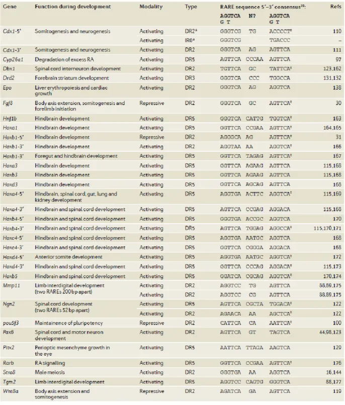
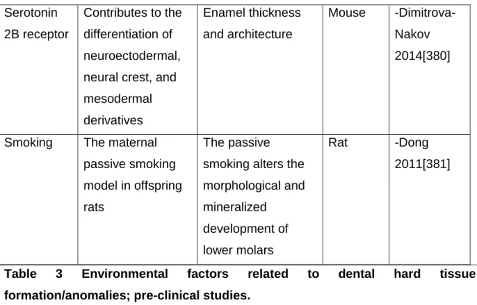
Table 1 Summary of phenotypes resulting from targeted inactivation of retinoid signaling pathway components in mice……….70 Table 2 RA response elements, either activating or repressing retinoid targets, and their respective physiological roles……….71 Table 3 Environmental factors related to dental hard tissue formation/anomalies; pre-clinical studies………80 Table 4 Human rare diseases and their phenotypes…………..……….………90
Liste des figures (List of figures)
Figure 1 The human and mouse dental morphology and a fate of dental
development………..……….24
Figure 2 Signaling cascades during epithelial-mesenchymal interaction..…….……..27
Figure 3 Ectodermal appendages development during its initial stages……….…….27
Figure 4 Normal enamel prism of lower incisor in mouse………...………31
Figure 5 Pattern of gene expression in the developing tooth……….35
Figure 6 Schematic representation of the Bmp and Tgf-β signaling pathways…...…40
Figure 7 TGF-β synthesis and activation………..……….43
Figure 8 Schematic representation of Fgf signaling pathway………….………45
Figure 9 Schematic representation of Wnt signaling pathway……….………..48
Figure 10 Schematic representation of Eda signaling pathway……….………50
Figure 11 Schematic representation of Notch, Wnt, and Shh signaling cascades….53 Figure 12 The fate of vitamin A transportation………..………60
Figure 13 Intracellular regulation of retinoid signaling……….64
Figure 14 RA signaling mechanism……….…...66
During mammalian evolution head morphogenesis is both a conserved and complex process. The vertebrate skull is a complexly designed, evolutionarily quite ancient structure. Within mammals, the overall shape of the skull shows important conserved components, species-specific variations, along with hereditarily conserved individual components[1]. Craniofacial and orodental developmental anomalies are often clear aspects of rare diseases or syndromes. These diseases encompass about 7000 different entities, affecting 4 million people in France, and almost 25 million in Europe. By definition, rare diseases affect less than one person in 2000 and for ~80%, are genetically driven. Among the over than 5000 known syndromes, more than 700 have a craniofacial and/or orodental phenotype[2, 3]. About 250 (of the 700) genetic diseases display cleft lip/palate symptoms. Novel rare diseases and corresponding mutated genes will thus be essential to understand genetic signaling pathways regulating craniofacial and dental development, and provide clues to understand organogenesis to develop future clinical approaches.
Tooth development/anomalies is an excellent model organogenesis to understand how a given mutated gene can alter tooth morphogenesis and/or terminal differentiation of post-mitotic cells, events leading to the formation and mineralization of dentin, enamel, cement. It is also a system to decipher/epigenetics, including genetic interactions with environment. The tooth is indeed an excellent marker of environmental assaults, which when taking place during the early mineralization process (prior tooth eruption within the oral cavity) can have permanent effects. A combination of genetic background and susceptibility to environment may modulate any given phenotype.
Mouse models dentition, despite intrinsic differences reproduces dental anomalies encountered in human rare diseases. These models are a powerful approach to increase our understanding of human disease, through advancing our understanding of fundamental biological mechanisms[4].
This PhD thesis will first present as background information dental development and the main pathways involved, with a focus on retinoids. It will then discuss dental anomalies in relationship with environmental assaults and rare diseases.
The experimental work (presented in published papers) begins with investigations on the role of environment, by examining the developmental effects of excess retinoic acid on enamel formation. The next two papers explore the craniofacial and orodental phenotype of 2 rare diseases transgenic animal models. Enamel and dental anomalies in latent transforming growth factor-β binding protein 3 (Ltbp3) mutant mice are examined. Lastly, a manuscript in preparation on the craniofacial and tooth abnormalities in Sparc-related modular calcium-binding protein 2 (Smoc2) mutant mice is presented.
These rare disease mouse models were selected based on sequencing data from families and patients enrolled in the Strasbourg University and Hospital Reference Center for orodental rare diseases. These investigations have taken place in collaboration with O-Rares, within the ERDF funding framework of the Interreg IV Offensive Sciences and the Interreg V RARENET projects.
CONTEXTE ET COMMENTAIRE
Part I
Teeth are composite organs with both an epithelial (the enamel organ) and a mesenchymal compartment (papilla, pulp). They use epithelio-mesenchymal interactions to drive their development. The same conserved signaling pathways that regulate most aspects of embryonic development are required for tooth development.
Odontogenesis starts as neural crest cells individualize and migrate towards the first branchial arch to interact with the oral ectoderm, and initiate in specific location and timing the beginning stages of tooth development. Tooth development then progresses through different stages from dental lamina to individual placodes, then through bud, cap, bell stages, to set up the crown and direct later root morphogenesis. Timely controlled terminal differentiation of odontoblasts and ameloblasts begins, leading to the patterning of dentin and enamel matrix. This is followed by further maturation in the mineralization processes. The alveolar bone and the periodontium develop concomitantly.
Most of our current knowledge on the molecular and genetic basis of tooth development has come from mouse studies. Extensive investigations into the molecular regulation of tooth formation have been carried out with many genetically engineered mouse models[5, 6].
Developmental anomalies include changes in the number, shape, size, and/or composite tooth structure. These changes may also be visualized through the color or structural integrity of dental hard tissues such as dentin, enamel, cementum, alveolar bone. In addition the eruption and/or resorption of teeth are also linked to stage-specific morphological and molecular events and pathways.
I.1 The mouse dentition
Mouse teeth have a unique, conserved organization. This is shown in Figure 1. In each quadrant, a single incisor is separated from three molars by a gap (or toothless region) called the diastema. Hence rodents such as mice display a reduced number of teeth (compared to humans). Mice also have a single set of teeth. Rodent teeth are considered to be deciduous teeth that do not undergo replacement[7, 8], whereas humans have two sets of teeth[9]. The potential for tooth replacement in mice appears to have been retained[8, 10, 11]. Rodent incisors are different from
humans because they grow continuously throughout the life of the animal, a property assigned to the presence of stem cells populations in their cervical loops. Enamel is present only on the labial side of the incisor, considered as a crown analogue. The lingual side is covered with dentin and represents the root analogue.
Figure 1 The human and mouse dental morphology and a fate of dental development.
(A) (B) The human permanent dentition, in comparison with the mouse. Mice have a toothless diastema separating incisor and molars in each half of the jaw.
(C) Dental development begins from a thickening of the epithelium, where the growing epithelium then forms bud. Then, the dental mesenchyme condenses underlying the tooth epithelial bud. At the morphogenesis stage, the epithelial tissue transforms to cap and bell shapes. The signaling centers, called primary and secondary enamel knots, in the enamel organ control the growth and the shape of the tooth. At the tooth differentiation stage, enamel-secreting ameloblasts and dentin-secreting odontoblasts mature from epithelial and mesenchymal cells, respectively. (source [12])
I.2 Sequential stages of odontogenesis
Odontogenesis occurs in sequential stages. To recapitulate, this begins with tooth initiation, induction of epithelial placodes (seen as localized thickening of the oral ectoderm), followed by bud, cap, and bell morphogenetic stages. Subsequently, the terminal differentiation of odontoblasts and ameloblasts is observed (Figure 1). Next, root formation and tooth eruption occur. The appearance of sub-regional tooth-types (incisors and molars) can be distinguished by the unique shape of the crown and root, as well as a defined cusp patterns. Studies of developing mouse teeth suggest that cusp initiation and patterning in tooth germs is a process that repeatedly re-utilizes conserved developmental pathways[13-17].
I.2.1 The origin of ectodermal tissue and appendages
During early embryogenesis the ectoderm is the external germ layer. The epidermis develops from the surface ectoderm[18]. Epidermis-derived structures include the skin and other stratified epithelia such as the oral epithelium. Subsequently, these tissues diversify to specialized structures (called ectodermal appendages) that include mammary glands, salivary glands, hair follicles, and teeth. These ectodermal appendages all develop through similar mechanisms, involving precise crosstalk between the epithelium and mesenchyme, and often sharing common morphological features during early organogenesis[19].
The cephalic neural crest
Teeth are ectomesenchymal organs derived from oral ectoderm and neural crest cells ectomesenchyme. The level of origin within the crest for lower incisors and lower and upper molars is identical, namely the caudal part of the midbrain/rostral hindbrain. These cells all migrate toward the first branchial arch. Cells originating from the forebrain and rostral midbrain also migrate towards the frontonasal process and the intermaxillary process to participate to the development of the upper incisors.
I.2.2 Initiation of dental development
Teeth are initiated from the dental lamina, a stripe of stratified epithelium, first visualized at mouse embryonic stage (E) 11. This dental lamina is of epithelial origin (oral ectoderm). Localized epithelial thickenings (placodes) within the dental lamina mark the initiating tooth, which then buds into the underlying mesenchyme.
Dental placode formation can be visualized by Pitx2 expression. This transcription factor serves as a specific, early marker of dental epithelium, whose expression persists in all epithelial cells of the developing tooth until crown morphogenesis[20]. Reciprocal epithelial–mesenchymal cross-tissue interactions induce and regulate tooth morphogenesis[21]. Tooth-generating oral regions have a so-called odontogenic potential.
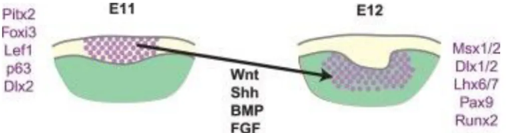
Classical tissue recombination experiments have shown that the odontogenic potential first resides in the epithelium at the placode stage[9]. Hence, mouse oral epithelial region (between E9 and E11.5) can induce tooth formation in non-dental mesenchyme[22, 23]. After E11.5 the odontogenic potential shifts to the dental mesenchyme, so this mesenchyme can induce non-dental epithelia to become teeth[24]. In parallel, the ability of the epithelium to induce tooth appears lost at this stage. The shift in odontogenic potential to mesenchyme temporally occurs at the placode stage[24, 25]. The dental lamina expresses many transcription factors and signal molecules such as fibroblast growth factors (FGFs), bone morphogenetic proteins (BMPs), sonic hedgehog (SHH), and WNT signals that regulate dental identity. Gene restricted to the placodes or early signaling centers include Pitx2 and
Foxi3, along with many other signaling pathway molecules (including Shh, Wnt, Bmp,
Fgfs-notably Fgf20)[12, 26, 27]. Early combined actions of these factors increase proliferation producing the tooth bud. Some of these factors are involved in mediating the shift of odontogenic potential to the mesenchyme (Figure 2) (reviewed in [21]).
Figure 2Signaling cascades during epithelial-mesenchymal interaction.
The capacity to initiate teeth first resides in the oral epithelium of E10 and E11 stage mouse embryos (dental lamina stage). This capacity shifts at the placode stage (E12) to the surrounding neural crest-derived mesenchyme. This is simultaneous with dental mesenchyme condensation. The epithelial/mesenchymal odontogenic potential was defined through tissue recombination experiments (source [21])
Teeth develop as epithelial appendages and share many of the same regulatory mechanisms with other ectodermally-derived organs during their initial formation and morphogenesis[12] (Figure 3). During odontogenesis, specific cells secrete signaling molecules at defined sites which often initiate defined transcriptional cascades[28].
Figure 3 Ectodermal appendages development during its initial stages.
Ectoderm-derived structures initiate from embryonic ectoderm, mainly due to mesenchymal-derived signals. Epithelial placodes appear and subsequently grow into the surrounding mesenchyme. These early developmental events are similar in all ectodermal organs. At later stages, epithelial buds undergo different morphogenetic configurations, resulting in the formation of more specialized structures. (source [29])
I.2.3 Dental morphogenesis
During the transition from the bud to cap stage, a signaling center called the primary enamel knot is formed from a transient cluster of dental epithelial cells[30]. The primary knot establishes the crown base, regulates the pattern of tooth cusps, and directs the projections of enamel and dentin on the occlusal surface. In teeth with multiple cusps (such as the molars) secondary enamel knots appear early at the bell stage, positioned at the tips of future cusps. The secondary knots coordinate cusp patterning by inducing terminal differentiation of the adjacent mesenchymal-derived odontoblasts. Additional enamel knots (which result in additional cusps) can form outside the zones of inhibition of previously formed enamel knots. This process is dynamic. The patterning of cusps is not predetermined, so the size, shape and location of the first-forming cusps regulates the appearance of later-forming cusps[15]. Under this patterning cascade model of cusp development[14], a small change early in tooth germ growth results in large alterations in the fully formed crown, particularly in the total number of cusps.
After performing their function, enamel knots disintegrate through cell apoptosis. Gradients of signaling molecules, including those belonging to the Bmp, Fgf, Wnt, Shh, and NF-κB signaling pathways[13], originate from and act around the knot, functioning as inducers, mediators, and inhibitors to determine correct formation of tooth shape and crown patterning. These reiterative signaling molecules are indispensable throughout the process of micropatterning (cusp size and cusp number) and macropatterning (tooth size and tooth number). In mice more than 80 genes have been reported to be expressed, in and surrounding the knot (http://bite-it.helsinki.fi), suggesting their complex integration and interactions. Alterations of
these fine-tuned interactions lead to alterations of cusp patterning and tooth morphology. These types of dental anomalies are observed in a number of animal models, and are sometimes seen in analogous patient rare disease cases.
I.2.4 How the tooth begins to differentiate into defined tooth lineages
Dentin and enamel are mineralized tissues produced at the late bell stage of tooth development by two different tooth-specific post-mitotic cells: the mesenchyme-derived odontoblasts that produce dentin and the epithelial ameloblasts that produce enamel matrix proteins. Morphological and molecular events occurring during dentinogenesis and amelogenesis will be described.
I.2.4.1 Dentin development (dentinogenesis)
Dentin is the substructure of enamel, and its flexibility reduces tooth damage by absorbing force. Dentin has about 60% mineral content and is a tissue very similar to bone. It has an intricate network of dentinal tubules and is filled with dentinal fluid and odontoblastic processes that are thought to play a role in the neurosensory function of teeth[31]. Dentin acts throughout life as a protective barrier, with reparative responses to environmental assaults. Dentin also can function to secrete sclerotic dentin, which upon attack from dental carries or chewing forces blocks and protects dentinal tubules. Sclerotic dentin appears translucent due to calcification, occurring with aging or injury.
Odontoblasts are specialized ciliated cells [32], who function in the formation of dentin. Odontoblast differentiation follows a defined temporospatial gradient from the cusp tip towards the cervical area of the tooth, an event induced by the inner dental epithelial (IEE) cells. Odontoblasts synthesize dentin matrix proteins [33]. Collagens (I, type I trimer, III, V, VI) are abundant odontoblast components. Non-collagen components of odontoblasts include osteonectin, osteocalcin, and SIBLINGs (Small Integrin-Binding Ligand, N-linked Glycoproteins), osteopontin, bone sialoprotein (BSP), dentin matrix protein 1 (DMP1), and dentin sialophosphosproteins (DSPP or DSP and DPP)[34]. Other molecules which contribute to dentin formation include the proteoglycans, serum proteins such as albumin, enamel proteins such as amelogenins and matrix metalloproteinases (MMPs)[35].
The collagen molecules communicate with a variety of non-collagenous proteins to aid initiate and regulate the mineral deposition in these tissues. DSPP is the most abundant and well-known non-collagenous protein. It is a highly phosphorylated protein, which can attach to the type 1 collagen fibril, regulating collagen deposition at specific sites[31, 33].
Mutations in either type I collagen or linked proteins can cause hereditary dentin defects like Dentinogenesis Imperfecta (DI). Many mutations are found in these manners and are often associated with Osteogenesis Imperfecta (OI), a group of hereditary defects associated with bone fragility/anomalies. By the way, only some collagen mutations result in dentin defects[31, 33].
I.2.4.2 Enamel development (amelogenesis)
Dental enamel is the hardest and the most densely mineralised tissue of the body (95% in weight) mainly composed of calcium hydroxyapatite crystallites. Its strength is not only linked to its great mineral content, but also to the sophisticated-arranged organization of enamel crystallites and substructures. Thus, enamel formation necessitates an elaborately regulated orchestration of cellular and chemical events during amelogenesis to properly make enamel with the certain mineral composition and organization[36]. Genetic aberrations or environmental disturbances during this process are able to cause developmental enamel defects[37, 38].
Ameloblasts differentiate, within the enamel organ, at bell stage, from IEE. Their differentiation follows the same temporospatial gradient as odontoblasts, but with a different timeframe. Amelogenesis proceeds in the presence of predentin/dentin and after disappearance of the basement membrane (stages when odontoblasts are functional).
Ameloblast synthesize, participate in mineralization and resorb the enamel matrix proteins during the maturation stage[37]. Amelogenesis can be segmented in five stages, including the initial or pre-secretory stage, secretory stage, transitional stage, maturation stage, and post-maturation stage. First, a layer of IEE adjacent to the underlying ectomesenchyme starts to differentiate into ameloblastic cells, they
withdraw from the cell cycle, elongate, polarize and develop the protein synthesis organites. Pre-secretory ameloblasts differentiate into secretory ameloblasts which deposit a protein matrix. Hence, the enamel matrix acts as a provisional protein scaffold on which enamel crystals are able to form[39]. Differentiating pre-ameloblasts stretch cytoplasmic projections through the basement membrane, which is then gradually degraded. These pre-secretory stage ameloblasts provide an appropriate environment for subsequent matrix and ion deposition. During the secretory stage, the ameloblasts become columnar-shape cells and secrete tremendous amounts of enamel matrix proteins such as Amelogenin (Amel), Ameloblastin (Ambn), and Enamelin (Enam).
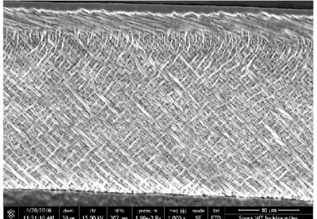
Figure 4 Normal enamel prism of lower incisor in mouse.
Scanning electron microscopic (SEM) analyses of lower incisors from 7 week-old wild-type (WT) mice shown the characteristic enamel rod/interrod structure. More detailed analyses of these enamel microstructure reveals a pattern of interlocking enamel prisms at the erupted portion of the incisor (where enamel is mature). This enamel prism pattern reveals normal thickness of decussations in its criss-crossing pattern. (Images from Supawich Morkmued (2016) collaborated with Pr Joseph Hemmerlé and Dr Eric Mathieu)
During initial enamel secretion, ameloblasts move away from the dentino-enamel junction (DEJ), to permit extension of dentino-enamel crystal ribbons and to enable
appositional growth (first as aprismatic enamel, then as prismatic enamel (ameloblasts with Tomes process), and finally as aprismatic surface enamel (stages in human enamel formation)[36]. This progression establishes the thickness of the enamel and serves as a mold for orientating enamel crystals. When the enamel reaches its maximum thickness, then secretory-stage ameloblasts withdraw their cytoplasmic processes and turn into a shorted columnar-shape transition-stage, under which a basement membrane reforms. Subsequently, the transition-stage ameloblasts further shorten into cuboidal maturation-stage ameloblasts, which begin adjusting between ruffle and smooth-ended cells at the surface of enamel. During this maturation stage, the enamel matrix proteins are then degraded by enamel proteases such as kallikrein-related peptidase 4 (KLK4) and matrix metalloproteinase 20 (MMP20). Thus, these proteases permit enamel to degrade matrix and to serve space for additional mineral deposition at the areas of future enamel crystallites, which increases the strength of the enamel[36, 37] (Figure 4). Finally, the enamel is able to exchange mineral ions of the saliva and oral fluid after eruption, which can affect the composition of the external layers of enamel. Hence, the enamel development is elaborately regulated and, still, highly susceptible to either environmental or genetic influences.
I.2.4.3 Dental Pulp
The tooth mesenchyme is subsequently termed dental pulp at the bell stage, as terminal differentiation of odontoblasts proceeds. The dental pulp is mainly a vital tissue comprised of odontoblastic cells, fibroblastic cells, blood vessels, nerves, and a multiplex extracellular matrix. The pulp provides a neurosensory function as well as a reparative potential of dentin[40]. Hence, the dental pulp can increase production of dentin, in a process called reparative or sclerotic dentin production, to protect and isolate essential vital pulp from the injury or noxious stimuli[41]. The pulp is maintained deposing small amounts of new dentin throughout life, as a part of pulp normal physiology[42]. The pulp chamber is gradually reduced with aging. It crucial to keep healthy dental pulp until the termination of root formation and development of the root walls. If the pulp dies in an immature tooth that lacks full root formation, the tooth will likely die. Enamel and dentin are of sufficient thickness to retain the massive forces transmitted from the crown during function.
I.2.4.4 Root formation
Root formation occurs after crown development. Outer dental epithelium of the prospective crown-root boundary, instead of differentiating into ameloblasts become Hertwig’s epithelial root sheath (HERS) –a structure that grows and migrates downward guiding the formation of root. This process also induces odontoblasts differentiation, producing root dentin. Essential regulators of bone and dentin differentiation, such as Osterix, appear to have roles during root elongation. Hence, dental-specific ablation of Osterix produces mice with short tooth roots[43]. Tgfbr2,
Bmp2, and Ptc1 also appear required for root elongation[44-46]. Thus, HERS has a
limited growth potential, which defines the length of the root. In addition, the expression of ameloblastin in HERS may trigger normal root differentiation[47]. The disintegration of HERS leads up to the formation of an epithelial mesh called epithelial rests of Malassez. These are odontogenic epithelium within the matrix of the periodontal ligament, supporting tissue-homeostasis or periodontal ligament regeneration potentially acting as a stem-cell like population[48].
Part II
Signaling pathways involved in tooth
development
Orchestrated signaling cascades regulate tooth development. Uncovering the molecular details of signaling networks regulating interactions between epithelial and mesenchymal cells within the epithelio-mesenchymal interactions during tooth morphogenesis has been the subject of intensive investigation (reviewed in [9]).
These events include developmentally programmed inter- and extra-cellular growth factor signaling, transcription factor DNA binding, and cell cycle modulation. These signals can be altered in pathological conditions, or by environmental agents - often by events adversely altering tooth patterning and/or differentiation[49].
II.1 Transcription factors critical in tooth patterning
The initial patterning, as well as the coordinated interplay of signals at each step of tooth development is greatly dependent on the actions of numerous transcription factors. For example, during the initiation of tooth development epithelial FGF8 and BMP4 within the oral ectoderm and dental lamina induce the expression of many transcription factors, including Barx1, Dlx1, Dlx2, Msx1, Msx2, Pax9, Pitx1, and
Pitx2[50-52]. The expression in the prospective mesenchyme of many non-classical
or divergent homeobox (HOX) -containing genes, such as Barx1, Dlx1, Dlx2, Dlx3,
Dlx4, Dlx5, Dlx6, Lhx6, Lhx7, Msx1, and Msx2[53-55] (Figure 5), led to the proposal
of the odontogenic homeobox code model. This model postulates that expression of a specific combinations of homeobox gene directs the formation of specific tooth types[56].
Figure 5 Pattern of gene expression in the developing tooth.
(a) The early dental signaling at the mandibular arch stage. Inductive signaling occurs between the epithelium and the mesenchyme, and within the epithelium at E10.5. Both positive auto-regulatory loops and mutual repression within the epithelium causes the formation of tight boundaries of gene expression, which establish the presumptive incisor and molar tooth fields. Members of different proteins in the epithelium regulate the expression of a variety of divergent homeobox genes. This results in a complicated pattern of gene expression in the mesenchyme in all directions.
(b) The early tooth homeobox model. The expression network of homeobox genes in the jaws produces a homeobox code that may determine tooth type. (source [57])
Pitx2
Paired-like homeodomain transcription factor 2 (Pitx2) is a member of the PITX homeobox family encoding a bicoid class of homeodomain proteins. This transcription factor[58] appears an important upstream regulator of the transcriptional hierarchy in early tooth development. At murine E8.5, prior to any morphological signs of tooth development, Pitx2 is expressed in the stomatodeal epithelium (the precursor to oral and dental epithelium). Accordingly, Pitx2 is considered to be the earliest transcription factor expressed during tooth development[51, 59]. Pitx2 hd-/-mandibular teeth arrested as tooth buds, and maxillary teeth arrested at the placode stage[60].
Dlx
Distal-Less/Dlx homeobox (Dlx) genes function as homeobox genes with explicit roles in patterning structures such as the developing limb[61]. Several studies have shown that alterations in DLX signaling pathways can cause variations in tooth number. Such studies indicate mechanisms that may have determined tooth number during mammalian evolution. An example of tooth loss in cypriniform fishes (including zebrafish) was correlated with the loss of dlx2a and dlx2b expression in the oral odontogenic epithelium[62]. Because DLX genes are required for tooth development in mice[63], changes in trans-acting regulators of DLX genes, which might be
downstream of FGF signaling, have been suggested as candidates responsible for the loss of fish pharyngeal oral teeth[62]. Additionally, a region in the upstream regulatory element of dlx2b was retained that conducts specific expression in the oral epithelium. The retention of this cis-regulatory element is believed to be due to its requirement in other tissues, being as Dlx genes have pleiotropic effects in other organs[64]. These studies suggest that tooth lost from specific regions may be relatively easy to reacquire during evolution[64].
The normal early developmental function of other distal-less genes such as
Dlx3 in odontogenesis remains unclear (potentially owing to functional redundancy
between other Dlx family members). The loss of Dlx3 in the neural crest diminishes both odontoblast differentiation and dentin production. Dlx3 mutant mice have brittle teeth and hypoplastic dentin. Dspp is decreased in Dlx3 mutant odontoblasts[65]. Hence Dlx3 appears essential in regulating dentin production. It also promotes the gene expression of enamel matrix proteins during amelogenesis[66]. In addition,
DLX3 is mostly involved cases of Tricho-dento-osseous (TDO) syndrome, an
autosomal dominant disorder characterized by abnormalities in the thickness and density of bones and teeth[67, 68].
Msx1
Msh homeobox 1 (Msx1) is a protein that in humans is encoded by the MSX1 gene[69]. Homozygous Msx1-deficient mice show complete secondary cleft palate, a failure of incisor development, and an arrest in molar development beginning at bud-stage[70].
Pax9
Pax9 is a member of the paired box (PAX) family of transcription factors that encodes for a paired domain-containing transcription factor which plays a major role in the development of mammal dentition. Pax9 expression has been revealed to
specifically mark the mesenchymal regions at prospective sites of tooth formation beginning at E10[50]. In human, it has been associated with selective tooth agenesis, which mainly involves the posterior teeth. In mouse, the lack of Pax9 causes an arrest in dental development beginning at the bud stage[71]. Molecularly Pax9 may
act together with Msx1, allowing the dental mesenchyme to maintain expression of Bmp4, which is crucial for establishing the enamel knot[72, 73]. Recently, it was shown that during early tooth formation, mesenchymal condensation alone could regulate expression of Msx1 and Pax9, as well as that of Bmp4[74].
Runx2
RUNX2 encodes a runt-domain containing transcription factor (Runx) that is critical for bone development, hence Runx2 mutation blocks skeletal ossification, severely disrupting tooth formation[75-77]. Runx2 is expressed in the dental mesenchyme during the bud and cap stages, and regulates Fgf signaling from the dental epithelium to mesenchyme[75, 78, 79]. Runx2 mutant mice show tooth developmental arrest at late bud stages[75], accompanied by large reduction or absence of expression of Fgf3 in the dental mesenchyme, and of Shh, Edar, and p21 in the enamel knot[79]. In addition, Runx2 controls continued tooth growth and morphogenesis beyond the cap stage through its activation of Fgf3 and Fgf10 expression in the dental papilla[79, 80].
MicroRNAs
MicroRNAs (miRNAs) are 19- to 25-nt noncoding small single-stranded RNAs that negatively regulate gene expression by binding target mRNAs, which is known to be essential for the fine-tuning signaling pathways in development. The involvement of miRNAs in various ectodermal derivatives has been demonstrated in skin[81, 82], hair[83], and teeth[84-87]. The presence of many miRNAs at a single site (potentially acting in a redundant manner) complicate the elucidation if their function. A clear strategy to elucidate miRNA function is the tissue-specific deletion of DICER, which abolishes all miRNAs processing. Functionally the deletion of dicer in using Pitx2-Cre (the earliest marker of tooth) changed both molar and incisor patterning, disrupted ameloblast development, and expanded the cervical loop[84]. Enamel-specific deletion of DICER revealed somewhat mild changes in tooth shape and enamel differentiation[86]. The expression of miRNAs in distinct regions of the mouse incisor and pulp was profiled using microarray experiments, laying the groundwork for future investigations[86, 87]. miRNA synthesis is tightly controlled, as it guides development
and can be altered in congenital malformations such as DiGeorge syndrome
(reviewed in [88]).
II.2 The TGF-β/BMP pathway
BMP
Bone morphogenetic proteins (BMPs) are a family of growth factors functioning to induce the formation of bone and cartilage. Because of the interactions between alveolar bone and tooth, they have obvious roles in tooth development. They also appear to have tooth-specific functions. BMPs play roles at multiple stages during odontogenesis. For example, BMP4, in particular, is an important mediator of signaling between epithelial and mesenchymal tissues[89]. Additionally, in the developing mouse tooth, several Bmp genes, including Bmp-2, -3, -4, and -7, are expressed in either epithelial or mesenchymal components[90] (Figure 6).
They have important physiological actions as dysregulated BMP signaling can lead to numerous congenital diseases such as Osteogenesis imperfecta, type XIII (OMIM; 614856) or Brachydactyly, type A2 (OMIM; 112600). BMP alterations mark diverse pathological processes including (for example) facial clefting, anophthalmia-microphthalmia, retinal dystrophy, myopia, poly- and/or syndactyly[91, 92].
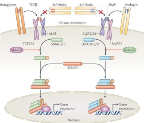
Figure 6 Schematic representation of the Bmp and Tgf-β signaling pathways. BMP is part of the TGF-β superfamily, its signals transduced into cells by a serine-threonine kinase transmembrane receptor binding with one of the 3 type I receptors (BMPR-IA, BMPR-IB, and Alk2). Its ligand-binding induces receptor in turn phosphorylating target intracellular Smads, mainly Smads-1. -5, and -8, in the cytoplasm. Tgf-β targets mainly Smads-2 and -3 through Tgf-β receptors and Alk5. These phosphorylated Smads (pSmads) then bind to the common Smad4 to control nuclear gene expression (source [93])
BMP roles in tooth initiation
The antagonism between Fgf and Bmp appears to determine the tooth-bud site and tooth type during initiation of the tooth bud. BMP signaling in the oral epithelium antagonizes FGF signaling, the later thought to determine the sites of tooth formation[50-52]. Thus mesenchymal BMP4 regulates Fgf8 expression[51] and is critical for the establishment of presumptive incisor and molar tooth field, for the
transition from the tooth bud to cap stage, and for induction of the epithelium enamel knot[72, 73]. The inactivation of activin or Bmpr1a (in either epithelium or mesenchyme) also results in the arrest of tooth development after the bud stage[94-96].
BMP roles in tooth number, tooth morphology, and size
Collectively the BMP signaling pathway regulates tooth formation. Overexpression of the BMP inhibitor Noggin arrests tooth development at the lamina to bud initiation stage of tooth morphogenesis[97]. While relatively little is known regarding the molecular mechanisms in which BMP alterations affect dental morphology, decreasing BMP signaling in the incisor region can lead an incisor to acquire a molar-like phenotype[98]. The relative size of mouse molars can also be influenced by overall BMP levels[99]. Specifically events of BMP activation and inhibition occur successively between developing teeth[100]. Mutation of the BMP inhibitor gremlin (Grem2) is associated with human tooth agenesis, microdontia, short tooth roots, taurodontism, sparse, and slow-growing hair[101]. Consistently, Grem2 mutant mice have small, malformed maxillary and mandibular incisors[102].
BMP roles in tooth differentiation
During odontoblastic differentiation, BMP2 mediates DSPP gene expression and odontoblast differentiation via the heterotrimeric transcription factor Y[103]. In addition, the down-stream effectors of BMP/TGF-β signaling, such as SMAD4 also play a role during tooth differentiation events[104].
Ameloblast differentiation is controlled by antagonistic actions of BMP4 and activin A[94]. The asymmetrically expression of the BMP inhibitor follistatin also regulates the labial-lingual patterning of enamel formation[105]. The continuous growth and enamel deposition in mouse incisors can be modulated by the net levels of Fgf, Activin, and Bmp signaling in the epithelial stem cell niche[106].
TGF-β
Transforming growth factors beta (TGF-β) is part of a super-family of growth factors that regulates a broad range of cell growth, differentiation, and extracellular morphogenetic events[107]. The essential functions of the TGF-β superfamily involve
actions during tooth crown patterning and tooth root development (Figure 6). Their importance is further highlighted in studies investigating dependent and Smad-independent pathways regulating tissue-tissue interactions during patterning of tooth crown and root (reviewed in [108]).
TGF-β roles in tooth differentiation
TGF-β proteins are thought to play an important role in the morphogenesis of developing teeth[109]. Both TGF-β1 and TGF-β3 are produced by the enamel organ and activated by components of the basement membrane[110]. Inhibiting TGF-β signaling stops ameloblast enamel secretion[111, 112] and inactivation of the Tgfbr2 gene increases odontogenic epithelial cell proliferation[113]. TGF-β-RII cKO and
TGF-β1 over-expression exhibits a tooth phenotype at the early stage of enamel
formation[112, 114]. Tgf-β receptor II conditional knockout mice also show enamel attrition with thinner crystals[114]. In addition, β-activating SMAD2, 3 and TGF-β-inhibiting SMAD7 are found both in the enamel epithelium and dental mesenchyme. Their mutations produce a variety of tooth phenotypes, such as a reduction of enamel related to their roles in ameloblastic function and tooth morphogenesis[109, 115].
In addition, dosage-dependent effects of TGF-β expression are observed. Transgenic overexpression of TGF-β1 in early secretory stage ameloblasts (via
dentin TGF-β1 transgene overexpression) triggers dentin adhesion process
detachment, ossification of dentin, and the formation of cyst-like structures formed from enamel matrix-like proteins[112]. An independent strategy of dental-targeted overexpression of TGF-β1 using a Dspp-Tgf-β1 transgene produced a dentin dysplasia-like phenotype[116]. TGF-β2 overexpression driven by an osteocalcin promoter (expressing in the dental mesenchyme) alters the dentin matrix, reducing dentin hardness and elasticity[117].
Modulation/activation of TGF-β via LTBP
In addition, TGF-β family interacting proteins play roles in the production and degradation of the extracellular matrix. TGF-βs are secreted in the form of latent high molecular mass complexes that contain other proteins [118]. Among their associated
factors are latent TGF-beta binding proteins (LTBPs) (Figure 7). To date, 4 members of the LTBP family (LTBP1, 2, 3, 4) are known. LTBPs are important regulators of TGF-β bioavailability and action. They also interact with other extracellular proteins, including microfibrils and elastic fibers [119]. The mouse LTBP polypeptide forms a complex with the TGF-β1 precursor [119]. LTBP3 allows latent TGF-β complexes to be targeted to connective tissue matrices and cells [120, 121]. The observation of bone abnormalities in Ltbp-3-null mice supports the role of LTBP3 in modulating TGF-β bioavailability [122]. Ltbp3 mutants, much like Ltbp1 null mice, develop discernible craniofacial abnormalities [123]. Human LTBP2 polymorphisms may underlie bone mineral density defects and fracture risk [124]. A recessive mutation in human LTBP4 causes multi-organ defects that include craniofacial malformations[125].
Figure 7 TGF-β synthesis and activation.
TGF-βs are synthesized as inactive precursors that contain unprocessed regions. Processing of inactive forms starts with a proteolytic cleavage that removes signal peptide from pre-pro-TGF-βs forms. After dimerization, TGF-βs are cleaved by proteases into terminal mature peptides and N-terminal Latency Associated Peptides. TGF-βs with Latency Associated Peptides form small latent complexes that are transported to extracellular matrix where they can further covalently bind to latent TGF-β binding protein (LTBP) to form a large latent complexes. LTBP is able to connect inactive TGF-β forms to extracellular matrix (ECM) proteins. This interaction is further supported by covalent transglutaminase-induced crosslinks. Activation of TGF-β starts with release of large latent complexes from ECM by proteases. Then, the mature protein is cleaved from LTBP, which is provided in vitro by acidic condition, pH or plasmin, or in vivo by thrombospondin. Once the active TGF-β family member is released from the ECM, it is capable of signaling. (Source [126])
LTBP3, has been defined as a member of the LTBP family, which serve to regulate TGF-β bioavailability and signaling by interacting with other extracellular proteins[119] (Figure 7). These proteins function to allow latent TGF-β protein complexes to have an altered conformation, and thus be targeted to connective tissue matrices and cells[121]. Mutations in human LTBP3 result in patients with short stature, vertebral and skull bone alterations, oligodontia[127], and mitral valve prolapse[128]. In a collaborative report examining patients with LTBP3 mutations, we observed a range of recessive hypomorphic mutations (producing alterations including gene deletion, nonsense, and aberrant splice mutations) causing developmental abnormalities such as short stature, brachyolmia, and hypoplastic Amelogenesis Imperfecta (AI) (OMIM; 601216)[129]. Investigating the dental phenotype of the Ltbp3-/- mouse model demonstrated very thin or absent enamel in both incisors and molars[129] and confirmed this animal model to be reliable to study the pathophysiology of Verloes Bourguignon syndrome. The full description of the tooth-specific morphological alterations in the Ltbp3-/- mutant mouse[130] is reported in this thesis in results section page 130.
II.3 FGFs
Fibroblast growth factors, or FGFs, are a vital family of numerous growth factors, with members involved in angiogenesis, wound healing, embryonic development, and various endocrine signaling pathways. FGFs are major actors in the processes of proliferation and differentiation of diverse cells and tissues (Figure 8) (for a general review see [131]; for skeletal-specific functions see [132]).