HAL Id: inserm-00836318
https://www.hal.inserm.fr/inserm-00836318
Submitted on 20 Jun 2013
HAL is a multi-disciplinary open access
archive for the deposit and dissemination of
sci-entific research documents, whether they are
pub-lished or not. The documents may come from
teaching and research institutions in France or
abroad, or from public or private research centers.
L’archive ouverte pluridisciplinaire HAL, est
destinée au dépôt et à la diffusion de documents
scientifiques de niveau recherche, publiés ou non,
émanant des établissements d’enseignement et de
recherche français ou étrangers, des laboratoires
publics ou privés.
during the differentiation process of Th1 and Th17 cells.
Patricia Luz-Crawford, Monica Kurte, Javiera Bravo-Alegría, Rafael
Contreras, Estefania Nova-Lamperti, Gautier Tejedor, Danièle Noël, Christian
Jorgensen, Fernando Figueroa, Farida Djouad, et al.
To cite this version:
Patricia Luz-Crawford, Monica Kurte, Javiera Bravo-Alegría, Rafael Contreras, Estefania
Nova-Lamperti, et al.. Mesenchymal stem cells generate a CD4+CD25+Foxp3+ regulatory T cell
pop-ulation during the differentiation process of Th1 and Th17 cells.. Stem Cell Research and Therapy,
BioMed Central, 2013, 4 (3), pp.65. �10.1186/scrt216�. �inserm-00836318�
R E S E A R C H
Open Access
Mesenchymal stem cells generate a CD4
+
CD25
+
Foxp3
+
regulatory T cell population during the
differentiation process of Th1 and Th17 cells
Patricia Luz-Crawford
1,4,5, Monica Kurte
4, Javiera Bravo-Alegría
4, Rafael Contreras
4, Estefania Nova-Lamperti
4,
Gautier Tejedor
1,2, Danièle Noël
1,2, Christian Jorgensen
1,2,3, Fernando Figueroa
4, Farida Djouad
1,2†and Flavio Carrión
4*†Abstract
Introduction: Mesenchymal stem cells (MSCs) are adult, multipotent, stem cells with immunomodulatory properties. The mechanisms involved in the capacity of MSCs to inhibit the proliferation of proinflammatory T lymphocytes, which appear responsible for causing autoimmune disease, have yet to be fully elucidated. One of the underlying mechanisms studied recently is the ability of MSCs to generate T regulatory (Treg) cells in vitro and
in vivo from activated peripheral blood mononuclear cells (PBMC), T-CD4+ and also T-CD8+ cells. In the present
work we investigated the capacity of MSCs to generate Treg cells using T-CD4+ cells induced to differentiate toward the proinflammatory Th1 and Th17 lineages.
Methods: MSCs were obtained from mouse bone marrow and characterized according to their surface antigen expression and their multilineage differentiation potential. CD4+T cells isolated from mouse spleens were induced to differentiate into Th1 or Th17 cells and co-cultured with MSCs added at day 0, 2 or 4 of the differentiation processes. After six days, CD25, Foxp3, IL-17 and IFN-γ expression was assessed by flow cytometry and helios and neuropilin 1 mRNA levels were assessed by RT-qPCR. For the functional assays, the ‘conditioned’ subpopulation generated in the presence of MSCs was cultured with concanavalin A-activated CD4+ T cells labeled with carboxyfluorescein succinimidyl ester. Finally, we used the encephalomyelitis autoimmune diseases (EAE) mouse model, in which mice were injected with MSCs at day 18 and 30 after immunization. At day 50, the mice were euthanized and draining lymph nodes were extracted for Th1, Th17 and Treg detection by flow cytometry. Results: MSCs were able to suppress the proliferation, activation and differentiation of CD4+T cells induced to differentiate into Th1 and Th17 cells. This substantial suppressive effect was associated with an increase of the percentage of functional induced CD4+CD25+Foxp3+regulatory T cells and IL-10 secretion. However, using mature Th1 or Th17 cells our results demonstrated that while MSCs suppress the proliferation and phenotype of mature Th1 and Th17 cells they did not generate Treg cells. Finally, we showed that the beneficial effect observed
following MSC injection in an EAE mouse model was associated with the suppression of Th17 cells and an increase in the percentage of CD4+CD25+Foxp3+T lymphocytes when administrated at early stages of the disease.
Conclusions: This study demonstrated that MSCs contribute to the generation of an immunosuppressive
environment via the inhibition of proinflammatory T cells and the induction of T cells with a regulatory phenotype. Together, these results might have important clinical implications for inflammatory and autoimmune diseases. Keywords: Mesenchymal stem cells, Th1, Th17, Immunosuppression, CD4+CD25+Foxp3+T cells
* Correspondence:fcarrion@uandes.cl
†
Equal contributors
4Laboratorio de inmunologia celular y molecular, Universidad de los Andes,
San carlos de apoquindo 2200, CP. 7620001, Las Condes, Santiago, Chile Full list of author information is available at the end of the article
© 2013 Luz-Crawford et al.; licensee BioMed Central Ltd. This is an Open Access article distributed under the terms of the Creative Commons Attribution License (http://creativecommons.org/licenses/by/2.0), which permits unrestricted use, distribution, and reproduction in any medium, provided the original work is properly cited.
Introduction
Mesenchymal stem cells (MSCs) are multipotent stromal cells characterized by their ability to differentiate into cells from mesodermal tissue making them an interest-ing cell source for application in regenerative medicine [1,2]. Another attractive potential of MSCs is their cap-acity to inhibit the proliferation of T and B lymphocytes, natural killer and dendritic cells both in vitro and in vivo [3-6]. These immunosuppressive abilities are mediated by different mechanisms specific for human or mouse MSCs, such as indoleamine 2,3-dioxygenase (IDO) or ni-tric oxide (NO), respectively, or overlapping suppressive factors, such as transforming growth factor β1 (TGF-β1), prostaglandin E2 (PGE2) and IL-10 among others [7-9]. Moreover, it has been shown that MSCs are able to generate CD4+CD25+highFoxp3+ T regulatory (Treg) cells in vitro, from activated human peripheral blood mononuclear cells (PBMC), mouse splenocytes or iso-lated T-CD4 cells. Indeed, MSCs promote the induction of CD4+CD25+high regulatory cells from human PBMC cells activated with IL-2 [10]. In the same line, Maccario et al. demonstrated that MSCs favor the differentiation of CD4+ T-cell subsets co-expressing CD25 and/or CTLA4, two markers of Treg cells [11]. These observa-tions were supported and extended by a study showing that direct MSC-T cell contact is required for Foxp3 and CD25Highexpression by CD4+ T cells; however, soluble factors produced by MSCs, such as TGF-β1 and PGE2, also played a non-redundant contribution in the ge-neration of CD4+CD25+Foxp3+ [8]. A role for other molecules including the human leukocyte antigen-G5 (HLA-G5) and the stress inducible enzyme heme-oxygenase-1 (HO-1) has also been described in the ge-neration of regulatory T cell phenotype mediated by human MSCs [12,13]. The ability of MSCs to induce such a regulatory phenotype in T cells was described both in vitro and in vivo. As an example, in a mouse ani-mal model of inflammatory bowel disease (IBD), the beneficial effect of injected human adipose derived MSCs (hASCs) on the clinical and histological scores of mice was associated with an increased number of CD4+ CD25+Foxp3+ and CD4+IL10+ cells in the lymph nodes [14]. The induction of a regulatory T cell phenotype population has also been shown in clinical applications. Indeed, a significant induction of CD4+CD25+Foxp3+cells was observed in two systemic lupus erythematous (SLE) patients [15]. Moreover, after autologous MSC administra-tion in patients with kidney transplantaadministra-tion, Perico et al. reported not only an increase in the number of CD4+
CD25+Foxp3+ cells but also a substantial decrease of the activity of CD4+ and CD8+ effectors as well as a subse-quent improvement in their renal function [16].
In the context of inflammatory diseases, T helper 1 (Th1) and Th17 T cell subsets are well known to
mediate inflammation [17,18]. Interestingly, we showed that MSCs inhibit human Th17 cell differentiation and function and induce a regulatory T cell phenotype [19]. This result revealed that, even under inflammatory con-ditions, MSCs exert in vitro anti-inflammatory effects through the induction of a regulatory T cell phenotype. However, the capacity of MSCs to generate functional Treg cells in vitro during the differentiation process or on fully differentiated Th1 and Th17 cells still remains to be elucidated. Therefore, in this study, we explored the capacity of MSCs to generate, in vitro, functional CD4+ CD25+Foxp3+ Treg cells under Th1 and Th17 inflamma-tory culture conditions. In parallel, in the experimental autoimmune encephalomyelitis (EAE) model, we assessed the percentage of regulatory T cells after MSC administra-tion at two different time points post-immunizaadministra-tion. The aim of this study was to determine whether MSCs are able to increase the percentage of regulatory T cells in vitro when co-cultured with either CD4+cells induced to diffe-rentiate into Th1 and Th1 or with fully diffediffe-rentiated Th1 and Th17 cells and in vivo in the EAE model.
Methods
Isolation and characterization of mouse mesenchymal stem cells
MSCs were isolated from eight- to ten-week-old C57BL/ 6 mice. Bone marrow cells were collected by flushing fe-murs and tibias and the cell suspension (1 × 10⁶cells/cm2) was plated in a modified minimum essential Eagle's medium (MEM) © (α-MEM, Gibco, Auckland, NZ) sup-plemented with 20% fetal bovine serum (FBS) (Hyclone, Thermo Fisher Scientific, Brebières, France), 2 mM glu-tamine and 100 U/mL penicillin with 100 mg/mL strepto-mycin (Gibco, Auckland, NZ) (α-20). At sub-confluence, cells were replated at a density of 20,000 cells/cm2and, after the second passage, MSCs were isolated by negative selection using a CD45+microbeads kit (Miltenyi Biotec, Bergisch-Gladbach, Germany). MSCs were characterized for expression of hematopoietic and mesenchymal cell an-tigens by fluorescence-activated cell sorting (FACS) ana-lysis and by their capacity to differentiate into adipogenic, chondrogenic and osteogenic lineages as previously de-scribed [20].
Th1 and Th17 differentiation and MSC cocultures
CD4+T cells from spleen of C57BL/6 mice were purified by negative selection using the CD4+T cell Isolation Kit MicroBeads (Miltenyi Biotec) according to the manufac-turer’s instructions. Purified CD4+ T cells were cultured in complete medium containing RPMI 1640 supplemen-ted with 10% heat-inactivasupplemen-ted FBS, 2 mM l-glutamine, 100 U/mL penicillin/100 μg/mL streptomycin. In a 24-well plate, 2 × 10⁶CD4+ T cells were cultured in the presence of 2.5 μg/ml coated antibodies against CD3 and
1.5 μg/ml CD28 (BD Biosciences, San Jose, CA, USA) under Th1 or Th17 differentiation conditions. Th1 cells were differentiated with 20 ng/ml of IL-12 (R&D Systems, Minneapolis, MN, USA) and 2.5 μg/ml IL-4 anti-bodies (BD Biosciences). Th17 cells were differentiated with 50 ng/ml IL-6 (R&D Systems, Minneapolis, MN, USA), 5 ng/ml of TGF-β1 (BioVision, Milpitas, CA USA) and 2.5 μg/ml of anti-IFN-γ and -IL-4 antibodies (BD Bio-sciences). MSCs were added at a MSC:Th ratio of 1:10 or 1:100 at day 0, 2 or 4 of the Th1 and Th17 differentiation process. After six days of culture, relative cell quantifica-tion was measured using the CellTiter-Glo™ luminescent cell viability assay (Promega, Charbonnières-les-Bains, France) and intracellular cytokine detection was measured by flow cytometry.
Analysis by flow cytometry
After six days of culture, T cells were stimulated for four hours with 50 ng/ml phorbolmyristate acetate (PMA) (Sigma-Aldrich, St Louis, MO, USA) and 1μg/ml ionomycin (Sigma-Aldrich), prior to the addition of 10 μg/ml brefeldin A (eBiosciences, San Diego, CA, USA). For the detection of surface markers, cells were stained with CD4-PEcy5 (eBiosciences) and CD25-PE-texas red (Invitrogen, Grand Island, NY, USA) and incubated for 20 minutes at 4°C in the dark. After two washing steps, we performed intracellular staining for IFN-γ-FITC, IL-17-PE or FoxP3-Alexa488 detection. For that purpose, cells were fixed and permeabilized using the Cytofix/ Cytoperm™ (BD Biosciences) kit according to the manu-facturer’s instructions. Acquisition was performed with a Coulter Epics-XL flow cytometer using the System II software (Coulter Corporation, Brea, CA, USA). Analysis was performed using the FCS express software (De novo softwares, Los Angeles, CA, USA).
Functional assay
After six days of co-culture at different times post-T-CD4+ activation (0, 2 and 4), the ‘conditioned’ Treg subpopulations generated from Th1 and Th17 cells cocultured with MSCs (cond-Th1 or cond-Th17 cells) were evaluated for their ability to suppress allogenic T cell proliferation. For that purpose, fresh T-CD4+ cells were labeled with carboxyfluorescein succinimidyl ester (CFSE) and cocultured with or without cond-Th1 or cond-Th17 cells at different ratios in the presence of 1 μg/ml of concanavalin A (ConA) (Sigma-Aldrich). After 72 hours, the proliferation of CD4+T cells was an-alyzed on a FACS Canto II using the BD FACSDiva soft-ware measured by flow cytometry.
Quantification of cytokines
Enzyme-linked immunosorbent assay (ELISA) from R&D Systems (Minneapolis, MN, USA) for IL-10 and
the Enzyme Immunoassay kit from Arbor Assays (Ann Arbor, MI, USA) for PGE2 were used. IL-10 and PGE2 were quantified from the supernatants of cocultures which were stored at −20°C until tested.
MSC purification from Th1 and Th17 cocultures
MSCs were washed three times with a PBS/0.05mM ethylenediamine tetraacetic acid (EDTA) buffer in order to detached lymphocytes from MSCs. Then, MSCs were trypsinized and resuspended in α-20 and cultured for two hours. After this time, MSCs were washed three times with a PBS/0.05mM EDTA to eliminate possible resting contamination with lymphocytes. The percentage of MSCs was 98%.
RT-qPCR analysis
Total RNA from purified MSCs and from Th1 and Th17 cells was extracted in the presence or absence of MSCs using the RNeasy mini kit (Qiagen S.A., Courtaboeuf, France). RNA (500 ng) was reverse transcribed using the Multiscribe reverse transcriptase (Applied Biosystems, Courtaboeuf, France). Quantitative PCR was performed using the SYBR Green I Master kit and a LightCycler® 480 Detection System, following the manufacturer’s rec-ommendations (Roche Applied Science, Meylan, France). Specific primers for IL-10, TGF-β1, Helios, Foxp3 and Nrp1 were designed using the Primer3 software [21]. Briefly, 50 ng cRNA were amplified and the analysis of mRNA expression level was performed using the Roche LightCycler® 480 software 1.5. Expression levels of tran-scripts were normalized to the housekeeping gene ribo-somal protein S9 (RPS9). For quantification, values were expressed as the relative mRNA level of specific gene ex-pression as obtained using the 2−ΔCtmethod.
EAE induction and treatment protocols
Female C57BL/6J mice, six- to eight-weeks old, were purchased from the Faculty of Medicine, Universidad de Chile (Santiago, Chile). All animals were housed and treated according to the guidelines of the Animal Ethical Committee of our Institution (Universidad de los Andes, Chile). Mice were immunized according to a previously published protocol using 50 μg MOG35-55 (LifeTein, Detroit, MI, USA), emulsified in Complete Freund’s Adjuvant (CFA; Difco, Detroit, MI, USA) containing 4 mg/ml Mycobacterium tuberculosis H35RA (strain H35Ra; Difco, Detroit, MI, USA) and injected subcu-taneously [22]. Immunization with MOG35-55 was followed by intraperitoneal administration of 350 ng per-tussis toxoid (Sigma-Aldrich) on days 0 and 2. Mice were injected with 1 × 10⁶MSCs on days 18 and 30 after immunization. Clinical score was recorded daily and
assigned according to a standard and validated 0 to 5 scale [23]. At day 50, the mice were euthanized and splenocytes and draining lymph nodes (drLN) were extracted and cultured in RPMI 1640 supplemented with 10% of heat-inactivated FCS, 2 mM L-glutamine and 100 U/mL penicillin/100 μg/mL streptomycin in 24 well plates for four hours with 50 ng/ml PMA and 1 μg/ml ionomycin (Sigma-Aldrich), in the presence of 10 μg/ml brefeldin A (eBiosciences) for intracellu-lar staining.
Statistical analysis
Results are expressed as the mean ± (SEM). Individual experiments were carried out between three and seven times to ensure reproducibility. Generated P values and post-analyses were performed with the Kruskal–Wallis test, considering non-normal distributions with small sample sizes and multiple groups and the Mann–Whitney test to compare between two groups. P-values <0.05 (*), P <0.01 (**) or P <0.001 (***) were considered statistically significant. Analysis and graphical representation were performed using Graph-Pad Prism™ software (Graphpad, San Diego, CA, USA).
Results
Phenotypic and functional characterization of murine MSCs
Murine bone marrow-derived MSCs were successfully isolated by negative selection with the CD45+ micro-beads isolation kit. After passage seven, MSCs had a stable fibroblast-like phenotype and were positive for the mesodermal surface antigens CD90, CD29, SCA-1 and CD44 and negative for the hematopoietic markers CD45 and CD11b as shown in Figure 1A. MSCs were also functionally capable of differentiating into adipocytes, chondrocytes and osteoblasts under inductive culture conditions showing their multipotent characteristics (Figure 1B).
MSCs inhibit the activation, proliferation and differentiation of Th1 and Th17 cells
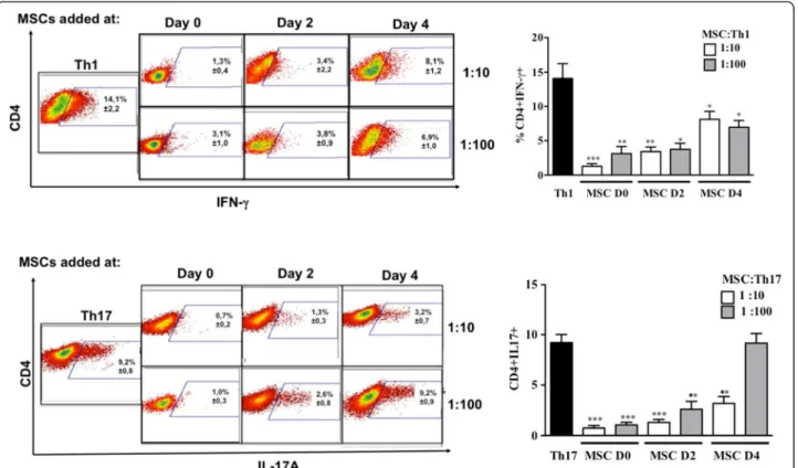
The effect of MSCs on the activation, proliferation and Th1 or Th17 differentiation was assessed using purified CD4+ T cells activated with anti-CD3/CD28 monoclonal antibodies (mAb) under Th1 or Th17 skewing conditions. MSCs were added at day 0, 2 or 4 post-activation. First, we showed that under Th1 and Th17 culture conditions, IFN-γ-producing cells and IL-17-producing cells, respec-tively, were generated after six days of culture (Figure 2).
Figure 1Surface antigens and differentiation potential of murine mesenchymal stem cells (MSCs). A) Phenotypic characterization of murine MSCs at passage nine to ten by FACS analysis. A representative histogram for each antigen is shown in red. B) Multilineage differentiation potential was assessed by their capacity to differentiate into chondrocyte (Col2 and aggrecan expression, up), adipocyte (Oil Red O staining; middle), and osteoblast (alizarin red S staining; bottom). FACS, fluorescence-activated cell sorting.
Under these conditions, the addition of MSCs at the beginning of the differentiation process (day 0) highly reduced the number of activated CD4+CD25+ T cells, whatever the ratio used (Figure 3A). This effect was still significant when MSCs were added at day 2. Interestingly, while MSC addition did not affect the activation state of fully differentiated Th1 and Th17 cells, they significantly suppressed the latter mature T cells when added at the highest ratio (Figure 3B). We then studied the effect of MSCs on the T cell differentiation process toward Th1 and Th17 and observed a significant inhibition of the per-centage of IFN-γ-producing Th1 and IL-17-producing Th17 cells. However, the percentage of IL-17-producing mature Th17 cells co-cultured with MSCs was not af-fected at the lowest MSC:T-cell ratio tested (Figure 2).
MSCs induce functional CD4+CD25+Foxp3+T cells during the differentiation process of Th1 and Th17 cells
Previous studies have shown that MSCs induce CD4+ CD25+Foxp3+ T regulatory cell phenotype from PBMC or CD4+ activated cells in vitro [8,10,11]. In the present study, we investigated the capacity of MSCs to induce functional Treg cells when co-cultured with T cells
induced to differentiate toward Th1 or Th17 cells. Flow cytometry analysis revealed that MSCs increased the percentage of CD4+CD25+Foxp3+T cells only when they were added at day 0 or 2 of the differentiation processes (Figure 4A). This latter increase of Foxp3 was confirmed at the RNA level (Figure 4B). Moreover, we showed an up-regulation of IL-10 production in the supernatants (Figure 5A). Since we demonstrated that MSCs sup-pressed the proliferation of CD4+T cells undergoing Th1 or Th17 differentiation we hypothesized that MSCs generate induced CD4+CD25+Foxp3+ T cells. To test that hypothesis, we investigated whether the CD4+CD25 +
Foxp3+ T cells obtained in the co-cultures with MSCs were natural (nTregs) or induced (iTregs) by determi-ning the expression levels of surface Nrp1 and helios, an ikaros family transcription factor [24-26]. We showed that CD4+CD25+Foxp3+ T cells generated in the co-cultures with MSCs expressed significantly lower levels of helios and Nrp1 when compared to the Th1 or Th17 differentiated T cells cultured in absence of MSCs. These results strongly suggest that MSCs might generate iTreg cells rather than stimulating the expansion of nTreg cells (Figure 4B).
Figure 2MSCs inhibit Th1 and Th17 differentiation depending on the stage of activation and cell ratio. The differentiation of Th1 and Th17 cells was measured by intracellular detection of IFN-γ and IL-17 positive cells, respectively, from purified CD4-T cells differentiated into Th1 and Th17 cells in the presence or absence of MSCs at different stages of activation and different MSC:Th ratios (left: representative dot plot: 1:10 upper panels and 1:100 lower panels). ** = P <0.01 and * = P <0.05, n = six for Th1 and n = seven for Th17. Values represent means ± SED for six and seven independent experiments for Th1 and Th17 cells, respectively. MSCD0, D2 and D4 = MSC added at day 0, 2 and 4 of the
Finally, in a functional test, we assessed the suppres-sive capacity of conditioned Th1 or Th17 (Th1cond or Th17cond) cells obtained after MSC addition at day 0 or 2 of the T cell differentiation processes (Figure 5B). By using different ratios of Thcond cells:CD4+ (1:1; 1:2 or 1:5), we showed a dose dependent capacity of the condi-tioned cells to inhibit the proliferation of activated CD4+ T cells (Figure 5B). In contrast, conditioned T cells ob-tained from the co-cultures of mature Th1 or Th17 cells with MSCs did not modulate the proliferation of ConA activated T cells (data not shown).
PGE2, TGF-β1 and IL-10 are up-regulated in the cocultures of MSCs with Th1- or Th-17 cells
Several studies have shown that soluble factors including PGE2, TGF-β1 and IL-10 are directly involved in Treg cell induction [27]. Therefore, we quantified PGE2 production in the supernatants of the co-cultures of MSCs with differentiating or mature Th1 or Th17 cells (Figure 6A). Compared to MSCs, Th1 or Th17 cells cultured alone, the secretion of PGE2 was significantly increased in the supernatants of the co-cultures. No significant difference was observed between the MSC: Figure 3MSCs inhibit the activation and the proliferation of Th1 and Th17 cells. A) MSCs inhibit the expression of CD25 in the early stage of activation of Th1 and Th17 cells. T cell activation was measured using purified CD4+T cells differentiated into Th1 or Th17 cells in the presence
or absence of MSCs according to the expression of the activation surface antigen CD25 (left: representative dot plot: 1:10 upper panels and 1:100 lower panels). B) MSCs suppress Th1 and Th17 cells independent of the stage of activation and cell ratio. Relative cell quantification was determined using purified CD4+T cells differentiated into Th1 or Th17 cells in the presence or absence of MSCs added at day 0, 2 or 4 after T cell
activation and at a MSCs:Th ratio (1:10 and 1:100), using the Cell Titer Glo™ luminescence assay. RLU = relative light units. MSCs, mesenchymal stem cells; Th, T helper.
T cell ratios tested or between differentiating or ma-ture Th1 or Th17 cells. Then, using quantitative PCR we measured the expression levels of these molecules in MSCs after co-culture with either differentiating or mature Th1 and Th17 cells. We found that all MSC co-cultures tested were able to increase the mRNA expression levels of TGF-β1 and IL-10 in MSCs (Figure 6B).
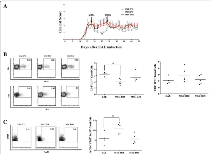
MSCs inhibit EAE progression and increased the CD4
+CD25+Foxp3+Treg population
Finally, we addressed whether MSCs could affect the generation of Th1, Th17 and CD4+CD25+Foxp3+ Treg cells in a T-cell-mediated autoimmune disease such as EAE. MSCs were administrated at day 18 and 30 after EAE induction in mice and the clinical scores were recorded daily until day 50. Compared to the control Figure 4MSCs generated iTreg cells during the differentiation process of Th1 and Th17 cells. A) Treg generation from Th1 and Th17 cells cocultured with MSCs at different stages of activation and MSC:Th ratios. Treg phenotype was determined according to the percentage of CD4 + CD25+ cells that express Foxp3 by flow cytometry (Dot plot upper A for Th1 and Upper B for Th17). B) Foxp3, Nrp 1 and helios mRNA expression levels weres quantified from Th1 and Th17 cells cocultured in the presence or absence of MSCs at different stage of activation and MSC:Th ratios (** = P <0.01 and * = P <0.05 ). iTreg, induced Treg; MSCs, mesenchymal stem cells; Nrp 1, neuropilin 1; Th, T helper; Treg, regulatory T cells.
group, we observed an early improvement of clinical score when MSCs were administrated at day 18 (Figure 7A). This beneficial effect was observed shortly after MSC injection and maintained for ten days. However, MSC ad-ministration at day 30 did not improve the EAE symp-toms. Then, the percentage of CD4+IFN-γ+, CD4+ IL-17+ and CD4+CD25+Foxp3+ T cells in lymph node cells and splenocytes were measured at day 50. We observed a sig-nificant decrease of Th17 cells and an increase of CD4+ CD25+Foxp3+ T cells only in the lymph nodes of EAE mice that received MSCs at day 18 (Figure 7B and 7C). Compared to the control group, the percentage of Th1 cells was not modified (Figure 7B) and the different T cell sub-populations studied were not affected in the spleens after the MSC treatments (data not shown).
Discussion
The capacity of MSCs to suppress the proliferation of T-cells has opened new perspectives for their clinical
use. Currently, it is well established that MSCs rely on different mechanisms to exert their suppressive proper-ties. One important mechanism is the capacity of MSCs to generate functional Treg cells, as shown in the last decade. The demonstration that direct MSC–T cell in-teractions induce T cells with regulatory properties has arisen from a number of studies describing the ge-neration of Treg populations with typical phenotypic characteristics [10,11,28]. Later on, Prevosto et al. de-monstrated that CD4+CD25+Foxp3+ cells, generated from PBMC after three days of culture with MSCs, were functional and inhibited the proliferation of alloantigen-activated T cells [29]. Another study established that MSCs were able to induce Treg cells from CD4+T
lym-phocytes via cell contact as well as a non-redundant contribution of PGE2 and TGF-β1 [8]. These and other studies confirmed that Treg cell generation was contact-and soluble factors-dependent contact-and priming of MSCs was required for an immunosuppressive effect [30]. However, Figure 5IL-10 production from supernatants of coculture of Th1 and Th17 cells with MSCs and functional assay. (A) IL-10 production was quantified in the supernatants of Th1 and Th17 cells co-cultured in the presence or absence of MSCs. * = P <0.05, comparing to Th1 and Th17 cells as control group. All the values represent means ± SED of three independent experiments. B) Functional effect of iTreg cells generated using Th1 and Th17 cells in cocultures with MSCs (conditioned Th1 or Th17, Th1condor Th17cond, respectively). The suppressive capacity of Th1condor Th17condwas assessed by following the number of cell divisions of ConA-stimulated T cells, which were labeled by CFSE. Different ratios of Thcond:CD4+(1:1; 1:2 or 1:5) were tested. ** = P <0.01 and * = P <0.05. All the values represent means ± SED of three independent experiments for Th1 and Th17 cells. Th1condand Th17cond= Th1 and Th17 cells that were co-cultured with MSCs added at different time points of the differentiation processes. CFSE, carboxyfluorescein succinimidyl ester; ConA, concanavalin A; iTreg, induced regulatory T cells; MSCs, mesenchymal stem cells; Th, T helper.
the MSC potential to stimulate the proliferation of na-tural Tregs or to induce Tregs when co-cultured with Th1 or Th17 cell subsets has not been investigated. There-fore, we explored the ability of MSCs to induce functional CD4+CD25+Foxp3+regulatory cells, when cultured in the presence of activated CD4+ T cells submitted to Th1 or Th17 skewing conditions. We first showed that MSCs strongly suppress the proliferation, activation and differen-tiation of Th1 and Th17 cells when added at early stages of the differentiation processes regardless of the cell ratio tested. However, MSCs added at the highest ratio in the presence of fully differentiated Th1 and Th17 cells were able to significantly reduce the percentage of the mature T cells as well as their proliferation rate. Moreover, we showed for the first time that under these culture con-ditions, MSCs were able to generate iTreg cells expres-sing low levels of Nrp 1 and helios. The generation of CD4+CD25+Foxp3+ regulatory T cells was not observed when MSCs were added to mature Th1 or Th17 cells.
Furthermore, we showed that the latter iTreg cells were functional and suppressed the proliferation of activated CD4+T cells.
It is now well established that the generation of Treg cells is mainly induced by the presence of TGF-β1 and IL-10 cytokines [31,32]. It has also been shown that PGE2 is involved in increased expression of Foxp3 in T-CD4+ cells [33] and that MSCs are able to produce
PGE2, TGF-β1 and IL-10 [8,29]. Therefore, we deter-mined the expression levels of IL-10 and TGF-β1 in MSCs and the concentration of IL-10 and PGE2 in the supernatants of MSCs co-cultured with Th1 or Th17 cells. In parallel to the Treg generation, we found that IL-10 production was significantly increased in superna-tants of the co-culture when MSCs were added at day 0 or 2 of the differentiation processes. Along the same line, under the same conditions we reported a significant increase of IL-10 and TGF-β1 mRNA expression level in MSCs. However, we did not find an increase of IL-10 Figure 6PGE2, IL-10 and TGF-β1 expression on MSCs co-cultured with Th1 and Th17 cells. A) PGE2 was quantified in the supernatants of MSCs co-cultured in the presence or absence of Th1 and Th17 cells. B) IL-10 and TGF-β1 expression was quantified in the supernatants of MSCs cultured in contact with Th1 and Th17 cells at different stages of the differentiation process (Day 0 and 4) ** = P <0.01, * = P <0.05, comparing to MSCs as the control group. All the values represent means ± SED of three independent experiments. MSCs, mesenchymal stem cells; PGE2, prostaglandin E2; TGF-β1, transforming growth factor β; Th, T helper.
production in the supernatants of MSCs when cultured with fully differentiated Th1 or Th17 cells. We also showed an increase of PGE2 production in the superna-tants of MSCs cultured with both differentiating or ma-ture Th1 and Th17 cells. This observation supports other studies revealing that PGE2, TGF-β1 and IL-10 may play a critical role in the induction of Treg cells by MSCs [8,19].
Finally, we used an experimental murine model of EAE to evaluate the effect of MSCs injected at different time points post-immunization on the percentage of Th1, Th17 and CD4+CD25+Foxp3+ Treg cells. Since a single injection of MSCs at an early stage of the disease has been shown to improve EAE symptoms [34], in this study we performed one injection of MSCs at different
stages of EAE. Our results demonstrated that MSCs were able to improve the clinical signs of EAE only when they were administrated at the peak of the disease. This therapeutic effect was associated with both a sig-nificant decrease of Th17 cell numbers and an increased percentage of CD4+CD25+Foxp3+ Treg cells in the lymph nodes. Interestingly, MSCs injected at the later time point neither diminished the percentage of Th17 cells nor increased the percentage CD4+CD25+Foxp3+ Treg cells. No effect was observed on Th1 cells.
Contradictory results have been previously reported following MSC injection showing either an increase of the Treg population associated with a therapeutic effect or describing an absence of any beneficial effect in dif-ferent animal models of autoimmune diseases [14,35,36]. Figure 7Therapeutic effects and Th1, Th17 and CD4 + CD25 + Foxp3 + Treg populations in experimental autoimmune
encephalomyelitis mice treated with MSCs. A) Early intravenous injection of MSCs ameliorates EAE. MSCs were injected at days 18 and 30 after immunization as shown by arrows (1 × 10⁶MSC/mice). B) CD4+IL17+and CD4+IFN-γ+cells in lymph nodes. MSCs significantly reduced the percentage of Th17 cells when injected at day 18 of the disease. C) CD4+CD25+Foxp3+T cells in lymph nodes. MSCs induce CD4+CD25+Foxp3+ cells when injected at day 18 of the disease. Control: EAE mice without treatment. MSCD18: EAE mice treated with MSCs injected 18 days post-immunization. MSCD30: EAE mice treated with MSCs injected 30 days post-immunization. * = P <0.05, compared to EAE untreated mice as control group. All the values represent means ± SED of n = 5 control condition and n = 6 for MSCs treated mice. EAE, experimental autoimmune encephalomyelitis; MSCs, mesenchymal stem cells; Th, T helper; Treg, regulatory T cells.
In the present study, we confirmed the generation of Treg cells following MSC injection in parallel with an improvement in the clinical signs of EAE.
Conclusion
Taken together, these data demonstrate that MSCs are able to induce functional Treg cells during the differenti-ation process of Th1 and Th17 cells. The generdifferenti-ation of Treg cells was associated with an increase of IL-10 pro-duction in the supernatant of the co-cultures and resulted in the inhibition of the proliferation, activation and differentiation of Th1 and Th17 cells. These data were confirmed in the EAE model, in which the thera-peutic effect of MSCs on disease progression was asso-ciated with an increase of the percentage of Treg cells when MSCs were administrated at a specific time point during disease progression.
Abbreviations
CFSE:Carboxyfluorescein succinimidyl ester; ConA: Concanavalin A; EAE: Experimental autoimmune encephalomyelitis; ELISA: Enzyme-linked immunosorbent assay; FACS: Fluorescence-activated cell sorting; FCS: Fetal calf serum; Fox: Forkhead box; IDO: Indoleamine 2,3-dioxygenase; IFN: Interferon; IL: Interleukin; iTreg: Induced Treg; MOG: Myelin oligodendrocyte glycoprotein; MSC: Mesenchymal stem cell; NO: Nitric oxide; Nrp1: Neuropilin 1; nTreg: Natural Treg; PBMC: Peripheral blood mononuclear cells; PBS: Phosphate-buffered saline; PCR: Polymerase chain reaction; PGE2: Prostaglandin E2; PMA: Phorbolmyristate acetate; Treg: Regulatory T cells; TGF-β1: Transforming growth factor β1; Th: T helper.
Competing interests
The authors declare that they have no competing interests. Authors’ contributions
PL-C participated in the performance of all experiments, interpreted and analyzed the results and wrote the manuscript; MK and JB-A performed the EAE experiments and participated in data analysis; RC participated in Th1 differentiation experiments and helped in data analysis; EN-L participated in Th17 differentiation experiments and helped in data analysis; GT performed PCR experiments for Treg characterization and participated in data analysis; DN participated in the conception and design of the experiments and critically revised the content of the manuscript; CJ participated in the conception and design of the experiments; FF participated in the design of the experiments and critically revised the content of the manuscript; FD and FC designed the research, interpreted the data and wrote the manuscript. All authors read and approved the final manuscript.
Acknowledgments
This work was supported by grant MED-03-2011 INOGTO201127 and grant FAI MED 002–10 INOGTO201042 from Universidad de los Andes and by Conicyt (Comision Nacional Ciencia y Tecnologia) that support Patricia Luz-Crawford PhD program and also her internship in Montpellier, France through the program “Becas Chile”. The work was also supported by the Inserm Institute, the University of Montpellier I and grants from the ‘Fondation pour la Recherche Médicale’ (project FRM 2011 ‘Comité Languedoc-Rousillon-Rouergue (LRR)’).
Author details
1
Inserm, U 844, Montpellier F-34091, France.2Université MONTPELLIER1, UFR de Médecine, Montpellier F-34000, France.3Service d'Immuno-Rhumatologie,
Hôpital Lapeyronie, Montpellier F-34295, France.4Laboratorio de inmunologia celular y molecular, Universidad de los Andes, San carlos de apoquindo 2200, CP. 7620001, Las Condes, Santiago, Chile.5Programa Doctorado en Biotecnología, Universidad Santiago de Chile. Avenida Libertador Bernardo O’Higgins 3363, CP. 9170022, Estación Central, Santiago, Chile.
Received: 9 November 2012 Revised: 21 March 2013 Accepted: 29 May 2013 Published: 4 June 2013
References
1. Bruder SP, Fink DJ, Caplan AI: Mesenchymal stem cells in bone development, bone repair, and skeletal regeneration therapy. J Cell Biochem 1994, 56:283–294.
2. Pittenger MF, Mackay AM, Beck SC, Jaiswal RK, Douglas R, Mosca JD, Moorman MA, Simonetti DW, Craig S, Marshak DR: Multilineage potential of adult human mesenchymal stem cells. Science 1999, 284:143–147. 3. Krampera M, Glennie S, Dyson J, Scott D, Laylor R, Simpson E, Dazzi F: Bone
marrow mesenchymal stem cells inhibit the response of naive and memory antigen-specific T cells to their cognate peptide. Blood 2003, 101:3722–3729.
4. Schena F, Gambini C, Gregorio A, Mosconi M, Reverberi D, Gattorno M, Casazza S, Uccelli A, Moretta L, Martini A, Traggiai E: Interferon-gamma-dependent inhibition of B cell activation by bone marrow-derived mesenchymal stem cells in a murine model of systemic lupus erythematosus. Arthritis Rheum 2010, 62:2776–2786.
5. Spaggiari GM, Capobianco A, Abdelrazik H, Becchetti F, Mingari MC, Moretta L: Mesenchymal stem cells inhibit natural killer-cell proliferation, cytotoxicity, and cytokine production: role of indoleamine 2,3-dioxygenase and prostaglandin E2. Blood 2008, 111:1327–1333. 6. Djouad F, Charbonnier LM, Bouffi C, Louis-Plence P, Bony C, Apparailly F,
Cantos C, Jorgensen C, Noel D: Mesenchymal stem cells inhibit the differentiation of dendritic cells through an interleukin-6-dependent mechanism. Stem Cells 2007, 25:2025–2032.
7. English K, Barry FP, Field-Corbett CP, Mahon BP: IFN-gamma and TNF-alpha differentially regulate immunomodulation by murine mesenchymal stem cells. Immunol Lett 2007, 110:91–100.
8. English K, Ryan JM, Tobin L, Murphy MJ, Barry FP, Mahon BP: Cell contact, prostaglandin E(2) and transforming growth factor beta 1 play non-redundant roles in human mesenchymal stem cell induction of CD4 + CD25(High) forkhead box P3+ regulatory T cells. Clin Exp Immunol 2009, 156:149–160.
9. Ren G, Zhang L, Zhao X, Xu G, Zhang Y, Roberts AI, Zhao RC, Shi Y: Mesenchymal stem cell-mediated immunosuppression occurs via concerted action of chemokines and nitric oxide. Cell Stem Cell 2008, 2:141–150.
10. Aggarwal S, Pittenger MF: Human mesenchymal stem cells modulate allogeneic immune cell responses. Blood 2005, 105:1815–1822. 11. Maccario R, Podesta M, Moretta A, Cometa A, Comoli P, Montagna D, Daudt L,
Ibatici A, Piaggio G, Pozzi S, Frassoni F, Locatelli F: Interaction of human mesenchymal stem cells with cells involved in alloantigen-specific immune response favors the differentiation of CD4+ T-cell subsets expressing a regulatory/suppressive phenotype. Haematologica 2005, 90:516–525. 12. Selmani Z, Naji A, Zidi I, Favier B, Gaiffe E, Obert L, Borg C, Saas P, Tiberghien P,
Rouas-Freiss N, Carosella ED, Deschaseaux F: Human leukocyte antigen-G5 secretion by human mesenchymal stem cells is required to suppress T lymphocyte and natural killer function and to induce CD4 + CD25highFOXP3+ regulatory T cells. Stem Cells 2008, 26:212–222. 13. Mougiakakos D, Jitschin R, Johansson CC, Okita R, Kiessling R, le Blanc K:
The impact of inflammatory licensing on heme oxygenase-1-mediated induction of regulatory T cells by human mesenchymal stem cells. Blood 2011, 117:4826–4835.
14. Gonzalez-Rey E, Anderson P, Gonzalez MA, Rico L, Buscher D, Delgado M: Human adult stem cells derived from adipose tissue protect against experimental colitis and sepsis. Gut 2009, 58:929–939.
15. Carrion F, Nova E, Ruiz C, Diaz F, Inostroza C, Rojo D, Monckeberg G, Figueroa FE: Autologous mesenchymal stem cell treatment increased T regulatory cells with no effect on disease activity in two systemic lupus erythematosus patients. Lupus 2010, 19:317–322.
16. Perico N, Casiraghi F, Introna M, Gotti E, Todeschini M, Cavinato RA, Capelli C, Rambaldi A, Cassis P, Rizzo P, Cortinovis M, Marasà M, Golay J, Noris M, Remuzzi G: Autologous mesenchymal stromal cells and kidney transplantation: a pilot study of safety and clinical feasibility. Clin J Am Soc Nephrol 2011, 6:412–422.
17. Lafaille JJ: The role of helper T cell subsets in autoimmune diseases. Cytokine Growth Factor Rev 1998, 9:139–151.
18. Bettelli E, Oukka M, Kuchroo VK: T(H)-17 cells in the circle of immunity and autoimmunity. Nat Immunol 2007, 8:345–350.
19. Ghannam S, Pene J, Torcy-Moquet G, Jorgensen C, Yssel H: Mesenchymal stem cells inhibit human Th17 cell differentiation and function and induce a T regulatory cell phenotype. J Immunol 2010, 185:302–312. 20. Carrion F, Nova E, Luz P, Apablaza F, Figueroa F: Opposing effect of
mesenchymal stem cells on Th1 and Th17 cell polarization according to the state of CD4+ T cell activation. Immunol Lett 2011, 135:10–16. 21. Rozen S, Skaletsky H: Primer3 on the WWW for general users and for
biologist programmers. In Bioinformatics Methods and Protocols: Methods in Molecular Biology. Edited by Krawetz S, Misener S. Totowa, NJ: Humana Press; 2000:365–386.
22. Iruretagoyena MI, Sepulveda SE, Lezana JP, Hermoso M, Bronfman M, Gutierrez MA, Jacobelli SH, Kalergis AM: Inhibition of nuclear factor-kappa B enhances the capacity of immature dendritic cells to induce antigen-specific tolerance in experimental autoimmune encephalomyelitis. J Pharmacol Exp Ther 2006, 318:59–67.
23. Domingues HS, Mues M, Lassmann H, Wekerle H, Krishnamoorthy G: Functional and pathogenic differences of Th1 and Th17 cells in experimental autoimmune encephalomyelitis. PLoS One 2010, 5:e15531. 24. Weiss JM, Bilate AM, Gobert M, Ding Y, Curotto de Lafaille MA, Parkhurst CN,
Xiong H, Dolpady J, Frey AB, Ruocco MG, Yang Y, Floess S, Huehn J, Oh S, Li MO, Niec RE, Rudensky AY, Dustin ML, Littman DR, Lafaille JJ: Neuropilin 1 is expressed on thymus-derived natural regulatory T cells, but not mucosa-generated induced Foxp3+ T reg cells. J Exp Med 2012, 209:1723–1742. S1.
25. Papatriantafyllou M: T cells: neuropilin 1 - distinguishing TReg cell subsets. Nat Rev Immunol 2012, 12:746.
26. Zabransky DJ, Nirschl CJ, Durham NM, Park BV, Ceccato CM, Bruno TC, Tam AJ, Getnet D, Drake CG: Phenotypic and functional properties of Helios + regulatory T cells. PLoS One 2012, 7:e34547.
27. Bacchetta R, Gregori S, Roncarolo MG: CD4+ regulatory T cells: mechanisms of induction and effector function. Autoimmun Rev 2005, 4:491–496. 28. Beyth S, Borovsky Z, Mevorach D, Liebergall M, Gazit Z, Aslan H, Galun E,
Rachmilewitz J: Human mesenchymal stem cells alter antigen-presenting cell maturation and induce T-cell unresponsiveness. Blood 2005, 105:2214–2219.
29. Prevosto C, Zancolli M, Canevali P, Zocchi MR, Poggi A: Generation of CD4+ or CD8+ regulatory T cells upon mesenchymal stem cell-lymphocyte interaction. Haematologica 2007, 92:881–888.
30. Djouad F, Plence P, Bony C, Tropel P, Apparailly F, Sany J, Noel D, Jorgensen C: Immunosuppressive effect of mesenchymal stem cells favors tumor growth in allogeneic animals. Blood 2003, 102:3837–3844.
31. Hori S, Takahashi T, Sakaguchi S: Control of autoimmunity by naturally arising regulatory CD4+ T cells. Adv Immunol 2003, 81:331–371. 32. Sakaguchi S: Naturally arising CD4+ regulatory t cells for immunologic
self-tolerance and negative control of immune responses. Annu Rev Immunol 2004, 22:531–562.
33. Baratelli F, Lin Y, Zhu L, Yang SC, Heuze-Vourc'h N, Zeng G, Reckamp K, Dohadwala M, Sharma S, Dubinett SM: Prostaglandin E2 induces FOXP3 gene expression and T regulatory cell function in human CD4+ T cells. J Immunol 2005, 175:1483–1490.
34. Zappia E, Casazza S, Pedemonte E, Benvenuto F, Bonanni I, Gerdoni E, Giunti D, Ceravolo A, Cazzanti F, Frassoni F, Mancardi G, Uccelli A: Mesenchymal stem cells ameliorate experimental autoimmune encephalomyelitis inducing T-cell anergy. Blood 2005, 106:1755–1761.
35. Gonzalez-Rey E, Gonzalez MA, Varela N, O'Valle F, Hernandez-Cortes P, Rico L, Buscher D, Delgado M: Human adipose-derived mesenchymal stem cells reduce inflammatory and T cell responses and induce regulatory T cells in vitro in rheumatoid arthritis. Ann Rheum Dis 2010, 69:241–248. 36. Schurgers E, Kelchtermans H, Mitera T, Geboes L, Matthys P: Discrepancy
between the in vitro and in vivo effects of murine mesenchymal stem cells on T-cell proliferation and collagen-induced arthritis. Arthritis Res Ther 2010, 12:R31.
doi:10.1186/scrt216
Cite this article as: Luz-Crawford et al.: Mesenchymal stem cells generate a CD4+CD25+Foxp3+regulatory T cell population during the
differentiation process of Th1 and Th17 cells. Stem Cell Research &
Therapy 2013 4:65.
Submit your next manuscript to BioMed Central and take full advantage of:
• Convenient online submission
• Thorough peer review
• No space constraints or color figure charges
• Immediate publication on acceptance
• Inclusion in PubMed, CAS, Scopus and Google Scholar
• Research which is freely available for redistribution
Submit your manuscript at www.biomedcentral.com/submit