HAL Id: tel-02910197
https://tel.archives-ouvertes.fr/tel-02910197
Submitted on 1 Aug 2020
HAL is a multi-disciplinary open access
archive for the deposit and dissemination of sci-entific research documents, whether they are pub-lished or not. The documents may come from teaching and research institutions in France or abroad, or from public or private research centers.
L’archive ouverte pluridisciplinaire HAL, est destinée au dépôt et à la diffusion de documents scientifiques de niveau recherche, publiés ou non, émanant des établissements d’enseignement et de recherche français ou étrangers, des laboratoires publics ou privés.
Epigenetic function of the amino-terminal domain of
CENP-A during mitosis
Defne Dalkara
To cite this version:
Defne Dalkara. Epigenetic function of the amino-terminal domain of CENP-A during mitosis. Cellular Biology. Université Grenoble Alpes, 2017. English. �NNT : 2017GREAV001�. �tel-02910197�
THÈSE
Pour obtenir le grade de
DOCTEUR DE LA COMMUNAUTE UNIVERSITE
GRENOBLE ALPES
Spécialité : Biologie cellulaire et moléculaire Arrêté ministériel : 25 mai 2016
Présentée par
Defne DALKARA
Thèse dirigée par Thierry GAUTIER
préparée au sein du laboratoire Institut pour l'Avancée des Biosciences, INSERM U1209 !et de l’École Doctorale Chimie et Sciences du Vivant
Étude des fonctions du domaine
amino-terminal de CENP-A
pendant la mitose
Thèse soutenue publiquement le 30 janvier 2017, devant le jury composé de :
Dr. Thierry GAUTIER
Institut Albert Bonniot, Grenoble, directeur de thèse
Pr. Hans GEISELMANN
Institut Jean Roger, Grenoble, président
Dr. Francesca PALLADINO
Laboratoire de Biologie Moleculaire de la Cellule, Lyon, rapporteur
Dr. Pierre JALINOT
Laboratoire de Biologie Moleculaire de la Cellule, Lyon, rapporteur
Dr. Dimitrios SKOUFIAS
Institut de Biologie Structurale, Grenoble, examinateur
Dr. Kiran PADMANABHAN
SUMMARY OF THE THESIS
The histone variant CENP-A epigenetically marks the centromere. The presence of CENP-A at the centromeres allows the recruitment of centromeric proteins that constitute the platform for functional kinetochores.
In human cells, the NH2-terminus of CENP-A and its phosphorylation at serine 7 in
mitosis has been reported to be crucial for the progression of mitosis. However, no phosphorylation of CENP-A in other metazoan species has been described. Here, we show
that the NH2-terminus of CENP-A, but not its primary sequence, is required for mitosis in
mouse embryonic fibroblast cells (MEFs). Our data show that the mitotic defects resulting from the depletion of the endogenous CENP-A can be rescued when MEFs expressing a
GFP-CENP-A mutant where the NH2-terminus of CENP-A was swapped with the
phosphorylatable tail of conventional histone H3. Conversely, no rescue was observed when the two phosphorylatable serines in the H3 tail mutant were replaced with alanines. Furthermore, a non-phosphorylatable fusion mutant of CENP-A where all seven serines in the amino-tail were replaced with alanines, was also unable to rescue the mitotic phenotype of CENP-A depleted cells.
We also identified that the first three serines of the tail of CENP-A as potential sites for phosphorylation. Additionally, we were able to link the phosphorylation of CENP-A amino-tail to the proper localization of the key centromeric protein CENP-C. These results suggest that mitotic CENP-A phosphorylation is a potentially common event in metazoans essential for mitotic progression.
In the second par of this work we wanted to unambiguously tie the NH2-terminus
function of CENP-A to mitosis. To achieve this, we wanted to remove the CENP-A
amino-tail only during mitosis and we devised a new method called the Hara-kiri approach
in order to answer the above question in human cells. The removal of the NH2-terminal
domain of CENP-A using the Hara-kiri approach at the onset of mitosis led to increased mitotic defects in cells. Taken collectively these data show that the CENP-A NH2-terminus is required during mitosis to assure proper cell division.
Keywords
RESUME DE LA THESE
Le variant d’histone CENP-A marque épigénétiquement le centromère. La présence de CENP-A au centromère permet le recrutement de protéines centromériques qui constituent la plateforme pour l’assemblage de kinétochores fonctionnels.
Dans les cellules humaines, l'extrémité amino-terminale de CENP-A ainsi que la phosphorylation de la sérine 7, ont été signalées comme étant cruciales pour la progression de la mitose. Cependant, aucune phosphorylation de CENP-A dans d'autres espèces de métazoaires n'a été décrite. Ici, nous montrons que le domaine NH2-terminal CENP-A, mais pas sa séquence primaire, est nécessaire pour la mitose dans les fibroblastes embryonnaires de souris (MEFs). Nos données montrent que les défauts mitotiques résultant de la déplétion de CENP-A endogène peuvent être restaurés lorsque les MEFs
expriment un mutant GFP-CENP-A dont l'extrémité NH2-terminal de CENP-A a été
échangée par la queue phosphorylable de l'histone canonique H3. Inversement, dans ce même mutant, lorsque l’on remplace les deux serines phosphorylables par des résidus alanines, les défauts mitotiques persistent. En outre, le mutant de fusion
non-phosphorylable de CENP-A, où les sept serines du domaine NH2-terminal ont été
remplacées par des résidus alanines, a été également incapable de restaurer le phénotype mitotique des cellules déplétées en CENP-A endogène.
Nous avons également identifié les trois premières sérines de la queue de CENP-A comme sites potentiels de phosphorylation. De plus, nos résultats montrent que l’absence de phosphorylation du domaine amino-terminal conduit à la délocalisation de la protéine centromérique CENP-C. Ces résultats suggèrent que la phosphorylation mitotique de CENP-A est un événement potentiellement fréquent chez les métazoaires et essentiel à la progression mitotique.
Dans la seconde partie de ce travail, nous avons voulu lier sans ambiguïté la
fonction du domaine NH2-terminal du CENP-A à la mitose. Nous avons conçu une
nouvelle méthode, appelée approche Hara-kiri, pour pouvoir éliminer le domaine NH2 -terminal seulement pendant la mitose. Ceci afin de répondre à la question ci-dessus dans les cellules humaines. L'élimination du domaine NH2-terminal du CENP-A en utilisant l'approche Hara-kiri en début de mitose a conduit à une augmentation des défauts mitotiques dans les cellules. Prises collectivement, ces données montrent que le domaine
NH2-terminal CENP-A est nécessaire pendant la mitose afin d’assurer le bon déroulement de la division cellulaire.
Mots-clés
ACKNOWLEDGEMENTS
I would like to thank Stefan Dimitrov for accepting me to his lab, always encouraging me during my PhD and his trust. I would also like to thank Thierry Gautier for his supervision. Thank you also to the past and current members of our team; especially to Damien and Véronique for giving me all the right advice and teaching me good bench manners at the very beginning and to Aysegul for being a good friend and supportive colleague.
I would like to express my gratitude to my wonderful family for their constant support and advice and for always being by my side, my parents for a science and exploration filled childhood that made me want to become a scientist and my sister for her companionship and perspective.
I would like to take the opportunity to thank the members of the jury, Pr. Geiselmann, Dr. Palladino, Dr. Jalinot and Dr. Skoufias for their time to evaluate this thesis.
Finally, I would like to give all my thanks to my amazing friends here in Grenoble and back in Turkey for always being there and for being the best “agony-aunts” a PhD student can ask for. Lastly, a quick shout-out to Puçpuç and Bipbip for always improving my mood and to everybody who read this sentence and smiled knowingly.
TABLE OF CONTENTS
SUMMARY OF THE THESIS ... 1
RESUME DE LA THESE ... 2 ACKNOWLEDGEMENTS ... 4 PART I INTRODUCTION ... 7 1. Chromatin ... 7 1.1. The Nucleosome ... 7 1.1.1. Histones ... 7
1.1.2. Nucleosome Core Particle and the Chromatosome ... 9
1.2. Chromatin Structure and Organization ... 10
1.2.1. Chromatin Higher-order Structures ... 10
1.2.2. The Metaphase Chromosome ... 12
1.2.3. Chromatin Remodelers ... 15
1.2.4. Histone Posttranslational Modifications ... 17
1.2.5. Histone Variants ... 20
1.1.1.1. H2A Family Variants ... 21
1.1.1.2. H2B Family Variants ... 23
1.1.1.3. H3 Family Variants ... 23
2. Mitosis ... 25
2.1. Stages of Mitosis ... 25
2.2. Regulation of Mitosis ... 26
2.3. The mitotic spindle ... 30
2.3.1. Microtubules ... 30
2.3.2. Centrosomes ... 30
2.3.3. The kinetochore ... 31
2.4. Chromosome dynamics during mitosis ... 32
2.4.1. Microtubule Attachment ... 32
2.4.2. Congression ... 33
2.4.3. Segregation ... 34
2.4.4. Spindle Assembly Checkpoint ... 35
3. Centromeric Chromatin ... 38
3.1. Centromere Determination ... 38
3.2. Centromeric Chromatin Structure ... 39
3.3. Centromeric Chromatin and Transcription ... 41
3.4. The Centromeric Histone Variant CENP-A ... 42
3.4.1. Structure and Conservation ... 42
3.4.2. Maintenance of CENP-A at the Centromeres ... 43
3.4.3. CENP-A and Kinetochore Assembly ... 45
3.4.4. Other Functions of CENP-A ... 47
3.4.5. CENP-A and Pathology ... 48
3.4.6. The Amino-terminal Domain of CENP-A ... 49
4. Objectives ... 51
PART II ... 52
5. Materials and Methods ... 52
5.1. Plasmid constructions ... 52
5.4. Nuclear isolations and immunoblotting ... 55
5.5. HeLa transfection and siRNA experiments ... 56
5.6. Live microscopy ... 56
PART III ... 57
6. Results ... 57
6.1. The phosphorylation of the NH2-terminus of mouse CENP-A is essential for mitotic progression ... 57
6.2. Investigating the function of the amino-terminal domain of CENP-A during mitosis using the “Hara-kiri” approach ... 70
PART IV ... 78
7. Discussion ... 78
7.1. A closer look into CENP-A depletion ... 78
7.2. The importance of the amino-terminal domain of CENP-A and its phosphorylation ... 79
7.3. The phosphorylation of CENP-A and CENP-C involvement ... 81
7.4. Mitotic function, centromere establishment or maintenance? ... 83
7.5. Perspectives ... 85
ANNEXES ... 87
8. Other publications ... 87
PART I INTRODUCTION
1. Chromatin
In eukaryotes DNA that is approximately 2m must be packaged in order to fit into a nucleus that is only 5 to 10 µm. To achieve this DNA of cells is tightly bound to small basic proteins called histones, resulting in a complex nucleoproteic structure known as
chromatin.
The term chromatin was first used in 1881 by W. Flemming who had observed the presence of thread like structures within the nucleus that had high affinity for dyes and thus named these structures “chroma”tin from the Greek word for colour. Three years later A. Kossel used for the first time the term histone to describe the basic proteins that resulted from acid extraction of nuclei. Nevertheless the discovery of DNA as the source of genetic information was to come much later in 1944 (Olins and Olins, 2003).
1.1. The Nucleosome
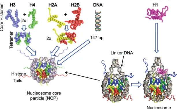
The basic unit of chromatin is the nucleosome. The nucleosome core particle (NCP) is 147bp of approximately 1.65 turns of super-helical DNA wrapped around a histone octamer. Linker protein histone H1, binds to the 10 to 90bp long linker DNA located at the entry/exit sites of the nucleosome core and the resulting structure is called the nucleosome (Luger et al., 1997).
1.1.1. Histones
Histones are small, highly basic proteins found in all eukaryotic cells. They are primordial for cell viability and are highly conserved during evolution from yeast to humans (Kim et al., 1988). Histones can be grouped into five families: H1, H2A, H2B, H3 and H4. Histones within the H1 family are called linker histones and differ from core histones that constitute the nucleosome core particle.
Genes encoding for conventional histones are generally present in multiple copies in the genome and are organized in clusters, for example in humans chromosomes 6 contains 55 histone genes and chromosome 1 contains nine. Similarly in mouse there is a large cluster on chromosome 13 with 51 genes and two smaller clusters on chromosome 3 and 11 (Marzluff et al., 2002). These genes exhibit atypical features for eukaryotic genes, such as the absence of introns and the derived mRNAs that end with a stem-loop structure instead of a poly-adenylation signal. It is also important to note that their transcription is
regulated to ensure that the mRNA levels increase dramatically during the S phase and fall sharply as soon as the DNA is replicated, restricting the expression of conventional histones to the S phase. In contrast, other subtypes whose expression is not coupled to replication are collectively called histone variants (Osley, 1991).
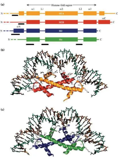
Figure 1.1 Histone families and histone fold interactions. a) Schematic view of the secondary structure of
the histones illustrating the central histone fold domain. Interaction of the histone-fold regions of the b) H2A–H2B dimer and c) H3–H4 dimer with approximately three turns of DNA. (adopted from Dutnall et al, 1997)
Core histones share a common overall structure organized into two domains (Figure 1.1a): a globular domain that contains the highly conserved histone fold domain and unstructured terminal domains, or tails, protruding out of the nucleosome core particle. (Arents et al., 1995.) The histone fold domain is an approximately 70 amino acid long region that is comprised of one larger central alpha helix (α2) separated by loops on each side from smaller alpha helixes (α1 and α3) and enables histone dimerization important for nucleosome formation via head to tail interaction of the histone fold domains (Figure 1.1b). Histone tails are less structured and poorly conserved domains, they differ in size
and presence of random coil elements within the histone families and are highly diverse (Figure 1.1a) (Luger et al., 1997; Sandman et al., 1990).
Linker histones are the most diverse family of histones, they are very rich in lysine and differ from core histones as they lack the histone fold domain. H1 family histones typically have a three domain structure: a highly conserved central globular domain (Albig et al., 1997), a short unstructured amino tail (20aa) and a long lysine rich unstructured carboxy terminal domain (100aa)(Hartman et al., 1977). As mentioned before histones H1 bind to linker DNA in the nucleosome for stabilization but are also more dynamic within chromatin. Partial depletion of H1 is not essential for survival in mice and other organisms but plays a crucial role in chromatin state and organization (Khochbin, 2001; Roque et al., 2005; Misteli et al., 2000; Bustin et al., 2005). Whereas simultaneous depletion of three somatic H1 subtypes was lethal in mice (Bednar et al., 2016).
1.1.2. Nucleosome Core Particle and the Chromatosome
The nucleosome core particle (NCP) is the crystalized part of the nucleosome that protects DNA and hence what remains after nuclease digestion. Histones are assembled in pairs of heterodimers H2A-H2B and H3-H4 that interacts with their histone fold domains through head to tail hydrophobic interactions. Both H3-H4 dimers assemble by an
interaction between the histone H3’s α2 and α3 helices, forming a tetramer (H3–H4)2. The
H2A-H2B dimers then bind to each side of the tetramer and complete the octamer formation (Figure 1.2). Though the highly basic structure of histones renders the octamer unstable, stability is obtained by DNA binding or high salt concentration. DNA bends around the histone octamer due to charge neutralization of the acidic DNA phosphate groups, hydrophobic interactions and by hydrogen bound formation between main-chain amide groups and phosphate oxygen atoms (Luger et al., 1997).
The chromatosome is the structure that results from histone H1 binding to the NCP. Though there’s multiple views on where within the nucleosome H1 binds, the widely accepted one is that it binds via its globular domain to the dyad of the nucleosome and its lysine rich carboxy-terminal tail interacts with linker DNA from each side of the nucleosome (Shaytan et al., 2015; Shaytan et al., 2016).
Figure 1.2 Nucleosome assembly: Histone assembly forms the NCP and is proceeded by H1 binding.
1.2. Chromatin Structure and Organization 1.2.1. Chromatin Higher-order Structures
Chromatin in cells can be organized differently depending on the level of compaction. The most relaxed conformation is called the nucleosome filament in which the succession of nucleosomes connected by linker DNA, forms an 11-nm wide linear structure, resembling “beads on a sting “ (Figure 1.4). This structure is observed under low salt conditions by electron microscopy (EM) on both endogenous and reconstituted chromatin. However, this primary structure is not initially found to be favored under physiological conditions (Thoma et al., 1979; Hansen, 2002; Horowitz-Scherer and Woodcock 2006), in fact in the presence of physiological salt concentrations, linear chromatin condenses into a helical rearrangement of nucleosomes, stabilized by linker histones and known as chromatin secondary structure or the 30-nm fiber (Woodcock and Dimitrov, 2001).
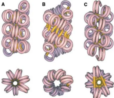
Chromatin organization and nucleosome arrangement is difficult to study and remains a controversial subject. Early EM studies of native chromatin fibers proposed two models for the 30-nm fiber. The one-start helix also known as the solenoid model consists of adjacent nucleosomes connected consecutively by bent linker DNA that fallows a helical trajectory with 6 to 8 nucleosomes per turn (Widom et al., 1985). The two-start helix model is where straight linker DNA connects adjacent nucleosomes and the two
nucleosome arrays are assembled in a zig-zag manner as nucleosome cores are helically arranged (Woodcock and Ghosh, 2010; Williams et al., 1986). The two-start model is divided to two models named the helical ribbon model and the cross-linker model (Figure 1.3) (Dorgio et al., 2004).
Figure 1.3 Proposed models for the 30-nm fiber organization. a) one-start solenoidal, b) two-start
cross-linked , and c) two-start helical ribbon. Upper views have the fiber axis running vertically; lower views are down the fiber axis. Linker DNA running between cores showed in yellow. (Adopted from Dorigo et al., 2004)
Further levels of chromatin compaction do exist, the most obvious being the maximum level of chromatin compaction achieved by the metaphase chromosome. Examples to chromatin tertiary structures are chromatin loops and other higher structures found in both metaphase chromosomes and specialized regions of interphase chromosomes, such as gene enhancers and insulators (Fraser and Grosveld 1998; Woodcock and Dimitrov 2001; Woodcock and Ghosh, 2010). However the lack of definitive information about chromatin secondary structure complicates inquiries to bona fide structures above the 30-nm fiber, in fact more recent studies suggest a paradigm shift. Small-angle X-ray scattering (SAXS) experiments were unable to find repetitive structures beyond the 10-nm fiber in isolated nuclei (Joti et al., 2012) or in mitotic chromosomes (Nishino et al., 2012). Cryo-electron microscopy studies of interphase (Bouchet-Marquis et al., 2006; Gan et al., 2013) and mitotic chromosomes (Eltsov et al., 2008), and electron spectroscopic imaging studies of mouse cells (Fussner et al., 2012), observed folded 10-nm fibers but no 30-nm fibers. Chromosome conformation capture experiments describe structures within interphase
associating domains” (TADs) (Dixon et al., 2012; Nora et al., 2012; Sexton et al., 2012; Dekker et al., 2013; Rao et al., 2014; Eagen et al., 2015).
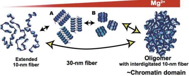
In light of these findings an alternative model has been proposed in which chromosomes are assembled through long-range interactions, so large oligomers are not assemblies of the 30-nm chromatin fibers, but rather are proposed to be an interdigitated polymer-melt like structure deriving from 11-nm nucleosomal arrays (Figure 1.4) (Maeshima et al., 2016).
Figure 1.4 Alternative model: In 1–2 mM Mg2+, the nucleosomal array folds into a folded 30-nm chromatin fiber structure. With further increases in Mg2+, the nucleosome arrays assemble into supramolecular oligomers rather then the 30-nm chromatin fibers. (adopted from Maeshima et al., 2016)
1.2.2. The Metaphase Chromosome
At the beginning of mitosis, chromatin adopts a conformation of maximum compaction that is the mitotic chromosome. The typical mitotic chromosome is X-shaped with two parallel chromatids held in proximity to each other by cohesins and role of condensin. The main functional elements of the chromosome are: the telomeres that protect the DNA at the ends of each chromatid and the centromeres, on which the kinetochore forms and constitutes the anchoring points for microtubules that mobilize chromatids during mitosis. The longest human chromosome is about 10 microns long and a little less than 2 μm wide and contains 250Mb of DNA in each chromatid (Marko, 2008).
Consistent with the uncertainty that surrounds chromatin higher order structures, the structure and the formation of mitotic chromosomes are not well known and widely discussed. The existing models can be loosely subdivided into three groups: hierarchical models of increasingly thicker coiled or looped fibers, loops-on-a-scaffold models and network models, which describe mitotic chromosomes as highly cross-linked gels.
The hierarchical folding type models suggest that the mitotic chromosome results from several levels of successive folding of 30 nm chromatin wrapping into thicker fibers which in turn are wound in the upper level of fiber subsequently coil to form thicker condensed chromatids (Belmont et al., 1987). The structure as strictly ordered hierarchy of increasing levels of coiling was criticized for being overly simplistic, the Belmont laboratory showed that mitotic chromosomes are folded to varying thickness and as an irregularly condensed fiber. They proposed an axial glue model where the chromatin fibers folds hierarchically, but without strict order around a longitudinal central axis enriched in codensins (Kireeva et al., 2004).
The other model is the radial loop/scaffold model, where analysis after histone removal from mitotic chromosomes revealed chromatin loops that emanate from a dense axial structure of average of ~80 kb and consists of condensins and topoisomerase II. This loop-axis structure can then coil to further shorten the chromosome (Boy de la Tour et al., 1988; Maeshima et al., 2003).
The network model was proposed by Poirier and Marko after they performed biophysical measurements by stretching chromosomes using micropipettes and applying restriction enzymes with different cutting frequencies to chromosomes prior to stretching. They observed that chromosomes can return to original length after being stretched up to ~5 fold and postulated that mitotic chromosomes consist of network or gel in which individual chromatin fibers are connected by crosslinking elements, presumably proteins distanced by 10 to 20 kb (Poirier et al., 2002a; Poirier et al., 2002b).
More recently Job Dekkers lab applied chromosome conformation capture methods, 5C and Hi-C to reassess some of these different models. Their findings revealed that contrary to interphase chromosomes, mitotic chromosomes don’t reveal any cell type-specific nor any locus-specific features and found that metaphase Hi-C data is mostly inconsistent with classic hierarchical models. In turn, they describe metaphase organization as a two-stage process: linear compaction by consecutive chromatin loops, potentially generated by cohesin and condensin complexes, followed by axial compression (Naumova et al., 2013). Chromatin Functional Organization
Euchromatin and Heterochromatin
When interphasic nuclei were observed by electron microscopy, two distinct functional states were assigned to chromatin: euchromatin, corresponding to the loose areas
associated with active transcription of genes, and heterochromatin, corresponding to regions of compact chromatin that is often transcriptionally inactive.
Euchromatin regions are relaxed and transcriptionally active during interphase, during replication have priority and only get compacted during mitosis. This compaction coincides with the cessation of mRNA synthesis during cell division (Santos-Rosa et al., 2002).
Heterochromatin protein HP1 is essential for heterochromatin formation, and heterochromatin can be defined by two separate states: constitutive and facultative. Constitutive heterochromatin is highly condensed, maintained and is often enriched in repetitive, late replicating DNA sequences and is poor in genes, such as those present in the centromeric, pericentromeric and subtelomeric regions (Greval et al., 2007).
Facultative heterochromatin state is more flexible, decondensation can occur outside of the S-phase and these regions can become transcriptionally active. Facultative heterochromatin formation is regulated by proteins such as Polycomb-group proteins and non-coding RNAs such as Xist. Xist RNA plays an essential role in X chromosome inactivation in mammalian females and is a good example of facultative heterochromatin function. Another interesting feature of facultative heterochromatin is that it’s capable of spreading to neighboring chromatin regions (Hines et al., 2009; Trojer and Reinberg, 2007; Greval et al., 2007).
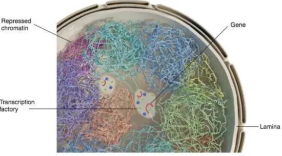
Heterochromatin is often enriched at the nuclear periphery, this preferential association with the nuclear lamina (lamin A, B and C) has been widely documented, these regions are called lamina-associated domains (LADs), typically 0.1-1 Mb in size (Figure 1.5) (Pickersgill et al., 2006; Guelen et al., 2008; Kind and van Steensel, 2010).
Chromosome Territories
Chromosome territories refer to the space occupied by chromosomes during interphase, although they don’t have a specific address within cell, large gene-poor chromosomes are more likely to located near the nuclear periphery, whereas small gene-rich chromosomes are located more internally. Chromosome territories don’t have specific inner compartmentalization regarding active genes within the territory, in fact it has been shown that in some gene dense regions such as the HOX gene cluster, chromatin loops protrude from the territory towards transcription factories (Figure 1.5) (Cremer and Cremer, 2010).
Figure 1.5 Schematic representation of the nucleus, different colors mark separate chromosome territories.
(Adopted from Dekker, 2015)
1.1. Chromatin Dynamics
Chromatin organization and structure is important for living cells but the highly compact state of chromatin can render the genome relatively inaccessible to protein machineries and these structures alone are not sufficient to meet the constantly alternating physiological and metabolic needs of eukaryotic organisms. Chromatin must be able to alternate freely between structural states and hence should be a highly dynamic structure that can reorganize and assure dynamic cellular processes to modulate chromatin structure and increase genome availability to regulatory proteins. Orchestrating such dynamics is complex, partial or complete remodeling of the nucleosomes allows access to molecular machineries of transcription, replication and DNA repair. Three mechanisms ensure this dynamism: chromatin modifiers, post-translational modifications of histones and incorporation of histone variants.
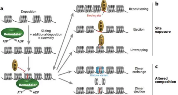
1.2.3. Chromatin Remodelers
Chromatin remodelers alter chromatin state in an ATP dependent manner by moving, ejecting, reassembling or restructuring nucleosomes. Based on their domain structures there are four families of remodelers: the SWI/SNF family, the ISW1 family, the CHD family and the INO80 family. All four families share some basic properties; a high affinity for nucleosomes, histone modification recognition sites, DNA dependent ATPase domains
capable of undoing DNA/histone interactions and finally domains and/or proteins for interaction with other chromatin or transcription factors. (Clapier and Cairns, 2009).
Figure 1.6 Different outcomes of chromatin remodeling. Remodelers can move already incorporated histone
octamers, generating room for additional deposition a) Remodeler activity on a nucleosome array can lead to separate outcomes: b) site exposure, where a previously inaccessible site (red) for a DNA-binding protein (DBP) is revealed by nucleosome sliding (repositioning), or nucleosome eviction (ejection), or localized unwrapping, and c) altered composition, where the actual content of the nucleosome is altered by dimer replacement or ejection. (Adopted from Clapier and Cairns, 2009)
The SWI/SNF (switching defective/sucrose nonfermenting) family remodelers have multiple function and activities involving nucleosome sliding and eviction but are not implicated in chromatin assembly (Mohrmann and Verrijzer, 2005). The ISWI (imitation switch) family remodelers modify nucleosome spacing and assist chromatin assembly dually where they can facilitate transcriptional activation or repression. (Corona and Tamkun, 2004). The CHD (chromodomain, helicase, DNA binding) family remodelers can promote or repress transcription by sliding and ejecting nucleosomes, this variability# may come from chromodomain diversity (Marfella et al., 2007). The INO80 (inositol requiring
80) family remodelers have diverse functions, including #promoting transcriptional
activation and DNA# repair and histone dimer replacement (Figure 1.6) (Bao and Shen, 2007).
1.2.4. Histone Posttranslational Modifications
Histones are the target of a large number of post-translational modifications (PTMs), these changes are associated with processes such as transcription, DNA repair or chromatin condensation. Modifications of the amino-tails were amongst the first observed and more extensively studied. The presence of one or more modified histones can alter the properties of nucleosomes but (Mutskov et al., 1998) it has been postulated that post-translational modifications of histones exert their effects not so much by altering the nucleosome but rather by the recruiting specific proteins. A further step of this model assumes that a combination of modifications rather than individual events recruit distinct proteins, proposing a sort of "histone code" (Strahl and Allis, 2000). This hypothesis remains widely debated, including lack of a clear causal link between post-translational modifications and the activation or repression of transcription (Henikoff and Shilatifard, 2011).
Although most previously known histone modifications were on the amino-tails of histones, new modifications have been located in the structured core and these modifications are on the histone-DNA interphase. These lateral surface modifications may in fact alter the free energy of histone-DNA interactions and hence nucleosome mobility (Cosgrove et al., 2004, Lawrence et al., 2016).
Principle Histone Modifications
Histone Acetylation
Histone acetylation is a dynamic process executed by two families of enzymes with antagonistic functions, histone acetyltransferases (HATs) catalyze the transfer of an acetyl group from acetyl CoA to the ε-amino terminal group of lysine residues, whereas histone deacetylases (HDACs) catalyze lysine de-acetylation where acetylated lysines are recognized via their bromodomain and are removed (Dhalluin et al., 1999; Jacobson et al., 2000).
There are two classes of HATs: type A HATs, are diverse, localized in the nucleus and are involved in histone acetylation within chromatin. Type B HATs are located in the cytoplasm and acetylate newly synthesized histones before these histones are deposited into chromatin (Sterner and Berger, 2000). Cytoplasmic acetylations can be needed for H4 deposition such as H4K5 or necessary due potential lack of access to structured domains once deposited such as with H4K91 acetylation (Ye et al., 2005).
HDACs are most frequently associated with transcriptional repression, deacetylation stabilizes chromatin structure and histone hypoacetylation is associated with repressed chromatin state (Grunstein, 1997; Katan-Khaykovich and Struhl, 2002). Histone acetylations also play a critical role in apoptosis, DNA repair and VDJ recombination (Kouzaride, 2007).
Histone Methylation
Histone methylation is the addition of one to three methyl groups on to the side chains of lysine and arginine residues. Lysine methylation is catalyzed by histone (lysine) methyltransferases (HKMT) and arginine methylation is catalyzed by protein arginine methyltransferases (PRMT). Contrary to acetylation and phosphorylation, histone methylation does not alter the charge of the histone. Each enzyme generally targets a specific residue and the impact of said methylation varies depending on residue that has been modified and the overall number of methyl groups transferred on said residue (Lachner and Jenuwein 2002; Lachner et al., 2003). Three lysines, in particular, are associated with activation of transcription when they are methylated: H3K4, H3K36 and H3K79; conversely, di- and tri-methylation of H3K9 and H3K27 is frequently associated with transcriptional repression and is a hallmark of heterochromatin, as they are recognized by HP1 and by the polycomb repressive complex respectively (Bannister et al., 2001). More so, H4K20me1 is associated with active transcription, H4K20me2 with DNA damage response and H4K20me3 with transcriptional repression when present at promoters (Wang et al., 2008; Botuyan et al., 2006).
For a long time it was thought that histone methylation was irreversible, the identification of the amine oxidase LSD1 (a.k.a KDM1) (Allis, 2007) as a histone demethylase changed this perception (Shi et al., 2004). Currently there are many demethylases that have been identified and grouped into six families KDM1 to KDM6. For instance, certain demethylases containing the Jumonji catalytic domains, can demethylate very stable trimethylated lysines as well as methylated arginines (Cloos et al., 2008).
Histone Phosphorylation
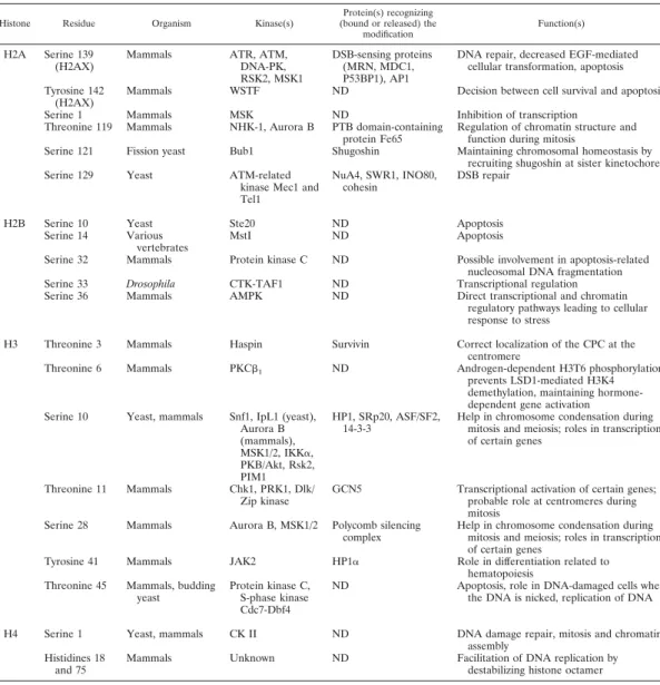
Phosphorylation is the addition of a phosphate group on a serine, threonine or tyrosine residue. Dedicated kinases transfer the phosphate group from ATP to the hydroxyl group of the amino acid side-chain, this introduces a negative charge to the histone. In chromatin,
phosphorylation seems to occupy a less prominent place when compared to acetylation or methylation. However lately, phosphorylation is being studied and linked to more functions (Table 1: Banerjee et al., 2011). Perhaps the most known phosphorylation event is that of H3S10 and to a lesser extent that of H3S28, both involved in chromosome condensation at the beginning of mitosis, mediated by Aurora kinases. (Hans and Dimitrov, 2001). Furthermore, in mammals phosphorylation at S139 of H2A.X, a variant of histone H2A, is associated with the repair of double-strand DNA breaks (Rogakou et al., 1998).
Dephosphorylation in turn is the removal of a phosphate group by hydrolysis, it is assured by phosphatases. For example, fallowing DNA damage repair, a chromatin-associated phosphatase, Wip1, dephosphorylates H2A.X at S139 and allowing the cell to pass from the cell cycle checkpoint (Macurek et al., 2010).
TABLE 1. Histone residues that are phosphorylateda
Histone Residue Organism Kinase(s)
Protein(s) recognizing (bound or released) the
modification
Function(s)
H2A Serine 139 (H2AX)
Mammals ATR, ATM, DNA-PK, RSK2, MSK1
DSB-sensing proteins (MRN, MDC1, P53BP1), AP1
DNA repair, decreased EGF-mediated cellular transformation, apoptosis Tyrosine 142
(H2AX)
Mammals WSTF ND Decision between cell survival and apoptosis Serine 1 Mammals MSK ND Inhibition of transcription
Threonine 119 Mammals NHK-1, Aurora B PTB domain-containing protein Fe65
Regulation of chromatin structure and function during mitosis
Serine 121 Fission yeast Bub1 Shugoshin Maintaining chromosomal homeostasis by recruiting shugoshin at sister kinetochore Serine 129 Yeast ATM-related
kinase Mec1 and Tel1
NuA4, SWR1, INO80, cohesin
DSB repair
H2B Serine 10 Yeast Ste20 ND Apoptosis Serine 14 Various
vertebrates
MstI ND Apoptosis
Serine 32 Mammals Protein kinase C ND Possible involvement in apoptosis-related nucleosomal DNA fragmentation Serine 33 Drosophila CTK-TAF1 ND Transcriptional regulation Serine 36 Mammals AMPK ND Direct transcriptional and chromatin
regulatory pathways leading to cellular response to stress
H3 Threonine 3 Mammals Haspin Survivin Correct localization of the CPC at the centromere
Threonine 6 Mammals PKCb1 ND Androgen-dependent H3T6 phosphorylation
prevents LSD1-mediated H3K4 demethylation, maintaining hormone-dependent gene activation Serine 10 Yeast, mammals Snf1, IpL1 (yeast),
Aurora B (mammals), MSK1/2, IKKa, PKB/Akt, Rsk2, PIM1 HP1, SRp20, ASF/SF2, 14-3-3
Help in chromosome condensation during mitosis and meiosis; roles in transcription of certain genes
Threonine 11 Mammals Chk1, PRK1, Dlk/ Zip kinase
GCN5 Transcriptional activation of certain genes; probable role at centromeres during mitosis
Serine 28 Mammals Aurora B, MSK1/2 Polycomb silencing complex
Help in chromosome condensation during mitosis and meiosis; roles in transcription of certain genes
Tyrosine 41 Mammals JAK2 HP1a Role in differentiation related to hematopoiesis
Threonine 45 Mammals, budding yeast
Protein kinase C, S-phase kinase Cdc7-Dbf4
ND Apoptosis, role in DNA-damaged cells when the DNA is nicked, replication of DNA H4 Serine 1 Yeast, mammals CK II ND DNA damage repair, mitosis and chromatin
assembly Histidines 18
and 75
Mammals Unknown ND Facilitation of DNA replication by destabilizing histone octamer
Ubiquitination and Sumoylation
Ubiquitin is a highly conserved eukaryotic protein of 8,6kDa. Ubiquitination is the addition of one or more ubiquitin molecules on the ε-amino group of lysine residues. This reaction is catalyzed by the combined activity of three enzymes: E1-activating, E2-conjugating and E3-E2-conjugating enzymes (Hershko et al., 1998). In the cytoplasm, ubiquitinated proteins are sent to the proteasome for degradation. Within chromatin, ubiquitination of histones does not lead to degradation. For example, mono-ubiquitination of histone H2B on K119 is associated with activation and elongation of transcription (Vitaliano-Prunier et al., 2008; Pavri et al., 2006) while the mono-ubiquitination of H2A on K120 or 123 is implicated in maintaining transcriptional repression by the polycomb
repressive complex (Wang et al., 2004). Ubiquitination is a reversible event and
polyubiquitin chains can be removed by specific isopeptidases called deubiquitinases
(Nijman et al., 2005).
Sumoylation is a similar process to ubiquitination where small ubiquitin like modifiers (SUMO) of ~12kDa are attached reversibly to lysine residues in a reaction catalyzed again by E1, E2 and E3 enzymes. Sumoylated lysines of histones have been reported to be subsequently ubiquitinated, acetylated or methylated, suggesting a cross-talk between sumoylation and other post-translational modifications (Hendriks et al., 2014).
Other Modifications
There is an increasing number of histone modifications being described and studied, such as ADP-ribosylation, deamination, propionylation, butyrylation, citrullination and crotonylation. These research show that histones are intensely modified both on their amino tails and their core domains (Tan et al., 2011).
1.2.5. Histone Variants
Histone variants are nonallelic isoforms of conventional histones and are present in all eukaryotic organisms. There are variants for all classes of histones, except for H4. Unlike canonical histones, the expression of histone variants is not restricted to the S phase but may occur throughout the cell cycle. Furthermore the expression of certain subtypes can even be restricted to a specific tissues or specific stages of development.
Genes encoding the histone variants are present only in one or two copies and are distributed throughout the genome. They all have at least one intron and a poly-adenylation signal.
The differences between histones and their variants can affect the overall stability of the nucleosome or the residues of the nucleosome that are exposed. Hence the incorporation of histone variants confer distinct structural and functional properties, and thus distinct roles, to the nucleosome (Marzluff et al., 2002; Kamakaka and Biggins, 2005).
1.1.1.1. H2A Family Variants
The H2A family includes the largest number of variants, including the H2A.Z and H2A.X variants, found in most eukaryotes, and H2A.Bbd and macroH2A variants, specific to vertebrates.
Histone variant H2A.Z
H2A.Z represents approximately 5-10% of total histone H2A and is highly conserved through evolution derived from a single origin, (Malik et al., 2003) sharing ~60% sequence homology with the canonical H2A. Its removal is lethal in Drosophila (Daal and Elgin, 1992) and mouse (Faast et al., 2001). Nucleosomes containing variant H2A.Z instead of histone H2A are less stable at the interaction between the H2A.Z/H2B dimer
and (H3/H4)2 tetramer, hence a less stable nucleosome, which may allow easier access to
the double helix of DNA (Abbott et al., 2001; Suto et al., 2000).
H2A.Z is involved in transcriptional activation and preventing heterochromatin formation (Meneghini et al., 2003). H2A.Z is enriched at eukaryotic promoters, specifically at the +1 and −1 nucleosomes, flanking the nucleosome free regions associated with RNA pol II-transcribed genes and facilitating transcription (Raisner et al., 2005; Zhang et al., 2005; Weber et al., 2014).
Posttranslational modifications of H2A.Z are also deterrent of its functions. For example mono-ubiquitination of H2A.Z is associated with facultative heterochromatin and X-inactivation while acetylation correlates with active genes (Sarcinella et al., 2007; Millar et al., 2006; Bruce et al., 2005). Genome wide deposition of H2A.Z is diverse, dynamic and regulated, in mouse trophoblast stem cells H2A.Z is lost from gene promoters and locates to the centromeres during mitosis (Nekrasov et al., 2012). H2A.Z in fission yeast is excluded from centromeric chromatin (Ogiyama et al., 2013). Interestingly,
even though its role may differ in these organisms, knockdown of H2A.Z leads to defects in chromosome segregation in both mice and fission yeast (Rangasamy et al., 2004; Kim et al., 2009).
Histone Variant H2A.X
H2A.X amounts up to a quarter of the total amount of histone H2A in the cell and
possesses a conserved C-terminal tail motif containing a phosphorylatable serine (S139). The phosphorylation occurs in response to DNA double-strand breaks, where foci, termed γ-H2A.X, appear within ten minutes after cells are exposed to ionizing radiation that introduces double strand breaks and disappear gradually within two hours (Rogakou et al., 1998). H2A.X in its phosphorylated form, γ-H2A.X, is believed to destabilize chromatin and impair H1 binding (Downs et al., 2000). It has been called the guardian of the genome as it recruits and triggers DNA repair mechanisms and induces chromatin remodeling near the break site (Thiriet and Hayes, 2005). However H2A.X is dispensable to the repair process since DNA repair was observed in H2AX-deficient cells and animals signaling the existence of independent repair pathways (Yuan et al., 2010).
Histone Variant MacroH2A
MacroH2A is atypical histone variant, although its amino-terminal domain shares ~60% similarity with canonical H2A, macroH2A is characterized by a large C-terminal extension that can measure up to 200 amino acids in length according to species, called the macrodomain. Nucleosomes containing macroH2A are less flexible and more hydrophobic (Chakravarthy et al., 2005), their presence in a nucleosome disturbs both the recruitment of transcription factors such as NF-kB, nucleosome remodeling by the SWI / SNF complex (Angelov et al., 2003) and that histone acetylation necessary for transcription (Doyen et al., 2006b). Hence macroH2A is associated with the suppression of transcription and is particularly enriched on the inactive X chromosome (Mietton et al., 2009).
Phylogenic distribution of macroH2A is not uniform and knock-out of this variant in mice isn’t lethal, however knock-out mice do exhibit impaired pre- and postnatal growth in addition to male reproductive impairments (Talbert et al., 2012; Pehrson et al., 2014).
Histone Variant H2A.B
The histone variant H2A.B, also known as H2A.Bbd is found, contrary to macroH2A, to be excluded from the inactive X chromosome (Bbd for Barr-deficient body) and is
associated with transcriptionally active chromatin (Chadwick and Willard, 2001). H2A.B shares only ~50 % sequence similarity with canonical H2A and has a truncated docking domain. H2A.B containing nucleosomes bind only ~120 bp of DNA and are less stable than conventional nucleosomes (Bao et al., 2004; Doyen et al., 2006a; Gautier et al., 2004). H2A.B nucleosomes also lack the small acidic region that forms on the surface of the canonical nucleosomes as well as K119 whose ubiquitination is linked to transcriptionally inactive regions (Zhou et al., 2007). More recently, H2A.B has also been found to localize at DNA replication and repair sites in elongating spermatids (Arimura et al., 2013).
1.1.1.2. H2B Family Variants
Contrary to the large diversity of H2A variants, few H2B variants have yet been identified. The most studied variants are the mammalian TH2B (Testis specific H2B) and H2B.FWT. TH2B shares 86% similarity with the canonical H2A and largely replaces conventional H2B in spermatocytes in humans, mouse and rats (Churikov et al., 2004). In humans, it is expressed throughout spermatogenesis, where it is found in at the telomeres (Zalensky et al., 2002). In mice, TH2B directs the final stages of histone to protamine transition in male germ cells and reassembles back after fertilization on the male genome during protamine to histone exchange (Montellier et al., 2013).
H2B.FWT is a surprisingly divergent variant and shares only 45% sequence identity with conventional H2B, sequence divergence is most pronounced in the amino-tail on account of the 42 amino acid extension of the H2B.FWT tail. The reconstitution of nucleosomes in vitro containing this variant showed H2B.FWT does not disturb the nucleosome remodeling by SWI / SNF, but its amino tail prevents H2B.FWT contribution during mitotic chromosome assembly. H2B.FWT is also associated with telomeric regions (Boulard et al., 2006).
1.1.1.3. H3 Family Variants
The H3 family includes two conventional subtypes H3.1 and H3.2, and three variant subtypes H3.3, H3.4 (a.k.a H3.t or TH3) and CenH3 (a.k.a CENP-A). With the exception of CenH3, all subtypes diverge from one another by only few residues. For instance H3.1 and H3.2 differ only by one residue.
Histone Variant H3.3
The histone variant H3.3 diverges from H3.1 by only five residues and from H3.2 by four residues. Nucleosomes containing these H3 subtypes are highly similar in structure. Intriguingly, H3.3 specific residues are highly conserved and are located on the accessible surfaces of the H3/H4 tetramer, including several PTM sites (Melters et al., 2015).
In mammals, H3.3 is coded by two genes H3F3A, H3F3B and disruption of either one of theses genes causes detrimental effects on development (Couldrey et al., 1999; Bush et al., 2013; Szenker et al., 2011). H3.3 has been associated with transcriptionally active chromatin (Ahmad and Henikoff, 2002; McKittrick et al, 2004) and is supposed to be a hallmark of active genes (Hake and Allis, 2006), although this role has recently been challenged given the presence of H3.3 in constitutive heterochromatin regions such as telomeres and pericentric chromatin (Szenker et al., 2011; Goldberg et al., 2010).
As mentioned before H3S10 and H3S28 phosphorylation are important for chromosome condensation at the beginning of mitosis. They first appear in prophase, persist until anaphase, and localize to peripheral regions of the condensed DNA. Strikingly, H3.3 is phosphorylated on S31 (H3.3 S31P) during mitosis but differs from H3S10P and H3S28P, in both timing and location, as it is present only in late prometaphase and metaphase (Hake et al., 2005).
Histone Variant H3.4
The variant H3.4, diverges from H3.1 by four residues and H3.2 by five residues, it has been identified as a testis specific variant. In rats, it represents up to 40% of total histone H3 throughout spermatogenesis (Meistrich et al., 1985). In humans, its expression is restricted to spermatocytes, where it potentially represents the majority histone H3 (Witt et al., 1996). H3.4 nucleosomes appear less stable than conventional nucleosomes, including a weaker interaction between the H3.4:H4 tetramer and H2A:H2B dimers. H3.4 has been also reported in other tissues, such as the brain and the embryo (Tachiwana et al., 2010).
Histone Variant CenH3
The variant CenH3 (CENP-A) is the centromere specific histone variant and is considered to be the epigenetic marker of centromeric chromatin. It is the central protein in this thesis work and will be thoroughly described in the final chapter of the introduction.
2. Mitosis
In eukaryotes the cell cycle of proliferating cells can be divided into two parts: interphase and mitosis. Mitosis is the part of the cell cycle where a eukaryotic cell equally divides its duplicated genetic material, resulting in two cells with the same genome. However interphase isn’t a stagnate state, it encapsulates specific stages during which both cell growth and DNA replication occur in preparation for cell division. Hence the cell cycle can be examined in four discrete phases. The M phase of the cycle corresponds to mitosis, which is usually followed by cytokinesis. The next phase is the G(ap)1 phase, which corresponds to the interval “gap” between mitosis and DNA replication where the cell is metabolically active and grows continuously. G1 is followed by the S(ynthesis) phase corresponding to DNA replication. The S phase is followed by the G2 phase, during which cell growth and protein synthesis continues in preparation for mitosis (Cooper GM (2000). "Chapter 14: The Eukaryotic Cell Cycle". The cell: a molecular approach.).
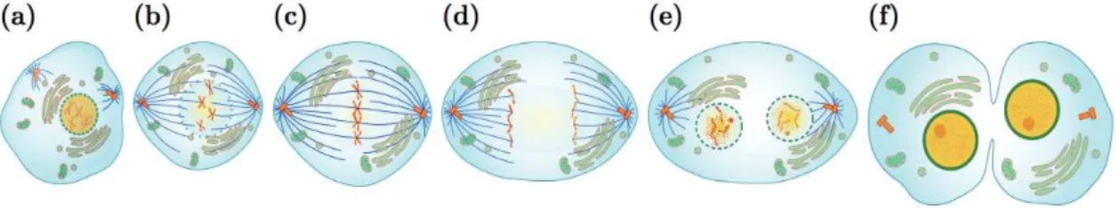
2.1. Stages of Mitosis
Mitosis is divided into five phases and starts with prophase (Figure 2.1a), during which the interphase chromatin condenses to form individualized chromosomes and transcription is widely paused (Kadauke et al, 2013). Animal cells contain a pair of centrosomes responsible for microtubule nucleation and anchoring. During prophase each centrosome migrates to the poles of the cell, forming a bridge of spindle fibers (Eddé et al., B.1990). The next stage is prometaphase (Figure 2.1b), the nuclear membrane disintegrates and the chromosomes that are now fully condensed migrate towards the cells equatorial plane. This movement is called congression. Kinetochore microtubules emerging from the centrosomes attach to the kinetochores and spindle microtubules move the chromosomes toward the center of the cell. The chromosomes align on the metaphase plate in metaphase (Figure 2.1c), during which the cell ensures the correct orientation and bipolar attachment of each chromosome to the mitotic spindle.
Metaphase is then followed by anaphase (Figure 2.1d), where the sister chromatid cohesion is broken and each chromatid pulled towards the opposing poles of the cell. In telophase (Figure 2.1e) the migration of chromatids is completed and the nuclear envelope reforms while microtubules of the mitotic spindle dissociate and chromatin decondensation occurs. Alongside telophase, the division of the cytoplasm called
leads to the near total separation of the two daughter cells. The disappearance remaining small dense structure, called the residual body marks the end of cell division.
Figure 2.1 Stages of mitosis. a) Prophase: chromosomes condense and the nuclear membrane disappears. b) Prometaphase: chromosome capture by microtubules. c) Metaphase: Alignment of chromosomes on the
metaphase plate. d) Anaphase: sister chromatids separate and migrate towards the poles. e) Telophase: chromosomes decondensation and nuclear membrane reformation. f) Cytokinesis: division of the cytoplasm. (by Marina Ruiz Villarreal)
2.2. Regulation of Mitosis
Ensuring proper progression of mitosis is exceptionally important as errors during cell division can have devastating consequences for the cell. Transcriptional arrest during mitosis mandates that all effectors and enzymes are readily available in the cell, thus activation of molecular pathways depend on protein turnover and posttranslational modifications. Two mechanisms ensure this control. The first is removal of effectors by proteolysis when their function is no longer needed, and the second is phosphorylation that serves as molecular on and off switch for many enzymes. Hence mitotic kinases play a central role in controlling mitosis.
Principle Kinases
This section only covers the role of these kinases during mitosis and does not envelope functions during the rest of the cell cycle.
CDK Family Kinases
Cyclin Dependent Kinases (CDKs) are among the most important conductors of the cell cycle. During mitosis, the predominant kinase family is Cdk1, of the twenty Cdks that have been identified in humans, Cdk1 is the only one that is essential for the cell cycle in all eukaryotic cells (Malumbres et al., 2009). Expressed during G2 phase, Cdk1 is phosphorylated on Tyr15 by Wee1 kinase and becomes inactive (Parker et al., 1992). Cdc25 phosphatase dephosphorylates Cdk1 and Cdk1 association with cyclin B allows the
activation of the kinase and triggers mitotic entry (Ferguson et al., 2005; Nilsson and Hoffmann, 2000).
The cyclinB/Cdk1 complex targets multiple proteins. The phosphorylation of lamins causes them to dissociate and depolymerize resulting in disintegration of the nuclear envelope. The phosphorylation of condensins induces chromosome condensation (Heald et al., 1990). Other targets include microtubule and kinesin-related proteins, promoting the assembly of the mitotic spindle and cohesins, located on chromosome arms (Kimura et al., 1998). Finally, during the transition from metaphase to anaphase, degradation of cyclin B initiated by the ubiquitin ligase anaphase promoting complex/cyclosome (APC/C), opens the way for completion of mitosis (Murray, 2004).
PLK family kinases
The polo-like kinases (PLK) are conserved serine/threonine kinases involved in the regulation of cell cycle progression through G2 and mitosis. Expressed during G2 phase, they are initially distributed throughout the cell but later localize from prophase to metaphase to the centrosomes and kinetochores. (Glover et al., 1998)
PLK1 mediates mitotic entry, both by direct activation of the cyclin B/Cdk1 complex and by phosphorylation of Cdc25c, which promotes its nuclear localization (Roshak et al., 2000). At the centrosomes, it promotes the recruitment of γ-tubulin by phosphorylating Asp, enabling centrosome maturation and microtubule nucleation (Petronczki et al., 2008). On the kinetochore, it contributes to recruitment of NDC80 complex involved in bipolar spindle formation (Wong and Fang, 2005). Finally, PLK1 participates in the metaphase to anaphase transition, either by directly phosphorylating the APC/C subunits (Golan et al., 2002) or by promoting the degradation of one of its inhibitors, Emi1.
NRK family kinases
NIMA (never in mitosis A) kinases have been identified in Aspergillus nidulans in a screen for cell cycle mutants. NIMA mutants remained arrested in G2 phase without ever entering into mitosis and acquired the name never in mitosis A. NIMA homologues are NRK family or NIMA-related kinases (O'Connell et al., 2003).
In vertebrates, the most likely functional homologue is the NEK2 kinase, essentially important in the maturation and separation of centrosomes and bipolar spindle formation. NEK2 controls centrosome separation (essential for the formation of bipolar spindles) by
phosphorylating proteins, such as Nap1, resulting in their displacement from the centrosomes (Bahe et al., 2005). It also regulates kinetochore microtubule attachment stability in mitosis via phosphorylation of NDC80 (Chen et al., 2002).
Aurora family kinases
Part of the serine/threonine kinases, Aurora kinase was first identified in Drosophila in a screening for defective chromosome segregation. Although in yeast one gene encodes for Aurora kinase, the family includes three members in mammals, Aurora A, Aurora B, and C. Aurora C has more limited expression and plays an important role during meiosis (Tang et al., 2006) and early embryonic development (Fernandez-Miranda et al., 2011; Schindler et al., 2012). Aurora A and B are involved in somatic mitosis and share some common characteristics: they are both expressed in G2 and M phases and their activation depends on their association with specific cofactors coupled to auto-phosphorylation. Lastly, they are both degraded at end of mitosis by APC/C dependent ubiquitination (Lindon et al., 2016).
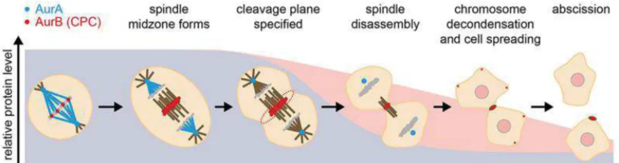
Aurora A mainly localizes at centrosomes and mitotic spindle poles throughout mitosis, assisting in centrosome maturation and separation, microtubule dynamics, and bipolar spindle assembly (Barr and Gergely, 2007). In contrast, Aurora B localizes at centromeres and chromosomes in early mitosis and locates to the spindle midzone/midbody during mitotic exit (Figure 2.2) (Adams et al., 2001; Schumacher et al., 1998).
Figure 2.2 Aurora kinase dynamics throughout mitosis (adopted from Lindon et al., 2016)
Aurora A
Aurora A contributes to mitotic entry and maturation of centrosomes. It initially displays a cytoplasmic localization, where it forms a complex with its cofactor Bora. The complex Aurora A/Bora in turn phosphorylates PLK1 (Macůrek et al., 2008; Seki et al.,
2008), which promotes mitotic entry (by PLK1 action on the complex cyclin B / Cdk1) and the phosphorylation and degradation of Bora. Aurora A then associates with another cofactor TPX2 (Chan et al., 2008) and the complex can locate to the centrosome (Figure 2.2). There, it allows the recruitment of pericentriolar material (PCM) (Hannak et al., 2001) and the mobilization of microtubule-associated proteins (Dutertre et al., 2002). It also phosphorylates the centrosomic fraction of Cdc25, maintaining the local activation of the cyclinB/Cdk1 (Dutertre et al., 2004).
More recent studies suggest also Aurora A involvement in cytokinesis by insuring the stability of astral microtubules and central spindle robustness (Lioutas and Vernos 2013; Reboutier et al., 2013).
Aurora B
Aurora B is a member of chromosomal passenger complex (CPC), a set of proteins characterized by their location profile during mitosis. This complex, in particular the protein INCENP (inner centromere protein), allows both the complete auto-activation of the kinase but also its proper localization. Aurora B associates with chromosome arms in prophase, where it participates in chromatin condensation by phosphorylating condensins and histone H3 at serine 10 (Giet and Glover, 2001; Lipp et al., 2007). It also assists in cohesin removal along chromosome arms, together with PLK1 (Giménez-Abián et al., 2004). During prometaphase and metaphase, Aurora B localizes to centromeres and oversees the attachment of microtubules to kinetochores by destabilizing incorrect attachments (Shang et al., 2003; Liu and Lampson, 2009; Tanaka et al., 2002; Lampson et al., 2004; Cimini et al., 2006; Knowlton et al., 2006; Lampson and Cheeseman, 2011) and activating the spindle assembly checkpoint (SAC). During telophase, Aurora B is located on the spindle midzone for required disassembly (Buvelot et al., 2003). Lastly during cytokinesis, it is located at the residual body where it contributes to the formation of the contractile ring (Figure 2.2) (Minoshima et al., 2003).
More recently a control mechanism involving Aurora B activity gradient has been proposed as a regulator of late mitosis. Complete separation of chromosomes and their arrival to the spindle poles is necessary for DNA decondensation and complete nuclear envelope reformation (NER). At the spindle midzone as chromosomes migrate to opposite poles, the lack of accessible substrates to become dephosphorylated by phosphatases ultimately drives mitotic exit. In this regard Aurora B phosphorylation gradient at the
spindle midzone would allow for the spatial regulation of several substrates involved in chromosome (de)condensation and NER, such as histone H3 (Hsu et al., 2000; Neurohr et al., 2011). DNA decondensation and NER are inhibited on lagging chromosomes/ chromatin bridges as a chance for possible reintegration (Afonso et al., 2014; Karg et al., 2015). This indicates a potential surveillance mechanism or “chromosome separation checkpoint” that controls the extent of anaphase chromosome separation before completion of telophase (Maiato et al., 2015).
2.3. The mitotic spindle 2.3.1. Microtubules
The mitotic spindle is composed of microtubules, a cylindrical structure formed by the association of thirteen protofilaments each composed of α- and β-tubulin heterodimers
(Nogales, 2001). Microtubules are polarized structures, the plus end only exposes α-tubulin and the minus end only exposes β- tubulin molecules. The protofilaments are of
parallel orientation so the overall structure also maintains polarity.
Microtubules are highly dynamic structures, their length varies by constant addition or removal of tubulin dimers at the polymer ends and microtubule polymerization alternates between slow and rapid depolymerization phases according to the local concentration of tubulin dimers (Desai and Mitchison, 1997). At a given end, the authorization of polymerization rate and frequency of depolymerization determine whether the microtubule is in growth or shrinkage. Each end may exhibit different dynamic properties, which gives the microtubule the opportunity to grow or shrink in a particular direction. When the dynamic properties of the two ends are such that one end is growing as the opposite end decreases as the tubulin dimers seem to “treadmill” over the microtubules to the other end, microtubule length remains constant (Margolis and Wilson, 1998).
2.3.2. Centrosomes
The centrosome is the major microtubule organizing center of the cell. It is duplicated during S phase and the resulting two centrosomes define the poles of the cell and hence that of the mitotic spindle during mitosis (Urbani and Stearns, 1999). Centrosomes consist of a pair of centrioles (themselves an assembly of nine microtubule triplets) surrounded by pericentriolar material (PCM).
A major component of PCM is the γ-tubulin ring complex (γ-TuRC) that nucleates microtubules (Zheng et al., 1995). During mitosis the nucleation capacity of the centrosome is increased by the recruitment of larger amounts of γ-TuRC (Khodjakov and Rieder, 1999, Piehl et al., 2004). This allows a rapid increase in the number of microtubules issued from the centrosome and thus the formation of the future mitotic spindle. However, the centrosome is not strictly necessary in this process, and a functional mitotic spindle can form in the absence of centrosomes (Mahoney et al., 2006). The centrosome also has an important role in cell cycle regulation (Doxsey et al., 2005).
2.3.3. The kinetochore
The kinetochore is a protein complex transiently associated with centromeres during mitosis. Spindle microtubules attach to chromosomes through kinetochores.
When observed by electron microscopy, the kinetochore has a structure revealing three layers: the inner kinetochore which is very closely associated with chromatin, the outer kinetochore, a thick region of 50 to 60 nm that interfaces with microtubules and central kinetochore, an electron sparse region between the inner and outer layers. A network of fibers, the fibrous ring is associated with the external surface of the kinetochore (McEwen et al., 2007).
The simplest kinetochore is that of Sacharromyces cerevisiae, it consists of about sixty proteins and contains only a single anchoring site for microtubule attachment (McAinsh et al., 2003; Westermann et al., 2007). Out of five of the main complexes in S. cerevisiae (CBF3, Ndc80, Mtw1, Spc105 and Dam) three of them, Ndc80, Mtw1 (called MIS12 in humans) and Spc105 (Knl1 humans) are conserved in higher eukaryotes (Musacchio and Salmon, 2007; Cheeseman and Desai, 2008; Welburn and Cheeseman, 2008). Kinetochores of higher eukaryotes are bulkier and each bind numerous microtubules (15 to 20 microtubules for the human kinetochore) and are formed of a repetition of the elementary module found in yeast (Joglekar et al., 2008). The constitutive centromere associated network (CCAN) is also conserved in all eukaryotes and is supposed to be the foundation on which the rest of the kinetochore proteins are assembled. Contrary to the other members of the kinetochore those of the CCAN are permanently associated to the centromeres and are not transitional like the rest of the kinetochore.
The elementary functional module kinetochore is the Knl1-MIS12-Ndc80 network (KMN), a 10-subunit supercomplex consisting of the three conserved complexes Ndc80,
Mtw1/MIS12 and Spc105/Knl1 (Cheeseman et al., 2006). In this network, the complex Ndc80 directly interacts with microtubules, with increasing affinity when combined with Mtw1/MIS12 and Spc105/Knl1 complexes (Figure 2.3). The 4-subunit Ndc80 complex forms an elongated structure with two globular heads at each end, separated by a long super-coiled region (Ciferri et al., 2007). One end of the cylinder, composed of globular regions Ndc80 and Nuf2, directly binds microtubules (Cheeseman et al., 2006; Wei et al., 2007); the other end composed of globular regions Spc24 and SPC25 (Wei et al., 2006), is associated with the inner kinetochore (Figure 2.3b). Each microtubule is bound to the kinetochore by several KMN complexes (Joglekar et al., 2006).
Figure 2.3 a) Schematic representation of the centromere b) Schematic view of the main interactions of the
KMN network (adopted from Pesenti et al., 2016)
The Mis12 complex interacts directly with the inner kinetochore via CENP-C (part of the CCAN). The interactions with the Ndc80 complex and with the 2-subunit Knl1 complex take place on adjacent binding sites at the opposite end of the Mis12 complex. They involve short linear motifs of the Nsl1 and Dsn1 subunits of the Mis12 complex, which engage RWD (RING finger, WD repeat, DEAD-like helicases) domains of the Ndc80 complex and the Knl1 complex (Pesenti et al., 2016).
2.4. Chromosome dynamics during mitosis 2.4.1. Microtubule Attachment
All chromosome movement during mitosis requires attachment to the mitotic spindle. Microtubules that attach to the kinetochore form K-fibers, these microtubules are oriented with their plus ends to the kinetochore and their minus ends to the centrosome.