HAL Id: hal-01622344
https://hal.umontpellier.fr/hal-01622344
Submitted on 14 Nov 2017HAL is a multi-disciplinary open access archive for the deposit and dissemination of sci-entific research documents, whether they are pub-lished or not. The documents may come from teaching and research institutions in France or abroad, or from public or private research centers.
L’archive ouverte pluridisciplinaire HAL, est destinée au dépôt et à la diffusion de documents scientifiques de niveau recherche, publiés ou non, émanant des établissements d’enseignement et de recherche français ou étrangers, des laboratoires publics ou privés.
Is nocturnal desaturation a trigger for neuronal damage
in chronic obstructive pulmonary disease?
François Alexandre, Nelly Heraud, Alain Varray
To cite this version:
François Alexandre, Nelly Heraud, Alain Varray. Is nocturnal desaturation a trigger for neuronal damage in chronic obstructive pulmonary disease?. Medical Hypotheses, Elsevier, 2015, 84 (1), pp.25 - 30. �10.1016/j.mehy.2014.11.009�. �hal-01622344�
Title: Is nocturnal desaturation a trigger for neuronal damage in chronic obstructive pulmonary disease?
Francois Alexandre1,2, Nelly Heraud2, Alain Varray1
1Movement To Health Laboratory, Euromov, University of Montpellier 1, Montpellier,
France. 2Clinique du Souffle La Vallonie, Fontalvie, Lodève, France
Corresponding author:
Francois Alexandre, Movement To Health (M2H), Euromov, University of Montpellier 1, 700 avenue du Pic Saint Loup, 34090 Montpellier, France. Phone: (+33) 434 432 632; Fax: (+33) 434 432 644; E-mail: [email protected]
Summary
Patients with chronic obstructive pulmonary disease (COPD) present many neurological disorders of unknown origin. Although hypoxemia has long been thought to be responsible, several studies have shown evidence of neuronal damage and dysfunction even in non-hypoxemic patients with COPD. Adaptive mechanisms protect the brain from hypoxia: when arterial oxygen tension (PaO2) decreases, the cerebral blood flow (CBF) increases, ensuring
continuously adequate oxygen delivery to the brain. However, this mechanism is abolished during non-rapid eye movement (NREM) sleep. Any drop in PaO2 during NREM sleep is
therefore not compensated by increased CBF, causing decreased cerebral oxygen delivery with subsequent brain hypoxia. Patients with may therefore be exposed to neuronal damage during this critical time. This mechanism is of vital importance for patients with COPD because of the potentially deleterious cortical effects. Nocturnal desaturation is quite frequent in COPD and affects approximately one out of two patients who are not hypoxemic during wakefulness. Although the prevalence of NREM sleep desaturation has never been specifically assessed in COPD, current data suggest that at least half of the nocturnal desaturation in desaturating patients occurs during NREM sleep. This review presents the rationale for the hypothesis that nocturnal desaturation during NREM sleep promotes neuronal damage and dysfunction in COPD.
Introduction
1
Chronic obstructive pulmonary disease (COPD) is one of the leading causes of death 2
worldwide. COPD is not restricted to the lung but instead has major systemic repercussions, 3
and it is now considered to be a multi-component syndrome. Accordingly, the predictors of 4
COPD survival include not only the degree of airflow obstruction, but also body mass index, 5
dyspnea, exercise capacity, peripheral muscle size and strength, and the level of hypoxemia. 6
Brain impairment is a well-documented secondary outcome of the disease, but surprisingly 7
this is often forgotten (1). Yet recent reports have irrefutably confirmed severe anatomical 8
brain impairment in COPD (2, 3) with significant functional repercussions that are not limited 9
to cognitive disorders; for example, both peripheral muscle strength (4) and driving abilities 10
(5) can be affected. The mechanisms of brain impairment are still not well understood, 11
however, and it is therefore essential to determine their origins so that they can be better 12
anticipated, with the ultimate goal being to prevent their deleterious effects. Although 13
hypoxemia has long been thought to be responsible for brain impairment (6), several studies 14
have shown evidence of neuronal damage and dysfunction even in non-hypoxemic patients 15
with COPD (2, 7, 8). Adaptive mechanisms protect the brain from hypoxia: when arterial 16
oxygen tension (PaO2) decreases, the cerebral blood flow (CBF) increases, ensuring
17
continuously adequate oxygen delivery to the brain (9-11). However, this mechanism is 18
abolished during non-rapid eye movement (NREM) sleep (12-14). Therefore, any drop in 19
PaO2 during NREM sleep is not compensated by increased CBF, causing decreased cerebral
20
oxygen delivery with subsequent brain hypoxia (15, 16). 21 22 Hypothesis 23 24 25
Given the high prevalence of nocturnal desaturation in patients with COPD (17-19) and the 26
absence of cerebrovascular O2 reactivity during NREM sleep stages (12-14), we hypothesize
27
that nocturnal desaturation during NREM sleep may act as a trigger for neuronal damage and 28
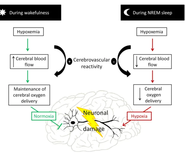
dysfunction in COPD (Figure 1). 29
30
Arguments to support the hypothesis
31
Anatomical brain impairment in COPD
32
Patients with COPD present many anatomical brain alterations. The neuronal damage in 33
COPD was first characterized as periventricular white matter lesions (20) and cerebral 34
metabolic abnormalities (21). Levels of N-acetyl aspartate, a marker of neuronal density, were 35
also reported to be lower in patients with COPD compared with healthy controls (21). More 36
recently, improvements in magnetic resonance imaging analyses through diffusion tensor 37
imaging and voxel-based morphometry have provided better descriptions of the structural 38
brain damage in COPD (2, 3, 22). Zhang et al. (22) found that gray matter volume was 39
reduced bilaterally in the frontal cortex, the cingulate cortex and the left insular cortex in 40
patients with COPD. They also identified gray matter deficits in many subcortical areas such 41
as the right thalamus and left amygdala (22). Hippocampal atrophy was observed and 42
associated with an increased level of serum S100b, a peripheral marker of glial cell 43
impairment (23). In addition, a high prevalence of cerebral microbleeds and small cerebral 44
vessel disease was found in patients with COPD (24), which is consistent with reports of both 45
the decrease in microstructural integrity and the increase in white matter ultrastructural 46
damage in several cortical and subcortical areas (2, 3). 47
48
Functional repercussions of brain impairment
The structural brain damage have several functional repercussions, although most studies 50
have focused on cognitive dysfunction (see Dodd et al. (25) for review). The results of 51
generic questionnaires such as the mini-mental state assessment (MMSE) or the Montreal 52
cognitive assessment (MoCA) have shown globally impaired cognitive function in patients 53
with COPD (23, 26). The P300 component of the event-related brain potential is a useful, 54
objective clinical tool to assess cognitive function. Two studies reported longer P300 latency 55
and lower P300 amplitude in patients with COPD compared with healthy controls, and both 56
these measures are known to reflect attention deficits and impaired decision-making processes 57
(7, 27). Another recent study found impairments in processing speed, working and episodic 58
memory, and executive functions (2), with memory and attention capacities being most 59
impaired (28). It should be noted that brain impairment is not systematic in COPD. Borson et 60
al. (29) found no differences in hippocampal volume or white matter lesions between patients 61
with COPD and healthy controls. Similarly, other studies have observed no significant 62
differences in MMSE scores between patients with COPD and healthy controls (30). Recent 63
studies have estimated the prevalence of cognitive dysfunction as being in the range of 27 to 64
36% (26, 31). 65
As noted above, the functional repercussions of neuronal damage are not restricted to 66
cognitive functioning. For instance, driving ability may be severely impaired in patients with 67
COPD (5, 32). Some studies have reported alterations in resting motor cortex excitability in 68
these patients (33, 34), and another found a lower level of voluntary activation of the knee 69
extensor muscles (35). In a recent study from our laboratory, we assessed the neural activity 70
of the motor cortex during maximal voluntary contractions of the knee extensors and recorded 71
lower motor cortical output in the patients with COPD (4). Taken together, these data support 72
the hypothesis that cerebral processes are involved in COPD muscle weakness. 73
Mechanisms responsible for brain impairment in COPD: an incomplete view
75
Although evidence of a major brain impairment in COPD has steadily accumulated since the 76
first study by Fix et al. (36), the trigger for these cerebral alterations remains unknown (37). A 77
model including vascular disease, inflammation, smoking and hypoxia was proposed to 78
explain COPD neuronal damage and dysfunction (25), yet Dodd et al. (2) reported the 79
persistence of neuronal damage and cognitive dysfunction in COPD even after controlling for 80
smoking and stroke risks. Another study found higher cognitive dysfunction in O2-dependent
81
patients with COPD than in non-dependent patients, with comparable level of systemic 82
inflammation in the two groups (29). This indicates that chronic inflammation, although 83
probably implicated (38), is not the main mechanism of the brain impairment. It also raises 84
doubt about the effectiveness of long-term O2 supplementation in preventing or correcting
85
cerebral alterations, perhaps because hypoxemia per se is not fully responsible for COPD 86
brain impairment. Indeed, while most studies have blamed chronic hypoxemia and 87
hypercapnia (6, 28), neither direct evidence nor explanatory mechanisms could be clearly 88
provided. Moreover, several studies have reported brain impairment and cognitive 89
dysfunction in non-hypoxemic patients with COPD, raising further doubt about the credibility 90
of this hypothesis (2, 7, 8). Although the evidence that hypoxia in vitro induces cellular 91
necrosis is indisputable, it has long been established that hypoxia exposure alone is not able to 92
induce neuronal damage in vivo. Indeed, a fall in arterial oxygen tension (PaO2) without
93
ischemia or a drop in blood pressure (which may cause ischemia) does not induce neuronal 94
death/necrosis (39-41). In this sense, it appears that abnormal perfusion is an essential 95
condition for neuronal damage in vivo. The exact mechanisms are examined in detail below. 96
97
Brain protection against hypoxemia during wakefulness: mechanisms and differences
98
between patients with COPD and healthy individuals
Cerebral autoregulation is the physiological mechanism that maintains constant cerebral 100
perfusion despite blood pressure changes within the normal range (42). However, during 101
changes in arterial blood gases, cerebral autoregulation mechanisms adjust cerebral blood 102
flow (CBF) through CBF velocity and regulate artery caliber to ensure adequate blood gas 103
delivery to the brain (43). When PaO2 decreases, CBF increases, and when arterial carbon
104
dioxide tension (PaCO2) increases, CBF increases. These responses are called cerebrovascular
105
O2 reactivity and cerebrovascular CO2 reactivity, respectively. This close coupling between
106
CBF and arterial blood gases was first described by Kety et al. (44). When PaO2 is decreased
107
in isocapnic conditions, the increase in global and regional CBF can reach up to 200% (9-11). 108
It is assumed that the adaptation of CBF during acute hypoxic exposure is protective, 109
maintaining the stable cerebral oxygen delivery that is a prerequisite for normal brain function 110
(9, 45-47). In contrast, it has been suggested that the increase in CBF in response to a PaCO2
111
increase causes CO2 washout from brain tissue in order to attenuate the level of central CO2
112
(10),therebypreventing the deleterious effects of excessive PaCO2 levels on brain tissue (48,
113
49). However, it is noteworthy that the deleterious effects were reported for PaCO2 levels
114
above 100 mmHg (49), values rarely reached in humans. 115
116
Effects of hypoxic exposure on CBF in healthy individuals 117
Isocapnia during acute hypoxic exposure is a very rare phenomenon in healthy humans. Acute 118
hypoxemia normally induces an increase in ventilation, which in turn leads to hypocapnia to 119
improve blood oxygenation (47, 50, 51). This occurrence of hypocapnia during hypoxic 120
exposure subsequently hampers the increase of CBF velocity caused by lower PaO2, as
121
hypoxemia and hypocapnia have opposite effects on CBF (52, 53). In addition, as CBF is 122
more sensitive to changes in PaCO2 than in PaO2, this heightens the risk of inadequate oxygen
123
delivery to the brain during acute hypoxemia-hypocapnia (53). Nevertheless, more recent 124
studies have shown that the modest increase in CBF velocity during acute hypoxemia-125
hypocapnia is compensated by a greater increase in the caliber of cerebral arteries to ensure 126
normal oxygen delivery to the brain (46, 50). 127
During chronic hypoxia in healthy humans, cerebral oxygen delivery also remains preserved 128
compared to sea-level values (47, 50, 54, 55). The mechanisms suggest that the adaptations 129
differ from those that occur during acute hypoxic exposure, since a progressive return of CBF 130
toward sea-level values has been observed after several days of hypoxic exposure (54-56). 131
This fall in CBF is nevertheless compensated by an increase in arterial oxygen content, 132
mainly due to respiratory acclimatization and a slight rise in hemoglobin concentration (55), 133
resulting in adequate cerebral oxygen delivery (47, 50, 54, 55). Thus, taken together, the 134
aforementioned findings do not support the occurrence of cerebral hypoxia under either acute 135
or chronic hypoxic exposure. 136
137
Effects of hypoxemia on CBF in patients with COPD 138
Patients with COPD can experience acute hypoxemia (e.g., during exercise-induced oxygen 139
desaturation) and the most severe patients may even experience chronic hypoxemia. In 140
patients with COPD acute and chronic hypoxemia are generally accompanied by normocapnia 141
or hypercapnia, rather than hypocapnia compared to healthy individuals. This is due to 142
ventilation-perfusion mismatch and impaired ventilatory muscle function (57). The 143
combination of hypoxemia and hypercapnia increases CBF much more than hypoxemia or 144
hypercapnia alone, since these two phenomena have cumulative vasodilator effects (56). It is 145
thus possible to speculate that the COPD brain is normally much more protected from 146
cerebral hypoxia than the healthy human brain, on condition that both acute and chronic 147
cerebrovascular reactivity are preserved in COPD. Many studies have provided evidence of 148
cerebrovascular CO2 reactivity in COPD (58-62), although this response seems mitigated
compared with that in healthy controls (59, 60). Regarding acute cerebrovascular O2
150
reactivity, one study reported a similar CBF increase in patients with COPD and healthy 151
controls in response to arterial oxygen saturation (SaO2) changes, supporting a preserved
152
response (58). In addition, another study reported higher CBF velocity and an increase in 153
cerebral artery diameter in recently exacerbated patients with COPD (63). More recently, 154
comparable levels of cerebral oxygen delivery during exercise-induced oxygen desaturation 155
were reported when the patients breathed room air and when they breathed oxygen to prevent 156
desaturation (64). This confirms the brain protection against acute hypoxemia in COPD. 157
Regarding the effect of chronic hypoxemia on CBF, a study reported higher CBF velocity and 158
larger cerebral artery diameter using transcranial Doppler in chronic hypoxemic patients with 159
COPD compared with healthy controls (65). This study showed that the mechanisms of 160
cerebral vasodilation persist in patients with COPD during chronic blood gas changes. In 161
addition, the highest CBF levels were found in the hypoxemic-hypercapnic patients, 162
indicating a possible cumulative effect of chronic hypoxemia-hypercapnia on the 163
cerebrovascular vasodilative response (65). Nevertheless, this response is sometimes difficult 164
to observe because of confounding factors (66, 67). For example, CBF is closely coupled with 165
cerebral metabolism: the lower the cerebral metabolism, the lower the CBF is. As patients 166
with COPD exhibit lower resting cerebral metabolism (21, 68, 69), the resting CBF can be 167
lowered in both hypoxemic (66, 67) and non-hypoxemic (62) patients. 168
To conclude, acute and chronic cerebrovascular O2 reactivity is preserved in COPD,
169
indicating that cerebral oxygen delivery is adequate during hypoxemia in patients with COPD 170
(64). Hence, hypoxemia per se does not induce cerebral hypoxia in COPD and this may 171
explain why the involvement of hypoxemia in triggering neuronal damage in COPD has never 172
been demonstrated. 173
Cerebrovascular reactivity during sleep compromises brain integrity in chronic respiratory
175
disorders
176
As the studies cited above have shown, changes in diurnal blood gases are well tolerated by 177
the brain through CBF adaptations. However, this mechanism may be hampered during sleep. 178
Contrary to the adaptations that occur in the waking state, the oxygen desaturation during 179
non-rapid eye movement (NREM) sleep is not accompanied by an increase in CBF (Figure 2). 180
Meadows et al. (13) reduced SaO2 from five to ten percent during slow-wave sleep in humans
181
and found an unexpected decrease in CBF during hypoxemia. This decoupling of CBF and 182
PaO2 has also been reported in patients with obstructive sleep apnea (OSA) (12, 70, 71).
183
While decreasing PaO2 by voluntary breath-holding increases CBF during the waking state
184
(72, 73), CBF tends to decrease during NREM sleep when PaO2 is decreased by sleep apnea,
185
increasing the risk of inadequate cerebral O2 delivery (70).
186
The absence of cerebrovascular O2 reactivity during NREM sleep makes it more difficult to
187
prevent cerebral hypoxia in patients with cardiorespiratory disorders who experience oxygen 188
desaturation during NREM sleep. Cerebral hypoxia has been reported during sleep in patients 189
with OSA (15, 16). It should be noted that this response seems very specific to the NREM 190
sleep stages, as cerebrovascular reactivity is not impaired during rapid eye movement (REM) 191
sleep (12). 192
In summary, cerebrovascular reactivity is impaired and even abolished during NREM sleep in 193
humans (13). In patients who experience hypoxemia or desaturation during NREM sleep, 194
cerebral hypoxia can occur and may induce neuronal damage (14). We thus propose that 195
nocturnal desaturation during NREM sleep can act as a trigger for neuronal damage and 196
cerebral dysfunction in patients with COPD (Figure 1). 197
198
Desaturation during NREM sleep in COPD: does it exist and, if so, does it matter?
Patients with COPD who are hypoxemic in the waking state usually become more hypoxemic 200
during sleep (74), but nocturnal desaturation can also occur in patients with COPD who are 201
normoxic while awake (17). The prevalence of patients with COPD who are normoxic while 202
awake and who spend at least 30% of the total sleep time (TST) with a mean pulsed oxygen 203
saturation (SpO2) below 90% ranges from 38 to 70% (17-19). Based on the same criteria, it is
204
also notable that nocturnal desaturation in COPD is observed in approximately half of the 205
patients undergoing long-term oxygen therapy because the diurnal flow rate is often 206
insufficient to prevent nocturnal desaturation (75). However, it is quite difficult to determine 207
the percentage of patients with COPD who desaturate during the NREM sleep stages because 208
all the studies to date have considered the total sleep time, thus including REM sleep. Indeed, 209
as the deepest desaturations occur during REM sleep, this sleep stage has logically been taken 210
as the main marker of COPD sleep abnormalities (74, 76, 77). Nevertheless, it is reasonable to 211
assume that desaturation during NREM sleep occurs in patients with nocturnal desaturation in 212
COPD, even though this has never been specifically assessed. The usual criterion to diagnose 213
sleep desaturation is oxygen desaturation for at least 30% of the total sleep time. As REM 214
sleep represents only approximately 13% of the total sleep time in COPD (78), more than half 215
of the desaturation time is likely to occur during NREM sleep in patients with COPD and 216
significant sleep desaturation. 217
218
Future studies to test the hypothesis
219
The abolition of cerebrovascular O2 reactivity during NREM sleep was demonstrated in
220
humans (13) and then specifically in patients who experience nocturnal desaturation (11). 221
Transcranial Doppler (58) or near-infrared spectroscopy (64) could be used to assess the 222
extent to which CBF decreases during NREM sleep desaturation in patients with COPD. 223
Moreover, a follow-up study with correction of NREM sleep desaturation in the desaturating 224
patients, for example by oxygen therapy, would be of great interest. A few studies assessed 225
the effects of oxygen therapy on cognitive function in COPD but the results were inconclusive 226
(79-82). In these studies, the oxygen flow rate was not adapted to the specific needs during 227
sleep which are often higher than during the waking state (75). Therefore, manually or 228
automated oxygen flow titration should be considered to accurately adjust oxygen delivery 229
during NREM sleep (83). Beyond hypothesis testing, the demonstration that preventing 230
NREM sleep desaturation improves cerebral function (or at least stops the decline in function) 231
would constitute a first step in developing new treatments for the neuronal damage and 232 dysfunction of COPD. 233 234 Conclusion 235
In summary, cerebrovascular reactivity to blood gas changes is a mechanism that prevents 236
brain hypoxia in awake humans. During NREM sleep, however, this reactivity is reduced or 237
even totally abolished. Consequently, any oxygen desaturation during this sleep stage will 238
favor neuronal damage. As abnormal blood oxygenation during NREM sleep is a common 239
feature in COPD and a decreased oxygen supply might not be compensated by an increase in 240
cerebral blood flow, the patient's brain is potentially exposed to hypoxic stress. The 241
hypothesis developed above considers NREM sleep desaturation as a potential trigger for 242
neuronal damage and dysfunction in COPD. 243
244
Acknowledgment
245
The authors wish to thank Dr Emilie Tremey for helpful discussions about the manuscript. 246
247
Conflict of interest statement
No conflict of interest, financial or otherwise, is declared by the authors. 249
250
Funding 251
No funding was received for this study. 252
253
References
254
[1] Barnes PJ , Celli BR. Systemic manifestations and comorbidities of COPD, Eur
255
Respir J. (2009); 33: 1165-1185.
256
[2] Dodd JW, Chung AW, van den Broek MD, Barrick TR, Charlton RA , Jones PW. 257
Brain structure and function in chronic obstructive pulmonary disease: a multimodal 258
cranial magnetic resonance imaging study, Am J Respir Crit Care Med. (2012); 186: 259
240-245. 260
[3] Ryu CW, Jahng GH, Choi CW, et al. Microstructural change of the brain in chronic 261
obstructive pulmonary disease: a voxel-based investigation by MRI, Copd. (2013); 10: 262
357-366. 263
[4] Alexandre F, Heraud N, Oliver N , Varray A. Cortical Implication in Lower Voluntary 264
Muscle Force Production in Non-Hypoxemic COPD Patients, PloS one. (2014); 9: 265
e100961. 266
[5] Orth M, Diekmann C, Suchan B, et al. Driving performance in patients with chronic 267
obstructive pulmonary disease, J Physiol Pharmacol. (2008); 59 Suppl 6: 539-547. 268
[6] Zheng GQ, Wang Y , Wang XT. Chronic hypoxia-hypercapnia influences cognitive 269
function: a possible new model of cognitive dysfunction in chronic obstructive 270
pulmonary disease, Med Hypotheses. (2008); 71: 111-113. 271
[7] Gupta PP, Sood S, Atreja A , Agarwal D. A comparison of cognitive functions in non-272
hypoxemic chronic obstructive pulmonary disease (COPD) patients and age-matched 273
healthy volunteers using mini-mental state examination questionnaire and event-274
related potential, P300 analysis, Lung India. (2013); 30: 5-11. 275
[8] Liesker JJ, Postma DS, Beukema RJ, et al. Cognitive performance in patients with 276
COPD, Respir Med. (2004); 98: 351-356. 277
[9] Harris AD, Murphy K, Diaz CM, et al. Cerebral blood flow response to acute hypoxic 278
hypoxia, NMR Biomed. (2013); 26: 1844-1852. 279
[10] Poulin MJ , Robbins PA. Influence of cerebral blood flow on the ventilatory response 280
to hypoxia in humans, Exp Physiol. (1998); 83: 95-106. 281
[11] Shapiro W, Wasserman AJ, Baker JP , Patterson JL, Jr. Cerebrovascular response to 282
acute hypocapnic and eucapnic hypoxia in normal man, J Clin Invest. (1970); 49: 283
2362-2368. 284
[12] Hajak G, Klingelhofer J, Schulz-Varszegi M, Sander D , Ruther E. Sleep apnea 285
syndrome and cerebral hemodynamics, Chest. (1996); 110: 670-679. 286
[13] Meadows GE, O'Driscoll DM, Simonds AK, Morrell MJ , Corfield DR. Cerebral 287
blood flow response to isocapnic hypoxia during slow-wave sleep and wakefulness, J 288
Appl Physiol. (2004); 97: 1343-1348.
289
[14] Corfield DR , Meadows GE. Control of cerebral blood flow during sleep and the 290
effects of hypoxia, Adv Exp Med Biol. (2006); 588: 65-73. 291
[15] Matsuo A, Inoue Y, Namba K , Chiba H. Changes in cerebral hemoglobin indices in 292
obstructive sleep apnea syndrome with nasal continuous positive airway pressure 293
treatment, Sleep Breath. (2011); 15: 487-492. 294
[16] Olopade C, Mensah E, Gupta R, et al. Noninvasive determination of brain tissue 295
oxygenation during sleep in obstructive sleep apnea: A near-infrared spectroscopic 296
approach, Sleep. (2007); 30: 1747-1755. 297
[17] Lacasse Y, Series F, Vujovic-Zotovic N, et al. Evaluating nocturnal oxygen 298
desaturation in COPD--revised, Respir Med. (2011); 105: 1331-1337. 299
[18] Levi-Valensi P, Weitzenblum E, Rida Z, et al. Sleep-related oxygen desaturation and 300
daytime pulmonary haemodynamics in COPD patients, Eur Respir J. (1992); 5: 301-301
307. 302
[19] Chaouat A, Weitzenblum E, Kessler R, et al. Sleep-related O2 desaturation and 303
daytime pulmonary haemodynamics in COPD patients with mild hypoxaemia, Eur 304
Respir J. (1997); 10: 1730-1735.
305
[20] van Dijk EJ, Vermeer SE, de Groot JC, et al. Arterial oxygen saturation, COPD, and 306
cerebral small vessel disease, J Neurol Neurosurg Psychiatry. (2004); 75: 733-736. 307
[21] Shim TS, Lee JH, Kim SY, et al. Cerebral metabolic abnormalities in COPD patients 308
detected by localized proton magnetic resonance spectroscopy, Chest. (2001); 120: 309
1506-1513. 310
[22] Zhang H, Wang X, Lin J, et al. Reduced regional gray matter volume in patients with 311
chronic obstructive pulmonary disease: a voxel-based morphometry study, AJNR Am J 312
Neuroradiol. (2013); 34: 334-339.
313
[23] Li J , Fei GH. The unique alterations of hippocampus and cognitive impairment in 314
chronic obstructive pulmonary disease, Respiratory research. (2013); 14: 140. 315
[24] Lahousse L, Vernooij MW, Darweesh SK, et al. Chronic obstructive pulmonary 316
disease and cerebral microbleeds. The Rotterdam Study, Am J Respir Crit Care Med. 317
(2013); 188: 783-788. 318
[25] Dodd JW, Getov SV , Jones PW. Cognitive function in COPD, Eur Respir J. (2010); 319
35: 913-922.
320
[26] Villeneuve S, Pepin V, Rahayel S, et al. Mild Cognitive Impairment in Moderate to 321
Severe Chronic Obstructive Pulmonary Disease: A Preliminary Study, Chest. (2012). 322
[27] Kirkil G, Tug T, Ozel E, Bulut S, Tekatas A , Muz MH. The evaluation of cognitive 323
functions with P300 test for chronic obstructive pulmonary disease patients in attack 324
and stable period, Clin Neurol Neurosurg. (2007); 109: 553-560. 325
[28] Schou L, Ostergaard B, Rasmussen LS, Rydahl-Hansen S , Phanareth K. Cognitive 326
dysfunction in patients with chronic obstructive pulmonary disease--a systematic 327
review, Respir Med. (2012); 106: 1071-1081. 328
[29] Borson S, Scanlan J, Friedman S, et al. Modeling the impact of COPD on the brain, 329
International journal of chronic obstructive pulmonary disease. (2008); 3: 429-434.
330
[30] Salik Y, Ozalevli S , Cimrin AH. Cognitive function and its effects on the quality of 331
life status in the patients with chronic obstructive pulmonary disease (COPD), Arch 332
Gerontol Geriatr. (2007); 45: 273-280.
333
[31] Singh B, Parsaik AK, Mielke MM, et al. Chronic obstructive pulmonary disease and 334
association with mild cognitive impairment: the Mayo Clinic Study of Aging, Mayo 335
Clin Proc. (2013); 88: 1222-1230.
336
[32] Karakontaki F, Gennimata SA, Palamidas AF, et al. Driving-Related 337
Neuropsychological Performance in Stable COPD Patients, Pulm Med. (2013); 2013: 338
297371. 339
[33] Mohamed-Hussein AA, Hamed SA , Abdel-Hakim N. Cerebral cortical dysfunction in 340
chronic obstructive pulmonary disease: role of transcranial magnetic stimulation, Int J 341
Tuberc Lung Dis. (2007); 11: 515-521.
342
[34] Oliviero A, Corbo G, Tonali PA, et al. Functional involvement of central nervous 343
system in acute exacerbation of chronic obstructive pulmonary disease A preliminary 344
transcranial magnetic stimulation study, J Neurol. (2002); 249: 1232-1236. 345
[35] Vivodtzev I, Flore P, Levy P , Wuyam B. Voluntary activation during knee extensions 346
in severely deconditioned patients with chronic obstructive pulmonary disease: benefit 347
of endurance training, Muscle Nerve. (2008); 37: 27-35. 348
[36] Fix AJ, Golden CJ, Daughton D, Kass I , Bell CW. Neuropsychological deficits 349
among patients with chronic obstructive pulmonary disease, Int J Neurosci. (1982); 350
16: 99-105.
351
[37] Rodriguez-Roisin R, Llufriu S , Fabbri LM. Changes in your breathing can change 352
your brain, Am J Respir Crit Care Med. (2013); 188: 763-764. 353
[38] Bratek A, Zawada K, Beil-Gawelczyk J, et al. Depressiveness, symptoms of anxiety 354
and cognitive dysfunctions in patients with asthma and chronic obstructive pulmonary 355
disease (COPD): possible associations with inflammation markers: a pilot study, J 356
Neural Transm. (2014).
357
[39] de Courten-Myers GM, Yamaguchi S, Wagner KR, Ting P , Myers RE. Brain injury 358
from marked hypoxia in cats: role of hypotension and hyperglycemia, Stroke. (1985); 359
16: 1016-1021.
360
[40] Miyamoto O , Auer RN. Hypoxia, hyperoxia, ischemia, and brain necrosis, Neurology. 361
(2000); 54: 362-371. 362
[41] Pearigen P, Gwinn R , Simon R. The effects in vivo of hypoxia on brain injury, Brain 363
Research. (1996); 725: 184-191.
364
[42] Paulson OB, Strandgaard S , Edvinsson L. Cerebral autoregulation, Cerebrovasc 365
Brain Metab Rev. (1990); 2: 161-192.
366
[43] Willie CK, Macleod DB, Shaw AD, et al. Regional brain blood flow in man during 367
acute changes in arterial blood gases, J Physiol. (2012); 590: 3261-3275. 368
[44] Kety SS , Schmidt CF. The Effects of Altered Arterial Tensions of Carbon Dioxide 369
and Oxygen on Cerebral Blood Flow and Cerebral Oxygen Consumption of Normal 370
Young Men, J Clin Invest. (1948); 27: 484-492. 371
[45] Binks AP, Cunningham VJ, Adams L , Banzett RB. Gray matter blood flow change is 372
unevenly distributed during moderate isocapnic hypoxia in humans, J Appl Physiol 373
(1985). (2008); 104: 212-217.
374
[46] Wilson MH, Edsell ME, Davagnanam I, et al. Cerebral artery dilatation maintains 375
cerebral oxygenation at extreme altitude and in acute hypoxia--an ultrasound and MRI 376
study, J Cereb Blood Flow Metab. (2011); 31: 2019-2029. 377
[47] Wolff CB. Cerebral blood flow and oxygen delivery at high altitude, High Alt Med 378
Biol. (2000); 1: 33-38.
379
[48] Vannucci RC, Towfighi J, Brucklacher RM , Vannucci SJ. Effect of extreme 380
hypercapnia on hypoxic-ischemic brain damage in the immature rat, Pediatr Res. 381
(2001); 49: 799-803. 382
[49] Zhou Q, Cao B, Niu L, et al. Effects of permissive hypercapnia on transient global 383
cerebral ischemia-reperfusion injury in rats, Anesthesiology. (2010); 112: 288-297. 384
[50] Imray C, Chan C, Stubbings A, et al. Time Course Variations in the Mechanisms by 385
Which Cerebral Oxygen Delivery Is Maintained on Exposure to Hypoxia/Altitude, 386
High Alt Med Biol. (2014).
387
[51] Richalet JP. Operation Everest III: COMEX '97, High Alt Med Biol. (2010); 11: 121-388
132. 389
[52] Fortune JB, Bock D, Kupinski AM, Stratton HH, Shah DM , Feustel PJ. Human 390
cerebrovascular response to oxygen and carbon dioxide as determined by internal 391
carotid artery duplex scanning, J Trauma. (1992); 32: 618-627; discussion 627-618. 392
[53] Norcliffe LJ, Rivera-Ch M, Claydon VE, et al. Cerebrovascular responses to hypoxia 393
and hypocapnia in high-altitude dwellers, J Physiol. (2005); 566: 287-294. 394
[54] Subudhi AW, Fan JL, Evero O, et al. AltitudeOmics: Effect of ascent and 395
acclimatization to 5260 m on regional cerebral oxygen delivery, Exp Physiol. (2014). 396
[55] Wolff CB, Barry P , Collier DJ. Cardiovascular and respiratory adjustments at altitude 397
sustain cerebral oxygen delivery -- Severinghaus revisited, Comp Biochem Physiol A 398
Mol Integr Physiol. (2002); 132: 221-229.
399
[56] Brugniaux JV, Hodges AN, Hanly PJ , Poulin MJ. Cerebrovascular responses to 400
altitude, Respir Physiol Neurobiol. (2007); 158: 212-223. 401
[57] Rabe KF, Hurd S, Anzueto A, et al. Global strategy for the diagnosis, management, 402
and prevention of chronic obstructive pulmonary disease: GOLD executive summary, 403
Am J Respir Crit Care Med. (2007); 176: 532-555.
404
[58] Bernardi L, Casucci G, Haider T, et al. Autonomic and cerebrovascular abnormalities 405
in mild COPD are worsened by chronic smoking, Eur Respir J. (2008); 32: 1458-406
1465. 407
[59] Clivati A, Ciofetti M, Cavestri R , Longhini E. Cerebral vascular responsiveness in 408
chronic hypercapnia, Chest. (1992); 102: 135-138. 409
[60] Hartmann SE, Pialoux V, Leigh R , Poulin MJ. Decreased cerebrovascular response to 410
CO2 in post-menopausal females with COPD: role of oxidative stress, Eur Respir J. 411
(2012); 40: 1354-1361. 412
[61] Sari A, Oshiata S, Toriumi T, et al. Cerebral blood flow and cerebral oxygen 413
consumption in patients with COPD on mechanical ventilation, Intensive Care Med. 414
(1992); 18: 455-458. 415
[62] Van de Ven MJ, Colier WN, Van der Sluijs MC, Kersten BT, Oeseburg B , Folgering 416
H. Ventilatory and cerebrovascular responses in normocapnic and hypercapnic COPD 417
patients, Eur Respir J. (2001); 18: 61-68. 418
[63] Yildiz S, Kaya I, Cece H, et al. Impact of COPD exacerbation on cerebral blood flow, 419
Clin Imaging. (2012); 36: 185-190.
420
[64] Vogiatzis I, Louvaris Z, Habazettl H, et al. Cerebral cortex oxygen delivery and 421
exercise limitation in patients with COPD, Eur Respir J. (2013); 41: 295-301. 422
[65] Albayrak R, Fidan F, Unlu M, et al. Extracranial carotid Doppler ultrasound 423
evaluation of cerebral blood flow volume in COPD patients, Respir Med. (2006); 100: 424
1826-1833. 425
[66] Antonelli Incalzi R, Marra C, Giordano A, et al. Cognitive impairment in chronic 426
obstructive pulmonary disease--a neuropsychological and spect study, J Neurol. 427
(2003); 250: 325-332. 428
[67] Ortapamuk H , Naldoken S. Brain perfusion abnormalities in chronic obstructive 429
pulmonary disease: comparison with cognitive impairment, Ann Nucl Med. (2006); 20: 430
99-106. 431
[68] Karakas E, Yildizhan M, Karakas O, et al. Examining cerebral metabolic 432
abnormalities in chronic obstructive pulmonary disease (COPD) patients by localized 433
proton magnetic resonance spectroscopy (MRS), Clin Ter. (2013); 164: e179-182. 434
[69] Sinha S, Kumar V, Jagannathan NR , Pandey RM. Proton magnetic resonance 435
spectroscopy of brain to study the cerebral metabolic abnormalities in COPD patients: 436
a case control study in north India, Indian J Chest Dis Allied Sci. (2009); 51: 15-19. 437
[70] Balfors EM , Franklin KA. Impairment of cerebral perfusion during obstructive sleep 438
apneas, Am J Respir Crit Care Med. (1994); 150: 1587-1591. 439
[71] Meyer JS, Ishikawa Y, Hata T , Karacan I. Cerebral blood flow in normal and 440
abnormal sleep and dreaming, Brain Cogn. (1987); 6: 266-294. 441
[72] Cross TJ, Kavanagh JJ, Breskovic T, Johnson BD , Dujic Z. Dynamic Cerebral 442
Autoregulation Is Acutely Impaired during Maximal Apnoea in Trained Divers, PloS 443
one. (2014); 9: e87598.
444
[73] Palada I, Obad A, Bakovic D, Valic Z, Ivancev V , Dujic Z. Cerebral and peripheral 445
hemodynamics and oxygenation during maximal dry breath-holds, Respir Physiol 446
Neurobiol. (2007); 157: 374-381.
447
[74] Weitzenblum E , Chaouat A. Sleep and chronic obstructive pulmonary disease, Sleep 448
Med Rev. (2004); 8: 281-294.
449
[75] Plywaczewski R, Sliwinski P, Nowinski A, Kaminski D , Zielinski J. Incidence of 450
nocturnal desaturation while breathing oxygen in COPD patients undergoing long-451
term oxygen therapy, Chest. (2000); 117: 679-683. 452
[76] Collop N. Sleep and sleep disorders in chronic obstructive pulmonary disease, 453
Respiration; international review of thoracic diseases. (2010); 80: 78-86.
454
[77] McNicholas WT, Verbraecken J , Marin JM. Sleep disorders in COPD: the forgotten 455
dimension, Eur Respir Rev. (2013); 22: 365-375. 456
[78] McSharry DG, Ryan S, Calverley P, Edwards JC , McNicholas WT. Sleep quality in 457
chronic obstructive pulmonary disease, Respirology. (2012); 17: 1119-1124. 458
[79] Thakur N, Blanc PD, Julian LJ, et al. COPD and cognitive impairment: the role of 459
hypoxemia and oxygen therapy, International journal of chronic obstructive 460
pulmonary disease. (2010); 5: 263-269.
461
[80] Heaton RK, Grant I, McSweeny AJ, Adams KM , Petty TL. Psychologic effects of 462
continuous and nocturnal oxygen therapy in hypoxemic chronic obstructive 463
pulmonary disease, Arch Intern Med. (1983); 143: 1941-1947. 464
[81] Incalzi RA, Chiappini F, Fuso L, Torrice MP, Gemma A , Pistelli R. Predicting 465
cognitive decline in patients with hypoxaemic COPD, Respir Med. (1998); 92: 527-466
533. 467
[82] Incalzi RA, Gemma A, Marra C, Muzzolon R, Capparella O , Carbonin P. Chronic 468
obstructive pulmonary disease. An original model of cognitive decline, Am Rev Respir 469
Dis. (1993); 148: 418-424.
470
[83] Lellouche F, Lipes J , L'Her E. Optimal oxygen titration in patients with chronic 471
obstructive pulmonary disease: a role for automated oxygen delivery?, Can Respir J. 472
(2013); 20: 259-261. 473
Figures
Figure 1: Hypoxemia during non-rapid eye movement (NREM) sleep promotes brain hypoxia and potentially damages brain tissue
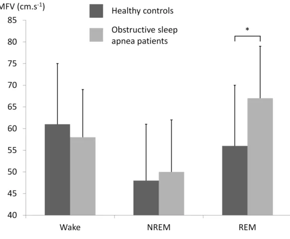
Figure 2: Mean flow velocity (MFV) in the right medial cerebral artery in healthy controls and patients with obstructive sleep apnea during wakefulness and sleep during which hypoxemia occurred: non-rapid eye movement sleep (NREM; stage 2) and rapid eye movement sleep (REM) of the second sleep cycle [Adapted from the data of Hajak et al. (12)]. Apnea-induced hypoxemia is compensated during REM sleep by an MFV increase, but this adjustment is totally abolished during NREM sleep, where MFV tends to decrease, mimicking the healthy controls' MVF kinetics. * p<0.05 between groups.