HAL Id: hal-03172911
https://hal.sorbonne-universite.fr/hal-03172911
Preprint submitted on 18 Mar 2021
HAL is a multi-disciplinary open access
archive for the deposit and dissemination of sci-entific research documents, whether they are pub-lished or not. The documents may come from teaching and research institutions in France or abroad, or from public or private research centers.
L’archive ouverte pluridisciplinaire HAL, est destinée au dépôt et à la diffusion de documents scientifiques de niveau recherche, publiés ou non, émanant des établissements d’enseignement et de recherche français ou étrangers, des laboratoires publics ou privés.
Anis Senoussi, Jean-Christophe Galas, André Estévez-Torres
To cite this version:
Anis Senoussi, Jean-Christophe Galas, André Estévez-Torres. Programmed mechano-chemical cou-pling in reaction-diffusion active matter. 2021. �hal-03172911�
Programmed mechano-chemical coupling in
reaction-diffusion active matter
Anis Senoussi,
∗Jean-Christophe Galas,
∗and Andr´
e Estevez-Torres
∗Sorbonne Universit´e, CNRS, Institut de Biologie Paris-Seine (IBPS), Laboratoire Jean Perrin (LJP), F-75005, Paris
E-mail: [email protected]; [email protected]; [email protected]
Abstract
2
Embryo morphogenesis involves a complex combination of pattern-forming
mech-3
anisms. However, classical in vitro patterning experiments explore only one
mecha-4
nism at a time, thus missing coupling effects. Here, we conjugate two major
pattern-5
forming mechanisms —reaction-diffusion and active matter— by integrating dissipative
6
DNA/enzyme reaction networks within an active gel composed of cytoskeletal motors
7
and filaments. We show that the strength of the flow generated by the active gel
con-8
trols the mechano-chemical coupling between the two subsystems. We use this property
9
to engineer the mechanical activation of chemical reaction networks both in time and
10
space, thus mimicking key aspects of the polarization mechanism observed in C.
el-11
egans oocytes. We anticipate that reaction-diffusion active matter may be useful to
12
investigate mechano-chemical transduction and to design new materials with life-like
13
properties.
14
Keywords: DNA programming, active matter, morphogenesis, pattern
for-15
mation, life-like material
16
Short title: Programmable reaction-diffusion active matter
Living embryos get their shape through a complex combination of chemical and
phys-18
ical processes that take place out of equilibrium. Basically, biochemical reaction networks
19
process information at particular points of space and time, while active gels generate
me-20
chanical forces and flows.1 These two generic processes are intertwined through diverse
21
chemo-mechanical and mechano-chemical couplings and both continuously consume
chem-22
ical energy. For instance, the pathway Rho GTPase brings about local contractions of the
23
actomyosin cortex in Xenopus,2 which regulates the cell cycle, while cytoplasmic flows of this
24
same cortex trigger the PAR reaction network in C. elegans, inducing embryo polarization.3
25
The development of in vitro dissipative molecular systems is key to produce non-equilibrium
26
materials and to investigate these processes in a controlled environment for testing
theo-27
retical predictions.4,5 On the chemical side, in vitro out-of-equilibrium reaction networks
28
produce spatio-temporal concentration patterns through reaction or reaction-diffusion
insta-29
bilities.6–14 On the mechanical side, active gels made of cytoskeletal motors and filaments
30
reconstituted in vitro4,15–24convert chemical energy into mechanical work and generate static
31
patterns and flows through hydrodynamic instabilities.25 More recently, efforts have focused
32
on coupling reaction networks with active fluids and gels, resulting in redox-controlled
self-33
oscillating gels26 and DNA-controlled passive27 and active gels.28–30 However, so far, only
34
one of the two systems could be maintained out of equilibrium, either the reaction network,26
35
or the gel,27–31 and thus only one of them could exhibit spatio-temporal self-organization.
36
This constraint suppresses the rich variety of mechano-chemical couplings that are essential
37
for pattern generation during development.1 It thus constitutes a major limitation for the
38
design of self-shaping materials inspired from embryogenesis.5
39
To create a functional mechano-chemical dissipative material we assembled two
dissipa-40
tive molecular subsystems, a chemical and a mechanical one. The chemical subsystem is
41
made of a network of DNA/enzyme reactions that produce single-stranded DNA (ssDNA)
42
molecules13 (Figure 1a). The mechanical subsystem is an active gel composed of bundles
43
of protein filaments propulsed by molecular motors15,17 (Figure 1b). Each subsystem is
b
a
c
d
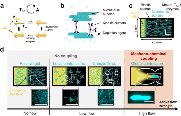
Nicking enzyme Polymerase + dNTP A T Microtubule bundles Kinesin clusters Depletion agent Plasticchannel Motors, Tenzymes AA &
A TAA
A
TAA
Chaotic flows
Passive gel Local contractions Global contraction
Active flow strength
Mechano-chemical coupling
No flow Low flow High flow
Nocoupling DNA A 1 mm 20 mm x y Propagating DNA front Nicking enzyme Polymerase + dNTP A T Microtubule bundles
Figure 1: In a reaction-diffusion active matter system, the coupling between the mechanical and the chemical subsystem is controlled by the strength of the active flow. (a) Scheme of the chemical subsystem involving the autocatalytic amplification of DNA strand A in the
presence of enzymes and template strand TAA. Harpoon-ended arrows denote ssDNA. (b)
Cartoon of the mechanical subsystem: an active gel formed by microtubules bundled together by a depletion agent and clusters of kinesin-1 motors. (c) Sketch of the channel in which the front propagation and the active gel dynamics were observed by fluorescence microscopy. Initially, A (yellow) is present only on the left side and the microtubule bundles (light blue) are aligned along x. (d) A mechano-chemical coupling between the two subsystems is achieved by increasing the strength of the flows generated by the active gel, which induces four different microtubule structures (light blue) and two DNA patterns (yellow). The white arrows represent the hydrodynamic flows generated by the active gel. Fluorescence images of the microtubules are represented for each morphology. Scale bars are 0.5 mm.
maintained out of equilibrium via the hydrolysis of high-energy compounds, respectively
45
deoxynucleosidetriphosphates (dNTPs) and adenosinetriphosphate (ATP).
46
The chosen chemical subsystem has four advantages. Firstly, due to DNA hybridization
47
rules, it can be easily reprogrammed into a variety of dissipative dynamics such as
oscilla-48
tions,32 bistability and excitability.33 Secondly, it can be maintained out of equilibrium in a
49
closed reactor for days.34 Thirdly, working in water at pH 7, it is a priori compatible with
50
other biochemical reactions.35 Lastly, it generates a variety of reaction-diffusion patterns
such as traveling fronts,36waves14and stationary patterns.37 In a first series of experiments,
52
the chemical subsystem encoded an autocatalytic loop that produces ssDNA species A —
53
the node— in the presence of ssDNA TAA —the template—, a polymerase and a nicking
54
enzyme13 (Figure 1a).
55
In the mechanical subsystem, the bundles are constituted of stabilized microtubule
fil-56
aments assembled together by attracting forces generated by the presence of a depletion
57
agent17 (Figure 1b). The motors are clusters of kinesin-1 and thus can bind several
micro-58
tubules at once.† Such an active gel generates macroscopic flows that, depending on the
59
concentration of motors and filaments, produce a diversity of microtubule morphologies:4,5
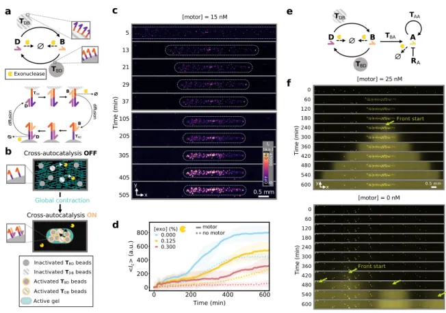
60
local contractions,15corrugations,24chaotic flows,17and global contractions.21In the
follow-61
ing, we demonstrate that, when mixed together, the two subsystems retain their ability to
62
undergo, respectively, chemical and mechanical instabilities that generate spatio-temporal
63
patterns. We further show that the strength of the active flow generated by the
mechani-64
cal subsystem controls the mechanochemical coupling between the two subsystems. Finally,
65
we take advantage of this property to design materials that mimick crucial aspects of a
66
mechanochemical patterning mechanism observed in C. elegans embryo.
67
To check whether the two subsystems remained functional when combined in an
opti-68
mized buffer (Figure S1), we tested the propagation of a DNA front through and active gel
69
undergoing local contractions. To do so, a solution containing all the components of the
70
chemical and mechanical subsystems, except the strand A, were filled into a microchannel.∗
71
An initial condition containing the same solution supplemented with A was injected on the
72
left side of the channel (Figure 1c). We recorded the spatiotemporal dynamics of each
subsys-73
tem by fluorescence microscopy thanks to the presence of a DNA intercalator that becomes
74
fluorescent upon binding to double-stranded DNA and of fluorescently-labeled microtubules
75
(SI Methods). In the chemical subsystem, we observed the propagation of a front of DNA
76
†Two types of clusters were used: biotinylated kinesin-1 (from D. melanogaster ) assembled together by
streptavidin,15or clusters made from SNAPtag modified kinesin-1 (from R. norvegicus) that spontaneously
multimerize24 (SI Methods).
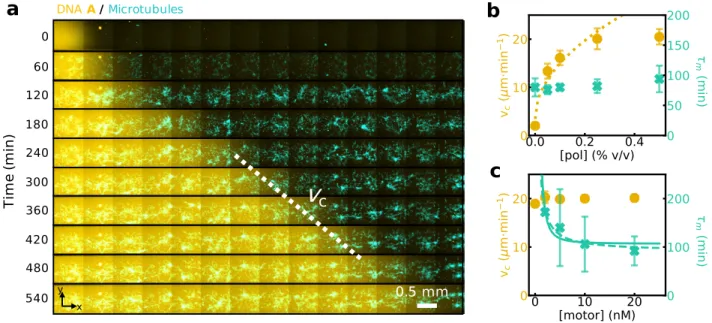
fluorescence with constant velocity vc= 20 µm/min across the whole length of the active gel,
77
i.e. > 1 cm (Figures 1d, 2a, S2 and Movie S1). Concomitantly, in the mechanical subsystem
78
the microtubules contracted locally with a characteristic time τm = 50 min into aggregates
79
with a typical size of 100 − 500 µm. The observation of a DNA front and microtubule
ag-80
gregates is in agreement with previous reports for each subsystem taken independently.15,36
81
b
a
c
v
c 0.5 mm 0 Ti m e (m in ) 60 120 240 360 480 180 300 420 540 x y DNA A/MicrotubulesFigure 2: A DNA/enzyme reaction-diffusion front propagates normally inside a locally-contracting cytoskeletal active gel and the dynamics of each subsystem can be independently tuned. (a) Time-lapse 2-color image of the fluorescence intensities associated to species A (yellow) and to the microtubule network (light blue) (see also Movie S1). The dotted line
indicates the velocity of the chemical front, vc. Plots of vc (yellow disks) and contraction
time of the active gel, τm, (blue crosses) for different concentrations of DNA polymerase (b)
and motors (c). The lines are fits to the data with vc∼ [pol]1/2(dotted line), τm∼ [motor]−1
(dashed line) and τm ∼ [motor]−2 (plain line). Error bars correspond to one standard
deviation from a triplicate experiment.
When the active gel produces local contractions, the dynamics of each subsystem can
82
be independently tuned. Increasing the polymerase concentration, [pol], increases the front
83
velocity, vc, until reaching a plateau at 22 µm/min (Figure 2b). In these conditions, the
84
characteristic contraction time τm remained constant. We find a scaling vc ∼ [pol]1/2, in
85
agreement with previous results36 and characteristic of Luther reaction-diffusion dynamics
86
where vc ∼ r
1/2
c , taking rc ∼ [pol] for the rate of the autocatalytic reaction as observed
in previous experiments.36 In turn, when the motor concentration, [motor], increases, τ m
88
decreases until reaching a plateau at 100 min (Figure 2c) and the size of microtubule
ag-89
gregates increases (Figure S3), while vc remains constant. A hydrodynamic model of a
90
contracting active gel5 yields τ−1
m ∼ ζ([motor]), where ζ is the strength of the gel activity,
91
which depends on the motor concentration, and two scalings are found in the literature18,38
92
yielding τm ∼ [motor]−α, with α = 1, 2 (Supplementary Text). Our data are compatible
93
with both scalings (Figure 2c). In summary, Figure 2 shows that the two subsystems are
94
both chemically and mechanically decoupled when the gel undergoes local contractions.
95
By varying the conditions, we can propagate the chemical front through active gels
under-96
going other spatial instabilities associated with different active flow strengths, as sketched
97
in Figure 1d. When the dGTP concentration was reduced (Figures S4-S5), chaotic flows
98
were observed in the mechanical subsystem during several hours before local contractions
99
occurred (Movie S2). Such flows did not modify the velocity of the chemical front because
100
transport remained dominated by Brownian diffusion.? When the length of microtubules
101
was increased using taxol and the motor concentration reduced, the active gel formed
corru-102
gations reminiscent of those previously reported in the absence of the chemical subsystem,24
103
again without perturbing the chemical front (Movie S3).
104
In contrast, a dramatic perturbation of the front propagation was observed when the
105
active gel underwent a global contraction, associated with large hydrodynamic flows
(Fig-106
ures 3 and S6-S11 and Movie S4). Global contractions were observed for long microtubules
107
and relatively high motor concentrations (Figure S7). When the gel contracted more rapidly
108
than the front propagated, the front moved faster and we distinguished 4 phases (Figure 3c).
109
During phase I, the active gel contracted rapidly towards the center of the channel,
accelerat-110
ing until reaching a maximum velocity vm = 400 µm/min at the end of phase I and dragging
111
DNA along, which formed a detached DNA islet ahead of the front. During phase II the gel
112
?The diffusivity of A due to the active flow, D
f, was estimated to be 10-fold smaller than the Brownian
diffusivity of A, DA, and thus vc ∼ (DA+ Df)1/2 ≈ D 1/2
A corresponds to a purely reaction-diffusion front
0 6 12 18 150 a c e b d f x y Time (min) 0.5mm Ti m e (m in ) 0 50 x y 0.5 mm DNA A /Microtubules g DNA A Microtubules
Figure 3: A globally-contracting active gel stretches and accelerates a reaction-diffusion front. (a) Time-lapse, 2-color fluorescence image with the DNA front in yellow and the microtubules in light blue. The inset shows the two fluorescence channels in separate images for the selected region. The white and yellow spots are dust particles concentrated by the contracting gel (see also Movie 4). (b) Position of the DNA front along x for different motor concentrations. (c) Time-lapse images of DNA fluorescence (color) at [motor] = 2.5 nM and 3 min per image. The extremities of the contracting gel are indicated with black markers. Roman numbers indicate the 4 phases described in the text. (d) Stroboscopic image averaged over 50 min showing the trajectories of fluorescent beads during gel contraction, the white arrows indicate the sense of the flow (see also Movie 5) and (e) plot of the bead velocity along x across the width of the channel for the beads in the red rectangle, 25 min after the beginning of the contraction. (f) Maximal front velocity during phase I (crosses) and steady-state velocity during phase IV (disks) for different motor concentrations. (g) Maximal front velocity during phase I vs. maximal gel contraction velocity. Error bars correspond to one standard deviation from a triplicate experiment.
decelerated and the DNA islet was diluted in the xy plane resulting in a front with a skewed
113
profile 5-fold wider than the initial one (Figure S6). Throughout phases I and II the P´eclet
114
number was greater than 1 (Figure S8), indicating that active convection predominated over
115
diffusion. Finally, when the active gel stopped contracting, the DNA front slowly recovered
a sigmoidal shape (phase III, Figure S6) and eventually reached a steady state with constant
117
velocity and width (phase IV).
118
In a control experiment with a passive dye, only phases I and II were observed,
indicat-119
ing that reaction was necessary for phases III and IV and that DNA islet formation was not
120
related to the binding of DNA to the active gel (Figure S10). We confirmed the last
interpre-121
tation by adding passive brownian beads to measure the hydrodynamic flow induced during
122
contraction. We observed two counter-rotating fluid rolls, symmetric along the central axis
123
of the channel, x, and producing water flows along x that reached +150 µm/min in the
124
center of the channel and −100 µm/min at its borders (Figures 3d,e and S11 and Movie S5).
125
Taken together, these results indicate that the stretching of the concentration profile of A
126
leading to the formation of the DNA islet during phase I was a purely hydrodynamic process.
127
The active gel contraction velocity, vm, was a sigmoidal function of the motor
concentra-128
tion (Figure S7). We quantified the two main regimes of front propagation with max(vcI) the
129
maximal velocity during phase I and vcIV the velocity at steady state in phase IV. Figure 3f
130
shows that the former strongly depended on the motor concentration while the latter was
131
independent. Finally, the linear relationship between max(vI
c) and vm is consistent with the
132
observation of a convection-dominated transport during phase I (Figure 3g). Taken together,
133
these results show that when the flows generated by the active gel are sufficiently fast there
134
is a mechano-chemical coupling between the gel and the reaction-diffusion front. This
cou-135
pling happens through hydrodynamics and can be interpreted as a time-dependent Taylor
136
dispersion.39
137
We have just seen that active flows can significantly modify heterogeneous concentration
138
profiles present in the chemical subsystem. Can they induce an asymmetry in an initially
139
homogeneous chemical subsystem? This is what happens in the C. elegans embryo, where
140
the active flow generated by the actomyosin cortex breaks the symmetry of an initially
141
homogeneous distribution of proteins. Later, this asymmetry is amplified by a
PAR-142
dependent bistable reaction network, leading to embryo polarization.3 The reaction-diffusion
active matter system developed here is a good candidate to mimick this process in a synthetic
144
material. To do so, we first need to implement a mechanism that couples a variation in
145
the microtubule concentration with a change in the concentration of a DNA species that is
146
inatially homogeneously distributed and second to engineer a chemical network that amplifies
147
this concentration change.
148
The first requirement was fulfilled by attaching DNA strands to ∼ 30 µm diameter
hydro-149
gel beads, which were trapped by the microtubule mesh and concentrated during contraction.
150
The second condition was satisfied by engineering a chemical subsystem whose kinetics
de-151
pend on the concentration of DNA-bead conjugates, and thus on the contraction state of the
152
gel. More precisely, the DNA autocatalytic loop was split into two nodes, B and D, that
153
cross-activate each other thanks to the templates TBD and TDB (Figure 4a). By attaching
154
each of these templates to a set of hydrogel beads and supplementing the medium with an
155
exonuclease that degrades B and D (Figure 4a) the cross-catalysis kinetics become
diffusion-156
controlled40and thus should depend on bead density. As a result, the beads brought together
157
in a contracted gel should activate faster, producing DNA that light them up in the presence
158
of a DNA intercalator dye (Figure 4b). Indeed, when both types of beads where embedded
159
in the active gel in the presence of a homogeneous, low concentration of D, they first reached
160
a high density as the gel contracted and later they became fluorescent (Figures 4c,d, S13 and
161
Movie S6). In the absence of contraction, the bead fluorescence amplification was delayed
162
(Figure S12) and its final amplitude reduced, both by a factor 2 (Figure 4d). As expected
163
for diffusion-controlled kinetics, the mechano-chemical DNA amplification dynamics slowed
164
down with increasing exonuclease concentration (Figure 4d).
165
To show that the activated beads may trigger downstream reactions in solution, the
166
previous system was supplemented with freely-diffusing templates TBA and TAA, that
re-167
spectively convert B into A and sustain the autocatalytic reaction of A described earlier.
168
In addition, to suppress the undesired self-activation of TAA a repressor strand RA was
169
added33 (Figure S15). In the presence of motors, the beads were all activated within 1 h at
dif fu sio n TDB TBD B D B D dif fu sio n TDB TBD B D a b e Inactivated TBDbeads Inactivated TDBbeads Active gel Activated TBDbeads Activated TDBbeads Cross-autocatalysis OFF Cross-autocatalysis ON d Exonuclease Global contraction c 405 505 105 205 Ti m e ( m in ) 5 305 0.5 mm x y 13 21 29 37 [motor] = 15 nM ON OF F 0 45000 Ic (a.u.) f motor no motor TDB TBD B D TBA A TAA RA Nicking enzyme Polymerase + dNTP A T [motor] = 0 nM 0 60 120 240 600 Ti m e ( m in ) 360 480 180 300 420 540 Front start [motor] = 25 nM 0.5 mm x y 0 60 120 240 600 Ti m e ( m in ) 360 480 180 300 420 540 Front start
Figure 4: A globally-contracting active gel triggers the activation of downstream reaction net-works with temporal and spatial control. (a) Scheme of the cross-autocatalytic DNA/enzyme network (top). Plain and dotted arrows indicate activation and degradation reactions, re-spectively. Harpoon-ended arrows correspond to ssDNA and disks indicate hydrogel beads
carrying templates Tij. Detailed mechanism of bead activation where the diffusion of B and
D between beads is indicated (bottom). (b) Sketch of the mechano-chemical activation of the reaction network in panel a through the contraction of the active gel (light blue) that brings the hydrogel beads (disks) close together, speeding up cross-catalysis. (c) Time-lapse images of DNA fluorescence from the template-bearing beads embedded in the active gel in the presence of motors. White dotted lines indicate the borders of the active gel and the channel walls are depicted in gray. (d) Average DNA fluorescence over the whole channel vs. time in the absence (dotted line) and in the presence (plain line) of motors for different exonuclease concentrations (colors). (e) Scheme of the bead-associated cross-autocatalytic network coupled to the autocatalysis of A in solution. Disks indicate templates linked to hydrogel beads. The blunt-ended arrow indicates repression. (f) Time-lapse images of DNA fluorescence in the channel for the network in panel e, in the presence (top) and in the ab-sence (bottom) of motors. The bright spots are the beads, the arrows indicate the start of the fronts of A, in yellow.
the center of the channel, where the gel contracted, and they triggered a controlled front of
171
A that propagated from the center of the channel to its extremities (Figures 4f and S14-S15
172
and Movie S7). In contrast, in the absence of gel contraction, the beads randomly activated
173
over the course of 5h, which was followed by the uncontrolled amplification of A. Taken
174
together, these results demonstrate that mechano-chemical coupling can be engineered to
175
trigger either temporal or spatio-temporal chemical instabilities in a synthetic material.
176
The coupling of chemical and mechanical self-organization is a key ingredient of biological
177
complexity, in particular during embryogenesis. We have demonstrated that it is possible to
178
couple two archetypal examples of these mechanisms, reaction-diffusion and active matter, in
179
a synthetic material. Our design is modular because it relies on two distinct subsystems with
180
well-characterized and predictable spatiotemporal behaviors: DNA/enzyme reactions and
ki-181
nesin/microtubule active gels. Considered independently, each subsystem reveals complex
182
dynamics and macroscopic organizations which are subject to intense scrutiny.14,15,17,19,21,24,35,37
183
When mixed-together, the coupling strength between the two subsystems is set by the
mag-184
nitude of the flow generated by the active gel. As a result, this system may be useful for
185
investigating self-organization when chemical and mechanical out-of-equilibrium processes
186
are intertwined. Finally, reaction-diffusion active matter provides a framework for the
ratio-187
nal engineering of functional out-of-equilibrium materials with life-like properties. On the
188
one hand, it could be advantageously combined with the wide array of methods in DNA
nan-189
otechnology, such as nanostructure design,41 logic gates,11 analyte detection42 or hydrogel
190
swelling.27 On the other hand, by using DNA-motor conjugates28–30 or photosensitive
mo-191
tors43 the system is extendable to chemo-mechanical as well as photo-mechanical couplings.
192
Acknowledgements
193
We thank H. Berthoumieux, V. Bormuth, M. Elez, A. Genot, G. Gines, N. Lobato-Dauzier,
194
A. Maitra, L. Robert, Y. Rondelez and R. Voituriez for insightful discussions and K. Furuta
and Z. Gueroui for their kind gift of kinesin plasmids. This work has been funded by the
196
European Research Council (ERC) under the European’s Union Horizon 2020 programme
197
(grant No 770940, T.), by the Ville de Paris Emergences programme (Morphoart,
A.E.-198
T.) and by MITI CNRS (J.-C. G.). The data that support the findings of this study are
199
available from the corresponding author upon reasonable request.
200
References
201
(1) Gross, P.; Kumar, K. V.; Grill, S. W. How Active Mechanics and Regulatory
Biochem-202
istry Combine to Form Patterns in Development. Annual Review of Biophysics 2017,
203
46, 337–356.
204
(2) Bement, W. M.; Leda, M.; Moe, A.; Kita, A.; Larson, M.; Golding, A.; Pfeuti, C.;
205
Su, K.-C.; Miller, A.; Goryachev, A.; von Dassow, G. Activator–inhibitor coupling
206
between Rho signalling and actin assembly makes the cell cortex an excitable medium.
207
Nature Cell Biology 2015, 17, 1471–1483.
208
(3) Goehring, N. W.; Trong, P. K.; Bois, J. S.; Chowdhury, D.; Nicola, E. M.; Hyman, A. A.;
209
Grill, S. W. Polarization of PAR proteins by advective triggering of a pattern-forming
210
system. Science 2011, 334, 1137–1141.
211
(4) Needleman, D.; Dogic, Z. Active matter at the interface between materials science and
212
cell biology. Nature reviews materials 2017, 2, 17048.
213
(5) Senoussi, A.; Vyborna, Y.; Berthoumieux, H.; Galas, J.-C.; Estevez-Torres, A. In
Out-214
of-Equilibrium Supramolecular Systems and Materials; Giuseppone, N., Walther, A.,
215
Eds.; Wiley-VCH, in press.
216
(6) Epstein, I.; Pojman, J. A. An introduction to nonlinear chemical reactions; Oxford
217
University Press: New York, 1998.
(7) Isalan, M.; Lemerle, C.; Serrano, L. Engineering gene networks to emulate Drosophila
219
embryonic pattern formation. PLoS biology 2005, 3 .
220
(8) Karzbrun, E.; Tayar, A. M.; Noireaux, V.; Bar-Ziv, R. H. Programmable on-chip DNA
221
compartments as artificial cells. Science 2014, 345, 829–832.
222
(9) Nakajima, M.; Imai, K.; Ito, H.; Nishiwaki, T.; Murayama, Y.; Iwasaki, H.; Oyama, T.;
223
Kondo, T. Reconstitution of Circadian Oscillation of Cyanobacterial KaiC
Phosphory-224
lation in Vitro. Science 2005, 308, 414–415.
225
(10) Loose, M.; Fischer-Friedrich, E.; Ries, J.; Kruse, K.; Schwille, P. Spatial regulators
226
for bacterial cell division self-organize into surface waves in vitro. Science 2008, 320,
227
789–792.
228
(11) Zhang, D. Y.; Seelig, G. Dynamic DNA nanotechnology using strand-displacement
229
reactions. Nature chemistry 2011, 3, 103.
230
(12) Chirieleison, S. M.; Allen, P. B.; Simpson, Z. B.; Ellington, A. D.; Chen, X. Pattern
231
transformation with DNA circuits. Nature chemistry 2013, 5, 1000.
232
(13) Montagne, K.; Plasson, R.; Sakai, Y.; Fujii, T.; Rondelez, Y. Programming an in vitro
233
DNA oscillator using a molecular networking strategy. Molecular systems biology 2011,
234
7 .
235
(14) Padirac, A.; Fujii, T.; Est´evez-Torres, A.; Rondelez, Y. Spatial waves in synthetic
236
biochemical networks. Journal of the American Chemical Society 2013, 135, 14586–
237
14592.
238
(15) Nedelec, F.; Surrey, T.; Maggs, A. C.; Leibler, S. Self-organization of microtubules and
239
motors. Nature 1997, 389, 305.
240
(16) Bendix, P. M.; Koenderink, G. H.; Cuvelier, D.; Dogic, Z.; Koeleman, B. N.;
241
Brieher, W. M.; Field, C. M.; Mahadevan, L.; Weitz, D. A. A quantitative analysis
of contractility in active cytoskeletal protein networks. Biophysical journal 2008, 94,
243
3126–3136.
244
(17) Sanchez, T.; Chen, D. T.; DeCamp, S. J.; Heymann, M.; Dogic, Z. Spontaneous motion
245
in hierarchically assembled active matter. Nature 2012, 491, 431.
246
(18) Foster, P. J.; F¨urthauer, S.; Shelley, M. J.; Needleman, D. J. Active contraction of
247
microtubule networks. Elife 2015, 4, e10837.
248
(19) Wu, K.-T.; Hishamunda, J. B.; Chen, D. T.; DeCamp, S. J.; Chang, Y.-W.; Fern´
andez-249
Nieves, A.; Fraden, S.; Dogic, Z. Transition from turbulent to coherent flows in confined
250
three-dimensional active fluids. Science 2017, 355, eaal1979.
251
(20) Kumar, N.; Zhang, R.; de Pablo, J. J.; Gardel, M. L. Tunable structure and dynamics
252
of active liquid crystals. Science advances 2018, 4, eaat7779.
253
(21) Torisawa, T.; Taniguchi, D.; Ishihara, S.; Oiwa, K. Spontaneous formation of a
glob-254
ally connected contractile network in a microtubule-motor system. Biophysical journal
255
2016, 111, 373–385.
256
(22) Ideses, Y.; Erukhimovitch, V.; Brand, R.; Jourdain, D.; Hernandez, J. S.; Gabinet, U.;
257
Safran, S.; Kruse, K.; Bernheim-Groswasser, A. Spontaneous buckling of contractile
258
poroelastic actomyosin sheets. Nature communications 2018, 9, 2461.
259
(23) Roostalu, J.; Rickman, J.; Thomas, C.; N´ed´elec, F.; Surrey, T. Determinants of polar
260
versus nematic organization in networks of dynamic microtubules and mitotic motors.
261
Cell 2018, 175, 796–808.
262
(24) Senoussi, A.; Kashida, S.; Voituriez, R.; Galas, J.-C.; Maitra, A.; Estevez-Torres, A.
263
Tunable corrugated patterns in an active nematic sheet. Proceedings of the National
264
Academy of Sciences 2019, 116, 22464–22470.
(25) Marchetti, M. C.; Joanny, J.-F.; Ramaswamy, S.; Liverpool, T. B.; Prost, J.; Rao, M.;
266
Simha, R. A. Hydrodynamics of soft active matter. Reviews of Modern Physics 2013,
267
85, 1143.
268
(26) Yoshida, R.; Takahashi, T.; Yamaguchi, T.; Ichijo, H. Self-oscillating gel. Journal of
269
the American Chemical Society 1996, 118, 5134–5135.
270
(27) Cangialosi, A.; Yoon, C.; Liu, J.; Huang, Q.; Guo, J.; Nguyen, T. D.; Gracias, D. H.;
271
Schulman, R. DNA sequence–directed shape change of photopatterned hydrogels via
272
high-degree swelling. Science 2017, 357, 1126–1130.
273
(28) Wollman, A. J.; Sanchez-Cano, C.; Carstairs, H. M.; Cross, R. A.; Turberfield, A. J.
274
Transport and self-organization across different length scales powered by motor proteins
275
and programmed by DNA. Nature nanotechnology 2014, 9, 44.
276
(29) Sato, Y.; Hiratsuka, Y.; Kawamata, I.; Murata, S.; Nomura, S.-i. M. Micrometer-sized
277
molecular robot changes its shape in response to signal molecules. Sci. Robot 2017, 2 .
278
(30) Keya, J. J.; Suzuki, R.; Kabir, A. M. R.; Inoue, D.; Asanuma, H.; Sada, K.; Hess, H.;
279
Kuzuya, A.; Kakugo, A. DNA-assisted swarm control in a biomolecular motor system.
280
Nature communications 2018, 9, 453.
281
(31) Bertrand, O. J. N.; Fygenson, D. K.; Saleh, O. A. Active, motor-driven
me-282
chanics in a DNA gel. Proceedings of the National Academy of Sciences 2012,
283
10.1073/pnas.1208732109 .
284
(32) Fujii, T.; Rondelez, Y. Predator–prey molecular ecosystems. ACS nano 2012, 7, 27–34.
285
(33) Montagne, K.; Gines, G.; Fujii, T.; Rondelez, Y. Boosting functionality of synthetic
286
DNA circuits with tailored deactivation. Nature communications 2016, 7, 13474.
(34) Urtel, G.; Van Der Hofstadt, M.; Galas, J.-C.; Estevez-Torres, A. rEXPAR: an
isother-288
mal amplification scheme that is robust to autocatalytic parasites. Biochemistry 2019,
289
58, 2675–2681.
290
(35) Van Der Hofstadt, M.; Galas, J.-C.; Estevez-Torres, A. Spatiotemporal Patterning of
291
Living Cells with Extracellular DNA Programs. ACS nano 2020,
292
(36) Zadorin, A. S.; Rondelez, Y.; Galas, J.-C.; Estevez-Torres, A. Synthesis of
pro-293
grammable reaction-diffusion fronts using DNA catalyzers. Physical review letters 2015,
294
114, 068301.
295
(37) Zadorin, A. S.; Rondelez, Y.; Gines, G.; Dilhas, V.; Urtel, G.; Zambrano, A.; Galas,
J.-296
C.; Estevez-Torres, A. Synthesis and materialization of a reaction-diffusion French flag
297
pattern. Nature chemistry 2017, 9, 990.
298
(38) Mart´ınez-Prat, B.; Ign´es-Mullol, J.; Casademunt, J.; Sagu´es, F. Selection mechanism
299
at the onset of active turbulence. Nature physics 2019, 15, 362.
300
(39) Vedel, S.; Bruus, H. Transient Taylor–Aris dispersion for time-dependent flows in
301
straight channels. Journal of Fluid Mechanics 2011, 691, 95–122.
302
(40) Gines, G.; Zadorin, A.; Galas, J.-C.; Fujii, T.; Estevez-Torres, A.; Rondelez, Y.
Micro-303
scopic agents programmed by DNA circuits. Nature nanotechnology 2017, 12, 351.
304
(41) Jones, M. R.; Seeman, N. C.; Mirkin, C. A. Programmable materials and the nature of
305
the DNA bond. Science 2015, 347, 1260901.
306
(42) Zhao, Y.; Chen, F.; Li, Q.; Wang, L.; Fan, C. Isothermal Amplification of Nucleic
307
Acids. Chemical Reviews 2015, 115, 12491–12545.
308
(43) Ross, T. D.; Lee, H. J.; Qu, Z.; Banks, R. A.; Phillips, R.; Thomson, M. Controlling
309
organization and forces in active matter through optically defined boundaries. Nature
310
2019, 572, 224–229.