Publisher’s version / Version de l'éditeur:
Thin Solid Films, 437, August, pp. 95-100, 2003
READ THESE TERMS AND CONDITIONS CAREFULLY BEFORE USING THIS WEBSITE. https://nrc-publications.canada.ca/eng/copyright
Vous avez des questions? Nous pouvons vous aider. Pour communiquer directement avec un auteur, consultez la première page de la revue dans laquelle son article a été publié afin de trouver ses coordonnées. Si vous n’arrivez pas à les repérer, communiquez avec nous à PublicationsArchive-ArchivesPublications@nrc-cnrc.gc.ca.
Questions? Contact the NRC Publications Archive team at
PublicationsArchive-ArchivesPublications@nrc-cnrc.gc.ca. If you wish to email the authors directly, please see the first page of the publication for their contact information.
NRC Publications Archive
Archives des publications du CNRC
This publication could be one of several versions: author’s original, accepted manuscript or the publisher’s version. / La version de cette publication peut être l’une des suivantes : la version prépublication de l’auteur, la version acceptée du manuscrit ou la version de l’éditeur.
For the publisher’s version, please access the DOI link below./ Pour consulter la version de l’éditeur, utilisez le lien DOI ci-dessous.
https://doi.org/10.1016/S0040-6090(03)00614-X
Access and use of this website and the material on it are subject to the Terms and Conditions set forth at
Crystallized SrFeO3-x films deposited by pulsed laser ablation without
in-situ substrate heating
Wang, Zhongke; Sasaki, Takeshi; Koshizaki, Naoto; Tunney, Jim; Post,
Michael
https://publications-cnrc.canada.ca/fra/droits
L’accès à ce site Web et l’utilisation de son contenu sont assujettis aux conditions présentées dans le site LISEZ CES CONDITIONS ATTENTIVEMENT AVANT D’UTILISER CE SITE WEB.
NRC Publications Record / Notice d'Archives des publications de CNRC:
https://nrc-publications.canada.ca/eng/view/object/?id=9cdb23ae-4adc-456c-92bd-899ec3d4c690 https://publications-cnrc.canada.ca/fra/voir/objet/?id=9cdb23ae-4adc-456c-92bd-899ec3d4c6900040-6090/03/$ - see front matter 䊚 2003 Elsevier Science B.V. All rights reserved. doi:10.1016/S0040-6090(03)00614-X
Crystallized SrFeO
3yxfilms deposited by pulsed laser ablation without
in-situ substrate heating
Zhongke Wang , Takeshi Sasaki , Naoto Koshizaki *, James J. Tunney , Michael L. Post
a a a, b bNanoarchitectonics Research Center (NARC), National Institute of Advanced Industrial Science and Technology (AIST), Central 5,
a
1-1-1 Higashi, Tsukuba, Ibaraki 305-8565, Japan
Institute for Chemical Process and Environmental Technology, National Research Council of Canada, Montreal Road, Ottawa,
b
Canada K1A 0R6
Received 20 January 2003; received in revised form 27 March 2003; accepted 11 April 2003
Abstract
Crystallized SrFeO3yx(0FxF0.5) films were deposited by pulsed laser ablation without in-situ substrate heating with the third harmonic of a Nd:YAG laser and off-axis configuration. The crystallinity of SrFeO3yx films strongly depended on the ambient pressure of Ar gas. The films deposited are amorphous below 10 Pa, and crystalline from 10 to 200 Pa. The films deposited at 30 Pa have the optimum stoichiometry of Sr to Fe, approximately 1.05. A stoichiometric and well-crystallized SrFeO film with3
a smooth surface was fabricated by post-annealing (300 8C in air) the amorphous films deposited at 1 Pa. 䊚2003 Elsevier Science B.V. All rights reserved.
Keywords: Laser ablation; SrFeO ; Thin films; Crystallization3
1. Introduction
Perovskite-type oxides exhibiting ionicandyor elec-tronic conductivity are interesting for their potential applications, such as high-temperature solid oxide fuel cells, battery electrodes, sensor materials w1,2x, and colossal magnetoresistance effect w3–5x. SrFeO perov-3 skite oxide has recently received special attention since SrFeO3yx (SFO, hereafter) exhibits a wide range of non-stoichiometry (0FxF0.5). This large non-stoichi-ometry is caused by the equilibrium compositional change with temperature and oxygen partial pressure, and the high oxygen mobility in the crystal lattice w6– 9x. Electric, magnetic and optical properties vary remarkably with the change in oxygen content in SFO w10–12x; hence, the gas-sensing properties are also studied w13–15x. Thin films are expected to have a rapid response of various properties originating from the large non-stoichiometry and are also highly desirable for integration as functional components with silicon
mon-*Corresponding author. Tel.: q81-298614879; fax: q81-298616355.
E-mail address:koshizaki.naoto@aist.go.jp (N. Koshizaki).
oliths. To meet these demands, a low-temperature thin-film processing technique is important.
Pulsed laser deposition (PLD) has proven highly successful in growing thin films of complex oxides with perovskite or perovskite-related structures. This tech-nique has also been used to prepare epitaxial SFO films w10,15–17x. To change the oxygen content and crystal-lographictexturing of the SFO epitaxial films, various kinds of single crystal substrates, deposition atmospheres and pressures, cooling atmospheres, and post-annealing conditions have been examined. However, the substrate temperature for SFO film deposition (640 to 700 8C) is common in these experiments. Therefore, the substrate temperature plays an important role for SFO epitaxial film growth. Furthermore, oxygen or ozone ambient during cooling or post-annealing is also required for obtaining highly oxidized SrFeO films.3
We investigated oxide thin film preparation techniques through nanoparticle-aggregated films by PLD with an inert gas ambient and in an off-axis configuration. In this case, an oxide target is ablated, mostly in Ar, and hence the target itself provides the oxygen. Therefore, the confinement of the ablated species in a small space is very important for promoting nanoparticle formation
Fig. 1. Schematic diagram of off-axis configuration for pulsed laser deposition.
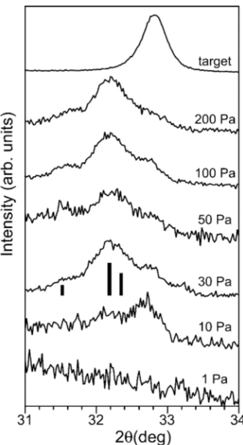
Fig. 2. XRD spectra of SFO films deposited on quartz glass substrates at 200 mJypulse under different pressures of Ar. The spectrum of the ceramic SrFeO target is also shown.3
Fig. 3. XRD spectra of SFO thin films deposited on quartz glass substrates at 10 Pa of Ar under different laser energies.
and simultaneous oxidation in the vapor phase. Argon has more confining power than oxygen, because the energy of the ejected species from the target is easily dissipated by the oxygen ambient through rotational energy excitation of diatomic oxygen molecules. By adopting this technique, we successfully obtained crys-tallized nanoparticle aggregated oxide films of Co O ,3 4 Fe O , LaFeO and BaTiO without any in-situ substrate2 3 3 3 heating w18–20x. However, a suitable pressure range exists for obtaining fully oxidized phase. In this article, we report the deposition of crystallized SFO films at room temperature by PLD. We also demonstrate well-crystallized film preparation by post-annealing an amor-phous film at a relatively low temperature deposited just below the crystallization pressure range.
2. Experimental details
Laser deposition was performed using the third har-monicof a Nd:YAG laser with a wavelength of 355 nm, pulse width of 5–7 ns, and repetition rate of 10 Hz. The laser beam was focused on the target surface at an incidence angle of 458. The beam spot diameter was approximately 2 mm on the target surface. The target was mounted in a rotating holder placed in a vacuum chamber evacuated to a base pressure below 5.5=10y4Pa. The films were 600 to 700 nm thick after 20 min deposition. A rotating quartz substrate was placed in an off-axis position, perpendicular to the target at a distance of 16 mm, as illustrated in Fig. 1. Argon gas was introduced into the chamber and the pressure was kept constant at the selected value between 1 to 200 Pa during deposition. All the films were deposited at room temperature without any in-situ substrate heat-ing. For comparison, the as-deposited films were post-annealed in air for two hours and then cooled naturally to room temperature.
The thickness of obtained films was measured with a profilometer (Tencor alpha-step profiler 300). The
crys-tal structure of the films was analyzed by X-ray diffrac-tion (XRD, Rigaku RAD-C) using Cu–Ka radiadiffrac-tion. The surface morphology of the films was observed by a scanning electron microscope (SEM, Hitachi S-800). The atomicratio of Sr to Fe in the films was determined by X-ray fluorescence measurement (XRF, Seiko SEA2010).
3. Results and discussion
3.1. Crystal structure and oxygen stoichiometry
Figs. 2 and 3 present the XRD patterns of a series of samples deposited at different pressures and laser
ener-Fig. 4. Enlarged XRD spectra of the SrFeO (110) peak from SFO3
films at 200 mJypulse under different pressures of Ar.
gies. XRD spectra with indexes shown at the top in these figures are from the ablation target that was used and coincide well with cubic perovskite SrFeO w21x.3 Fig. 2 shows that the crystallinity of SFO films strongly depends on the ambient pressure of Ar gas. The sample deposited at 1 Pa exhibited no diffraction peaks and was amorphous. Just one weak peak near the (110) peak of the target appeared in the sample prepared at 10 Pa. At pressures from 30 to 200 Pa, strong but broad diffraction peaks comprising a few components were recorded at the lower angle side of the (110) peak from the target. Very weak and broad peaks were also detected near the (200) and (211) peaks of the target. Fig. 3 shows that the peak intensity increased with the laser energy, indicating that the film crystallinity was improved at the higher energies. The film deposited using 300 mJypulse irradiation had almost a cubic perovskite structure.
Fig. 4 illustrates the enlarged spectra in the region corresponding to the SrFeO (110) peak in Fig. 2. At3 least three components were observed from most of the spectra. Increasing the ambient pressure reduced the intensity of the component at approximately 32.88, although the components near 32.2 and 31.68 became more evident. Three bars near the spectrum at 30 Pa in Fig. 4 indicates the peak position and relative intensity
of the orthorhombicbrownmillerite SrFeO . phase in2 5 the JCPDS database w22x. These three peaks, from the lower angle side, correspond to (200), (141) and (002) reflections from SrFeO . . The peak splitting and shifting2 5 to lower angles by reduction of SrFeO are due to loss3 of symmetry caused by oxygen vacancies and the resultant cell volume expansion that is as large as 7% w16x.
SrFeO3yxexhibits a wide range of non-stoichiometry (2.5F3yxF3.0). Within this range, there are four phases at approximately 3yx (2.50 (brownmillerite, orthorhombic), 2.75 (orthorhombic), 2.87 (tetragonal) and 2.97 (cubic)) w7,9x. The intermediate 3yx value corresponds to the coexistence range of the closest end components at both sides. Nemudry et al. studied the electrochemical oxidation reactions of SrFeO2.5in detail and monitored XRD spectrum variations with charge transfer to the sample w7x. Initially, only the three peaks plotted as bars in Fig. 4 were observed in the same angle range. Along with the electrochemical oxidation, the intensity of these three peaks relative to the peak at 32.88 changed monotonically, and finally, when fully oxidized, only the peak from SrFeO (110) remained.3 This monotonic change can thus be utilized to estimate the non-stoichiometric value 3yx of our samples from the XRD spectra shown in Fig. 4, though our samples have broader peaks due to the small size of constituent particulates in the films. The 3yx values estimated were 2.74 (10 Pa), 2.65 (30, 50 and 100 Pa), and 2.62 (200 Pa). As the deposition pressure increases from 10 to 200 Pa, the product gradually changes to a less oxidized composition.
Previously, in our studies on the preparation of nan-oparticle-aggregated films by laser ablation, we found similar pressure dependences for simple oxides, such as cobalt oxide or iron oxide, under the same configuration for deposition without any substrate heating w18,19x. At low pressure, the products were amorphous. With pres-sure increase, a crystalline film of Co O or Fe O (the3 4 2 3 same composition as the target) formed concurrently with nano-sized particulates. Further pressure increases resulted in products with mixtures of Co O and CoO,3 4 or of Fe O and FeO, i.e. a less oxidized composition.2 3 For SFO, the product changed from amorphous to crystallized SrFeO;2.74, and finally to crystallized SrFeO;2.62, with the pressure increase, though the prod-uct at 10 Pa was not the same composition as the target. This is probably because the higher oxidation state of tetravalent iron (Fe4q) is unstable w10,11,23,24x, and, therefore, an oxygen deficiency is inevitably introduced for SFO perovskite thin films without a strongly oxidiz-ing environment w16x.
3.2. Sr-to-Fe stoichiometry
Figs. 5 and 6 present the wSrxywFex atomicratio by XRF analysis in the films deposited at different Ar
Fig. 5. Argon pressure dependence of wSrxywFex atomicratio for the deposited SFO films by 200 mJypulse irradiation.
Fig. 6. wSrxywFex atomicratio in the deposited films vs. different laser energies for SFO films deposited at various Ar pressures.
pressures. The film stoichiometry is closely related to the ambient pressure. Analysis by XRF indicated that the films deposited at 30 Pa have optimum stoichiome-try, i.e. an atomicratio of wSrxywFex close to unity (1.05) irrespective of the laser energy. The atomic ratio wSrxy wFex exceeded 1.1 below 10 Pa, and was less than 0.9 above 50 Pa. The Sr deficiency in the film arose from the deposition process. This Sr deficiency could be different from the Li deficiency due to the high Li volatility during deposition of LiNbO films w25x. Res-3 puttering, another possible mechanism resulting in non-stoichiometric products of metallic components, is not considered since this is generally observed in UV laser ablation under high vacuum with very high laser fluence w26x. In our previous study on LaFeO and BaTiO3 3 perovskite film preparation by laser ablation w20x, the heavy A-site ions (La, Ba) became rich compared to the light B-site ions (Fe, Ti) in the deposited films at lower pressures (approx. 1 Pa). This trend was also observed for SFO films, as shown in Fig. 5. Even with a change in laser energy, this phenomenon is still almost the same, as shown in Fig. 6 where similar SryFe ratios are evident. Monte Carlo simulation of the laser depo-sition process w27x suggests that this could be due to the mass difference between Sr and Fe. The vaporization process played a crucial role in determining the film microstructure and stoichiometry of complex oxides during the deposition. The ambient gas pressure affects the kineticenergy of the species arriving on the substrate surface due to the collision of ablated species, including atoms, ions, molecules and nanoparticles in the gas phase. High ambient gas pressure results in a small plume and low kineticenergy of the species in the
plume due to the successive collisions between species. Conversely, at low pressure, fewer collisions occur in the larger plume, and the lighter species travel further without collision. Collision between flying species plays an important role in stoichiometry of thin films prepared by pulsed laser ablation w28x. There is thus an optimum pressure range for fabricating crystalline films with the appropriate stoichiometry between A and B ions. From these results, this pressure range is from 30 to 50 Pa for deposition of stoichiometric SrFeO3yxfilms.
3.3. Film morphology
All the deposited films were brownish yellow, though they varied from transparent to opaque as the Ar pressure increased. This reflected the aggregate formation at higher pressure. Fig. 7 presents SEM photographs of the SFO film surfaces deposited at 10 Pa at different laser pulse energies. Many particles were observed, though the number of particles decreased as the laser energy decreased. The particles observed on thin films deposited by pulsed laser ablation originated mainly from two sources: the droplets directly dislodged in liquid phase from the target and the particles segregated from the thin film matrix on the substrate. The amount of droplets can be easily decreased by using a lower laser energy, because there is generally a threshold laser fluence below, which the droplets are scarcely observable w29x. The particles observed in Fig. 7 were mainly droplets coming from the target, though they disappeared below 150 mJypulse and a smooth surface was obtained.
The films deposited at 1 Pa were amorphous as shown in Fig. 2. SEM observation also revealed droplets on these films, indicating that the droplets on the films were not crystalline. Therefore, the droplets were not
Fig. 7. SEM images of SFO films deposited under 10 Pa of Ar at 250 mJypulse and 150 mJypulse.
Fig. 8. XRD patterns of the films deposited at 1 Pa using 200 mJypulse and post-annealed in air for 2 h at various temperatures.
produced directly from the original bulk material of the target, such as fragments, but due to modified material from the target during ablation. The crystallinity of SFO films obtained at higher pressures is thus not due to the particulates from the target.
3.4. Effect of post-annealing on crystal structure Crystallized SFO films were obtained by laser abla-tion at pressures of 10 to 200 Pa of Ar without any in situ substrate heating or post-annealing. However, as shown in Fig. 2, the as-deposited films were not per-fectly crystallized, because the film is a nanoparticle-aggregated film. To further promote crystallization of SFO films, we studied the effect of post-annealing on the films deposited at 1 Pa (amorphous) and 100 Pa (crystallized) with 200 mJypulse. The films were annealed in air for 2 h at 300 to 500 8C. Fig. 8 illustrates the XRD spectrum change caused by post-annealing of the amorphous films deposited at 1 Pa. The spectrum of the amorphous, as-deposited film was transformed to
that of a cubic perovskite structure even by annealing at 300 8C in air. When the film was annealed at 400 8C or higher, a small amount of SrCO was generated due3 to the reaction of excess strontium oxide (Fig. 5) with carbon dioxide in air. The crystal structure of the main component in the annealed SFO films still corresponded to cubic perovskite.
Conversely, for the films deposited at 100 Pa, anneal-ing at 300 8C did not lead to complete oxidation to cubic phase, and just a slight increase of the cubic component was observed. Further annealing at 400 or 500 8C yielded cubic phase as the main component, though the peak intensity was still very low compared to the case at 1 Pa, indicating less crystallinity. SrCO3 was also generated and the peak became stronger with increasing annealing temperature. This clearly indicates that an amorphous iron component still remained in the film, since the Sr-to-Fe stoichiometric ratio is approxi-mately 0.8. SFO films once crystallized in nanoparticles probably have to be annealed in oxygen or ozone at least at 600 8C to obtain crystalline films as in the bulk case w15,16x.
By post-annealing at 300 8C, the amorphous film deposited at 1 Pa changed to the well crystallized SFO film while for the film deposited at 100 Pa the crystal-lization did not proceed further. As shown in Fig. 2, the pressure 1 Pa was just below the threshold pressure that determined whether the product would become an amor-phous film or a crystallized nanoparticle- aggregated film. Therefore, the film deposited at 1 Pa might contain smaller nucleus-like substances that would act as nucle-ation sites and lower the activnucle-ation energy for crystalli-zation, resulting in the lower crystallization temperature.
Conversely, once larger crystallized nanoparticles were present and aggregated, as in the film deposited at 100 Pa, the crystallization process might be similar to the bulk case. The contrast of the post-annealing effect for the SFO films could be explained in this way.
4. Summary
In summary, we have successfully obtained crystalline SrFeO3yxfilms by pulsed laser ablation without in-situ substrate heating or post-annealing. The crystallinity and stoichiometry of SrFeO3yxfilms depends on the ambient pressure of the Ar gas. The product changed from an amorphous form to crystallized SrFeO;2.74, and finally to crystallized SrFeO;2.62, with the pressure increase. The films deposited at 30 Pa have the optimum stoichi-ometry with an atomicratio of wSrxywFex close to unity (approx. 1.05), irrespective of laser energy. Stoichio-metric crystallized SrFeO films with smooth surfaces3 can be fabricated by post-annealing the amorphous films at 300 8C in air. Regarding the morphology, particulates observed by SEM on surfaces of films deposited at laser energies above 200 mJypulse were not present below 150 mJypulse.
Acknowledgments
Author Z. Wang, a JSPS fellow, would like to acknowledge the support from the Japan Society for the Promotion of Science.
References
w1x C.V.G. Reddy, S.V. Manorama, V.J. Rao, A. Lobo, S.K. Kulkarni, Thin Solid Films 348 (1999) 261.
w2x H.Y. Tu, Y. Takeda, N. Imanishi, O. Yamamoto, Solid State Ionics 100 (1997) 283.
w3x S. Jin, T.H. Tiefel, M. McCormack, R.A. Fastnacht, R. Ramesh, L.H. Chen, Science 264 (1994) 413.
w4x C.N.R. Rao, B. Raveau, Colossal Magnetoresistance, Charge Ordering and Related Properties of Mangnaese Oxides, World Scientific Press, Singapore, 1998.
w5x S. Pignard, K. Zhang, Y. Leprince, K. Han, H. Vincent, J.P. Senateur, Thin Solid Films 391 (2001) 21.
w6x M. Schmidt, J. Phys. Chem. Solids 61 (2000) 1363.
w7x A. Nemudry, M. Weiss, I. Gainutdinov, V. Boldyrev, R. Schol-¨ lhorn, Chem. Mater. 10 (1998) 2403.
w8x M. Schmidt, S.J. Campbell, J. Solid State Chem. 156 (2001) 292.
w9x J.P. Hodges, S. Short, J.D. Jorgensen, X. Xiong, B. Dabrowski, S.M. Mini, C.W. Kimball, J. Solid State Chem. 151 (2000) 190.
w10x N. Hayashi, T. Terashima, M. Takano, J. Mater. Chem. 11 (2001) 2235.
w11x T. Koslowski, Phys. Chem. Chem. Phys. 1 (1999) 3017. w12x K.L. Saenger, J. Solid State Chem. 158 (2001) 320.
w13x M.L. Post, J.J. Tunney, D. Yang, X. Du, D.L. Singleton, Sens. Actuators B 59 (1999) 190.
w14x Y. Wang, J. Chen, X. Wu, Mater. Lett. 49 (2001) 361. w15x T. Yu, Y.F. Chen, Z.G. Liu, L. Sun, S.B. Xiong, N.B. Ming,
Z.M. Ji, J. Zhou, Appl. Phys. A64 (1997) 69.
w16x H. Yamada, M. Kawasaki, Y. Tokura, Appl. Phys. Lett. 80 (2002) 622.
w17x B.W. Sanders, M.L. (1993). Post, Laser Ablation in Materials Processing Fundamentals and Applications, in: B. Braren, J.J. Dubowski, D.P. Norton (Eds.), Materials Research Society Symposium Proceedings Boston, U.S.A., December 1-4, 1992, 285 427.
w18x N. Koshizaki, A. Narazaki, T. Sasaki, Scripta Mater. 44 (2001) 1925.
w19x L. Zbroniec, T. Sasaki, N. Koshizaki, Appl. Surf. Sci. 197 (2002) 883.
w20x J.W. Yoon, T. Sasaki, N. Koshizaki, Appl. Phys. A 76 (2003) 641.
w21x Powder diffraction file, 2001 JCPDS-International Centre for Diffraction Data, PCPDFWIN v. 2.2. 40-0905.
w22x Powder diffraction file, 2001 JCPDS-International Centre for Diffraction Data, PCPDFWIN v. 2.2. 70-0836.
w23x J. Mizusaki, M. Okayasu, S. Yamauchi, K. Fueki, J. Solid State Chem. 99 (1992) 166.
w24x Y. Takeda, K. Kanno, T. Takada, O. Yamamoto, M. Takano, N. Nakayama, Y. Bando, J. Solid State Chem. 63 (1986) 237. w25x G. Balestrino, S. Martellucci, P.G. Medaglia, A. Paoletti, G. Petrocelli, A. Tebano, A. Tucciarone, F. Gelli, E. Giorgetti, S. Sottini, L. Tapfer, Appl. Phys. Lett. 78 (2001) 1204.
w26x M. Joseph, N. Sivakumar, P. Manoravi, S. Vanavaramban, Solid State Ionics 144 (2001) 339.
w27x T.E. Itina, W. Marine, M. Autric, J. Appl. Phys. 82 (1997) 3536.
w28x K.L. Saenger, J. Appl. Phys. 70 (1991) 5629.
w29x L. Chen, in: D.B. Chirsey, G.K. Hubler (Eds.), Pulsed Laser Deposition of Thin Films, John Wiley and Sons Inc. Press, New York, 1994, p. 167.