Antigen structure affects cellular routing through DC-SIGN
The MIT Faculty has made this article openly available.
Please share
how this access benefits you. Your story matters.
Citation
Jarvis, Cassie M. et al. "Antigen structure affects cellular routing
through DC-SIGN." Proceedings of National Academy Sciences 116,
30 (July 2019): 14862-14867 © 2019 National Academy of Sciences
As Published
http://dx.doi.org/10.1073/pnas.1820165116
Publisher
National Academy of Sciences
Version
Final published version
Citable link
https://hdl.handle.net/1721.1/123339
Terms of Use
Article is made available in accordance with the publisher's
policy and may be subject to US copyright law. Please refer to the
publisher's site for terms of use.
Antigen structure affects cellular routing through
DC-SIGN
Cassie M. Jarvisa, Daniel B. Zwickb, Joseph C. Grimc, Mohammad Murshid Alama, Lynne R. Prostb, Jaye C. Gardinerd, Soyeong Parkd, Laraine L. Zimdarsd, Nathan M. Shererd, and Laura L. Kiesslinga,b,c,1
aDepartment of Chemistry, Massachusetts Institute of Technology, Cambridge, MA 02139;bDepartment of Biochemistry, University of Wisconsin–Madison,
Madison, WI 53706;cDepartment of Chemistry, University of Wisconsin–Madison, Madison, WI 53706; anddDepartment of Molecular Virology
and Oncology, University of Wisconsin–Madison, Madison, WI 53706
Edited by Angela M. Gronenborn, University of Pittsburgh School of Medicine, Pittsburgh, PA, and approved June 10, 2019 (received for review November 27, 2018)
Dendritic cell (DC) lectins mediate the recognition, uptake, and processing of antigens, but they can also be coopted by pathogens for infection. These distinct activities depend upon the routing of antigens within the cell. Antigens directed to endosomal compart-ments are degraded, and the peptides are presented on major histocompatibility complex class II molecules, thereby promoting immunity. Alternatively, HIV-1 can avoid degradation, as virus engagement with C-type lectin receptors (CLRs), such as DC-SIGN
(DC-specific ICAM-3–grabbing nonintegrin) results in trafficking to
surface-accessible invaginated pockets. This process appears to
en-able infection of T cellsin trans. We sought to explore whether
antigen fate upon CLR-mediated internalization was affected by antigen physical properties. To this end, we employed the ring-opening metathesis polymerization to generate glycopolymers that each display multiple copies of mannoside ligand for DC-SIGN, yet differ in length and size. The rate and extent of glycopolymer
in-ternalization depended upon polymer structure—longer polymers
were internalized more rapidly and more efficiently than were shorter polymers. The trafficking, however, did not differ, and both short and longer polymers colocalized with transferrin-labeled early endosomes. To explore how DC-SIGN directs larger particles, such as pathogens, we induced aggregation of the polymers to access par-ticulate antigens. Strikingly, these parpar-ticulate antigens were diverted to the invaginated pockets that harbor HIV-1. Thus, antigen structure has a dramatic effect on DC-SIGN–mediated uptake and trafficking. These findings have consequences for the design of syn-thetic vaccines. Additionally, the results suggest strategies for tar-geting DC reservoirs that harbor viral pathogens.
HIV
|
C-type lectin|
polymer|
antigen|
endocytosisD
endritic cells (DCs) are sentinels of the innate immune system: They sense pathogens in the periphery and mount adaptive responses to enable immunity. Glycans are key determi-nants of pathogen recognition by DCs. Their importance is reflected in the multiplicity of carbohydrate-binding protein (lectin) receptors present on the surface of DCs (1, 2). Lectins can recognize antigenic glycans to facilitate pathogen inter-nalization and subcellular trafficking to endosomal compart-ments (3); pathogens are then degraded, and antigenic peptides are loaded onto major histocompatibility complexes for presentation to T cells.Despite the apparent function of lectins in mediating immunity, viruses have developed mechanisms to coopt lectins for dissemi-nation (4–6). The complicated relationship between HIV-1 and the lectin DC-SIGN (DC-specific ICAM-3–grabbing nonintegrin) serves as an example. DC-SIGN is a member of the family of Ca2+ -dependent lectin receptors (C-type lectin receptors; CLRs) that recognizes high mannose- and fucose-substituted glycans. DC-SIGN binds mannosylated gp120 on the HIV-1 envelope to me-diate internalization (7–9). Curiously, the virus is not routed to the traditional endocytic pathway, nor is it degraded; instead, it is trafficked to subcellular compartments near the cell surface (10). These“invaginated pockets,” which are enriched in tetraspanins
such as CD81, can facilitate viral transfer to T cells (i.e., trans infection) (11, 12). Similarly, Siglec-1 (13), another DC lectin, is implicated in facilitating HIV-1 trans infection of T cells (14). Thus, HIV-1 exploits lectin trafficking to avoid degradation and to promote infection of CD4+T cellsin trans (7).
These studies indicate that DCs can take up glycosylated an-tigens and traffic them, either to the endosomes or to in-vaginated pockets. In the case of HIV-1, recent studies implicate actin dynamics (11). Multivalent glycosylated antigens like HIV-1 can promote the clustering of immune receptors to facilitate an-tigen uptake, as well as changes in the actin cytoskeleton (15–18). We postulated that glycosylated antigens of different size or density could vary in their ability to promote lectin-mediated up-take and perhaps even trafficking (reviewed in ref. 19). Though this possibility seemed feasible, we could not find examples in which antigens displaying the same epitope were trafficked to distinctly different cellular locations.
The binding of multivalent antigens to immune receptors promotes receptor clustering, and the extent of clustering can influence downstream signaling and alter actin polymerization and stabilization (20, 21). DC-SIGN is a tetrameric lectin, and the binding of multivalent antigens results in their uptake. Spe-cifically, anti–DC-SIGN antibodies that can cluster DC-SIGN were internalized (22–24). Similarly, carbohydrate-substituted den-drimers undergo internalization (25), and DC-SIGN–expressing
Significance
Dendritic cells (DCs) express cell-surface lectins that bind to car-bohydrates displayed on the surface of pathogens. The binding of pathogens to these lectins results in internalization to endo-somal compartments, where the pathogens are destroyed and an immune response is initiated. HIV-1 can subvert this process— lectin engagement routes HIV-1 to cellular compartments that allow the virus to evade destruction. We synthesized glycopol-ymers to test whether the size and physical properties of the antigens impact trafficking. Small polymers trafficked to endo-somes, as expected. Alternatively, large particulate polymers localized in the nonendosomal compartments occupied by HIV-1. These data indicate that antigen structure affects rout-ing by DC lectins. Our findrout-ings can be exploited to direct cargo to different compartments.
Author contributions: C.M.J., D.B.Z., J. C. Grim, and L.L.K. designed research; C.M.J., D.B.Z., J. C. Grim, M.M.A., L.R.P., J. C. Gardiner, S.P., and L.L.Z. performed research; C.M.J., J. C. Grim, J. C. Gardiner, S.P., L.L.Z., and N.M.S. contributed new reagents/analytic tools; C.M.J., D.B.Z., J. C. Grim, M.M.A., L.R.P., N.M.S., and L.L.K. analyzed data; and C.M.J., D.B.Z., J. C. Grim, and L.L.K. wrote the paper.
The authors declare no conflict of interest. This article is a PNAS Direct Submission. Published under thePNAS license.
1To whom correspondence may be addressed. Email: [email protected].
This article contains supporting information online atwww.pnas.org/lookup/suppl/doi:10. 1073/pnas.1820165116/-/DCSupplemental.
cells appeared to exhibit a preference for taking up higher-generation glycodendrimers. The role of DC-SIGN clustering in antigen internalization was not apparent, however, as anti–DC-SIGN Fab fragments that presumably do not cluster the lectin were also internalized (22–24). Moreover, the role of antigen structure in trafficking was unclear.
Polymeric antigens can promote changes like receptor-capping in B cells, which depends on the actin cytoskeleton (20, 26). We therefore used the ring-opening metathesis polymerization (ROMP) to generate polymers of defined length that act as ligands for DC-SIGN. The longer polymers were internalized more effi-ciently than their shorter counterparts, but all of these soluble antigens were routed to endosomal compartments. To access an-tigens that more effectively mimic the features of pathogens, we aggregated the polymers to form large particulate antigens. Re-markably, the particulate antigens were not directed to endo-somes; their trafficking paralleled that of HIV-1 in that they localized in invaginated pockets that contain CD81 (10). Thus, control over antigen structure provides access to different DC compartments. These findings suggest that the intrinsic, bulk properties of antigens can direct their localization in DCs. More-over, they suggest that antigen structure can be tailored for syn-thetic vaccines or to target viruses subverting immune surveillance.
Results
Internalization of Glycopolymers by DC-SIGN.
Carbohydrate-substituted polymers have been used to cluster cell-surface re-ceptors, including transmembrane lectins (refs. 20, 26 and 27; reviewed in ref. 28). As the length of the polymer increases, so does its ability to cluster cell-surface receptors (20, 26, 27, 29); therefore, polymers of controlled length can reveal the importance of receptor clustering in a given process (19, 28), The length of glycan-substituted polymers can be controlled by using modern polymerization chemistry—as was first demonstrated by using ROMP (Fig. 1) (30). We therefore used ROMP to generate gly-copolymers to probe the effects of receptor clustering on DC-SIGN–mediated internalization and trafficking.
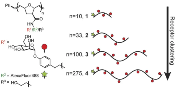
ROMP effected by defined metal carbene catalysts is a living polymerization in which the initiation rates can exceed those of propagation (31, 32). Control over the length of the resulting polymer is achieved by altering the ratio of catalyst initiator to monomer (31, 33). We generated a panel of glycopolymers of defined lengths (DP= 10, 33, 100, and 275) using a functional group-tolerant ruthenium catalyst that affords polymers of con-trolled length and narrow polydispersity. The polymers were functionalized to display an aryl mannoside, a ligand that is su-perior to alkylmannosides for binding to DC-SIGN and enables rapid synthesis of multivalent DC-SIGN ligands. We reasoned that the multivalent presentation of this ligand would afford a highly dense display of mannose residues, as is seen on man-nosylated viruses, such as HIV (34). The polymers also carried a fluorophore, which was used to monitor their cellular binding,
uptake, and localization. The mannoside epitope density was held constant between all four polymers (∼35 mol%), such that the effects of glycopolymer length (and thus receptor clustering), and not ligand density, could be assessed.
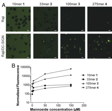
We tested whether these glycopolymers were internalized by cells expressing DC-SIGN. To analyze the role of DC-SIGN in the absence of other mannose-binding lectins, we compared glycopolymer uptake using Raji cells (a human B-cell line that does not express DC-SIGN) and a stably transfected Raji cell line that expresses DC-SIGN (Raji/DC-SIGN). Cells were treated with fluorophore-labeled glycopolymers of defined length, and internalization was assessed by confocal microscopy. The glycopolymers were internalized only by the cells expressing DC-SIGN (Fig. 2A). Moreover, polymer internalization was dose-dependent, further supporting the role for a specific interaction with DC-SIGN (SI Appendix, Fig. S1).
To assess how the extent of DC-SIGN clustering influences internalization, Raji or Raji/DC-SIGN cells were treated with polymers of different lengths, such that the mannose–ligand concentration was constant. The uptake of the longest polymer4 (DP= 275) was ∼10-fold higher than of the next longest 3 (DP = 100), which in turn was∼10-fold higher than for 2 (DP = 33) and 1 (DP = 10) (Fig. 2B). These findings indicate that receptor clustering indeed affects the efficiency of antigen internalization: The longer the polymer, the greater the extent of internalization.
Endocytic Trafficking of Glycopolymer Probes. Given that longer
glycopolymers were more efficiently taken up than their shorter counterparts (Fig. 2), we assessed whether there were differences in glycopolymer trafficking. Mannosylated dendrimers had been shown to be internalized into endosomal compartments (35), and we assumed that the glycopolymers would be routed to early endosomes. We therefore assessed whether the glycopolymers colocalized with transferrin, a marker for early endosomes. We employed Raji/DC-SIGN cells because they provide the means to dissect DC-SIGN’s role in the uptake of these antigens, as this receptor is the only cell-surface C-type lectin expressed. To de-termine if uptake and trafficking is similar in DCs, we also tested monocyte-derived DCs (moDCs). We found that each polymer colocalized with transferrin in either moDCs (Fig. 3B and C) or Raji/DC-SIGN cells (SI Appendix, Fig. S2 A and B). Trafficking occurred via energy-dependent, DC-SIGN–mediated uptake (SI Appendix, Fig. S2 A–C). Treatment with anti–DC-SIGN
anti-bodies abrogated uptake in Raji/DC-SIGN cells, although in-hibition of polymer uptake in moDCs was incomplete (SI Appendix, Fig. S2 C and D). Together, our data indicate that DC-SIGN mediates polymer internalization and trafficking, but suggest that other redundant receptors can affect uptake when DC-SIGN is inhibited. These data also show that polymer length modulates the efficiency of polymer internalization to endo-somal compartments that lead to antigen processing.
Differences in Localization of Particulate and Soluble Glycopolymers.
The glycopolymers used in the aforementioned studies are sol-uble, but pathogens encountered by DCs (such as viruses and bacteria) are particulate. We therefore compared DC-SIGN– mediated internalization and trafficking for soluble vs. particu-late antigens. To access particuparticu-late antigens, we aggregated glycopolymers1–4 by long-term incubation of the compounds in solution. Analysis of the resulting polymers (termed 1p–4p) by dynamic light scattering (DLS) indicated that the length of the polymers influenced the particulate sizes that resulted. Specifi-cally, glycopolymer2p (354 nm) afforded the smallest particles, and the longest polymers, 3p and 4p, gave rise to the largest particles (432 and 402 nm, respectively). Despite being exposed to conditions that led to the aggregation of the longer polymers, glycopolymer 1p (10mer) did not aggregate, and its size was similar to that of the soluble polymers (1.4 nm) (SI Appendix, Table S1). Additionally, application of transmission electron microscopy (TEM) to further characterize polymer size and
Fig. 1. Glycopolymer probes of DC-SIGN endocytosis. A panel of glycopol-ymers 1–4 of defined length were synthesized bearing an aryl mannoside ligand (red) for DC-SIGN as well as Alexa Fluor 488 (green) for visualization.
Jarvis et al. PNAS | July 23, 2019 | vol. 116 | no. 30 | 14863
CHE MISTRY IMMUNOLO GY AND INFLAMMATION
structure revealed that both soluble and aggregate polymers were globular in shape, but vastly different in size (Fig. 3A).
The trafficking of the particulate antigens differed markedly from that observed for the soluble polymers. Specifically, the particulate antigens were almost completely excluded from transferrin-labeled endosomes (Fig. 3 D and E). Only glycopol-ymer1p colocalized with the endosomal marker transferrin, as it was similar in size to soluble glycopolymers1–4 and exhibited the same trafficking patterns. In contrast, glycopolymers2p–4p did not colocalize with transferrin; rather, they were localized to discrete puncta near the cell surface. We quantified the extent of colocalization over time in individual cells and observed that, even at early timepoints, glycopolymers 2p–4p were not trafficked to transferrin-positive endosomes. Thus, they appear to be directly routed to nonendosomal compartments. Additionally, while sol-uble polymers continued through the antigen-presentation path-way, these aggregates failed to colocalize not only with transferrin, but also with other endosomal markers, including Rab5 (a marker of early endosomes), Rab7 (a marker of late endosomes), and LAMP-2 (a marker of late endosomes/lysosomes) (Fig. 3F). Up-take and trafficking of the particulate antigens 2p–4p to these compartments occurred via energy-dependent, DC-SIGN–medi-ated uptake (SI Appendix, Fig. S3). The trafficking phenotype of particulate polymers is the same across Raji/DC-SIGN cells (SI
Appendix, Fig. S3 C and D) and moDCs (Fig. 3 D and E).
Therefore, while polymer uptake is not completely dependent on DC-SIGN in moDCs, our findings indicate that DC-SIGN can mediate the nonendosomal trafficking in both cell types.
Particulate Antigens Traffic to CD81+Compartments with HIV-1.The
trafficking of the glycopolymer aggregates is reminiscent of the trafficking of HIV-1 in DCs. Only a subpopulation of HIV-1 internalized into DCs is routed to degradative com-partments (36, 37). In contrast, HIV-1 is predominantly found in
membrane-proximal, surface-accessible invaginated pockets. These locations are identified by the presence of the tetraspanin CD81 (10). To test whether the internalized glycopolymers2p– 4p are trafficked to surface-accessible compartments, we added Trypan blue, a dye that can quench Alexa Fluor 488 emission and is not cell-permeable (38, 39). The fluorescence emission of the particulate antigens was quenched, suggesting that these polymer aggregates are present in a cell-surface-accessible compartment (Fig. 4A and B).
The trafficking data suggest that glycopolymer aggregates2p–4p are routed similarly to HIV-1. To evaluate this possibility, we examined whether2p–4p colocalized with CD81 in Raji/DC-SIGN cells (Fig. 4C). The soluble glycopolymers 1–4 did not colocalize with CD81. In contrast, CD81 localized with the fluorescence of particulate glycopolymers2p–4p. We next compared the routing of glycopolymers and fluorescently labeled HIV-1 virus-like par-ticles (VLPs) by analyzing their localization with CD81. VLPs resemble native virions and thus can be used to model viral uptake and trafficking from cell to cell at viral synapses (40–42). To cir-cumvent potential effects of CD81–mCherry expression on poly-mer or HIV-1 uptake and trafficking, we examined antigen colocalization with endogenous CD81 in moDCs and Raji/DC-SIGN cells. While the soluble polymer4 showed no appreciable colocalization, the aggregate4p and HIV-1 VLPs colocalized with dense pockets of CD81 in membrane-proximal compartments (Fig. 4D and E andSI Appendix, Fig. S4). To further validate this finding, we coincubated cells with HIV-1 VLPs and polymer and found that aggregate4p directly colocalizes with HIV-1, but sol-uble polymer4 does not (Fig. 4 F and G). These data confirm that aggregated polymers undergo the same route of internal traf-ficking as HIV-1.
Discussion
We designed glycopolymer antigens to probe DC-SIGN en-docytosis, and their application indicated that glycopolymer in-ternalization depends upon its valency (length)—longer glycopolymers are internalized more readily than shorter glyco-polymers. These results suggest that receptor clustering pro-motes DC-SIGN–mediated internalization. DC-SIGN is a tetramer with carbohydrate-recognition domain (CRD) binding sites separated by ∼40 Å (43, 44). Relatively small multivalent ligands may be able to cluster DC-SIGN and engage multiple CRDs for enhanced avidity (43, 45), but the shortest glycopol-ymer 1 (DP = 10) would likely only cluster two DC-SIGN tet-ramers at most. Glycopolymer1’s short (2.6 nm) length suggests it would not be capable of spanning the 4-nm distance between CRDs within a single DC-SIGN tetramer. That glycopolymer1 is internalized supports previous findings that minimal DC-SIGN clustering can induce antigen internalization (46). Still, the up-take of this short polymer is significantly less efficient than that of the longer polymers, which can engage more binding sites and nucleate larger clusters. These data are consistent with a role for DC-SIGN clustering in antigen uptake.
Although the efficiency of uptake for the soluble glycopol-ymers varied with length, the trafficking of these mannosylated conjugates did not. All soluble mannosylated polymers were targeted to early endosomes. Since antigen routing to endosomes is an integral component of efficient antigen presentation in DCs (47–50), these findings suggest that soluble multivalent ligands can effectively deliver antigenic epitopes to the endosome for access to antigen presentation pathways. We anticipate that higher-valency polymers may be more effective scaffolds for targeted synthetic vaccines than those glycoconjugates less ca-pable of clustering DC-SIGN. This advantageous property of the higher-valency polymers synergizes with their demonstrated ability to deliver multiple copies of antigenic peptides and therefore promote higher levels of T-cell activation (29).
In striking contrast to the results with soluble polymers, the particulate antigens are almost entirely excluded from transferrin-labeled endosomes. Instead, particulate glycopolymers were routed to distinct CD81+ compartments and colocalized with HIV-1
Fig. 2. Comparison of fluorophore-labeled glycopolymer uptake in DC-SIGN–positive (Raji/DC-SIGN) and DC-SIGN–negative (Raji) cells. (A) Cells were treated with glycopolymers 1–4 (40 μM in mannoside ligand for 1 and 2; 10μM in mannoside ligand for 3 and 4) for 30 min at 37 °C, and in-ternalization was visualized via confocal microscopy. Higher concentrations of glycopolymers 1 and 2 were required to visualize internalization. (Scale bars, 10μm.) (B) Cells were treated with 1–4 (0.5, 5, 50, and 150 μM in mannoside ligand) for 40 min and placed on ice, and flow cytometry was performed. The resulting fluorescence (arbitrary units) was normalized to account for the average number of fluorophores appended to each polymer.
particles. Another DC lectin, dectin-1, appears to make similar distinctions, as it preferentially internalizes particulate antigen over soluble antigen (51). Intriguingly, despite their distinct trafficking patterns, both small and large aggregates alike are internalized via energy-dependent receptor-mediated uptake mechanisms. Antigen internalization depends only on DC-SIGN in Raji/DC-SIGN cells and partially on the lectin in moDCs. Consistent with these data, prior studies have shown that HIV-1 internalization and transmission is inhibited byα–DC-SIGN blocking antibodies in Raji/DC-SIGN cells, but not in DCs, implying that other lectins are capable of facilitating viral up-take when DC-SIGN is unavailable (52, 53). The polymer/HIV-1–trafficking phenotypes observed in Raji/DC-SIGN cells are identical in moDCs, indicating that DC-SIGN can route anti-gen to a distinct subcellular location based on the antianti-gen’s structure (summarized in SI Appendix, Fig. S7). Previous studies have demonstrated that changes in antigen size and shape can affect the extent of receptor-mediated internaliza-tion and signaling (reviewed in ref. 54), but this study shows how those features influence antigen trafficking to different compartments.
Superresolution microscopy has shown that DC-SIGN exists on the cell surface in stable, loosely packed ∼80-nm diameter
nanodomains that can be clustered into larger microdomains (55). These microdomains can range in size from 300 nm to 1.5μm, with an average spacing of 400 nm (56, 57). The polymer aggregates include species larger than HIV-1, but in the size range of many pathogens. Both HIV-1 and aggregate polymers can bridge several DC-SIGN nanodomains, and aggregates can even span multiple microdomains. These antigen attributes could potentiate unique internalization mechanisms. In immune cells, engagement and clustering of such receptor domains can lead to changes in actin organization (15–18, 58, 59). Recent studies have revealed that HIV-1 endocytosis into DCs andtrans infection of CD4+T cells at the viral synapse depend critically on actin-remodeling factors—in particular, dynamin-2 (11, 60). Similarly, the internalization of particulate antigens in our studies was partially dependent on dynamin-2 activity (SI Appendix, Fig. S5). In contrast, the endocy-tosis of soluble antigens did not depend on dynamin-2 until later steps of trafficking. Particulate polymers also elicited responses as-sociated with HIV-induced DC-SIGN signaling, including expres-sion of IL-12B and IL-6 and activation of Raf-1 (SI Appendix, Fig. S6A–C) (61, 62). Consistent with what has been observed with the lectin dectin-1, only particulate, and not soluble, polymers induce signaling (SI Appendix, Fig. S6A). These findings indicate that DC-SIGN can make similar distinctions in signaling based on antigen properties (51).
HIV-1 and particulate polymers may stimulate more profound changes in actin remodeling than soluble polymers, giving rise to their differential trafficking. The larger polymers appear to be capable of engaging, organizing, and signaling through DC-SIGN in a similar manner as HIV-1, resulting in alternative trafficking to the invaginated pockets. These findings suggest that the particulate polymer aggregates can be used to elucidate the mechanisms governing HIV’s divergent trafficking.
With the ability to control glycopolymer features, we found that the physical characteristics of a DC-SIGN–binding antigen affect its fate. In addition to the utility of polymers for un-derstanding antigen-uptake mechanisms, our findings provide opportunities to customize materials to serve as highly effective scaffolds for synthetic vaccines. By tailoring the properties of the polymer, one could direct antigenic cargo to specific compart-ments for classical presentation or cross presentation (48, 49). Our results also suggest strategies to target sites where viruses evade immune surveillance. In HIV-1–infected individuals, the persistence of HIV-1 in latently infected cells is a major obstacle toward completely eradicating the virus (63). Materials such as particulate glycopolymers could facilitate the identification of latently infected cells and aid in elucidating the mechanism un-derlying HIV’s ability to escape immune detection.
Materials and Methods
Polymer Synthesis and Characterization. A complete description of ligand and polymer synthesis and characterization is provided inSI Appendix. Production of Fluorescently Labeled VLPs. HEK293T cells plated in 10-cm tissue-culture dishes were cotransfected to express plasmids encoding HIV-1 Gag-mCherry, HIV-1 Rev, and HIV-1 CCR5 tropic Env. Forty-eight hours post-transfection, viral supernatants were collected, filtered through a 0.2-μm filter, and spun through a 0.1% sucrose cushion in a Beckman Coulter LE80K ultra-centrifuge at 14,000 rpm at 4 °C for 2 h to sediment the VLPs. Pellet was resuspended in 10 mL of culture medium (DMEM, 10% FBS, 1% PenStrep, and 1%L-glutamine), filtered through a 0.2-μm sterile filter, aliquoted, and stored at−80 °C until use.
Cell Stimulation and Transfections. MoDCs, Raji, or Raji/DC-SIGN cells were suspended at 1× 10⁶to 4× 10⁶cells/mL in 1 mM CaCl
20.5 mM MgCl2, and
PBS (pH 7.4) containing 0.1% BSA. Where indicated, cells were pretreated at 37 °C for 15 min with inhibitors: Dynasore (80μM), Genistein (100 μM), or vehicle (0.1% DMSO). With inhibitor present, cells were treated with in-dicated polymers at a concentration of 10–40 μM mannose for indicated time periods before immunofluorescence analysis. For the expression of mCherry fusion constructs, Rab 5 and Rab 7 cDNA (a gift of M. Sandor, University of Wisconsin–Madison, Madison, WI) and human CD81 cDNA (Open Biosystems) were cloned into pmCherry-N1 vector (Clontech). Raji/DC-SIGN Fig. 3. Trafficking of soluble and particulate glycopolymers. (A) TEM of
soluble and particulate 275mer polymers. (B and D) Soluble (B) or particulate (D) glycopolymers (green) were added (mannose concentration of 10μM) to moDCs at 37 °C for 30 min, and their trafficking to transferrin-labeled early endosomes (red) was monitored via confocal microscopy. (C and E) Polymer/ transferrin colocalization in B and D, respectively, was assessed at the in-dicated time points. Pearson’s coefficient was obtained by the Colocalization Threshold plugin in ImageJ using n> 50 cells per treatment. Error bars represent the 95% confidence interval of the mean for representative data from at least two independent experiments. P values represent colocaliza-tion of the 10mer compared with each polymer at indicated time point. *P< 0.001. (F) Polymer/Rab5, Rab7, or LAMP-2 colocalization was assessed in Raji/ DC-SIGN cells either transfected with mRFP or Rab 5/7 mRFP or stained for LAMP-2 after treatment with polymer for 30 min at 37 °C. [Scale bars: 100 nm (A) and 5μm (B, D, and F).]
Jarvis et al. PNAS | July 23, 2019 | vol. 116 | no. 30 | 14865
CHE MISTRY IMMUNOLO GY AND INFLAMMATION
cells were electroporated with 1 μg of indicated plasmid by using the Eppendorf Multiporator in iso-osmolar electroporation buffer using a 90-μs, 1,200-V pulse. Cells were then transferred to prewarmed culture medium and utilized 48 h posttransfection.
Immunofluorescence and Confocal Microscopy. Where indicated, cells were incubated with 8μg/mL Cy3-transferrin (Jackson ImmunoResearch) for 30 min in 1% BSA/RPMI. Cells were washed and treated with polymer for the indicated time periods at either 4 °C or 37 °C and then placed on ice prior, washed, and transferred to LabTek II Chambered Coverglass (#1.5). For HIV VLP in-ternalization, cells were pretreated overnight with LPS and then incubated with HIV-1::Gag-mCherry particles for 24 h. In CD81 immunostaining experi-ments, cells were treated with polymer or HIV/VLP, fixed in 4% para-formaldehyde, washed, permeabilized in 0.05% saponin/PBS, and stained with mouseα-hCD81 (1.3.3.22) (Santa Cruz Biotechnology) and either Alexa Fluor 594– or Dylight 649–α-mouse IgG secondary antibody (Jackson Immuno-Research). The same procedure was used for LAMP2 colocalization experiments
using ratα-LAMP2 primary antibody (Santa Cruz Biotechnology) and Alexa Fluor 594 donkeyα-rat IgG secondary antibody (Thermo Fisher Scientific). For the Trypan blue quenching experiments, fluorescence images were acquired before and after a 2-min incubation with Trypan blue (0.1%; Bio-Rad), which was added carefully to minimize sample movement. Cells were imaged on a Nikon A1R confocal microscope [60× oil immersion lens, 1.4 numerical aperture (NA)] or an RPI spinning-disk confocal microscope (63× oil immersion lens, 1.4 NA). Images were despeckled or background subtraction was performed be-fore colocalization. Colocalization analysis was performed in Fiji by using the Colocalization Threshold or Coloc 2 plugin to obtain the Pearson’s Coefficient and Mander’s coefficient of antigen overlap with the different markers. All P values reported were acquired from the Mann–Whitney u test performed by using GraphPad Prism.
ACKNOWLEDGMENTS. This research was supported by National Institutes of Health Grant AI055258. We thank the following people for assistance with TEM and confocal microscopy: Nikki Watson and Wendy Salmon of the Fig. 4. Comparison of trafficking of particulate and soluble antigens to HIV. (A) Soluble and particulate fluorophore-labeled glycopolymers (green; 10μM mannose) were added to Raji/DC-SIGN cells at 37 °C for 30 min. Fluorescence quenching by Trypan blue (2-min exposure) was monitored via confocal mi-croscopy. Arrowheads indicate intracellular fluorescence quenched by Trypan blue. (Scale bars, 10μm.) (B) Polymer fluorescence before and after Trypan blue treatment was measured in ImageJ (normalized to untreated). Error bars represent the SEM of at least two experiments for n> 50 cells. (C and D) Polymers or fluorescently labeled HIV-1 VLPs (green) were added at 37 °C to Raji/DC-SIGN cells transfected with CD81-mCherry (C) or moDCs stained for endogenous CD81 (D). Colocalization with CD81 was monitored by confocal microscopy. Arrowheads indicate regions of colocalization. (E) Quantitation of colocalization in moDCs in D, utilizing the Mander’s coefficient of antigen fluorescence overlapping with CD81. Error bars represent the SEM for at least two experiments for n> 35 cells. ns, not significant. (F) Polymer (green) and HIV (red) were coincubated in moDCs to assess colocalization. The plasma membrane was stained with wheat germ agglutinin (blue). (G) The overlap of polymer and HIV VLP fluorescence intensity (from F) was compared over the region indicated by a white line. (Scale bars, 5μm.) *P < 0.01; ***P < 0.0001. A.U., arbitrary units.
W. M. Keck Biological Imaging Facility at the Whitehead Institute; Dr. Michelle Grevstad of the University of Wisconsin–Madison (UW–Madison) Biochemistry Optical Core; and Lance Rodenkirch, director of the UW–Madison Keck
Laboratory for Biological Imaging. We also thank Dr. J. Hank, Dr. K. McDowell, and Prof. P. Sondel of UW–Madison for assistance in obtaining blood and helpful conversations.
1. C. G. Figdor, Y. van Kooyk, G. J. Adema, C-type lectin receptors on dendritic cells and Langerhans cells. Nat. Rev. Immunol. 2, 77–84 (2002).
2. W. I. Weis, R. Kahn, R. Fourme, K. Drickamer, W. A. Hendrickson, Structure of the calcium-dependent lectin domain from a rat mannose-binding protein determined by MAD phasing. Science 254, 1608–1615 (1991).
3. M. J. Robinson, D. Sancho, E. C. Slack, S. LeibundGut-Landmann, C. Reis e Sousa, Myeloid C-type lectins in innate immunity. Nat. Immunol. 7, 1258–1265 (2006). 4. B. Tassaneetrithep et al., DC-SIGN (CD209) mediates dengue virus infection of human
dendritic cells. J. Exp. Med. 197, 823–829 (2003).
5. C. P. Alvarez et al., C-type lectins DC-SIGN and L-SIGN mediate cellular entry by Ebola virus in cis and in trans. J. Virol. 76, 6841–6844 (2002).
6. E. G. Cormier et al., L-SIGN (CD209L) and DC-SIGN (CD209) mediate transinfection of liver cells by hepatitis C virus. Proc. Natl. Acad. Sci. U.S.A. 101, 14067–14072 (2004). 7. T. B. Geijtenbeek et al., DC-SIGN, a dendritic cell-specific HIV-1-binding protein that
enhances trans-infection of T cells. Cell 100, 587–597 (2000).
8. D. S. Kwon, G. Gregorio, N. Bitton, W. A. Hendrickson, D. R. Littman, DC-SIGN-mediated internalization of HIV is required for trans-enhancement of T cell in-fection. Immunity 16, 135–144 (2002).
9. D. C. DeLucia, C. R. Rinaldo, G. Rappocciolo, Inefficient HIV-1 trans infection of CD4+T cells by macrophages from HIV-1 nonprogressors is associated with altered
membrane cholesterol and DC-SIGN. J. Virol. 92, e00092-18 (2018).
10. H. J. Yu, M. A. Reuter, D. McDonald, HIV traffics through a specialized, surface-accessible intracellular compartment during trans-infection of T cells by mature dendritic cells. PLoS Pathog. 4, e1000134 (2008).
11. M. M. Ménager, D. R. Littman, Actin dynamics regulates dendritic cell-mediated transfer of HIV-1 to T cells. Cell 164, 695–709 (2016).
12. E. Garcia et al., HIV-1 trafficking to the dendritic cell-T-cell infectious synapse uses a pathway of tetraspanin sorting to the immunological synapse. Traffic 6, 488–501 (2005). 13. N. Izquierdo-Useros et al., Siglec-1 is a novel dendritic cell receptor that mediates HIV-1 trans-infection through recognition of viral membrane gangliosides. PLoS Biol. HIV-10, e1001448 (2012).
14. J. Martinez-Picado et al., Identification of Siglec-1 null individuals infected with HIV-1. Nat. Commun. 7, 12412 (2016).
15. G. Gabbiani, C. Chaponnier, A. Zumbe, P. Vassalli, Actin and tubulin co-cap with surface immunoglobulins in mouse B lymphocytes. Nature 269, 697–698 (1977). 16. J. Braun, P. S. Hochman, E. R. Unanue, Ligand-induced association of surface
immu-noglobulin with the detergent-insoluble cytoskeletal matrix of the B lymphocyte. J. Immunol. 128, 1198–1204 (1982).
17. G. F. Schreiner, K. Fujiwara, T. D. Pollard, E. R. Unanue, Redistribution of myosin ac-companying capping of surface Ig. J. Exp. Med. 145, 1393–1398 (1977).
18. J. Braun, K. Fujiwara, T. D. Pollard, E. R. Unanue, Two distinct mechanisms for re-distribution of lymphocyte surface macromolecules. I. Relationship to cytoplasmic myosin. J. Cell Biol. 79, 409–418 (1978).
19. L. L. Kiessling, J. E. Gestwicki, L. E. Strong, Synthetic multivalent ligands as probes of signal transduction. Angew. Chem. Int. Ed. Engl. 45, 2348–2368 (2006).
20. E. B. Puffer, J. K. Pontrello, J. J. Hollenbeck, J. A. Kink, L. L. Kiessling, Activating B cell signaling with defined multivalent ligands. ACS Chem. Biol. 2, 252–262 (2007). 21. G. Roelants, L. Forni, B. Pernis, Blocking and redistribution (“capping”) of antigen
receptors on T and B lymphocytes by anti-immunoglobulin antibody. J. Exp. Med. 137, 1060–1077 (1973).
22. P. J. Tacken, M. Ter Huurne, R. Torensma, C. G. Figdor, Antibodies and carbohydrate ligands binding to DC-SIGN differentially modulate receptor trafficking. Eur. J. Im-munol. 42, 1989–1998 (2012).
23. N. K. Dakappagari et al., Conformational HER-2/neu B-cell epitope peptide vaccine designed to incorporate two native disulfide bonds enhances tumor cell binding and antitumor activities. J. Biol. Chem. 280, 54–63 (2005).
24. N. K. Dakappagari et al., Intracellular delivery of a novel multiepitope peptide vaccine by an amphipathic peptide carrier enhances cytotoxic T-cell responses in HLA-A*201 mice. J. Pept. Res. 65, 189–199 (2005).
25. J. J. García-Vallejo et al., Multivalent glycopeptide dendrimers for the targeted de-livery of antigens to dendritic cells. Mol. Immunol. 53, 387–397 (2013).
26. J. E. Gestwicki, C. W. Cairo, L. E. Strong, K. A. Oetjen, L. L. Kiessling, Influencing re-ceptor-ligand binding mechanisms with multivalent ligand architecture. J. Am. Chem. Soc. 124, 14922–14933 (2002).
27. J. E. Gestwicki, L. E. Strong, L. L. Kiessling, Visualization of single multivalent receptor-ligand complexes by transmission electron microscopy. Angew. Chem. Int. Ed. Engl. 39, 4567–4570 (2000).
28. L. L. Kiessling, J. C. Grim, Glycopolymer probes of signal transduction. Chem. Soc. Rev. 42, 4476–4491 (2013).
29. N. R. Bennett, D. B. Zwick, A. H. Courtney, L. L. Kiessling, Multivalent antigens for promoting B and T cell activation. ACS Chem. Biol. 10, 1817–1824 (2015). 30. M. Kanai, K. H. Mortell, L. L. Kiessling, Varying the size of multivalent ligands: The
dependence of concanavalin A binding on neoglycopolymer length. J. Am. Chem. Soc. 119, 9931–9932 (1997).
31. T. L. Choi, R. H. Grubbs, Controlled living ring-opening-metathesis polymerization by a fast-initiating ruthenium catalyst. Angew. Chem. Int. Ed. Engl. 42, 1743–1746 (2003).
32. R. R. Schrock, High oxidation state alkylidene and alkylidyne complexes. Chem. Commun. (Camb.), 2773–2777 (2005).
33. C. W. Bielawski, R. H. Grubbs, Living ring-opening metathesis polymerization. Prog. Polym. Sci. 32, 1–29 (2007).
34. L. R. Prost, J. C. Grim, M. Tonelli, L. L. Kiessling, Noncarbohydrate glycomimetics and glycoprotein surrogates as DC-SIGN antagonists and agonists. ACS Chem. Biol. 7, 1603–1608 (2012).
35. A. Berzi et al., Pseudo-mannosylated DC-SIGN ligands as immunomodulants. Sci. Rep. 6, 35373 (2016).
36. A. Moris et al., Dendritic cells and HIV-specific CD4+ T cells: HIV antigen presentation, T-cell activation, and viral transfer. Blood 108, 1643–1651 (2006).
37. L. Jones, D. McDonald, D. H. Canaday, Rapid MHC-II antigen presentation of HIV type 1 by human dendritic cells. AIDS Res. Hum. Retroviruses 23, 812–816 (2007). 38. J. Rejman, V. Oberle, I. S. Zuhorn, D. Hoekstra, Size-dependent internalization of
particles via the pathways of clathrin- and caveolae-mediated endocytosis. Biochem. J. 377, 159–169 (2004).
39. L. Thiele, H. P. Merkle, E. Walter, Phagocytosis and phagosomal fate of surface-modified microparticles in dendritic cells and macrophages. Pharm. Res. 20, 221–228 (2003). 40. J. C. Gardiner, E. J. Mauer, N. M. Sherer, HIV-1 gag, envelope, and extracellular
de-terminants cooperate to regulate the stability and turnover of virological synapses. J. Virol. 90, 6583–6597 (2016).
41. D. Gheysen et al., Assembly and release of HIV-1 precursor Pr55gagvirus-like particles
from recombinant baculovirus-infected insect cells. Cell 59, 103–112 (1989). 42. N. Izquierdo-Useros et al., Capture and transfer of HIV-1 particles by mature dendritic
cells converges with the exosome-dissemination pathway. Blood 113, 2732–2741 (2009). 43. K. C. Garber, K. Wangkanont, E. E. Carlson, L. L. Kiessling, A general glycomimetic strategy yields non-carbohydrate inhibitors of DC-SIGN. Chem. Commun. (Camb.) 46, 6747–6749 (2010).
44. D. A. Mitchell, A. J. Fadden, K. Drickamer, A novel mechanism of carbohydrate rec-ognition by the C-type lectins DC-SIGN and DC-SIGNR. Subunit organization and binding to multivalent ligands. J. Biol. Chem. 276, 28939–28945 (2001).
45. I. Sutkeviciute et al., Unique DC-SIGN clustering activity of a small glycomimetic: A lesson for ligand design. ACS Chem. Biol. 9, 1377–1385 (2014).
46. P. Liu et al., Low copy numbers of DC-SIGN in cell membrane microdomains: Impli-cations for structure and function. Traffic 15, 179–196 (2014).
47. E. M. Bertram, R. G. Hawley, T. H. Watts, Overexpression of rab7 enhances the kinetics of antigen processing and presentation with MHC class II molecules in B cells. Int. Immunol. 14, 309–318 (2002).
48. C. D. Platt et al., Mature dendritic cells use endocytic receptors to capture and present antigens. Proc. Natl. Acad. Sci. U.S.A. 107, 4287–4292 (2010).
49. B. Chatterjee et al., Internalization and endosomal degradation of receptor-bound antigens regulate the efficiency of cross presentation by human dendritic cells. Blood 120, 2011–2020 (2012).
50. L. C. Bonifaz et al., In vivo targeting of antigens to maturing dendritic cells via the DEC-205 receptor improves T cell vaccination. J. Exp. Med. 199, 815–824 (2004). 51. H. S. Goodridge et al., Activation of the innate immune receptor Dectin-1 upon
formation of a‘phagocytic synapse’. Nature 472, 471–475 (2011).
52. C. Boggiano, N. Manel, D. R. Littman, Dendritic cell-mediated trans-enhancement of human immunodeficiency virus type 1 infectivity is independent of DC-SIGN. J. Virol. 81, 2519–2523 (2007).
53. A. Granelli-Piperno et al., Dendritic cell-specific intercellular adhesion molecule 3-grabbing nonintegrin/CD209 is abundant on macrophages in the normal human lymph node and is not required for dendritic cell stimulation of the mixed leukocyte reaction. J. Immunol. 175, 4265–4273 (2005).
54. N. Oh, J.-H. Park, Endocytosis and exocytosis of nanoparticles in mammalian cells. Int. J. Nanomedicine 9 (suppl. 1), 51–63 (2014).
55. M. S. Itano et al., Super-resolution imaging of C-type lectin spatial rearrangement within the dendritic cell plasma membrane at fungal microbe contact sites. Front. Phys. 2, 46 (2014). 56. A. K. Neumann, N. L. Thompson, K. Jacobson, Distribution and lateral mobility of
DC-SIGN on immature dendritic cells–Implications for pathogen uptake. J. Cell Sci. 121, 634–643 (2008).
57. A. Cambi et al., Microdomains of the C-type lectin DC-SIGN are portals for virus entry into dendritic cells. J. Cell Biol. 164, 145–155 (2004).
58. P. K. Mattila et al., The actin and tetraspanin networks organize receptor nano-clusters to regulate B cell receptor-mediated signaling. Immunity 38, 461–474 (2013). 59. A. Aderem, D. M. Underhill, Mechanisms of phagocytosis in macrophages. Annu. Rev.
Immunol. 17, 593–623 (1999).
60. W. Hübner et al., Quantitative 3D video microscopy of HIV transfer across T cell vi-rological synapses. Science 323, 1743–1747 (2009).
61. S. I. Gringhuis, J. den Dunnen, M. Litjens, M. van der Vlist, T. B. Geijtenbeek, Carbohydrate-specific signaling through the DC-SIGN signalosome tailors immunity to Mycobacterium tuberculosis, HIV-1 and Helicobacter pylori. Nat. Immunol. 10, 1081–1088 (2009). 62. A. Prasad, R. Kulkarni, S. Jiang, J. E. Groopman, Cocaine enhances DC to T-cell HIV-1
transmission by activating DC-SIGN/LARG/LSP1 complex and facilitating infectious synapse formation. Sci. Rep. 7, 40648 (2017).
63. D. M. Margolis, J. V. Garcia, D. J. Hazuda, B. F. Haynes, Latency reversal and viral clearance to cure HIV-1. Science 353, aaf6517 (2016).
Jarvis et al. PNAS | July 23, 2019 | vol. 116 | no. 30 | 14867
CHE MISTRY IMMUNOLO GY AND INFLAMMATION