Survival and Morbidity of Preterm Children Born at 22
Through 34 Weeks’ Gestation in France in 2011
Results of the EPIPAGE-2 Cohort Study
Pierre-Yves Ancel, PhD; François Goffinet, PhD; and the EPIPAGE-2 Writing GroupIMPORTANCEUp-to-date estimates of the health outcomes of preterm children are needed for assessing perinatal care, informing parents, making decisions about care, and providing evidence for clinical guidelines.
OBJECTIVES To determine survival and neonatal morbidity of infants born from 22 through 34 completed weeks’ gestation in France in 2011 and compare these outcomes with a comparable cohort in 1997.
DESIGN, SETTING, AND PARTICIPANTSThe EPIPAGE-2 study is a national, prospective, population-based cohort study conducted in all maternity and neonatal units in France in 2011. A total of 2205 births (stillbirths and live births) and terminations of pregnancy at 22 through 26 weeks’ gestation, 3257 at 27 through 31 weeks, and 1234 at 32 through 34 weeks were studied. Cohort data were collected from January 1 through December 31, 1997, and from March 28 through December 31, 2011. Analyses for 1997 were run for the entire year and then separately for April to December; the rates for survival and morbidities did not differ. Data are therefore presented for the whole year in 1997 and the 8-month and 6-month periods in 2011.
MAIN OUTCOMES AND MEASURES Survival to discharge and survival without any of the following adverse outcomes: grade III or IV intraventricular hemorrhage, cystic periventricular leukomalacia, severe bronchopulmonary dysplasia, retinopathy of prematurity (stage 3 or higher), or necrotizing enterocolitis (stages 2-3).
RESULTS A total of 0.7% of infants born before 24 weeks’ gestation survived to discharge: 31.2% of those born at 24 weeks, 59.1% at 25 weeks, and 75.3% at 26 weeks. Survival rates were 93.6% at 27 through 31 weeks and 98.9% at 32 through 34 weeks. Infants discharged home without severe neonatal morbidity represented 0% at 23 weeks, 11.6% at 24 weeks, 30.0% at 25 weeks, 47.5% at 26 weeks, 81.3% at 27 through 31 weeks, and 96.8% at 32 through 34 weeks. Compared with 1997, the proportion of infants surviving without severe morbidity in 2011 increased by 14.4% (P < .001) at 25 through 29 weeks and 6% (P < .001) at 30 through 31 weeks but did not change appreciably for those born at less than 25 weeks. The rates of antenatal corticosteroid use, induced preterm deliveries, cesarean deliveries, and surfactant use increased significantly in all gestational-age groups, except at 22 through 23 weeks.
CONCLUSIONS AND RELEVANCEThe substantial improvement in survival in France for newborns born at 25 through 31 weeks’ gestation was accompanied by an important reduction in severe morbidity, but survival remained rare before 25 weeks. Although improvement in survival at extremely low gestational age may be possible, its effect on long-term outcomes requires further studies. The long-term results of the EPIPAGE-2 study will be informative in this regard.
JAMA Pediatr. 2015;169(3):230-238. doi:10.1001/jamapediatrics.2014.3351 Published online January 26, 2015. Corrected on March 9, 2015.
Editorial page 207 Supplemental content at jamapediatrics.com
Author Affiliations: Obstetrical,
Perinatal, and Pediatric Epidemiology Team, Epidemiology and Biostatistics Sorbonne Paris Cité Research Center (U1153), INSERM, Paris, France (Ancel, Goffinet); Paris Descartes University, Paris, France (Ancel, Goffinet); Clinical Research Unit, Center for Clinical Investigation P1419, Cochin Broca Hotel–Dieu Hospital, Paris, France (Ancel); Maternité Port-Royal, Hospital University Department Risks in Pregnancy, Cochin Brocha Hotel–Dieu Hospital, Paris, France (Goffinet).
Group Information: The members of
the EPIPAGE-2 Writing Group and other EPIPAGE-2 Study Group Collaborators are listed at the end of the article.
Corresponding Author: Pierre-Yves
Ancel, PhD, Obstetrical, Perinatal, and Pediatric Epidemiology Team, Epidemiology and Biostatistics Sorbonne Paris Cité Research Center (U1153), INSERM, 53 avenue de l’Observatoire, 75014 Paris, France (pierre-yves.ancel@inserm.fr).
Original Investigation
P
revious cohort studies1-7suggest that survival ofin-fants born before 27 weeks’ gestation has improved dur-ing the past 2 decades. However, disability rates re-main high at these gestational ages.8-10Moreover, countries
differ substantially in their organization of care, available re-sources, national laws, and cultural preferences regarding pro-vision of proactive care. Therefore, to provide the best avail-able information for parents and medical staff to use in making treatment decisions, mortality and morbidity must be moni-tored, and studies should be conducted in countries with dif-ferent attitudes toward active care at early gestational ages.
By far, most of the important cohort studies11-13in the
field have focused exclusively on infants born before 27 weeks’ gestation. However, even though infants born between 27 and 31 weeks are at lower relative risk of adverse outcomes, they represent a much larger proportion of pre-term births. Hence, in absolute numbers, they account for most children with deficits.
We present the results of the EPIPAGE-2 (Etude Epidémi-ologique sur les Petits Ages Gestationnels 2) study, a national cohort of infants born at a gestational age of 22 through 34 weeks in France in 2011. Our objectives were to study sur-vival and sursur-vival without severe neonatal morbidities. We also looked at perinatal interventions and compared the out-comes with those of a similar cohort from 1997, the EPIPAGE-1 study.13Our hypothesis was that survival and survival
with-out severe morbidity have improved during the past 15 years in France, except for extremely preterm infants.
Methods
Ethics
Recruitment and data collection occurred only after families had received information and agreed to participate in the study. This study was approved by the National Data Protection Au-thority (Commission Nationale de l’Informatique et des Lib-ertés) and by the appropriate ethics committees (Consulta-tive Committee on the Treatment of Information on Personal Health Data for Research Purposes and Committee for the Pro-tection of People Participating in Biomedical Research). Par-ticipants provided oral informed consent.
Study Design and Population Study
EPIPAGE-2 is a national, prospective, population-based study scheduled to follow up preterm children to the age of 12 years. Infants born at 22 through 34 completed weeks’ gestation in France were eligible for inclusion. Only one region, which ac-counts for 2% of all births in France, did not participate. The study began March 28, 2011. Recruitment took place at birth in all maternity units in the participating regions. The num-ber of infants required according to our sample size calculations14was provided by an 8-month recruitment
pe-riod for births at 22 through 26 weeks, a 6-month pepe-riod for 27 through 31 weeks, and a 5-week period for 32 through 34 weeks. During recruitment, members of the regional coordinating committees visited all maternity units to ensure the identifi-cation of all eligible children.
The births in our study population were defined to com-prise live births, stillbirths, and terminations of pregnancy for maternal (severe maternal diseases) or fetal (severe growth re-striction and oligohydramnios) reasons other than congeni-tal anomalies. In all, 2381 births were eligible at 22 through 26 weeks, 3478 at 27 through 31 weeks, and 1376 at 32 through 34 weeks, with 176, 221, and 142 parental refusals, respectively. The study thus included 2205 births at 22 through 26 weeks, 3257 at 37 through 31 weeks, and 1234 at 32 through 34 weeks. Total births in the 25 French regions in 2011 (National Insti-tute of Statistics and Economic Studies; http://www.insee.fr /fr/) were used to estimate preterm birth rates, taking into ac-count months of inclusion and differences in recruitment periods according to gestational age at birth.
Data Collection
In each center, one obstetric and one pediatric study coordi-nator were responsible for data acquisition, validation, and quality control. Data were collected from medical records and obstetric and neonatal staff. Data on stillbirths and termina-tions of pregnancy were collected at the time of delivery. Data on live-born infants were collected prospectively during hos-pitalization until discharge or death. Gestational age was de-fined as the best obstetric estimate combining last menstrual period and ultrasonogram assessment. Extensive data were col-lected about pregnancy, delivery, and the neonatal period to investigate pregnancy complications, decisions about termi-nations of pregnancy, the child’s condition at birth, neonatal diseases, organization of care, treatment, and attitudes to-ward care. Only selected perinatal data were considered for this study: level of care of the institution, antenatal corticosteroid use, vaginal or cesarean delivery, indicated preterm delivery (defined as a birth after induction of labor or cesarean deliv-ery before the onset of labor), and use of surfactants and post-natal corticosteroids. Questionnaires were completed online, with a secure interface that protected the confidentiality and privacy of data and personal information. The EPIPAGE coor-dination team used a centralized system to monitor and vali-date inclusion and data collection at the national level.
Outcome Measures
The primary outcome was infant survival, defined as the num-ber of children discharged home alive. The secondary out-come was survival to discharge without severe neonatal mor-bidity. Severe neonatal morbidity was defined as any of the following outcomes: severe intraventricular hemorrhage (IVH), defined as IVH associated with ventricular dilatation (grade III IVH) and intraparenchymal hemorrhage (ie, large unilateral pa-renchymal hyperdensity or a large unilateral porencephalic cyst)15; cystic periventricular leukomalacia (cPVL) (ie,
peri-ventricular white matter echolucencies at ultrasonography); stages II and III necrotizing enterocolitis, according to the stag-ing of Bell et al16; stage 3 or higher retinopathy of
prematu-rity, according to the international classification17and/or
la-ser treatment; and/or severe bronchopulmonary dysplasia (BPD), defined as administration of oxygen for at least 28 days plus need for 30% or more oxygen and/or mechanical venti-latory support or continuous positive airway pressure at 36
weeks’ postmenstrual age.18Among infants admitted to the
neonatal intensive care unit (NICU), 98.0% of survivors born before 33 weeks, 88.0% of those born at 33 weeks, and 66.0% of those born at 34 weeks had at least one cranial ultrasono-gram assessment. Survivors at 33 and 34 weeks without cra-nial ultrasonogram assessment were considered not to have severe cerebral lesions, and those with no funduscopic exami-nation were considered not to have severe retinopathy of prematurity.
Comparison of French Birth Cohorts Between 1997 and 2011
In 1997, the EPIPAGE-1 study, a comparable prospective, popu-lation-based cohort study, took place in 9 regions of France.13,19
Eligible infants for this comparison are those born alive be-tween 22 and 34 weeks’ gestation from the 1997 and 2011 co-horts in the same 9 regions. Outcome measures are survival to discharge and survival without neonatal morbidity. The lat-ter was defined as above, except for BPD (oxygen supplemen-tation at 36 weeks’ postmenstrual age) because information on its severity was not available in 1997. Cohort data were col-lected from January 1 through December 31, 1997, and from March 28 through December 31, 2011.14Analyses for 1997 were
run for the entire year and then separately for April to Decem-ber; the rates for survival and morbidities did not differ. Data are therefore presented for the whole year in 1997 and the 8-month and 6-month periods in 2011.
Statistical Analysis
Data reported on the birth certificate (ie, gestational age at birth, vital status at birth, and neonatal death) were available and us-able without parental consent for children not included in the study. We compared the survival of the participating and non-participating (because of parent refusal) newborns. Then, for the infants whose parents agreed to participate, we analyzed survival to discharge, severe neonatal morbidity, survival with-out severe morbidity, and some obstetric and neonatal inter-ventions, according to gestational age. To examine trends over time, we compared survival and neonatal morbidity in the EPIPAGE-1 study19with those in the EPIPAGE-2 study
accord-ing to gestational age. For each week of gestation, we report exact 95% binomial CIs of survival rates and their differ-ences. All tests were 2-sided; P < .05 was considered statisti-cally significant. All statistical analyses were performed with SAS statistical software, version 9.3 (SAS Institute Inc).
Results
Preterm Birth Rates
In 2011, preterm birth rates were 4.4 per 1000 total stillbirths and live births and 2.1 per 1000 live births before 27 weeks’ ges-tation, 8.4 and 7.5 at 27 through 31 weeks, and 17.8 and 17.3 at 32 through 34 weeks, respectively.
Comparison of Study Participants and Nonparticipants
Participation rates were 92.6% among infants born at 22 through 26 weeks, 93.6% among those at 27 through 31 weeks, and 89.7% among those at 32 through 34 weeks. In each
gesta-tional age group, the proportion of live births was slightly higher among participants than nonparticipants. Survival among live births did not differ significantly between the 2 groups (eTable 1 in the Supplement).
Status at Birth and Perinatal Deaths
The proportion of live-born infants increased with gesta-tional age from 13.5% at 22 weeks to 98.5% at 34 weeks (Table 1). Only one infant born at 22 through 23 weeks (ie, 0.1% of all births and 0.7% of live births) survived to discharge (Table 1). Survival rates were 14.4% of all births and 31.2% of live births at 24 weeks, 41.8% and 59.1% at 25 weeks, 59.6% and 75.3% at 26 weeks, 73.7% and 86.3% at 27 through 28 weeks, 88.0% and 96.6% at 29 through 31 weeks, and 96.7% and 98.9% at 32 through 34 weeks, respectively (Table 1). Among infants who died, the proportion whose deaths followed a decision to limit intensive care varied from 80.9% at 22 through 24 weeks and 70.3% at 25 through 26 weeks to 57.0% at 27 through 31 weeks. The median age at death was the day of birth for infants born at 22 through 24 weeks, 5 days (interquartile range, 1-16 days) for those born at 25 through 26 weeks, and 7 days (interquar-tile range, 1-22 days) for those born at 27 through 31 weeks.
Perinatal Interventions
Among the live-born infants at 22 weeks, 36.2% were born in level III hospitals compared with 61.8% at 23 weeks, 77.4% at 24 weeks, 85.0% at 25 through 26 weeks, 84.8% at 27 through 31 weeks, and 50.1% at 32 through 34 weeks (Table 2). Among extremely preterm infants not born in a level III hospital, 54.3% were postnatally transferred to a NICU; this proportion var-ied from 4.3% at 22 through 23 weeks to 45.3% at 24 weeks and 90.7% at 25 through 26 weeks. Postnatal transfers to a NICU reached 91.8% at 27 through 29 weeks but decreased to 56.3% at 30 through 31 weeks and 17.0% at 32 through 34 weeks. The percentage of infants exposed to antenatal corticosteroids was very low at 22 (1.8%) and 23 (12.3%) weeks but increased to 56.7% at 24 weeks and 78.4% at 25 through 26 weeks (Table 2). Cesarean rates were 6.3% at 22 through 23 weeks and 13.5% at 24 weeks compared with 34.0% at 25 weeks and 59.9% at 26 weeks; this rate reached 69.8% at 27 through 31 weeks. Few of the infants born at 22 through 23 weeks were admitted to NICUs (6.1%); this percentage increased to 60.8% at 24 weeks, 91.9% at 25 weeks, 95.6% at 26 weeks, and 98.9% at 27 through 31 weeks. Among infants admitted to the NICU, 96.7% of those born at 24 through 26 weeks received surfactant and 24.0% re-ceived postnatal corticosteroids.
Neonatal Morbidity
Of survivors at 24 through 26 weeks, 12.9% had severe IVH, 2.4% had cPVL, 25.6% had severe BPD, 6.0% had retinopathy of prematurity stage 3 or higher, and 5.1% had stage 2 or 3 nec-rotizing enterocolitis (Table 3). In this group, 299 infants (59.2% of survivors and 34.1% of live births) were discharged home without severe neonatal morbidity. The percentage of such sur-vivors ranged from 0% at 23 weeks to 65.3% (47.5% of live births) at 26 weeks (Figure 1). Of the 206 infants who survived with severe neonatal conditions, 23.8% had 2 or more condi-tions. Of survivors at 27 through 31 weeks, 2254 (87.6%; 81.3%
of live births) were discharged home without severe neonatal morbidity; the percentage of survivors ranged from 71.9% (57.6% of live births) at 27 weeks to 93.5% (90.6% of live births) at 31 weeks (Figure 1). Among the 320 infants with severe neo-natal morbidities, 8.4% had 2 or more morbidities. At 32 through 34 weeks, 1080 infants (97.9% of survivors and 96.8% of live births) were discharged home without severe neonatal morbidity. One infant had 2 severe conditions.
Trends Between 1997 and 2011
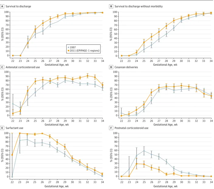
Among infants born alive at 22 through 23 weeks in the 9 re-gions studied in 1997, none survived in 1997 or 2011, and the chance of survival at 24 weeks did not change between the studies (Figure 2A and eTable 2 in the Supplement). Survival increased in these regions by 11.2% (95% CI, −0.5% to 22.9%) at 25 weeks, 18.1% (95% CI, 8.2% to 28.1%) at 26 weeks, 12.8% (95% CI, 4.8% to 20.8%) at 27 weeks, 12.3% (95% CI, 6.1% to 18.6%) at 28 weeks, 7.1% (95% CI, 2.7% to 11.5%) at 29 weeks, 4.7% (95% CI, 1.4% to 8.0%) at 30 weeks, and 2.1% (95% CI, −0.2% to 4.4%) at 31 weeks. Although median age at death did not change at 22 through 24 weeks, it increased significantly at 25 through 26 weeks. Between 1997 through 2011, the rates of antenatal corticosteroid use, indicated preterm deliveries, and surfactant use increased significantly in all gestational-age groups, except at 22 through 23 weeks (Figure 2C-E and eTable 3 in the Supplement).
Survival without neonatal morbidity did not change sig-nificantly at 24 weeks between 1997 (2.4%) and 2011 (7.4%) (Figure 2B). It increased by 16.2% (95% CI, 6.7% to 25.8%) at 25 weeks, 19.0% (95% CI, 9.1% to 28.8%) at 26 weeks, 16.3% (95% CI, 6.4% to 26.2%) at 27 weeks, 17.8% (95% CI, 9.2% to 26.5%) at 28 weeks, 16.6% (95% CI, 8.7% to 24.5%) at 29 weeks,
6.3% (95% CI, 0.7% to 11.8%) at 30 weeks, and 5.9% (95% CI, 1.8% to 10.1%) at 31 weeks. Among survivors at 24 through 26 weeks, the rates of necrotizing enterocolitis (P = .005), BPD (P = .004), cPVL, and severe retinopathy of prematurity de-creased between 1997 and 2011, although not significantly for the cPVL (P = .07) and severe retinopathy of prematurity (P = .11) (eTable 4 in the Supplement). At 27 through 31 weeks, the prevalence of cPVL decreased by 3% (P < .001) and BPD by 4% (P < .001). Only cPVL decreased among infants born at 32 through 34 weeks (P = .03) (eTable 4 in the Supplement).
Discussion
The results of the EPIPAGE-2 study, a national, prospective, population-based cohort study of births at 22 through 34 weeks’ gestation, indicate that survival and survival without severe neonatal morbidity improved significantly between 1997 and 2011 for infants born at 25 through 31 weeks. By contrast, nei-ther survival nor survival without morbidity improved for in-fants born before 25 weeks.
The strengths of the EPIPAGE-2 study include the popu-lation-based cohort design and prospective enrollment of in-fants born prematurely in France in 2011. Standardized defi-nitions of outcomes and systematic and prospective collection of all information available (eg, all cranial ultrasonograms) from a national sample of more than 8000 preterm births (22-34 weeks’ gestation) allowed us to look at the effects associated with a wide range of gestational ages on survival and on ma-jor neonatal morbidities in our population. The accuracy of the gestational age estimates was improved by the very high rate (>98%) of women with early ultrasonogram assessments. Table 1. Vital Status at Birth, Deaths, and Survival by Gestational Age in 2011
Gestational Age, wk No. (%) of Events All Infants (N = 6696) TOPa(n = 214) Stillbirths a (n = 1313) Live Birthsa (n = 5169) Deaths in Maternity Wardb (n = 289) Deaths in NICUb (n = 413) Survival to Dischargeb,c (n = 4467) 22 430 53 (12.3) 319 (74.2) 58 (13.5) 56 (96.6) 2 (3.4) 0 23 414 43 (10.4) 282 (68.1) 89 (21.5) 82 (92.1) 6 (6.7) 1 (1.1) [0-3.3] 24 404 40 (9.9) 178 (44.1) 186 (46.0) 73 (39.2) 55 (29.6) 58 (31.2) [24.5-37.8] 25 435 28 (6.4) 99 (22.8) 308 (70.8) 25 (8.1) 101 (32.8) 182 (59.1) [53.6-64.6] 26 522 24 (4.6) 85 (16.3) 413 (79.1) 18 (4.4) 84 (20.3) 311 (75.3) [71.1-79.5] 22-26 2205 188 (8.5) 963 (43.7) 1054 (47.8) 254 (24.1) 248 (23.5) 552 (52.4) [49.4-55.4] 27 478 11 (2.3) 67 (14.0) 400 (83.7) 9 (2.3) 62 (15.5) 329 (82.3) [78.5-86.0] 28 526 6 (1.1) 63 (12.0) 457 (86.9) 6 (1.3) 40 (8.8) 411 (89.9) [87.2-92.7] 29 561 4 (0.7) 48 (8.6) 509 (90.7) 6 (1.2) 17 (3.3) 486 (95.5) [93.7-97.3] 30 761 5 (0.7) 75 (9.9) 681 (89.5) 2 (0.3) 19 (2.8) 660 (96.9) [95.6-98.2] 31 931 0 69 (7.4) 862 (92.6) 8 (0.9) 18 (2.1) 836 (97.0) [95.8-98.1] 27-31 3257 26 (0.8) 322 (9.9) 2909 (89.3) 31 (1.1) 156 (5.4) 2722 (93.6) [92.7-94.5] 32 281 0 10 (3.6) 271 (96.4) 1 (0.4) 4 (1.5) 266 (98.2) [96.6-99.8] 33 363 0 9 (2.5) 354 (97.5) 1 (0.3) 2 (0.6) 351 (99.2) [98.2-100] 34 590 0 9 (1.5) 581 (98.5) 2 (0.3) 3 (0.5) 576 (99.1) [98.4-99.9] 32-34 1234 0 28 (2.3) 1206 (97.7) 4 (0.3) 9 (0.7) 1193 (98.9) [98.3-99.5]
Abbreviations: NICU, neonatal intensive care unit; TOP, termination of pregnancy for maternal and fetal reasons (other than congenital anomalies). aRelated to all births.
bRelated to live births.
One limitation is that 7% of eligible infants were not in-cluded because of parental refusal. However, the survival sta-tus of all patients, including those who refused to participate, was available. Furthermore, the percentage of survival in these 2 groups did not differ significantly. Therefore, the effect of this selection was very slight.
These 2 EPIPAGE studies13,19made it possible to
deter-mine the changes in mortality and morbidity between 1997 through 2011. We studied neonatal conditions known to be prognostic for long-term outcomes. Although the studies had a common design, more extensive data were collected in 2011 than in 1997. Hence, we may have underestimated changes be-tween the 2 periods for survival without morbidity and mor-bidity rates in general. However, because we restricted our comparisons to severe neonatal conditions, defined similarly in each study, we assume that the influence of this difference was slight.
One important result of our study is that less than 1% of infants born at 22 through 23 weeks survived. In this popula-tion, 80.9% of deaths occurred after a decision to limit
inten-sive care, mostly within the first day of life. The general policy in France is not to intervene before 24 weeks’ gestation; in-fants born earlier receive palliative but not intensive care.20We
compared French results with those of large contemporary in-ternational studies1,2,4,6,7,21conducted in the middle to late
2000s (eTable 5 in the Supplement). The more active perina-tal management at the limit of viability in other countries has resulted in higher survival rates than those in our popu-lation at extremely preterm gestational ages.1,2,4,6,7At 24
weeks, survival remained low in France, reflecting the lack of consensus and heterogeneity of perinatal management for these infants. In this group, as among those born at 22 through 23 weeks, deaths occurred within a day of birth after a decision to limit intensive care. This timing contrasts with the timing of death in those countries that report high rates of perinatal interventions and survival.1,2,6,7Active
perinatal interventions and survival became more frequent in France at 25 weeks, but survival rates remained higher in the United States, Japan, and Sweden up to a gestational age of 27 weeks.5,6
Table 2. Perinatal Characteristics and Obstetric and Neonatal Interventions by Gestational Age in 2011a
Gestational Age, wk Multiple Birthb Birth Weight, Median (IQR), gc Birth in Level III Maternityb Antenatal Corticosteroid Useb Indicated Preterm Deliveryb,d Cesarean Deliveryb Surfactant Usee Postnatal Corticosteroid Usef Length of Hospital Stay, Median (IQR), wke 22 20/58 (34.5) 490 (438-523) 21/58 (36.2) 1/57 (1.8) 8/57 (14.0) 5/57 (8.8) 1/2 (50.0) 0/2 (0) 0 23 31/89 (34.8) 570 (510-620) 55/89 (61.8) 10/81 (12.3) 8/88 (9.1) 4/87 (4.6) 5/7 (71.4) 0/7 (0) 147 24 52/186 (28.0) 680 (618-730) 144/186 (77.4) 101/178 (56.7) 20/182 (11.0) 24/178 (13.5) 108/112 (96.4) 30/109 (27.5) 119 (109-141) 25 121/308 (39.3) 760 (700-830) 258/308 (83.8) 225/298 (75.5) 71/303 (23.4) 103/303 (34.0) 270/278 (97.1) 75/273 (27.5) 104 (90-123) 26 114/413 (27.6) 860 (750-940) 355/413 (86.0) 328/407 (80.6) 153/400 (38.3) 246/411 (59.9) 375/389 (96.4) 78/379 (20.6) 92 (82-105) 22-26 338/1054 (32.1) 750 (633-860) 833/1054 (79.0) 665/1021 (65.1) 260/1030 (25.2) 382/1036 (36.9) 759/788 (96.3) 183/770 (23.8) 98 (87-119) 27 135/400 (33.8) 970 (806-1070) 347/400 (86.8) 315/389 (81.0) 183/382 (47.9) 277/396 (69.9) 347/388 (89.4) 53/373 (14.2) 81 (70-98) 28 142/457 (31.1) 1090 (950-1220) 400/457 (87.5) 386/452 (85.4) 224/446 (50.2) 320/456 (70.2) 364/448 (81.3) 32/432 (7.4) 70 (62-84) 29 149/509 (29.3) 1240 (1050-1370) 449/509 (88.2) 424/503 (84.3) 274/492 (55.7) 356/508 (70.1) 327/501 (65.3) 23/487 (4.7) 59 (51-70) 30 208/681 (30.5) 1370 (1160-1530) 593/681 (87.1) 561/668 (84.0) 376/655 (57.4) 488/678 (72.0) 312/673 (46.4) 12/658 (1.8) 50 (43-60) 31 294/862 (34.1) 1540 (1310-1710) 678/862 (78.7) 713/841 (84.8) 465/827 (56.2) 578/854 (67.7) 324/841 (38.5) 8/830 (1.0) 41 (36-50) 27-31 928/2909 (31.9) 1260 (1040-1500) 2467/2909 (84.8) 2399/2853 (84.1) 1522/2802 (54.3) 2019/2892 (69.8) 1674/2851 (58.7) 128/2780 (4.6) 55 (44-70) 32 125/271 (46.1) 1710 (1520-1939) 162/271 (59.8) 220/264 (83.3) 130/257 (50.6) 177/269 (65.8) 54/264 (20.5) 2/261 (0.8) 34 (28-40) 33 124/354 (35.0) 1920 (1710-2120) 175/354 (49.4) 271/345 (78.6) 163/336 (48.5) 202/354 (57.1) 57/346 (16.5) 0/341 (0) 26 (21-32) 34 197/581 (33.9) 2150 (1920-2370) 267/581 (46.0) 376/569 (66.1) 265/564 (47.0) 280/578 (48.4) 38/561 (6.8) 0/563 (0) 16 (12-22) 32-34 446/1206 (37.0) 1985 (1720-2230) 604/1206 (50.1) 867/1178 (73.6) 558/1157 (48.2) 659/1201 (54.9) 149/1171 (12.7) 2/1165 (0.2) 23 (16-32) Abbreviation: IQR, interquartile range.
a
Data are presented as number of events/number in group (percentage) unless otherwise indicated. Denominators vary according to the number of missing data for each variable.
b
Related to live births.
cBirth weight is missing for 6 infants born at 23 through 26 weeks' gestation
and 2 infants born at 27 through 31 weeks' gestation. d
Indicated preterm delivery: birth after induction of labor or cesarean delivery before the onset of labor.
eEstimated in days among survivors: only for length of hospital stay. f
Related to infants admitted to neonatal intensive care units: only for surfactant use and postnatal corticosteroid use.
International comparisons emphasize that the potential for survival among extremely preterm infants is 10% to 50% higher than our results. They also suggest that active man-agement of extremely preterm infants can improve survival for those born at higher gestational ages. In France, the extension of withholding care to less premature infants, because of fears about immediate and long-term adverse outcomes, might also explain our results at 25 through 27 weeks. However, results of comparisons such as those noted above should be interpreted with caution because differ-ences in gestational age measurement and in the distinction between stillbirths and live births cannot be excluded.22
One way to clarify the role of these issues would be to design multinational cohort studies with standardized methods. In addition, meta-analysis of outcomes using patient-level data might allow better assessment of country-level differences in outcomes.
There is a widespread consensus that the aim of neonatal care should be to resuscitate infants with a reasonable likeli-hood of an acceptable quality of life, but identification of strat-egies for better outcomes remains difficult. Uncertainty about long-term outcomes at the limit of viability influences treat-ment decisions at extremely low gestational ages in France. The results of previous studies6,23-25of trends in short-term
morbidity and longer-term outcomes of infants born at
gesta-tional ages close to this limit make it difficult to predict the ef-fect of a more proactive management of these infants on their survival without morbidity. Hence, consideration of this po-tential effect must examine the possible and problematic na-ture of the trade-off between improved survival and in-creased risk of severe long-term adverse health outcomes for infants born before 25 weeks.
Table 3. Severe Neonatal Morbidity According to Gestational Age Among Survivors to Discharge in 2011a
Gestational Age, wk
No. of Events/No. in Group (%) Grade III
IVH or IPH Cystic PVL Severe BPD Severe ROP Severe NEC
No. of Severe Neonatal Morbidities
0 1 ≥2 23 0/1 (0) 0/1 (0) 1/1 (100.0) 0/1 (0) 1/1 (100.0) 0/1 (0) 0/1 (0) 1/1 (100.0) 24 13/58 (22.4) 1/58 (1.7) 19/51 (37.3) 10/58 (17.2) 3/57 (5.3) 21/51 (41.2) 17/51 (33.3) 13/51 (25.5) 25 26/180 (14.4) 4/182 (2.2) 47/168 (28.0) 17/180 (9.4) 10/181 (5.5) 90/165 (54.5) 57/165 (34.5) 18/165 (10.9) 26 32/310 (10.3) 8/311 (2.6) 64/292 (21.9) 6/308 (1.9) 14/310 (4.5) 188/288 (65.3) 83/288 (28.8) 17/288 (5.9) 23-26 71/549 (12.9) 13/552 (2.4) 131/512 (25.6) 33/547 (6.0) 28/549 (5.1) 299/505 (59.2) 157/505 (31.1) 49/505 (9.7) 27 26/326 (8.0) 9/327 (2.8) 49/311 (15.8) 4/325 (1.2) 17/323 (5.3) 220/306 (71.9) 74/306 (24.2) 12/306 (3.9) 28 16/404 (4.0) 10/406 (2.5) 30/391 (7.7) 1/408 (0.2) 19/402 (4.7) 315/380 (82.9) 58/380 (15.3) 7/380 (1.8) 29 24/477 (5.0) 10/482 (2.1) 16/466 (3.4) 1/482 (0.2) 17/483 (3.5) 399/459 (86.9) 57/459 (12.4) 3/459 (0.7) 30 17/651 (2.6) 10/654 (1.5) 13/644 (2.0) 0/655 (0) 21/651 (3.2) 572/629 (90.9) 56/629 (8.9) 1/629 (0.2) 31 16/819 (2.0) 9/823 (1.1) 12/821 (1.5) 1/830 (0.1) 19/831 (2.3) 748/800 (93.5) 48/800 (6.0) 4/800 (0.5) 27-31 99/2677 (3.7) 48/2692 (1.8) 120/2633 (4.6) 7/2700 (0.3) 93/2690 (3.5) 2254/2574 (87.6) 293/2574 (11.4) 27/2574 (1.0) 32 2/251 (0.8) 3/253 (1.2) 0/261 (0) 0/261 (0) 2/260 (0.8) 236/242 (97.5) 6/242 (2.5) 0/242 (0) 33 1/350 (0.3) 4/350 (1.1) 0/342 (0) 0/345 (0) 6/339 (1.8) 317/328 (96.6) 11/328 (3.4) 0/328 (0) 34 4/574 (0.7) 2/574 (0.3) 0/560 (0) 0/564 (0) 2/544 (0.4) 527/533 (98.9) 5/533 (0.9) 1/533 (0.2) 32-34 7/1175 (0.6) 9/1177 (0.8) 0/1163 (0) 0/1170 (0) 10/1143 (0.9) 1080/1103 (97.9) 22/1103 (2.0) 1/1103 (0.1) Abbreviations: BPD, bronchopulmonary dysplasia; IPH, intraparenchymal
hemorrhage; IVH, intraventricular hemorrhage; NEC, necrotizing enterocolitis; PVL, periventricular leukomalacia; ROP, retinopathy of prematurity.
aDenominators vary according to the number of missing data for each variable.
Figure 1. Survival Without Severe Neonatal Morbidity in 2011
100 80 60 40 20 90 70 50 30 10 0 22 23 24 25 26 27 28 29 30 31 33 34 Sur viv al , % (95% CI) Gestational Age, wk 32 Related to survivors Related to live births Related to all births
For each week of gestation, percentages and exact 95% binomial CIs (error bars) are presented.
Results of our trend study during a 15-year period (1997-2011) reveal that survival without morbidity increased by 14.1% for infants born at 25 through 29 weeks. This finding indicates that 1 of every 7 infants had a more favorable out-come in 2011 compared with 1997. Hence, the total number of children surviving without short-term and perhaps also long-term severe adverse outcomes has increased over time.
Conclusions
Few other population-based studies from around the world provide up-to-date estimates of short-term prognosis of extremely, very, and moderately preterm infants and of changes during the past decade. International comparisons
help to estimate the potential for survival and to identify appropriate interventions; they thus reveal areas for improvement in each country. In particular, they reveal that improvement in survival at extremely low gestational age is possible in France and in countries with similar practices. This finding should encourage health care professionals to reassess their attitudes toward care at extremely low gesta-tional ages. This reassessment should include a complete analysis of neonatal morbidity and long-term sequelae, which have not yet been sufficiently evaluated, although they remain important fac tors in dec ision making. EPIPAGE-2 should provide further information on them as the children it studies age. Finally, specificities in the organization of care, health policies, laws, and available resources of each country must also be part of this dis-cussion.
Figure 2. Comparison of Survival Rates and Obstetric and Neonatal Interventions in 1997 and 2011
100 80 60 40 20 90 70 50 30 10 0 100 80 60 40 20 90 70 50 30 10 0 100 80 60 40 20 90 70 50 30 10 0 100 80 60 40 20 90 70 50 30 10 0 100 80 60 40 20 90 70 50 30 10 0 100 80 60 40 20 90 70 50 30 10 0 22 23 24 25 26 27 28 29 30 31 33 34 % (95% CI) Gestational Age, wk 32 1997 2011 (EPIPAGE-1 regions) 22 23 24 25 26 27 28 29 30 31 33 34 % (95% CI) Gestational Age, wk 32 22 23 24 25 26 27 28 29 30 31 33 34 % (95% CI) Gestational Age, wk 32 22 23 24 25 26 27 28 29 30 31 33 34 % (95% CI) Gestational Age, wk 32 22 23 24 25 26 27 28 29 30 31 33 34 % (95% CI) Gestational Age, wk 32 22 23 24 25 26 27 28 29 30 31 33 34 % (95% CI) Gestational Age, wk 32 Survival to discharge
A B Survival to discharge without morbidity
Antenatal corticosteroid use
C D Cesarean deliveries
Surfactant use
E F Postnatal corticosteroid use
ARTICLE INFORMATION
Accepted for Publication: November 18, 2014. Published Online: January 26, 2015.
doi:10.1001/jamapediatrics.2014.3351.
The EPIPAGE-2 Writing Group includes Pierre
Kuhn, PhD; Bruno Langer, MD; Jacqueline Matis, MD; Xavier Hernandorena, MD; Pierre Chabanier; Laurence Joly-Pedespan, MD; Bénédicte Lecomte, MD; Françoise Vendittelli, PhD; Michel Dreyfus, MD; Bernard Guillois, MD; Antoine Burguet, PhD; Pierre Sagot, MD; Jacques Sizun, MD; Alain Beuchée, MD; Florence Rouget, MD; Amélie Favreau, MD; Elie Saliba, PhD; Nathalie Bednarek, PhD; Patrice Morville, MD; Gérard Thiriez, PhD; Loïc Marpeau, MD; Stéphane Marret, PhD; Gilles Kayem, PhD; Xavier Durrmeyer, MD; Michèle Granier, MD; Olivier Baud, PhD; Pierre-Henri Jarreau, PhD; Delphine Mitanchez, PhD; Pascal Boileau, PhD; Pierre Boulot, MD; Gilles Cambonie, PhD; Hubert Daudé, MD; Antoine Bédu, PhD; Fabienne Mons, PhD; Jeanne Fresson, PhD; Rachel Vieux, PhD; Corine Alberge, MD; Catherine Arnaud, PhD; Christophe Vayssière, MD; Patrick Truffert, PhD; Véronique Pierrat, PhD; Damien Subtil, PhD; Claude D’Ercole, MD; Catherine Gire, MD; Umberto Simeoni, MD; André Bongain, PhD; Loïc Sentilhes, PhD; Jean-Christophe Rozé, PhD; Jean Gondry, MD; André Leke, PhD; Michel Deiber, MD; Olivier Claris, PhD; Jean-Charles Picaud, PhD; Anne Ego, PhD; Thierry Debillon, PhD; Anne Poulichet, MD; Eliane Coliné, MD; Anne Favre, MD; Olivier Fléchelles, MSc; Sylvain Samperiz, MD; Duksha Ramful, MD; Bernard Branger; Valérie Benhammou, PhD; Laurence Foix-L’Hélias, PhD; Laetitia Marchand-Martin, MSc; Monique Kaminski, MSc.
Affiliations of The EPIPAGE-2 Writing Group:
University Hospital, Strasbourg, France (Kuhn, Langer, Matis); La Côte Basque Hospital, Bayonne, France (Hernandorena); University Hospital, Bordeaux, France (Chabanier, Joly-Pedespan); University Hospital Estaing, Clermont-Ferrand, France (Lecomte, Vendittelli); Department of Gynecology and Obstetrics, University Hospital, Caen, France (Dreyfus); Department of Neonatal Pediatrics and Intensive Care, University Hospital, Caen, France (Guillois); Department of Neonatal Pediatrics, University Hospital, Dijon, France (Burguet); Department of Gynecology and Obstetrics, University Hospital, Dijon, France (Sagot); University Hospital, Brest, France (Sizun); Department of Pediatrics, University Hospital, Inserm-Irset U 1085, Rennes, France (Beuchée, Rouget); Department of Neonatal Pediatrics and Intensive Care, University Hospital, Tours, France (Favreau); INSERM U 930, François Rabelais University, Tours, France (Saliba); Department of Neonatal Pediatrics, University Hospital, Reims, France (Bednarek, Morville); Department of Neonatal Pediatrics, University Hospital, Besançon, France (Thiriez); Department of Gynecology and Obstetrics, University Hospital, Rouen, France (Marpeau); Department of Neonatal Pediatrics and Intensive Care, Rouen University Hospital-Laboratory of microvascular endothelium and neonatal brain lesions, Rouen, France (Marret); Department of Obstetrics and Gynecology, Louis Mourier Hospital, University Hospitals Paris Nord Val de Seine (HUPNVS)), Assistance Publique-Paris Hospitals (APHP), Paris Diderot University, Paris, France (Kayem); Department of Neonatal Pediatrics
and Intensive Care, CHI, CRC, Créteil, France (Durrmeyer); Department of Neonatal Pediatrics, Sud Francilien Hospital, Evry, France (Granier); Neonatal intensive care unit, Robert Debré Hospital, INSERM, UMR 676, Paris, France (Baud); Department of Neonatal Pediatrics and Intensive Care, Cochin Hotel Dieu Hospital, Paris, France (Jarreau); Department of Neonatal Pediatrics, Trousseau Hospital, Paris, France (Mitanchez); Department of Neonatal Pediatrics, Poissy Saint Germain University Hospital, Poissy, France (Boileau); Department of Obstetrics and Gynecology, Arnaud de Villeneuve Hospital, Montpellier, France (Boulot); Department of Neonatal Pediatrics and Intensive Care, Arnaud de Villeneuve Hospital, Montpellier, France (Cambonie); CAMSP, University Hospital, Montpellier, France (Daudé); Department of Neonatal Pediatrics, Mère-Enfant Hospital, Limoges, France (Bédu, Mons); Department of Medical Information, Adolphe Pinard Maternity Unit, Nancy, France (Fresson); Department of Neonatal Pediatrics and Intensive Care, Adolphe Pinard Maternity Unit, Nancy, France (Vieux); UMR 1027 INSERM, Paul-Sabatier Toulouse III University, Toulouse, France (Alberge, Arnaud); Department of Obstetrics and Gynecology, Toulouse, France (Vayssière); Department of Neonatal Pediatrics, Jeanne de Flandres Hospital, Lille, France (Truffert, Pierrat); Department of Gynecology and Obstetrics, Jeanne de Flandre Hospital, Lille, France (Subtil); Department of Gynecology and Obstetrics, Nord Hospital, Marseille, France (D’Ercole); Department of Neonatal Pediatrics and Intensive Care, Nord Hospital, Marseille, France (Gire); Department of Neonatal Pediatrics and Intensive Care, La Conception Hospital, Marseille, France (Simeoni); Department of Gynecology and Obstetrics, Archet Hospital, Nice, France (Bongain); Department of Obstetrics and Gynecology, Angers University Hospital, Angers, France (Sentilhes); Department of Neonatal Medicine, Angers University Hospital and INSERM CIC 004, Nantes, France (Rozé); Department of Obstetrics and gynecology, Amiens, France (Gondry); Department of Pediatrics, Amiens, France (Leke); Department of Pediatrics, Chambéry, France (Deiber); Department of Neonatal Pediatrics and Intensive Care, University Hospital, Lyon, France (Claris); Department of Neonatal Pediatrics and Intensive Care, La Croix Rousse Hospital, Lyon, France (Picaud); INSERM CIC003, University Hospital, Grenoble, France (Ego); Department of Neonatal Pediatrics, University Hospital, Grenoble, France (Debillon); University Hospital, Pointe à Pitre, Guadeloupe, France (Poulichet, Coliné); Department of Neonatal Pediatrics and Intensive Care, Cayenne Hospital, Cayenne, Guyane, France (Favre); University Hospital, Fort de France, Martinique (Fléchelles); Department of Neonatal Pediatrics and Intensive Care, University Hospital Felix Guyon, Saint-Denis, La Réunion, France (Samperiz, Ramful); Fédération des Réseaux de Santé en Périnatalité [FFRSP], Nantes, France (Branger); Inserm UMR1153, Perinatal and Pediatric Epidemiology Team, Paris, France (Benhammou, Foix-L’Hélias, Marchand-Martin, Kaminski).
Author Contributions: Dr Ancel had full access to
all the data in the study and takes responsibility for the integrity of the data and the accuracy of the data analysis.
Study concept and design: All authors. Acquisition, analysis, or interpretation of data: All authors.
Drafting of the manuscript: All authors. Critical revision of the manuscript for important intellectual content: All authors.
Statistical analysis: All authors. Obtained funding: Ancel. Study supervision: All authors.
Conflict of Interest Disclosures: None reported. Funding/Support: This study was supported by the
French Institute of Public Health Research/Institute of Public Health and its partners the French Health Ministry, the National Institute of Health and Medical Research, the National Institute of Cancer, and the National Solidarity Fund for Autonomy; grant ANR-11-EQPX-0038 from the National Research Agency through the French Equipex Program of Investments in the Future; and the PremUp Foundation.
Role of the Funder/Sponsor: The funding source
had no role in the design and conduct of the study; collection, management, analysis, and
interpretation of the data; preparation, review, or approval of the manuscript; and the decision to submit the manuscript for publication.
Additional Contributions: We are grateful for the
participation of all families of preterm infants in the EPIPAGE-2 cohort study and for the cooperation of all maternity and neonatal units in France.
The EPIPAGE-2 Study Group Collaborators
include Alsace: D. Astruc, P. Kuhn, B. Langer, J. Matis (Strasbourg), C. Ramousset; Aquitaine: X. Hernandorena (Bayonne), P. Chabanier, L. Joly-Pedespan (Bordeaux), M. J. Costedoat, A. Leguen; Auvergne: B. Lecomte, D. Lemery, F. Vendittelli (Clermont-Ferrand); Basse-Normandie: G. Beucher, M. Dreyfus, B. Guillois (Caen), Y. Toure; Bourgogne: A. Burguet, S. Couvreur, J. B. Gouyon, P. Sagot (Dijon), N. Colas; Bretagne: J. Sizun (Brest), A. Beuchée, P. Pladys, F. Rouget (Rennes), R. P. Dupuy (St-Brieuc), D. Soupre (Vannes), F. Charlot, S. Roudaut; Centre: A. Favreau, E. Saliba (Tours), S. Leclercq; Champagne-Ardenne: N. Bednarek, P. Morville (Reims), M. Palot; Franche-Comté: G. Thiriez (Besançon), C. Balamou; Haute-Normandie: L. Marpeau, S. Marret (Rouen), C. Barbier RM; Ile-de-France: G. Kayem (Colombes), X. Durrmeyer (Créteil), M. Granier (Evry), M. Ayoubi, A. Baud, B. Carbonne, L. Foix L’Hélias, F. Goffinet, P. H. Jarreau, D. Mitanchez (Paris), P. Boileau (Poissy), C. Duffaut, E. Lorthe; Languedoc-Roussillon: P. Boulot, G. Cambonie, H. Daudé (Montpellier), A. Badessi, N. Tsaoussis; Limousin: A. Bédu, F. Mons (Limoges), C. Bahans; Lorraine: M. H. Binet, J. Fresson, J. M. Hascoët, A. Milton, O. Morel, R. Vieux (Nancy), L. Hilpert; Midi-Pyrénées: C. Alberge, C. Arnaud, C. Vayssière (Toulouse), M. Baron; Nord-Pas-de-Calais: M. L. Charkaluk, V. Pierrat, D. Subtil, P. Truffert (Lille), C. Delaeter; PACA et Corse: C. D’Ercole, C. Gire, U. Simeoni (Marseille), A. Bongain (Nice), M. Deschamps, C. Grangier; Pays de Loire: B. Branger (FFRSP), J. C. Rozé, N. Winer (Nantes), V. Rouger, C. Dupont; Picardie: J. Gondry, G. Krim (Amiens), B. Baby; Rhône-Alpes: M. Debeir (Chambéry), O. Claris, J. C. Picaud, S. Rubio-Gurung (Lyon), C. Cans, A. Ego, T. Debillon (Grenoble), H. Patural (Saint-Etienne), A. Rannaud; Guadeloupe: E. Janky, A. Poulichet, J. M. Rosenthal (Point à Pitre), E. Coliné; Guyane: A. Favre (Cayenne), N. Joly; Martinique: S.
Châlons (Fort de France), V. Lochelongue; La Réunion: P. Y. Robillard (Saint-Pierre), S. Samperiz, D. Ramful (Saint-Denis); Inserm UMR S953: P. Y. Ancel, V. Benhammou, B. Blondel, M. Bonet, A. Brinis, M. L. Charkaluk, M. Durox, L. Foix-L’Hélias, F. Goffinet, M. Kaminski, G. Kayem, B. Khoshnood, C. Lebeaux, L. Marchand-Martin, V. Pierrat, M. J. Saurel-Cubizolles, D. Tran, L. Vasante-Annamale, J. Zeitlin.
Correction: This article was corrected on March 9,
2015, to fix errors in Group Information and Figure 2.
REFERENCES
1. Fellman V, Hellström-Westas L, Norman M, et al;
EXPRESS Group. One-year survival of extremely preterm infants after active perinatal care in Sweden. JAMA. 2009;301(21):2225-2233.
2. Costeloe KL, Hennessy EM, Haider S, Stacey F,
Marlow N, Draper ES. Short term outcomes after extreme preterm birth in England: comparison of two birth cohorts in 1995 and 2006 (the EPICure studies). BMJ. 2012;345:e7976.
3. Field DJ, Dorling JS, Manktelow BN, Draper ES.
Survival of extremely premature babies in a geographically defined population: prospective cohort study of 1994-9 compared with 2000-5. BMJ. 2008;336(7655):1221-1223.
4. Stoll BJ, Hansen NI, Bell EF, et al; Eunice
Kennedy Shriver National Institute of Child Health and Human Development Neonatal Research Network. Neonatal outcomes of extremely preterm infants from the NICHD Neonatal Research Network. Pediatrics. 2010;126(3):443-456.
5. Bode MM, D’Eugenio DB, Forsyth N, Coleman J,
Gross CR, Gross SJ. Outcome of extreme prematurity: a prospective comparison of 2 regional cohorts born 20 years apart. Pediatrics. 2009;124 (3):866-874.
6. Doyle LW, Roberts G, Anderson PJ; Victorian
Infant Collaborative Study Group. Outcomes at age 2 years of infants < 28 weeks’ gestational age born in Victoria in 2005. J Pediatr. 2010;156(1):49-53.e1.
7. Itabashi K, Horiuchi T, Kusuda S, et al. Mortality
rates for extremely low birth weight infants born in Japan in 2005. Pediatrics. 2009;123(2):445-450.
8. Marlow N, Wolke D, Bracewell MA, Samara M;
EPICure Study Group. Neurologic and developmental disability at six years of age after extremely preterm birth. N Engl J Med. 2005;352 (1):9-19.
9. Herber-Jonat S, Schulze A, Kribs A, Roth B,
Lindner W, Pohlandt F. Survival and major neonatal complications in infants born between 22 0/7 and 24 6/7 weeks of gestation (1999-2003). Am J Obstet Gynecol. 2006;195(1):16-22.
10. De Groote I, Vanhaesebrouck P, Bruneel E, et al;
Extremely Preterm Infants in Belgium (EPIBEL) Study Group. Outcome at 3 years of age in a population-based cohort of extremely preterm infants. Obstet Gynecol. 2007;110(4):855-864.
11. Blencowe H, Cousens S, Oestergaard MZ, et al.
National, regional, and worldwide estimates of preterm birth rates in the year 2010 with time trends since 1990 for selected countries: a systematic analysis and implications. Lancet. 2012;379(9832):2162-2172.
12. Saigal S, Doyle LW. An overview of mortality
and sequelae of preterm birth from infancy to adulthood. Lancet. 2008;371(9608):261-269.
13. Larroque B, Ancel PY, Marret S, et al; EPIPAGE
Study group. Neurodevelopmental disabilities and special care of 5-year-old children born before 33 weeks of gestation (the EPIPAGE study): a longitudinal cohort study. Lancet. 2008;371 (9615):813-820.
14. Ancel PY, Goffinet F; EPIPAGE 2 Writing Group.
EPIPAGE 2: a preterm birth cohort in France in 2011. BMC Pediatr. 2014;14:97.
15. Volpe JJ. Brain injury in premature infants:
a complex amalgam of destructive and
developmental disturbances. Lancet Neurol. 2009; 8(1):110-124.
16. Bell MJ, Ternberg JL, Feigin RD, et al. Neonatal
necrotizing enterocolitis: therapeutic decisions based upon clinical staging. Ann Surg. 1978;187(1):1-7.
17. International Committee for the Classification
of Retinopathy of Prematurity (ICCROP). The
International Classification of Retinopathy of Prematurity revisited. Arch Ophtalmol (Paris). 2005;123(7):991-999.
18. Jobe AH, Bancalari E. Bronchopulmonary
dysplasia. Am J Respir Crit Care Med. 2001;163(7): 1723-1729.
19. Larroque B, Bréart G, Kaminski M, et al; Epipage
Study group. Survival of very preterm infants: Epipage, a population based cohort study. Arch Dis Child Fetal Neonatal Ed. 2004;89(2):F139-F144. 20. Moriette G, Rameix S, Azria E, et al; Groupe de
réflexion sur les aspects éthiques de la
périnatologie. Very premature births: dilemmas and management: second part: ethical aspects and recommendations [in French]. Arch Pediatr. 2010; 17(5):527-539.
21. de Waal CG, Weisglas-Kuperus N, van
Goudoever JB, Walther FJ; NeoNed Study Group; LNF Study Group. Mortality, neonatal morbidity and two year follow-up of extremely preterm infants born in The Netherlands in 2007. PLoS One. 2012;7 (7):e41302.
22. Joseph KS, Liu S, Rouleau J, et al; Fetal and
Infant Health Study Group of the Canadian Perinatal Surveillance System. Influence of definition based versus pragmatic birth registration on international comparisons of perinatal and infant mortality: population based retrospective study. BMJ. 2012; 344:e746.
23. Serenius F, Källén K, Blennow M, et al; EXPRESS
Group. Neurodevelopmental outcome in extremely preterm infants at 2.5 years after active perinatal care in Sweden. JAMA. 2013;309(17):1810-1820.
24. Moore T, Hennessy EM, Myles J, et al.
Neurological and developmental outcome in extremely preterm children born in England in 1995 and 2006: the EPICure studies. BMJ. 2012;345: e7961.
25. Hintz SR, Kendrick DE, Wilson-Costello DE,
et al; NICHD Neonatal Research Network. Early-childhood neurodevelopmental outcomes are not improving for infants born at <25 weeks’ gestational age. Pediatrics. 2011;127(1):62-70.