Advance Access publication 19 July 2013
An investigation into the mechanical and aesthetic properties of
new generation coated nickel-titanium wires in the as-received
state and after clinical use
T. Gerard Bradley
*
, David W. Berzins
*
, Nicholas Valeri
*
, Jessica Pruszynski
**
,
Theodore Eliades
***
and Christos Katsaros
****
*Department of Developmental Sciences, Marquette University School of Dentistry, Milwaukee, WI, USA, **Division of Biostatistics, Medical College of Wisconsin, Milwaukee, USA, ***Department of Orthodontics and Paediatric Dentistry, Center of Dental Medicine, University of Zurich, Switzerland, ****Department of Orthodontics and Dentofacial Orthopedics, Medical School, University of Bern, Switzerland
Correspondence to: T. Gerard Bradley, Marquette University School of Dentistry, 1801 W. Wisconsin Ave, Milwaukee, WI 53233, USA. E-mail: [email protected]
SUMMAry
BACKGrOUND/OBJECTIVES: The purpose of this study was to compare the mechanical, structural, and
aes-thetic properties of two types of aesaes-thetic coated nickel-titanium (NiTi) wires compared with comparable regular NiTi wires in the as-received state and after clinical use.
MATErIAlS/METhODS: Sixty one subjects were randomly assigned to four groups (N = 61), two groups of
coated wires and two groups of comparable, non-coated controls (n = 15/group). The period in the mouth ranged from 4 to 12 weeks after insertion. In total, 121 wires (61 retrieved and 60 as-received) were used in the study. The percentages of coating retention and loss were extrapolated from scans. A brief survey of five questions with three choices was given to all patients. Differential scanning calorimetry (DSC) and three-point bending tests were done on as-received and used wires.
rESUlTS: The surface characterization by the percentage of resin remaining indicated that most wires in
both test groups lost a significant amount of coating. A patient survey indicated that this was a noticeable feature for patients. DSC analysis of the wires indicated that the metallurgical properties of the coated wires were not similar to the uncoated wires in the as-received condition. Three-point bending results indicate a wide variation in test results with large standard deviations among all the groups.
lIMITATIONS: The extent of coating loss requires investigating, as do the biological properties of the
detached coating.
CONClUSIONS: Both wires lost a significant amount of aesthetic coating after varying periods in the mouth.
The metallurgical testing of these findings may indicate that these wires perform differently in the mouth.
Introduction
The orthodontic profession is constantly seeking to improve and optimize the aesthetics of orthodontic wires since the introduction of aesthetic brackets. Nickel-titanium (NiTi) wires since their introduction to orthodontics (Andreasen and Hilleman, 1971) have been extensively researched
in vitro and are used as an initial levelling and aligning
archwire because of its properties of springback and superelasticity (Burstone et al., 1985; Miura et al., 1986;
Leu et al., 1990; Bishara et al., 1995; Bradley et al., 1996; Biermann et al., 2007; Berzins and Roberts 2010). Aesthetic wires are usually either coated NiTi wires or composite wires of reinforced polymers. Shape memory polymers have wide application in space technology and are being used currently in medicine and industrial applications (Jung and Cho, 2010; Hu et al., 2012). These wires have enormous potential for clinical application in orthodontics,
and polyphenylene, a self-reinforced polymer composite, is close to being introduced to orthodontic practice (Burstone
et al., 2011; Goldberg et al., 2011). However, these wires are still at the experimental stage. A fibre reinforced polymer is in clinical use (BioMers Products, LLC, Jacksonville, FL, USA) that is manufactured using a pultrusion process with a photo-cured resin (Gopal et al., 2005); however, these wires may be more likely to crack during bending and have been shown to deliver less consistent forces compared with alloy wires (Chang, 2012). The coated wires, which are currently available, either have an epoxy resin, polytetraflouroethylene (Teflon; Neumann et al., 2002), or a low reflectivity rhodium coating (Iijima et al., 2012) applied to the surface. Atomized Teflon particles are used to coat the wire using clean compressed air as a transport medium, which is then further heat treated in a chamber furnace (Husmann et al., 2002). The rhodium coating is applied
by using a plasma-immersion ion implantation technique (Sridharan et al., 2004). The coated wires are found to be routinely damaged from mastication and activation of enzymes (Kusy, 1997). These wires have been shown to deliver lower forces in loading and unloading (Elayyan
et al., 2010; Alavi and Hosseini, 2012; Iijima et al., 2012;
Kaphoor and Sundareswaran, 2012). Poor colour stability has also been reported (Silva et al., 2013) and up to 25 per cent of the coating lost after 33 days in vivo (Elayyan et al., 2008). The coating itself, the process of its application and the fact that the NiTi component of the wire may be smaller to accommodate the thickness of the coating (Kaphoor and Sundareswaran, 2012), may account for these altered properties. The studies mentioned above concentrated on
in vitro testing and comparison of these wires with the
uncoated version. Only one previous study has investigated coated wires after clinical use (Elayyan et al., 2008). Therefore, the purpose of this study was to investigate key characteristics of four different types of Food and Drug Administration approved and commercially available wires in the as-received condition and after clinical use in the mouth.
Materials and methods
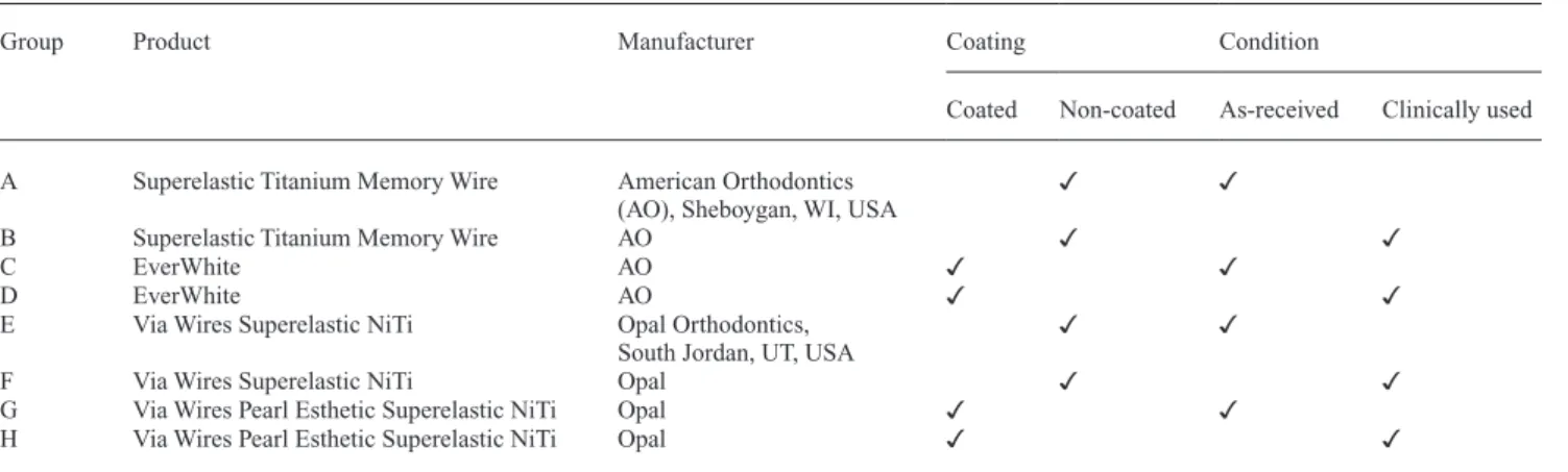
Following approval from the Institutional Review Board at Marquette University HR-2347, 61 subjects with written informed consent were randomly allocated to four groups of 0.016 × 0.022 inches NiTi wires when that wire type was indicated in treatment, two of which were coated and two were non-coated controls of the same wire (manufacturer personal communication; Table 1). Group H had 16 subjects. American 022 Mini Masters Low Profile MBT (American Orthodontics, Sheboygan, WI, USA) metal brackets were selected for the study. The inclusion criteria included a full complement of teeth with an age range between 9 and 20 years. Wires were only placed in the maxillary arch
only with conventional elastic ligation. In total, 32 females and 29 males were included in the study. The wires were placed in the patients’ mouth and left for a period from 4 to 12 weeks to accomplish the clinical goals of the wire. After wire retrieval, the wires were washed under running water, wiped with gauze soaked with Birex (Biotrol, Earth City, MO, USA), and placed in a plastic bag with a number and letter identifier. A survey, described below, was given to the patient. The wires were then analysed in the following manner.
Coating retention
The percentages of coating retention and loss were extrapolated from scans (Epson Expression 1680, model G780B, Nagano, Japan) of the wire. The clinically retrieved aesthetic wires were scanned (n = 31) at 1200 dpi (4 × 4 inches), 24-bit depth, and saved in TIF format (Supplementary Figure 1). A light green background was used to provide contrast between the white wire coating, silver wire, and black shadow. The TIF images were processed through Matlab (R2011b, The Mathworks, Inc., Natick, MA, USA) with imread to import the image and
csvwrite to save the image with three numerical values for
each pixel—red, green, and blue (RGB). A local application was developed using Delphi (2010, Embarcadero, San Francisco, CA, USA) to evaluate and tabulate the RGB value for each pixel. Approximate RGB values for wire, coating, and background were established by selecting a point in the image and inspecting the respective RGB values for that point. An evaluation percentage was calculated with the percent of wire divided the sum of percentages of wire and coating. For the final results, an unused wire was processed as a control for each group. Then, the percentage of wire, coating, and background and the evaluation percentage were computed. The independent
t-test was used to compare the percentage in the D group to
Table 1 Wire groups. NiTi, nickel-titanium.
Group Product Manufacturer Coating Condition
Coated Non-coated As-received Clinically used A Superelastic Titanium Memory Wire American Orthodontics
(AO), Sheboygan, WI, USA ✓ ✓
B Superelastic Titanium Memory Wire AO ✓ ✓
C EverWhite AO ✓ ✓
D EverWhite AO ✓ ✓
E Via Wires Superelastic NiTi Opal Orthodontics,
South Jordan, UT, USA ✓ ✓
F Via Wires Superelastic NiTi Opal ✓ ✓
G Via Wires Pearl Esthetic Superelastic NiTi Opal ✓ ✓
H Via Wires Pearl Esthetic Superelastic NiTi Opal ✓ ✓
the percentage in the H group. The control values were not included in the analysis.
Survey
A brief survey of five questions with three choices was given to all patients in the study (Figure 1). These data were analysed to determine the patient’s perspective on the aesthetics and surface texture of the wire. The survey was completed by the patient at the time of the wire retrieval. Due to the low cell counts, the chi-square test was invalid and the Fisher’s exact test was used instead for statistical analysis.
Differential scanning calorimetry
In conventional differential scanning calorimetry (DSC; Model 822e, Mettler-Toledo Inc., Columbus, OH, USA), two small crucibles, one empty and the other with the mate-rial to be tested, are heated at the same rate and the differ-ences in thermal energy to the crucibles to maintain equal heating are plotted as a function of temperature over the scanning range to yield the DSC thermogram. Seven wires from each group (n = 56) were evaluated whereby 5 mm sec-tions from the midline area of the wire were cut using a low-speed water-cooled diamond saw (Isomet, Buehler Ltd, Lake Bluff, IL, USA) with care to avoid mechanical stress and heating that could alter the structure of the wire. The wire section was weighed to the nearest 0.01 mg, placed in an alu-minium crucible, and sealed. The temperature of the cruci-bles was scanned from −100 to +100°C with liquid nitrogen as a coolant and nitrogen gas for purging, at 10°C per minute for heating and cooling. With associated software, the areas of the peaks were analysed and any changes in transitions, crystallization, or structural transformation identified. The
DSC plots were qualitatively and quantitatively evaluated using the manufacturer’s software. A one-way analysis of variance followed by Tukey’s honestly significant differ-ence (HSD) test, if indicated, was performed on the thermal property measurements. Statistical models were run only for relevant comparisons (i.e. as-received versus clinically used wires of a given brand and comparison of coated versus non-coated).
Three-point bend test
A 20 mm segment from the straight portion of the arch form was sectioned from each wire (n = 121). Mechanical testing was carried out based on American National Standard/American Dental Association Specification No. 32 for Orthodontic Wires. A three-point bend test on a uni-versal testing machine (Model 5500R, Instron, Norwood, MA, USA) at 37 ± 2°C using a deflection span of 14 mm and a crosshead speed of 2 mm/min. Activation–deactiva-tion curves of each specimen were obtained during deflec-tion to 3.1 mm. Stiffness, flexural modulus, and force values at 1, 2, and 3 mm were determined from each load-ing and unloadload-ing curve. A one-way analysis of variance followed by Tukey’s HSD test, if indicated, was performed on the activation modulus, stiffness, deactivation modu-lus, and stiffness, and force values at 1, 2, and 3 mm. As with the DSC data, statistical models were run only for rel-evant comparisons (i.e. as-received versus clinically used wires of a given brand and comparison of coated versus non-coated).
Results
Coating retention
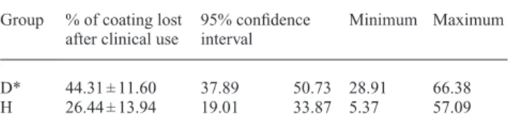
The D group (American Orthodontics EverWhite) had a mean percentage loss of 44.31 per cent with a standard deviation of 11.60 per cent (Table 2). The H group (Opal Via Pearl) had a mean percentage loss of 26.44 per cent with a standard deviation of 13.94 per cent. Using the
t-test, a test statistic of 3.87 (P < 0.0001) was calculated
indicating a significant difference between the D and H groups.
PARTICIPANT SURVEY
1. The wire color was very close to the color of my teeth and I was very pleased with its appearance.
Very pleased Pleased Disappointing
2. The wire color changed from the time it was placed in my mouth to the time of its removal. It became
Brighter Did not change Darker
3. While brushing my teeth the wire as compared to the bracket or pad was. Easier to keep clean The same Harder to keep clean 4. The wire had a smooth texture when it was initially placed.
Agree Not sure Disagree
5. The wire surface texture (smoothness or cracks) did not change during treatment and looked the same at the time of wire removal.
Agree Not sure Disagree Figure 1 Patient survey.
Table 2 The percentage of the coating (mean values and
standard deviations) lost after retrieval from the mouth.
Group % of coating lost
after clinical use 95% confidence interval Minimum Maximum D* 44.31 ± 11.60 37.89 50.73 28.91 66.38 H 26.44 ± 13.94 19.01 33.87 5.37 57.09 *EverWhite (D) lost significantly (P < 0.0001) more coating than Via Pearl (H).
Survey
All patients with wires placed were given the survey to complete at the time of the wire removal (Figure 1). For all questions, the hypothesis of an association between the question response and the group (B, D, F, and H) was tested. This would determine whether the group influenced the patient’s response to the question. Due to the low cell counts, the chi-square test was invalid and the Fisher’s exact test was used instead. Two of the five questions showed significance in answers to the groups. These two questions were on the colour aesthetic change and the texture of the wire at the time of wire removal (Supplementary Table 1). In question 2, about a third of the patients in both aesthetic groups felt that the wire had darkened in appearance during the period it was in the mouth. In question 5, nearly a half of the patients in groups D and H felt that the wire had not changed in its texture and looked the same at the time of the wire removal.
Differential scanning calorimetry
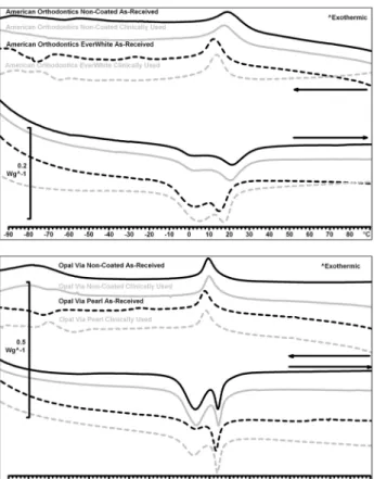
Statistical significance was found in many thermal proper-ties between groups A and C, the American Orthodontics NiTi non-coated wire and its coated equivalent wire in the as-received state, as well as between groups E and G, the Via wire and its coated equivalent in the as-received state. The American Orthodontics EverWhite wire had a lower austenitic finish temperature (20.6°C; Table 3 and Figure 2) compared with the non-coated American Orthodontics ver-sion (29.8°C). The difference in austenitic finish tempera-tures in the Opal Via wires are less drastic (Table 3). The aesthetic Pearl wire had an Af of 15.4°C compared with 18.2°C for the non-coated wire (Figure 2). With regard to whether clinical use altered the thermal properties of the wires, no statistically significant (P > 0.05) differences were observed between as-received and clinically used wires of the same product (Table 3).
Three-point bend test
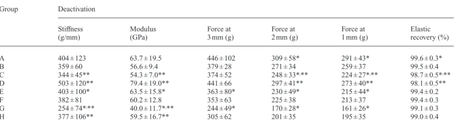
Tables 4 and 5 display the bending values during activation and deactivation, respectively, while Figure 3 shows typical bending curves for all groups. Statistically significant differ-ences in all activation and deactivation stiffnesses and forces were found between group E and G, the Opal Via Wire non-coated wire and its non-coated equivalent Opal Via Pearl wire in the as-received state. The as-received coated and non-coated American Orthodontics wires differed in deactiva-tion force at 1 and 2 mm as well as elastic recovery. None of the uncoated NiTi wires displayed differences in bending values, but the coated types showed statistically significant differences in the as-received state and after clinical use in a majority of stiffness and force parameters. Specifically, both EverWhite and Via Pearl displayed greater activation and deactivation stiffness after clinical use compared with the as-received condition. Similarly, EverWhite’s activation and deactivation force values were greater in the clinically used wire for five of the six values, whereas that was the case for two values for Via Pearl.
Discussion
Both wire types lost a significant amount of coating after use in the areas of archwire engagement, but the Opal Via Pearl wires showed better retention of the coating with a 26.44 per cent loss versus 44.31 per cent for American Orthodontics EverWhite. The American Orthodontics EverWhite wire and Opal Via Pearl wire were used clinically for an average of 48 and 55 days, respectively. While the Opal Via Pearl wire was used for an average of 7 days longer than the EverWhite wire, it still maintained more coating than the EverWhite type. In addition, some wires from both manufacturers that were used for the longest period of time showed lower than average coating loss, and conversely some wires that were used for the shortest period of time showed higher
Table 3 Differential scanning calorimetry measured temperature and enthalpy changes (mean values and standard deviations) for phase
transformations during heating and cooling of archwires.
Group Heating Cooling
Heating onset,
°C First peak temperature, °C Second peak temperature, °C Heating endset, °C Change in enthalpy, J/g Cooling onset, °C Cooling endset, °C Change in enthalpy, J/g A −7.3 ± 0.7* 3.6 ± 0.8 20.7 ± 0.5* 29.8 ± 1.8* 4.6 ± 0.5* 27.5 ± 2.0* 8.8 ± 0.9* 2.0 ± 0.5* B −7.1 ± 1.4 3.6 ± 0.8 20.9 ± 0.6 30.2 ± 2.1 4.6 ± 0.6 27.5 ± 1.9 8.6 ± 0.4 1.9 ± 0.3 C −10.8 ± 1.0* 2.5 ± 2.3 14.5 ± 1.7* 20.6 ± 2.2* 8.6 ± 1.4* 18.5 ± 2.2* 5.2 ± 1.5* 2.5 ± 0.4* D −10.8 ± 1.3 2.6 ± 2.2 14.3 ± 1.4 19.9 ± 1.5 9.0 ± 0.8 17.3 ± 1.8 4.7 ± 1.1 4.7 ± 0.4 E −6.2 ± 0.6 3.5 ± 0.5 14.4 ± 0.4* 18.3 ± 0.6* 13.0 ± 0.6* 15.3 ± 0.7 4.9 ± 0.3* 3.1 ± 0.4 F −6.4 ± 0.3 3.4 ± 0.3 14.2 ± 0.3 17.8 ± 0.5 13.1 ± 0.6 15.2 ± 0.5 5.0 ± 0.4 3.1 ± 0.3 G −6.3 ± 0.7 1.7 ± 0.9 12.5 ± 0.8* 15.4 ± 0.8* 6.6 ± 1.6* 12.7 ± 1.1 2.8 ± 1.1* 2.7 ± 0.2 H −7.4 ± 0.5 0.9 ± 0.8 12.0 ± 1.0 14.8 ± 1.2 9.0 ± 1.4 11.6 ± 1.1 2.2 ± 0.7 2.4 ± 0.2 No statistically significant (P > 0.05) differences in a given measure were observed between as-received and clinically used wires of the same product. *Indicates a statistically significant (P < 0.05) difference between as-received wires from the same manufacturer [i.e. coated versus non-coated from American Orthodontics (A versus C) or coated versus non-coated from Opal Orthodontics (E versus G)] for a given measure.
than average coating loss. Therefore, it appears that time of clinical use does not directly relate to the amount of coating loss and that coating loss is due to some other mechanical or chemical irritants and could be patient related. The loss reported is higher than had been previously reported with a loss of 25 per cent with a different wire type (Elayyan et al., 2008). From visual observation of the wires after retrieval
from the mouth, the resin is primarily lost where it was engaged in the bracket and the resin was less damaged in the inter-bracket span (Supplementary Figure 1). This suggests that the engagement of the wire in the bracket, where friction and force systems on the wire are high caused the resin to be peeled off. This is consistent with the findings as suggested previously (Lim et al., 1994; Kusy 2002; Elayyan et al., 2008). This is an interesting finding for two reasons as it may be expected to impact friction as the surface defects related are at the edges of the brackets, which may impede the archwire sliding. Secondly, these wires are more expensive and marketed for improved aesthetics, yet from the survey it was clear that about half of the patients were aware of colour and texture change over time. Rosvall et al. (2009) found that patients are willing to pay more for improved aesthetics but these results indicate that these wires may not be adequate to address the aesthetic demands of patients.
There was a statistical difference in the DSC results between both coated wires compared with the comparable non-coated wire. This difference was more pronounced in the EverWhite than the Opal Pearl wire, which indicates that both may behave differently in the mouth than the uncoated version, which is contrary to manufacturer claims. The difference was less in the clinically retrieved wires, which may be explained by the fact that a significant amount of the coating was lost during the clinical trial. The EverWhite wire had a lower austenitic finish temperature (20.6°C) compared with the non-coated American Orthodontics version (29.8°C). This large temperature difference indicates that the aesthetic wire may be superelastic or force/stress dependent for phase transformation and that the non-coated variant may be heat activated or temperature dependent. This variance can cause the wires to have differing forces and behaviours and can alter their clinical use. This large of a difference in austenite finish temperature would indicate that either the coating process dramatically alters the wire or the coated and non-coated wire
Figure 2 Differential scanning calorimetry thermograms of the wires tested.
Table 4 Bending values (mean values and standard deviations) during activation for wires.
Group Activation Stiffness (g/mm) Modulus (GPa) Force at 1 mm (g) Force at 2 mm (g) Force at 3 mm (g) A 438 ± 133 69.0 ± 21.0 393 ± 95 467 ± 90 486 ± 107 B 413 ± 101 65.1 ± 15.9 361 ± 60 425 ± 53 421 ± 35 C 388 ± 51* 61.2 ± 8.1* 356 ± 43* 411 ± 49* 417 ± 50* D 614 ± 171* 96.9 ± 26.9* 471 ± 85* 517 ± 85* 507 ± 81* E 460 ± 142** 72.5 ± 22.4** 375 ± 61** 401 ± 80** 411 ± 90** F 438 ± 124 69.1 ± 19.6 368 ± 51 393 ± 65 397 ± 70 G 272 ± 79*,** 42.8 ± 12.4*,** 252 ± 50*,** 284 ± 50*,** 286 ± 55** H 462 ± 92* 72.9 ± 14.5* 341 ± 55* 367 ± 63* 357 ± 68
*Indicates a statistically significant (P < 0.05) difference between as-received and clinically used wires of the same product for a given measure (i.e. A versus B, C versus D, E versus F, and G versus H).
**Indicates a statistically significant (P < 0.05) difference between as-received wires from the same manufacturer [i.e. coated versus non-coated from American Orthodontics (A versus C) or coated versus non-coated from Opal Orthodontics (E versus G)] for a given measure.
products are not the same stock wire, despite claims to the contrary. The difference in austenitic finish temperatures in the Opal Via wires was less drastic. The aesthetic Pearl wire had an
Af of 15.4°C compared with 18.2°C for the non-coated wire.
The lower Af of the coated wire may cause a slight difference in force compared with the non-coated wire since the force applied depends partially on the Af and the deviation from the ambient temperature. The coating itself rather than the process of applying the coating seems to be the direct cause of these differences in values in the heating and cooling portions of the scan. There does not appear to be any significant thermal differences between the non-coated and coated wires before and after clinical use. These findings correlate with a previous DSC study on uncoated archwires (Biermann et al., 2007). Although not to a level of statistical significance, it may be observed that the heating and cooling enthalpies of the coated wires were generally greater in the clinically retrieved wires compared with the as-received wires. This may be explained by the fact that a significant amount of the coating was lost during clinical use and the coating was no longer able to act as an insulator for the heat transfer measurement involved.
The three-point bending test can be used to verify the pres-ence of superelasticity, can differentiate between wires with this property, and mimic the clinical deflection in the mouth (Miura et al., 1986). In agreement with others (Kaphoor and Sundareswaran, 2012), there was significant variation between the coated and non-coated wires in the as-received state with Opal’s Via Wires showing a large difference and to a lesser extent the American Orthodontics’ wire and its coated equivalent. This is in accordance with the thermal property data, which showed differences in the coated and non-coated wires, further bolstering the thought that either the coating process affects the properties of the wires or different stock wire is used for each product and it is not simply coating the product the manufacturers already market (as per personal communication). The interesting finding in this study, which is in contrast to the one other clinical study (Elayyan et al., 2008) is that the as-received aesthetic wires often had sig-nificantly lower stiffness and force values than the same wires after clinical use. It appears that the coating in this instance
Table 5 Bending (mean values and standard deviations) values during deactivation for wires.
Group Deactivation Stiffness
(g/mm) Modulus (GPa) Force at 3 mm (g) Force at 2 mm (g) Force at 1 mm (g) Elastic recovery (%)
A 404 ± 123 63.7 ± 19.5 446 ± 102 309 ± 58* 291 ± 43* 99.6 ± 0.3* B 359 ± 60 56.6 ± 9.4 379 ± 28 271 ± 34 259 ± 37 99.5 ± 0.4 C 344 ± 45** 54.3 ± 7.0** 374 ± 52 248 ± 33*,** 224 ± 27*,** 98.7 ± 0.5*,** D 503 ± 120** 79.4 ± 19.0** 441 ± 66 297 ± 41** 273 ± 40** 98.1 ± 0.5** E 403 ± 100* 63.5 ± 15.8* 363 ± 80* 230 ± 49* 215 ± 44* 99.4 ± 0.2 F 382 ± 81 60.2 ± 12.8 353 ± 63 225 ± 38 213 ± 37 99.4 ± 0.3 G 254 ± 74*,** 40.0 ± 11.7*,** 244 ± 49* 170 ± 28* 161 ± 26* 99.1 ± 0.3 H 377 ± 106** 59.5 ± 16.7** 305 ± 62 201 ± 35 195 ± 35 99.0 ± 0.4
*Indicates a statistically significant (P < 0.05) difference between as-received wires from the same manufacturer [i.e. coated versus non-coated from American Orthodontics (A versus C) or coated versus non-coated from Opal Orthodontics (E versus G)] for a given measure.
**Indicates a statistically significant (P < 0.05) difference between as-received and clinically used wires of the same product for a given measure (i.e. A versus B, C versus D, E versus F, and G versus H).
0 50 100 150 200 250 300 350 400 450 0 0.5 1 1.5 2 2.5 3 For ce (g) Deflection (mm)
Opal Via Non-Coated As-Received Opal Via Non-Coated Clinically Used Opal Via Pearl As-Received Opal Via Pearl Clinically Used
0 100 200 300 400 500 600 0 0.5 1 1.5 2 2.5 3 Fo rc e (g ) Deflection (mm)
American Orthodontics Non-Coated As-Received American Orthodontics Non-Coated Clinically Used American Orthodontics EverWhite As-Received American Orthodontics EverWhite Clinically Used
had an effect on the force levels and as more of the coating was lost the wire began to behave more like the non-coated wires that had greater stiffness and force values as-received. Overall, both the bending and thermal property data show the clinical effects on the coated wires may be different than the non-coated controls. The increased rate and extent of coating loss may give rise to the necessity for investigating the bio-logical properties of the detached fraction of coating.
Conclusions
1. Both wires lost a significant amount of aesthetic coating after 4–12 weeks in the oral cavity, and improvements to coating techniques or alternative wires must be explored for better aesthetics.
2. Patient satisfaction with these wires declined signifi-cantly as the coating was lost while in the mouth. 3. Some DSC parameters were different between
as-received coated and non-coated archwires, indicating a difference in clinical behaviour is expected.
4. Coated wires were generally less stiff and produced lower forces compared with their non-coated counterparts. 5. Aesthetic wires that were clinically retrieved showed
greater stiffness and force values compared with as-received wires.
6. Non-coated wires were not affected by clinical exposure since no differences in bending or thermal properties between as-received and clinically retrieved wires were observed.
Supplementary material
Supplementary material is available at European Journal of Orthodontics online.
Acknowledgements
The authors wish to thank Opal and American Orthodontics for their generous donation of materials and Mr Tom Wirtz from Marquette University Informatics for developing the program to scan the wire coating.
References
Alavi S, Hosseini N 2012 Load-deflection and surface properties of coated and conventional superelastic orthodontic archwires in conventional and metal-insert ceramic brackets. Dental Research Journal 9: 133–138 Andreasen G F, Hilleman T B 1971 An evaluation of 55 cobalt substituted
Nitinol wire for use in orthodontics. Journal of the American Dental Association 82: 1373–1375
Berzins D W, Roberts H W 2010 Phase transformation changes in thermo-cycled nickel-titanium orthodontic wires. Dental Materials 26: 666–674 Biermann M C, Berzins D W, Bradley T G 2007 Thermal analysis of as-received and clinically retrieved copper-nickel-titanium orthodontic archwires. The Angle Orthodontist 77: 499–503
Bishara S E, Winterbottom J M, Sulieman A H, Rim K, Jakobsen J R 1995 Comparisons of the thermodynamic properties of three
nickel-titanium orthodontic archwires. The Angle Orthodontist 65: 117–122
Bradley T G, Brantley W A, Culbertson B M 1996 Differential scanning calorimetry (DSC) analyses of superelastic and nonsuperelastic nickel-titanium orthodontic wires. American Journal of Orthodontics and Dentofacial Orthopedics 109: 589–597
Burstone C J, Liebler S A, Goldberg A J 2011 Polyphenylene polymers as esthetic orthodontic archwires. American Journal of Orthodontics and Dentofacial Orthopedics 139: e391–e398
Burstone C J, Qin B, Morton J Y 1985 Chinese NiTi wire–a new orthodon-tic alloy. American Journal of Orthodonorthodon-tics 87: 445–452
Chang J 2012 The effect of water storage on bending properties of esthetic, fiber-reinforced composite orthodontic wires. A Thesis submitted to the Faculty of the Graduate School, Marquette University
Elayyan F, Silikas N, Bearn D 2008 Ex vivo surface and mechanical proper-ties of coated orthodontic archwires. European Journal of Orthodontics 30: 661–667
Elayyan F, Silikas N, Bearn D 2010 Mechanical properties of coated super-elastic archwires in conventional and self-ligating orthodontic brackets. American Journal of Orthodontics and Dentofacial Orthopedics 137: 213–217
Goldberg A J, Liebler S A, Burstone C J 2011 Viscoelastic proper-ties of an aesthetic translucent orthodontic wire. European Journal of Orthodontics 33: 673–678
Gopal R et al. 2005 Fiber reinforced composite and method of forming the same. US Patent 7,758,785
Hu J, Zhu Y, Huang H, Lu J 2012 Recent advances in shape–memory poly-mers: structure, mechanism, functionality, modeling and applications. Progress in Polymer Science 37: 1720–1763
Husmann P, Bourauel C, Wessinger M, Jäger A 2002 The frictional behav-ior of coated guiding archwires. Journal of Orofacial Orthopedics 63: 199–211
Iijima M et al. 2012 Effect of coating on properties of esthetic orthodontic nickel-titanium wires. The Angle Orthodontist 82: 319–325
Jung Y C, Cho J W 2010 Application of shape memory polyurethane in orthodontic. Journal of Materials Science. Materials in Medicine 21: 2881–2886
Kaphoor A A, Sundareswaran S 2012 Aesthetic nickel titanium wires–how much do they deliver? European Journal of Orthodontics 34: 603–609 Kusy R P 1997 A review of contemporary archwires: their properties and
characteristics. The Angle Orthodontist 67: 197–207
Kusy R P 2002 Orthodontic biomaterials: from the past to the present. The Angle Orthodontist 72: 501–512
Leu L, Fournelle R, Brantley W, Ehlert T 1990 Evidence of R structure in superelastic NiTi orthodontic wires. Journal of Dental Research 69: 313 Lim K F, Lew K K, Toh S L 1994 Bending stiffness of two aesthetic ortho-dontic archwires: an in vitro comparative study. Clinical Materials 16: 63–71
Miura F, Mogi M, Ohura Y, Hamanaka H 1986 The super-elastic prop-erty of the Japanese NiTi alloy wire for use in orthodontics. American Journal of Orthodontics and Dentofacial Orthopedics 90: 1–10 Neumann P, Bourauel C, Jäger A 2002 Corrosion and permanent fracture
resistance of coated and conventional orthodontic wires. Journal of Materials Science. Materials in Medicine 13: 141–147
Rosvall M D, Fields H W, Ziuchkovski J, Rosenstiel S F, Johnston W M 2009 Attractiveness, acceptability, and value of orthodontic appliances. American Journal of Orthodontics and Dentofacial Orthopedics 135: 276.e1–e12
Silva D L, Mattos C T, Araujo M V A, Ruellas A C 2013 Color stability and fluorescence of different orthodontic esthetic archwires. The Angle Orthodontist 83: 127–132
Sridharan K, Anders S, Nastasi M, Walter KC, Anders A, Monterio OR, Ensinger W 2004 Nonsemiconductor application of PIII&D. In: Anders A, (ed.) Handbook of Plasma Immersion Ion Implantation and Deposition, Weinheim, Wiley-VCH, pp. 553–637