HAL Id: hal-01399928
https://hal.univ-reunion.fr/hal-01399928
Submitted on 21 Nov 2016
HAL is a multi-disciplinary open access
archive for the deposit and dissemination of
sci-entific research documents, whether they are
pub-lished or not. The documents may come from
teaching and research institutions in France or
abroad, or from public or private research centers.
L’archive ouverte pluridisciplinaire HAL, est
destinée au dépôt et à la diffusion de documents
scientifiques de niveau recherche, publiés ou non,
émanant des établissements d’enseignement et de
recherche français ou étrangers, des laboratoires
publics ou privés.
Antimicrobial activity of extracts from Crotalaria
bernieri Baill. (Fabaceae)
Herizo Lalaina Andriamampianina, Danielle Aurore Doll Rakoto, Thomas
Petit, Heriniaina Ramanankierana, Hanitra Randrianarivo, Victor Jeannoda
To cite this version:
Herizo Lalaina Andriamampianina, Danielle Aurore Doll Rakoto, Thomas Petit, Heriniaina
Ra-manankierana, Hanitra Randrianarivo, et al.. Antimicrobial activity of extracts from Crotalaria
bernieri Baill. (Fabaceae). African Journal of Microbiology Research, Academic Journal, 2016, 10
(31), pp.1229 - 1239. �10.5897/AJMR2016.8186�. �hal-01399928�
Vol. 10(31), pp. 1229-1239, 21 August, 2016 DOI: 10.5897/AJMR2016.8186
Article Number: 934391E60068 ISSN 1996-0808
Copyright © 2016
Author(s) retain the copyright of this article http://www.academicjournals.org/AJMR
African Journal of Microbiology Research
Full Length Research Paper
Antimicrobial activity of extracts from Crotalaria
bernieri Baill. (Fabaceae)
Herizo Lalaina Andriamampianina
1, Danielle Aurore Doll Rakoto
1, Thomas Petit
3,4, Heriniaina
Ramanankierana
2, Hanitra Ranjana Randrianarivo
1and Victor Louis Jeannoda
1*
1
Laboratory of Applied Biochemistry to Medical Sciences, Fundamental and Applied Biochemistry Department, Faculty
of Sciences, University of Antananarivo, Antananarivo, Madagascar.
2
Centre National de la Recherche pour l’Environnement (CNRE), Antananarivo, Madagascar.
3Laboratoire de Chimie des Substances Naturelles et Sciences des Aliments (LCSNSA), Saint Pierre,
La Réunion, France.
4
UMR Qualisud, IUT de La Réunion, Saint Pierre, La Réunion, France.
Received 27 June, 2016; Accepted 25 July, 2016This work was designed to study the antimicrobial activity of Crotalaria bernieri Baill. (Fabaceae).
Extracts from leaf, root, pod and seed using hexane, ethyl acetate and methanol were tested in vitro for
their activity against 17 bacteria, 5 fungi (3 yeasts and 2 molds) using disc diffusion and micro dilution
methods. At the concentration of 1 mg/disc, all the extracts exhibited antimicrobial activity depending
on the plant part and the extraction method used. The most sensitive germs were Salmonella enteridis,
Streptococcus pyogenes and Candida guilliermondii with inhibition zone diameter (IZD) of 11 mm, 15
mm and 13 mm respectively. Most of extracts showed, broad activity spectrum varying from one extract
to another. Minimum inhibitory concentration (MIC), minimum bactericidal concentration (MBC) and
minimum fungicidal concentration (MFC) of all extracts were recorded. Ten extracts displayed an
excellent effect (MIC < 100 µg/ml), 8 a moderate effect (MIC from 100 to 500 µg/ml), 5 a weak effect (MIC
from 500 to 1000 µg/ml) and the others were ineffective (MIC > 1000 µg/ml). Leaf methanol extracts were
the most efficient and Gram positive bacteria the most sensitive. All extracts had bactericidal (MBC/MIC
≤ 4) or fungicidal action (MFC/MIC ≤ 4) in certain microorganisms and bacteriostatic (MBC/MIC > 4) or
fungistatic action (MFC/MIC > 4) in others. Antimicrobial activity might be due to tannins, polyphenols,
steroids, triterpenes and flavonoids that were present in most of the plant organs, but alkaloids in leaf
and pod and saponosides in root might also be involved. C. bernieri with the effectiveness of all its
parts, the variety of its secondary metabolites, the great number of sensitive pathogen microorganisms
and its ubiquity make this plant species an interesting source of antimicrobial agents.
Key words: Crotalaria bernieri, antimicrobial activity, disc diffusion method, microdilution method, minimum
inhibitory concentration, minimum bactericidal concentration, minimum fungicidal concentration.
INTRODUCTION
Antimicrobial resistance is one of the world’s most
serious public health problems. There is an urgent need
*Corresponding author. E-mail: victor_jeannoda@yahoo.fr.
Author(s) agree that this article remains permanently open access under the terms of the Creative Commons Attribution License 4.0 International License
1230 Afr. J. Microbiol. Res
Figure 1. Crotalaria bernieri (a) the whole plant; (b) flower; (c) fruits (Source: the authors).
to find new disposable and affordable remedies to face
this problem (Zongo et al., 2011). Many studies led to
systematic screening of plant extracts as a source of
anti-bacterial compounds (Dalmarco et al., 2010; Stefanovic
and Comic, 2011). Several Crotalaria species have been
reported to display antimicrobial properties. For example,
Crotalaria madurensis is active against Bacillus subtilis,
Staphylococcus aureus, Escherichia coli and Candida
albicans (Bhakshu et al., 2008), Crotalaria capensis
against Salmonella typhimurium (Dzoyem et al., 2014),
Crotalaria burhia against B. subtilis and S. aureus
(Sandeep et al., 2010; Mansoor et al., 2011), Crotalaria
juncea against S. aureus (Chouhan and Sushil, 2010),
Crotalaria pallida against E. coli and Pseudomonas sp
(Pelegrini et al., 2009), and Cladophora trichotoma
against
Alternaria
solani
(Ravikumar and Rajkumar, 2013).The purpose of this study was to assess the
antimicrobial activity of C. bernieri
by testing plant part’s
extracts obtained in different methods on pathogen
bacteria and molds. C. bernieri is one of the 53 Crotalaria
species growing in Madagascar, an annual herb which is
found in open vegetation, grassy places and roadsides in
most regions of Madagascar (Peltier, 1959). It flowers on
July to October and December to March (Polhill, 1982;
Dupuy et al., 2002).
MATERIALS AND METHODS Plants
C. bernieri (Figure1) were harvested in Ibity, District of Antsirabe, Region of Vakinankaratra, 200 km from Antananarivo region. The plant was collected in April and July, 2013 and was identified by Polhill R.M. Voucher specimens (Herizo R. 010) of C. bernieri were deposited in the herbarium of Plant Biology and Ecology Department of the Faculty of Sciences of the University of Antananarivo.
Microorganisms strains
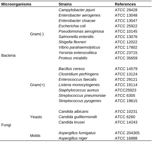
The microorganisms used in this study consisted of 17 strains of bacteria (10 Gram (-) and 7 Gram (+)), 3 yeasts and 2 molds (Table 1). These strains were obtained from the collections of Laboratoire de Chimie des Substances Naturelles et Sciences des aliments (LCSNSA) of La Réunion University. They were maintained on agar slant at 4°C and cultured on a fresh appropriate agar plate during 24 h prior to antimicrobial tests.
Chemicals for antimicrobial assay
Commonly used pre-impregnated discs, from Bio-Rad F 92430 Marnes-la-Coquette were chosen as antimicrobial references
Figure 1: Crotalaria bernieri a) the whole plant: b) flowers; c) fruits
Source: the authors
a
c
b
Andriamampianina et al. 1231
Table 1. Bacterial, yeast and mold strains used to study antimicrobial activities.
Microorganisms Strains References
Bacteria
Gram(-)
Campylobacter jejuni ATCC 29428
Enterobacter aerogenes ATCC 13048
Enterobacter cloacae ATCC 13047
Escherichia coli ATCC 25922
Pseudomonas aeruginosa ATCC 10145
Salmonella enteridis ATCC 13076
Shigella flexneri ATCC 12022
Vibrio parahaemolyticus ATCC 17802
Yersinia enterocolitica ATCC 23715
Proteus mirabilis ATCC 35659
Gram(+)
Bacillus cereus ATCC 14579
Clostridium perfringens ATCC 13124
Enterococcus faecalis ATCC 29121
Listeria monocytogenes ATCC 19114
Staphylococcus aureus ATCC25923
Streptococcus pneumoniae ATCC 6305
Streptococcus pyogenes ATCC 19615
Fungi
Yeasts
Candida albicans ATCC 10231
Candida guilliermondii ATCC 6260
Candida krusei ATCC 14243
Molds Aspergillus fumigatus ATCC 204305
Aspergillus niger ATCC 16888
Table 2. Abbreviations designating the different extracts tested.
(Camara et al.,2013; Rakholiya et al., 2014): amoxicillin 25 µg, chloramphenicol 30 µg, penicillin 6 µg as antibiotics and miconazole 50 μg as antifungal.
Preparation of extracts
The dried leaves, seeds, seed pods, and roots of the plant were grounded into powder. The resulting powder (100 g) was extracted successively with 4x500 mL of hexane, ethyl acetate and methanol for 24 h under stirring at room temperature. After filtration using a Whatman filter paper, each combined extract was evaporated under reduced pressure to dryness. The dry residues, dissolved in
hexane, ethyl acetate and sterile distilled water, constituted hexane, ethyl acetate and methanol extracts respectively (Table 2).
Phytochemical screening
The reactions for the detection of chemical groups were those developed by Fong et al. (1977) and Marini-Bettolo et al. (1981).
Antimicrobial assays
Antimicrobial activity test
The in vitro antimicrobial activity of the extracts was determined using disc diffusion method described by Pyun and Shin (2006) and Ngameni et al. (2009). Two mL of inoculum corresponding to 0.5 MacFarland (108 CFU/ml) was uniformly spread on the surface of Columbia Agar medium (for Streptococcus); Mueller-Hinton Agar (MHA) for the other bacteria and Potato Dextrose Agar (PDA) for yeasts. Sterilized filter paper discs 6 mm diameter (BioMérieux, REF 54991) were impregnated with 10 μL of each extract to the concentration of 100 mg/mL (1 mg/disc). The soaked discs were then placed on the surface of the agar and incubated at 37°C during 24 h for bacteria, or at 25°C for yeasts. The inhibition zone diameter (IZD) was measured and the results were interpreted by
Extracts Hexane Extract Ethyl acetate Extract Methanol Extract
Leaves LHE LEA LME
Seeds SHE SEA SME
Pods PHE PEA PME
1232 Afr. J. Microbiol. Res
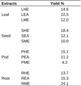
Table 3. Extraction yields of C. bernieri extracts.
Extracts Yield % Leaf LHE 14.6 LEA 22.5 LME 12.0 Seed SHE 18.4 SEA 12.1 SME 10.0 Pod PHE 15.1 PEA 11.2 PME 4.2 Root RHE 13.7 REA 15.3 RME 24.1
means of the scale used by Ponce et al. (2003) and Celikel and Kavas, (2008) stating that bacteria are not sensitive for IZD less than 8 mm, sensitive for IZD of 9 to 14 mm, very sensitive for IZD of 15 to 19 mm and extremely sensitive for IZD larger than 20 mm. Antifungal activity was evaluated by a method described by Favel et al. (1994). One ml of each extract was added to 19 ml of medium culture of PDA and maintained at 45°C. The mixture is then poured into Petri dishes and dried for 15 min at 37°C. 10 µl of each tested microorganism corresponding to 0.5 MacFarland were spread on the medium surface. IZD were measured after incubation at 25°C for 72 h. Negative controls were prepared by using the same solvents employed to dissolve the plant extract samples while the reference antibiotics were used as positive controls. All the experiments were performed in triplicate. The results were expressed as mean values ± standard deviations (mm ± SD).
MIC, MBC and MFC determination
For extracts showing antibacterial activity in the disc diffusion method (IZD ≥ 8 mm), MIC (minimum inhibitory concentration), MBC (minimum bactericidal concentration) MFC (minimum fungicidal concentration) were determined by microdilution method (Kuete et al., 2009).
The concentration of each extract was adjusted to 25 mg/ml. This was serially diluted two-fold to obtain concentration ranges of 0.024 to 25 mg/ml. Each concentration was added in a well (96-well microplate) containing 95 μl of Mueller-Hinton broth (MHB) and 5 μl of inoculum (standardized at 0.5 MacFarland). A positive control containing bacterial culture without the extract and a negative control containing only the medium, were also analyzed. The plates were covered with sterilized aluminum foil, and then incubated at 25°C (yeasts and molds) or at 37°C (bacteria) for 24 h. The MIC of each extract was detected following addition 40 µl of 0.2 mg/ml p-iodonitrotetrazolium chloride and incubation at 25°C (yeasts and molds) or at 37°C (bacteria) for 30 min (Kuete et al., 2009). Viable bacteria reduced the yellow dye to a pink color. MIC was defined as the lowest sample concentration that prevented this change and exhibited complete inhibition of bacterial growth. For the determination of
MBC and MFC, 5 μl from each well that showed no change in color was transferred on MHA or PDA plate and incubated at 25°C (yeasts and molds) or at 37°C (bacteria) for 24 h. The lowest concentration at which no growth occurred on the agar plates corresponded to the MBC or MFC.
The ratios MBC/MIC and MFC/MIC were calculated for each extract, to determine the nature of the effect. The extract is bactericidal or fungicidal for MBC/MIC or MFC/MIC ≤ 4 and bacteriostatic or fungistatic when these ratios are >4 (Djeussi et al., 2013; Bouharb et al., 2014; Chamandi et al., 2015).
Statistical analyses
Results were expressed as mean values ± standard deviations of three separate determinations. One-way analysis of variance (ANOVA) which was followed by Newman Keuls comparison test with Staticf® software was used for statistical analysis. Statistical estimates were made at confidence interval of 95%.
RESULTS
Extraction yields
The extractive yield of different parts of C. bernieri with
different solvents varied from 4.2 (PME) to 24.1% (RME)
(Table
3).
Qualitative phytochemical analysis
The
major
secondary
metabolites
identified
in
the different
organ extracts are presented in Table 4. Tannins,
polyphenols, steroids, triterpenes and unsaturated sterols
occurred in all the C. bernieri organs. Flavonoids were
found in all organs except root. Alkaloids were present
only in leaf and pod while saponins only in root. Iridoïds,
leucoanthocyanins, and quinones were not detected in all
parts of C. bernieri.
Antimicrobial activity
At 1 mg/disc, a concentration generally used for
antimicrobial activity assessment in plants (Sandeep et
al., 2010; Govindappa et al., 2011; Linthoingambi and
Mutum, 2013; Marimuthu et al., 2014), the large majority
of C. bernieri extracts (16 of 22) inhibited the
microorganism growth with IZD ranging from 8 to 15 mm
(Tables 5 to 7). However, activity depended on the
microorganism, the plant parts and extraction method
used. The most sensitive germs were S. enteridis
(IZD=11 mm), S. pyogenes (IZD=15 mm) and C.
guilliermondii (IZD=13 mm) in Gram (-)
bacteria, Gram (+)
bacteria and yeasts, respectively. Gram (-) strains C.
jejuni and E. coli, E. faecalis, Gram (+) L. monocytogenes
and the two molds A. fumigatus and A. niger were
resistant to all the extracts. REA, with an IZD of 15 mm
against S. pyogenes, displayed the highest antibacterial
activity.
Andriamampianina et al. 1233
Table 4. Phytochemical screening of C. bernieri extracts.
Chemical groups Tests Leaf Seed Pod Root
a b c d e f g h i j k l Alkaloids Mayer - - + - - - + - - - Wagner - - + - - - + - - - Dragendorff - - + - - - + - - - Confirmatory test (solubility in ethanol) - - + - - - + - - - Saponins Foam test - - - + Confirmatory test (hemolytic test) - - - + Flavonoids Willstätter - + + - + + - - + - - - Leucoanthocyanins Bate-Smith - - - - Tannins and Polyphenols Gelatin 1% + - + - + + - - - + + + Gelatin-salt 10% + - + + + + - + - + + + FeCl3 - + + - - + - - + - - + Quinones Borntrager - - - - Steroids Liebermann-Burchard + + - + + + + + - + + - Iridoïds Hot HCl - - - - Triterpenes Liebermann-Burchard + - - + + + + + - + + - Unsaturated sterols Salkowsky + - - + + + + + - + + -
+: positive result; -: negative result a: LHE; b: LEA; c: LME; d: SHE; e: SEA; f: SME; g: PHE; h: PEA; i: PME; j: RHE; k: REA; l: RME.
In yeasts, most of leaf extracts were active against the
three Candida strains tested, but seed and pod extracts
were active only against C. guilliermondii. Antibiotics
used as references in this study (amoxicillin 25 µg,
chloramphenicol 30 µg, penicillin 6 µg and miconazole 50
µg) were more effective than most of C. bernieri extracts.
MIC, MBC, MFC and MBC or MFC/MIC ratio values are
presented in Tables 8 to 10. MIC ranged from 0.048 to 25
mg/ml. MIC maximum values registered was 12.5 mg/mL
except for RHE on S. pyogenes (MIC=25 mg/ml).
Concerning MBC or MFC, maximum values for all
extracts were 25 mg/ml except for root extracts on some
Gram (+) bacteria and C. guilliermondii (MBC>25 mg/ml).
The ratio MBC or MFC/MIC varied from 1 to more than
100.
The most sensitive microorganism were P. mirabilis in
Gram (-)
bacteria (MIC=MBC=0.097 mg/ml), B. cereus
(MIC=0.048 mg/ml, MBC=0.195 mg/ml) and S. pyogenes
(MIC=MBC=0.048 mg/ml) in Gram (+) bacteria and C.
guilliermondii (MIC=MFC=0.048 mg/ml) in yeasts.
All methanol extracts were active. This is also the case
for ethyl acetate extracts except LEA. As to hexane
extracts, PHE and RHE were efficient but not LHE and
SHE. Pod extracts had the broadest spectrum of activity
with 10 sensitive microorganisms and seed extracts the
narrowest ones with 8 sensitive microorganisms.
DISCUSSION
The present study shows that the C. bernieri extracts
inhibited the growth of most tested microorganisms,
indicating the presence of antimicrobial compounds in all
parts of the plant. Phytochemical screening showed the
presence of diverse secondary metabolites, reported to
have antimicrobial property. At this stage of the work,
results did not yet allow to state whether the same or
different compounds are involved in the different parts of
the plant. However, they suggested that C. bernieri
antimicrobial activity might be mainly due to tannins,
polyphenols, steroids, triterpenes and flavonoids, which
were present in all or most of the plant organs. Alkaloids
might also be concerned in leaves and pods and
saponosides in root.
C. bernieri extracts showed generally a broad
antimicrobial spectrum. They were capable of inhibiting
the growth of different Gram (-) and Gram (+) bacterial
strains as well as some yeasts. However, each extract
1234 Afr. J. Microbiol. Res
Table 5. In vitro Antimicrobial Activity (IZD in mm) of extracts (1 mg/disc) on Gram (-) bacteria.
Extracts/ controls Cj Ea Ec Esc Pa Se Sf Vp Ye Pm Leaf LHE - - - - LEA - - - - LME - 10.00±0.01 - - 9.33±0.47 - - 7.00±0.01 - 8.33±0.47 Seed SHE - - - 7.00±0.01 - - SEA - - 10.00±0.01 - - - - 9.00±0.01 - 8.00±0.01 SME - - - 8.00±0.01 - Pod PHE - 9.67±0.47 - - - - PEA - - 9.00±0.01 - - - - 10.67±0.01 - - PME - - - 11.00±0.82 8.00±0.01 9.00±0.82 8.00±0.01 8.00±0.01 Root RHE - - - - 10±0.01 8.00±0.01 - - - - REA - - - 9.00±0.01 - - RME - - - 9.00±0.01 - - - 9.00±1.41 PC Amx 45.00 - - 23.00 10.00 27.00 25.00 - 10.00 - Chl 38.00 25.00 25.00 30.00 15.00 32.00 30.00 - 38.00 - Pen 40.00 - - - 25.00 NC Hex - - - - EtOAc - - - - Sdw - - - -
Cj: C. jejuni; Ea: E. aerogenes; Ec: E. cloacae; Esc: E. coli; Pa: P. aeruginosa; Se: S. enteridis; Sf: S. flexneri; Vp: V. parahaemolyticus; Ye: Y.
enterocolitica; Pm: P. mirabilis PC: Positive control (Amx: Amoxicillin 25µg; Chlor: Chloramphenicol 30µg; Pen: Penicillin 6µg); NC: Negative
control (Hex: Hexane; EtOAc: Ethyl acetate; Sdw: sterile distilled water); −: No activity.
displayed a specific activity spectrum that could be due
to difference between the chemical nature and
concentration of bioactive compounds in extracts. The
results obtained with microdilution method were more
reliable than those with disc diffusion. That might be due
to the fact that bioactive compounds were in direct
contact with germs in liquid medium whereas they
diffused little or not at all in solid medium.
There was no consensus on the acceptable level of
inhibition for natural products (Benko and Crovella,
2010). For Dalmarco et al. (2010), for crude extracts and
fractions, a MIC lower than 100 µg/mL was considered
as an excellent effect, from 100 to 500 µg/ml as
moderate, from 500 to 1000 µg/mL as weak, and over
1000 µg/ml as inactive. According to Kouitcheu et al.
(2013), when a crude extract was used, the MIC values
of 8 mg/mL or below against any microorganism tested
was considered as active.
If the scale adopted by Dalmarco et al. (2010) was
used as a reference, 10 extracts displayed an excellent
effect, 8 a moderate effect, 5 a weak effect then the
remaining extracts were inactive. Excellent effects were
observed on P. mirabilis (RME), S. enteridis (PME), B.
cereus (LME, PEA, REA), S. pneumoniae (LME), S.
pyogenes (REA), C. albicans (LME) and C. guilliermondii
(LME, SEA). Moderate effects, were found against E.
aerogenes (SEA), P. mirabilis (LME), P. aeruginosa
(LME), C. perfringens (LME), S. aureus (LME), S.
pyogenes (RME, PEA, LME) and C. guilliermondii
(SME). Weak effects were observed on E. aerogenes
(SEA), P. aeruginosa (LME), S. aureus (REA) and S.
pneumoniae (REA, RME).
The most efficient extracts were RME (MIC=MBC=
0.097 mg/ml) against Y. enterolytica in Gram (-) bacteria,
REA (MIC=MBC=0.048 mg/ml) against S. pyogenes in
Gram (+) bacteria and LME (MIC=MFC=0.048 mg/ml)
against C. guilliermondii. Some of the extracts were very
effective against some organisms (LME against B.
cereus, S. pneumoniae,
C.
albicans
and
C.
guilliermondii,
REA against B. cereus and S. pyogenes) while others
were totally inactive (SME against S. pneumoniae and S.
pyogenes).
However, if the interpretation of Kouitcheu et al.,
(2013) was taken into account, only nine extracts had
MIC higher than 8 mg/mL on some germs, which means
that all the other extracts of C. bernieri used showed
Andriamampianina et al. 1235
Table 6. In vitro Antimicrobial Activity (inhibition zone diameter in mm) of extracts (1 mg/disc) on Gram (+) bacteria.
Plant
parts/controls Extracts Bc Cp Ef Lm Sa Spn Spy
Leaf LHE - - - - 7.00±0.01 - - LEA - - - - LME 9.00±0.01 8.00±0.01 - - 10.00±0.01 12.67±1.25 12.33±1.70 Seed SHE - - - - 7.00±0.01 - - SEA - - - - 9.00±0.82 11.33±0.47 9.00±0.01 SME - - - 8.33±1.25 8.00±0.01 Pod PHE - 9.67±1.25 - - 7.00±0.01 - - PEA 11.33±0.47 8.33±0.47 - - 10.33±0.47 13.00±0.82 12.00±1.41 PME - - - - 7.00±0.01 8.00±0.01 7.00±0.01 Root RHE - - - 8.00±0.82 REA 11.33±0.94 7.00±0.01 - - 11.00±0.01 13.00±0.82 15.00±0.01 RME 9.00±1.41 10.00±0.01 - - 8.00±0.01 13.00±0.83 12.00±1.41 PC Amx 15.00 27.00 - - 37.00 26.00 32.00 Chl 38.00 30.00 - 30.00 30.00 25.00 22.00 Pen 15.00 - - - 35.00 23.00 25.00 NC Hex - - - - EtOAc - - - - Sdw - - - -
Bc: B. cereus; Cp: C. perfringens; Ef: E. faecalis; Lm: L. monocytogenes; Sa: S. aureus; Spn: S. pneumoniae; Spy: S. pyogenes Amx: Amoxicillin 25 µg; PC: Positive control (Amx: Amoxicillin 25 µg; Chlor: Chloramphenicol 30 µg; Pen: Penicillin 6 µg; NC: Negative control (Hex: Hexane; EtOAc: Ethyl acetate; Sdw: sterile distilled water); −: No activity.
Table 7. In vitro Antimicrobial Activity (inhibition zone diameter in mm) of extracts (1mg/disc) on
yeasts and molds
Plant part Extract Yeast Mold
Ca Cg Ck Af An Leaf LHE - 7.00±0.01 - - - LEA - 7.00±0.01 7.00±0.01 - - LME 8.00±0.01 8,67± 0.94 7.00±0.01 - - Seed SHE - 7.00±0.01 - - - SEA - 13.00±0.82 - - - SME - 11.00±0.01 - - - Pod PHE - 7.00±0.01 - - - PEA - 7.00±0.01 - - - PME - 7.00±0.01 - - - Root RHE - 8.00±0.01 - - - REA - 7.00±0.01 - - - RME - 8.00±0.82 - - - PC Mic 18.00 30.00 33.00 28.00 23.00 NC Hex - - - - - EtOAc - - - - - Sdw - - - - -
Ca: C. albicans; Cg: C. guilliermondii; Ck: C. krusei; Af: A. fumigatus; An: A. niger PC: Positive control (Mic: Miconazole 50µg); NC: Negative control (Hex: Hexane; EtOAc: Ethyl acetate; Sdw: sterile distilled water); −: No activity.
1236 Afr. J. Microbiol. Res
Table 8. MIC and MBC values (mg/mL) of C. bernieri extracts (1mg/disc) on Gram(-)
bacteria
Gram(-) Bacteria Extracts MIC (mg/ml) MBC (mg/ml) MBC/MIC
Enterobacter aerogenes LME 0.195 25 128.21
Enterobacter cloacae SEA 0.781 25 32.01
PEA 6.25 25 4.00
Pseudomonas aeruginosa LME 0.781 25 32.01
RHE 0.195 25 128.21
Salmonella enteridis
PME 0.097 12.5 128.87
RHE 12.5 12.5 1.00
RME 12.5 25 2.00
Shigella flexneri PME 3.125 25 8.00
Vibrio parahaemolyticus
SEA 1.562 6.25 4.00
PEA 1.562 25 16.01
PME 3.125 6.25 2.00
REA 1.562 1.562 1.00
Yersinia enterolitica SME 1.562 12.5 8.00
PME 1.562 12.5 8.00 Proteus mirabilis LME 0.195 0.781 4.01 SEA 1.562 3.125 2.00 PME 1.562 0.781 0.50 RME 0.097 0.097 1.00
antimicrobial activities.
All the extracts had bactericidal action (MBC/MIC
≤ 4)
in certain bacteria and bacteriostatic action (MBC/MIC >)
4) in other ones. For example LME was bactericidal
against B. cereus and C. perfringens but bacteriostatic
against S. aureus and S. pneumoniae. The comparison
of A. bernieri extract activities to foreign Crotalaria
species was not easy because antimicrobial activity was
assessed under different conditions (other microorganism
strains and extract doses used).
Compared to available data, the IZD of C. bernieri
extracts were generally of the same order of magnitude
as those of leaf ethyl acetate extract from C. madurensis
against B. subtilis and S. aureus (IZD=14 mm), M. luteus
(IZD=12 mm), E. coli and C. albicans (IZD=10 mm
(Bhakshu et al., 2008) and leaf ethanol extract from C.
pallida against X. axanopodis (IZD=16 mm), E.
coli(IZD=14 mm) and C. michiganensis (IZD=13 mm)
(Govindappa et al., 2011). Root methanol extract from C.
burhia was more efficient with an IZD of 18 mm against
B. subtilis and P. aeruginosa (Sandeep et al., 2010).
If comparison was based on antimicrobial indexes,
LME (MIC=0.781 mg/ml, MBC=25 mg/ml) and REA
(MIC=0.195 mg/ml, MBC=25 mg/ml) were more efficient
against P. aeruginosa than the leaf methanol extract from
C. quartiniana (MIC=MBC=37.5 mg/ml) (Omori et al.,
2011). The leaf hexane extract from C. retusa (MIC=0.125
mg/ml, MBC=37.5 mg/ml) (Maregesi et al., 2008) was
less active against B. cereus than LME (MIC=0.097
mg/ml, MBC=0.195 mg/ml), PEA and REA (MIC=0.048
mg/ml, MBC=0.195 mg/ml). By contrast, C. bernieri
extracts were less active on P. mirabilis (MIC between
0.097 and 1.56 mg/ml) than a peptide isolated from C.
pallida seeds (MIC=0.030 mg/ml) (Pelegrini et al., 2009).
Compared to the antibacterial activities from other plant
extracts, several C. bernieri extracts were more efficient
than methanolic aerial part extracts of Inula viscosa
against B. subtilis (MIC=25 mg/ml, MBC=50 mg/ml) and S.
aureus (MIC=12.5 mg/ml, MBC=50 mg/ml) (Larbi et al.,
2016). By contrast, tuber ethyl acetate extract of
Tropaeolum pentaphyllum against E. coli (MIC=0.02
mg/ml, MBC=0.64 mg/ml), P. aeruginosa (MIC=0.04
mg/ml, MBC=0.64 mg/ml) (da Cruz et al., 2016) and
organic extract (aerial parts) of Rapanea parvifolia against
E.
faecalis
(MIC=0.03
mg/ml,
MBC=0.06
mg/ml) (Suffredini
et al., 2006) were more efficient.
Andriamampianina et al. 1237
Table 9. MIC and MBC values (mg/ml) of C. bernieri extracts (1mg/disc) on Gram(+) bacteria.
Gram (+) Bacteria Extract MIC (mg/ml) MBC (mg/mL) MBC/MIC
Bacillus cereus LME 0.097 0.195 2.01 PEA 0.048 0.195 4.06 REA 0.048 0.195 4.06 RME 1.562 1.562 1.00 Clostridium perfringens LME 0.195 0.390 2.00 PHE 12.5 25 2.00 PEA 6.25 12.5 2.00 RME 6.25 ˃25 - Staphylococcus aureus LME 0.195 6.25 32.05 SEA 6.25 12.5 2.00 PEA 3.125 25 8.00 REA 0.781 25 32.01 RME 12.5 25 2.00 Streptococcus pneumoniae LME 0.097 3.125 32.22 SEA 3.125 12.5 4.00 SME 12.5 25 2.00 PME 1.562 6.25 4.00 REA 0.781 ˃25 - RME 0.781 ˃25 - Streptococcus pyogenes LME 0.195 1.562 8.01 SEA 1.562 12.5 8.00 SME 12.5 25 2.00 PEA 0.195 0.781 4.01 RHE 25 ˃25 - REA 0.048 0.048 1.00 RME 0.195 12.5 64.10
Table 10. MIC and MBC values (mg/ml) of C. bernieri extracts (1mg/disc) on
yeasts.
Yeasts Extraits MIC (mg/ml) MFC (mg/ml) MFC/MIC
Candida albicans LME 0.097 0.195 2.01
Candida guilliermondii LME 0.048 0.048 1.00 SEA 0.048 1.562 32.54 SME 0.195 25 128.21 RHE 12.5 ˃25 - RME 12.5 ˃25 -
On fungi, LME (MIC=0.097 mg/ml, MFC=0.195 mg/ml)
was more efficient than leaf methanolic extract of Myrtus
nivellei against C. albicans (MIC=4.5 mg/ml) (Touaibia
and Chaouch, 2015) whereas LME, SEA and SME
against C. guilliermondii (MIC=0.08 mg/ml, MFC=0.32
mg/ml) were less efficient than ethyl acetate extract of T.
pentaphyllum (da Cruz et al., 2016).
Conclusion
1238 Afr. J. Microbiol. Res
as a source of interesting natural wide spectrum
antimicrobial molecules. All its parts were efficient and
could be easily found in significant amounts for the plant
grows in fields, in the vicinity of homes, on roadsides and
can be cultivated. Furthermore, according to our survey
of local populations, C. bernieri is consumed by zebus
but no cases of poisoning have yet been reported. At
present, our works are concerned with the isolation of
pure compounds from different extracts of C. bernieri and
the elucidation of their structures in order to better
evaluate their pharmacological activity.
In view of later therapeutic use of C. bernieri, study on
various experimental models of animals is also on going
to assess the harmful effects it might have.
Conflict of Interests
The authors have not declared any conflict of interests.
ACKNOWLEDGMENT
The authors are grateful to the Laboratoire de Chimie des
Substances Naturelles et Sciences des aliments
(LCSNSA) Saint Pierre, La Réunion and the Centre
National de Recherche sur l’Environnement (CNRE) for
their helpful support to this work.
REFERENCES
Benko IAM, Crovella S (2010). Ethnobotanical bioprospection of candidates for potential antimicrobial drugs from brazilian plants. Curr. Protein Pept. Sci. 11(3):189-194.
Bhakshu LM, Venkata RK, Venkataraju RR (2008). Medicinal properties and antimicrobial activity of Crotalaria madurensis Var. Kurnoolica. Ethnobot Leaflets. (1):104
Bouharb H, El Badaoui K, Zair T, El amri J, Chakir S, Alaoui T (2014). Sélection de quelques plantes médicinales du Zerhoun (Maroc centrale) pour l’activité antibactérienne contre Pseudomonas
aeruginosa. J. Appl. Biosci. 78:6685-6693.
Camara M, Dieng A, Boye CSB (2013). Antibiotic Susceptibility of
Streptococcus Pyogenes Isolated from Respiratory Tract Infections in
Dakar, Senegal. Microbiol Insights.6:71-75.
Celikel N, Kavas G (2008). Antimicrobial properties of some essential oils against some pathogenic microorganisms. Czech J. Food Sci. 26(3):174-181.
Chamandi G, Olama Z, Holail H (2015). Antimicrobial effect of Propolis from different geographic origins in Lebanon. Int. J. Curr. Microbiol. App. Sci. 4(4):328-342.
Chouhan H, Sushil KS (2010). Antibacterial Activity of Seed and Flower Parts of Crotalaria juncea Linn. Am-Euras. J. Sci. Res. 5(3):212-215. da Cruz RC, Denardi LB, Mossmann NJ, Piana M, Alves SH and de
Campos MMA (2016). Antimicrobial Activity and Chromatographic Analysis of Extracts from Tropaeolum pentaphyllum Lam. Tubers. Molecules, 21(566):1-11.
Dalmarco JB, Dalmarco EM, Koelzer J, Pizzolatti MG, Fröde TS (2010). Isolation and identification of bioactive compounds responsible for the anti-bacterial efficacy of Lotus corniculatus var. São Gabriel. Int. J. Green Pharm. 4:108-114.
Djeussi DE, Noumedem JAK, Seukep JA, Fankam AG, Voukeng IK, Tankeo SB, Nkuete AHL, Kuete V (2013). Antibacterial activities of selected edible plants extracts against multidrug-resistant Gram bacteria. BMC Complement. Altern. Med. 13(164):1-8.
Dupuy DJ, Labat JN, Rabevohitra R, Villiers JF, Bosser J, Morat J (2002). The Leguminosae of Madagascar. Royal Botanic Gardens, Kew, Richmond, United Kingdom, pp. 243-288.
Dzoyem JP, Mc Gaw LJ, Eloff JN (2014). In vitro antibacterial, antioxidant and cytotoxic activity of acetone leaf extracts of nine under investigated Fabaceae tree species leads to potentially useful extracts in animal health and productivity. BMC Complement Altern
Med. 14(147):1-7.
Favel A, Steinmetz MD, Regli EV, Olivier RE, Balandsard G (1994). In
vitro antifungal activity of triterpenoid saponins. Planta Med.
60:50-53.
Fong EHS, Tin-Wa M, Farnsworth NR Dobberstein RH (1977). Phytochemical screening methods. Rev. Department of pharmacognosy and pharmacology. College of pharmacy. University of Illinois.
Govindappa M, Bharath N, Shruthi HB, Sadananda TS, Sharanappa P (2011). Antimicrobial, antioxidant and in vitro antiinflammatory activity and phytochemical screening of Crotalaria pallida Aiton. Afr. J. Pharm. Pharmacol. 5(21):2359-2371.
Kouitcheu LBM, Tamesse JL, Kouam J (2013). The anti-shigellosis activity of the methanol extract of Picralima nitida on Shigella
dysenteriae type I induced diarrhoea in rats. BMC Complement.
Altern. Med. 13(211):4-11.
Kuete V, Fozing DC, Kapche WFGD, Mbaveng AT, Kuiate JR, Ngadjui BT, Abega ZBM (2009). Antimicrobial activity of the methanolic extract and compounds from Morus mesozygia stem bark. J. Ethnopharmacol. 124(3):551-555.
Larbi SK, Meddah B, Meddah ATT, Sonnet P (2016). The antibacterial effect of two medicinal plants Inula viscosa, Anacyclus valentinus (Asteraceae) and their synergistic interaction with antibiotic drugs. J. Fundam Appl. Sci. 8(2):244-255.
Linthoingambi W, Mutum SS (2013). Antimicrobial activities of different solvent extracts of Tithonia diversifolia (Hemsely) A. Gray. Asian J. Plant Sci. Res. 3(5):50-54.
Mansoor H, Muhammad A, Al-Quriany F, Tahira N, Muhammad SAA, Adnan Y, Nargis N (2011). Medicinal flora of the Cholistan desert. Pak. J. Bot. 43: 39-50.
Maregesi SM, Pieters L, Ngassapa OD, Apers S, Vingerhoets R, Cos P, Vanden BDA, Arnold JV (2008). Screening of some Tanzanian medicinal plants from Bunda district for antibacterial, antifungal and antiviral activities. J. Ethnopharmacol. 119:58-66.
Marimuthu MM, Aruldass CA, Sandrasagaran UM, Mohamad S, Ramanathan S, Mansor SM, Murugaiyah V (2014). Antimicrobial activity and phytochemical screening of various parts of Ixora
coccinea. J. Med. Plants Res. 8(10):423-429.
Marini-Bettolo GB, Nicoletti S, Patami M (1981). Plant screening by chemical and chromatographic procedure under field conditions. J. Chromatogr. 218: 113-217.
Ponce AG, Fritz R, del Valle C and Roura SI (2003). Antimicrobial activity of essential oils on the native microflora of organic Swiss chard. Lebensm. Wiss. Technol. 36:679-684.
Pyun MS, Shin S (2006). Antifungal effects of the volatile oils from allium plants against trichophyton species and synergism of the oils with ketoconazole. Phytomedicine, 13(6):394-400.
Ngameni B, Kuete V, Simo IK, Mbaveng AT, Awoussong PK, Patnam RR, Ngadjui BT (2009). Antibacterial and antifungal activities of the crude extract and compounds from Dorstenia Turbinate (Moraceae). S. Afr. J. Bot. 75: 256-261.
Omori EO, Richard MM, Lawrence A, Paul OO (2011). Evaluation of methanolic extracts of six medicinal plants used by herbal practitioners in central province Kenya. Int. J. Pharm. Sci. Res. 2(4): 867-874.
Pelegrini PB, Farias LR, Saude AC, Costa FT, Bloch CJ, Silva LP, Oliveira AS, Gomes CE, Sales MP and Franco OL (2009). A novel antimicrobial peptide from Crotalaria pallida seeds with activity against human and phytopathogens. Curr Microbiol, 59(4): 400-404. Peltier MAG (1959). Notes sur les Légumineuses-Papilionoidées de
Madagascar et des Comores (suite). J. Agric. Trop. Bot. Appl. 6:267-289.
Polhill RM (1982). Crotalaria in Africa and Madagascar. Royal Botanic Gardens Kew, A. A. Bakema, Rotterdam, pp.1-89.
Study of Hydroalcoholic Extracts of Momordica charantia L. against Foodborne Pathogens. Indian J. Pharm. Sci. 76(2):148-156. Ravikumar MC, Rajkumar HG (2013). Antifungal activity of plants
extracts against Alternaria solani, the causal agent of early blight of tomato. Arch. Phytopathol. Plant Protect. 46(16):1-7.
Sandeep K, Shrivastava B, Khajuria RK (2010). Antimicrobial activity of
Crotalaria burhia Buch.-Ham. Indian J. Nat. Prod. Resour. 1(4):481
484.
Stefanovic O, Comic L (2011). Inhibitory effect of Cytisus nigricans L. and Cytisus capitatus Scop. on growth of bacteria. Afr. J. Microbiol. Res. 5(27):4725-4730.
Suffredini IB, Pacienca MLB, Varella AD, Younes RN (2006). Antibacterial Activity of Brazilian Amazon Plant Extracts. Braz. J. Infect. Dis. 10(6):400-402.
Andriamampianina et al. 1239
Touaibia M, Chaouch FZ (2015). Propriétés antioxydantes et antimicrobiennes des extraits de Myrtus nivellei Batt et Trab. obtenus in situ et in vitro. Phytothérapie, Lavoisier SAS pp1-7.
Zongo C, Savadogo A, Somda MK, Koudou J, Traore AS (2011). In vitro evaluation of the antimicrobial and antioxidant properties of extracts from whole plant of Alternanthera pungens H.B. & K. and leaves of Combretum sericeum G. Don. Int. J. Phytomed. 3:182-191.