A
PPLIED ANDE
NVIRONMENTALM
ICROBIOLOGY, Apr. 2009, p. 2545–2553
Vol. 75, No. 8
0099-2240/09/$08.00
⫹0 doi:10.1128/AEM.02211-08
Copyright © 2009, American Society for Microbiology. All Rights Reserved.
Use of Calcofluor White for Detection, Identification, and
Quantification of Phytoplanktonic Fungal Parasites
䌤
Serena Rasconi, Marle
`ne Jobard, Lionel Jouve, and Te
´lesphore Sime-Ngando*
Laboratoire Microorganismes: Ge´nome et Environnement, UMR CNRS 6023, Universite´ Blaise Pascal (Clermont-Ferrand II),
F-63177 Aubie`re Cedex, France
Received 25 September 2008/Accepted 8 February 2009
We propose a routine protocol based on size fractionation of pelagic samples and the use of the fluorochrome
calcofluor white (which binds to
-1,3 and -1,4 polysaccharides) for diagnosing, identifying, and counting
chitinaceous fungal parasites (i.e., the sporangia of chytrids) of phytoplankton. The protocol was applied to
freshwater samples collected during different seasons (spring and summer/autumn) in two lakes whose trophic
statuses varied. Because few samples were collected (i.e., two dates per site), the findings are considered
preliminary and mainly a “proof of concept” rather than a valid comparison of sites versus seasons. The results
from the proposed protocol indicate higher diversity of infected host and parasite communities than in
previous studies. Chytrid epidemics were omnipresent, infecting diverse phytoplankton host communities,
primarily diatoms, chlorophytes, and colonial and filamentous cyanobacteria. The diversity and numerical
abundance of sporangia and of hosts, and the prevalence of infection (range, <1 to 24% of total host cells) as
well, increased from the oligotrophic Lake Pavin to the eutrophic Lake Aydat, while the temporal changes in
parasites were apparently more influenced by the host community composition. We conclude that the proposed
protocol offers a valid method for the quantitative ecology of chytrid epidemics in aquatic ecosystems and food
web dynamics.
Fungal infections are recurrent in aquatic ecosystems (15,
42, 46). Organisms belonging to the order of Chytridiales (i.e.,
chytrids) are known mainly as phytoplankton parasites (10, 22,
44). Recently, we have unveiled a large reservoir of unexpected
fungal diversity in freshwater lakes, primarily of chytrids (31–
33). Parasitic chytrids and other zoosporic true fungi sensu
Barr (3) produce zoospores and are often host specific, highly
infective, and extremely virulent (13, 16). They are considered
relevant not only for the evolution of their hosts but also for
the population dynamics and successions of phytoplankton
communities (14, 48). Studies of chytrid fungal parasitism
car-ried out in the English Lake District indicate that infection of
diatoms, desmids, and other green algae is fairly common (10).
Significant chytrid parasitism has also been recorded in other
lakes, affecting primarily the diatom Asterionella formosa (12,
29, 37, 48). However, fungal parasitism on plankton has rarely
been studied and has mostly been restricted to descriptive
taxon-omy. Full descriptions of parasitic chytrids have been given since
the middle of the last century (8–9) but even today, their impact
on the dynamics of host populations and the related
biogeochemi-cal cycling and energetics remain largely unknown (16), mainly
because of methodological difficulties (17).
Various approaches have been used to study fungal
para-sites, but routine techniques for reliably identifying and
count-ing these organisms are lackcount-ing in the context of aquatic
mi-crobial ecology (31–33). Some of them have been misidentified
as protistan bacterivorous nanoflagellates, e.g., flagellates in
the group of Bicosoeca, which are attached to phytoplankton
but do not harm the algae (31–33). A few studies have
dem-onstrated epidemic outbreaks of chytrids on phytoplankton,
but mainly with particular emphasis on host-parasite
interac-tions and coevolution (21, 30, 48). Thus far, observainterac-tions of
parasitic fungi were obtained by using phase-contrast light
microscopy with live or Lugol’s iodine preserved samples (30,
39, 48). Such conventional microscopy allows observation of
fungal sporangia or similar forms (especially in laboratory
cul-tures) but is a poor approach for characterizing chytrid
para-sites in natural samples, at the complex community level. For
example, a simple light microscopy observation of fungal
rhi-zoidal systems, i.e., a pertinent criterion for identifying chytrids
(10, 13, 44), is very difficult, a situation which usually leads to
confusion of chytrids with protistan flagellates such as
cho-anoflagellates (31–33). Most chytrids occupy the most basal
branch of the kingdom Fungi, a finding consistent with
cho-anoflagellate-like ancestors (25, 26).
Methodological limitations for the study of the ecological
dynamics of chytrid populations can be overcome with
epiflu-orescence microscopy coupled to a specific fluorochrome
tar-geting molecular tracers (i.e., some types of polysaccharides)
of chitinaceous fungal structures, primarily sporangia, the
pres-ence of an opercule, and the extent of the rhizoidal system. We
propose here a simple procedure based on the fluorochrome
calcofluor white (CFW) and epifluorescence microscopy for
diagnosing, identifying, and counting parasitic chytrids within
phytoplanktonic communities.
MATERIALS AND METHODS
Sites and sampling.Samples were collected in two freshwater lakes, which differed in trophic status and were located in the French Massif Central. Lake
* Corresponding author. Mailing address: Laboratoire
Microorgan-ismes: Ge
´nome et Environnement, UMR CNRS 6023, Universite
´
Blaise Pascal (Clermont-Ferrand II), F-63177 Aubie
`re Cedex, France.
Phone: 33 473 497 836. Fax: 33 473 407 670. E-mail: telesphore.sime
-ngando@univ-bpclermont.fr.
Pavin (45°29⬘41⬙N, 002°53⬘12⬙E) is an oligotrophic deep volcanic mountain lake (Zmax⫽ 92 m), characterized by small surface (44 ha) and small drainage basin
(50 ha) areas. Lake Aydat (45°39⬘48⬙N, 002°59⬘04⬙E) is a small eutrophic lake (Zmax⫽ 15 m, surface area ⫽ 60 ha), with a larger catchment area (3 ⫻ 104ha).
For both lakes, samples were taken near the center of the lake, at the point of maximum depth. Lake Pavin was first sampled on 26 October 2006 for testing different methodological protocols, and both lakes were subsequently sampled twice in 2007 for the application of the optimal protocol retained (i.e., the size-fractionated community approach using 1% CFW final concentration; see below) to the natural community dynamics. The latter samples were collected during the spring and autumn (22 March and 2 October 2007) for the oligotro-phic Lake Pavin, and during spring and late summer (22 May and 28 August 2007) for the eutrophic Lake Aydat. All samples were collected in triplicates, i.e., from three independent sampling operations per sampling date.
During all sampling occasions, ca. 21 liters of the whole euphotic layers of the two lakes (estimated from Secchi depths) was sampled manually using a flexible plastic tube (diameter, 4 cm) provided by a rope connecting the ballasted bottom of the tube with a surface manipulator. With this system, the euphotic water column samples are collected by simple capillarity. This technique of sampling is rapid, easy, and inexpensive (43). Analytic samples were thus considered as integrated samples representative of the euphotic layers of the lakes, i.e., 0 to 20 m for Lake Pavin and 0 to 5 m for Lake Aydat. Collected samples were immediately prefiltered through a 150-m-pore-size nylon filter (to eliminate the predatory metazoan zooplankton) when poured into clean transparent recipients
previously washed with the lake water and then transferred immediately to the laboratory for processing.
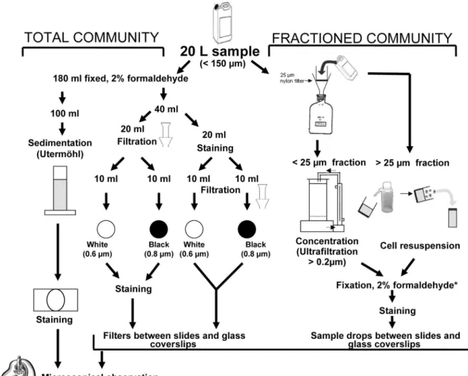
Protocols tested for concentrating and staining fungal parasites.Back in the laboratory, different procedures were tested on the experimental samples col-lected on 26 October 2006 in Lake Pavin for concentrating phytoplankton cells before the staining tests of chytrids. Concentrating phytoplankton is essential for an accurate assessment of morphological characteristics (general shape of spo-rangia, the rhizoidal system, the attachment of peduncle, and the presence of operculum), which include typical identification keys of fungal parasites. The sample processing is summarized in Fig. 1.
Concentration of host cells.Two different approaches were tested on samples collected on 26 October 2006 in Lake Pavin: the total community approach and the size-fractionated community approach (Fig. 1). For the former approach, 180-ml portions of experimental samples were fixed with formaldehyde (2% final concentration), and aliquots were concentrated in three different ways: (i) by simple gravity according to the Utermo¨hl method (47) before the chyrids were stained, (ii) by vacuum pressure on two different filters before staining directly onto filters, and (iii) by vacuum pressure on the same two types of filters but after being stained in solution. For the Utermo¨hl method, 100 ml of fixed samples was settled for at least 24 h. For each of the two filter-vacuum pressure methods, 10 ml of fixed samples was filtered onto polycarbonate white filters (pore size, 0.6 m; catalog no. DTTP02500 [Millipore]) and Nuclepore polycarbonate black filters (pore size, 0.8m; catalog no. 110659 [Whatman]) by using gentle vacuum (20 kPa).
FIG. 1. Sample partitioning and different concentration procedures tested for CFW staining and epifluorescence microscopy observation of
phytoplankton parasitic chytrids.
*
, the fixation step for the fractionated community approach is facultative, i.e., can be avoided when observations
are made without delay.
For the size-fractionated community approach, 20 liters of the experimental samples was passed through a 25-m-pore-size nylon filter (Fig. 1). Large phy-toplankton cells in the⬎25-m size fraction were collected by washing the filter with 40 ml of 0.2-m-pore-size-filtered lake water and fixed with formaldehyde (2% final concentration) before staining and analysis. Nanoplanktonic cells in the⬍25-m size fraction (i.e., the 20-liter filtrate) were concentrated ⬃20-fold by ultrafiltration to a volume of⬃1 liter using a high-performance concentration/ diafiltration system (model DC 10LA; Amicon, Epernon, France) equipped with a reusable hollow fiber cartridge (0.2-m cutoff, surface area of 0.45 m2
; Ami-con) and an entry pressure of 0.9 bar. About 180 ml of the ultrafiltrate retentate was then fixed with formaldehyde (2% final concentration) before staining and analysis. The size-fractionated community approach gave optimal results and was used for the samples collected in 2007 in both Lake Pavin and Lake Aydat.
Staining of parasites.Sporangia and rhizoids of parasitic chytrids in concen-trated subsamples were stained with the fluorochrome CFW (C40H44N12O10S2
Fluorescent Brightener 28; Sigma catalog no. F3543). CFW is used as whitening agent by the paper industry and selectively binds to cellulose and chitin. The dye fluoresces when exposed to UV light and offers a very sensitive method for direct microscopic examination of skin scrapings, hairs, nails, and other clinical speci-men for fungal elespeci-ments (19, 20). Here, we have adapted the technique to environmental aquatic samples. Before staining, a stock solution of CFW was prepared as modified from an original protocol (20; http://www.mycology .adelaide.edu.au/) by adding 35 mg of CFW to 7 ml of sterile distilled water and a few drops of 10 N NaOH to increase the pH to between 10 and 11, because CFW does not dissolve well in neutral solutions. The final volume of stock solution was then adjusted to 10 ml with sterile distilled water, divided into small aliquots (150l), and stored in the dark at ⫺20°C until use.
For the experimental samples collected on 26 October 2006 in Lake Pavin, four final concentrations of CFW (20, 10, 3, and 1% [vol/vol]) relative to the stock solution were tested. For the Utermo¨hl method, settled phytoplankton cells were stained by replacing an appropriate volume of the supernatant water with the stock solution of CFW directly into the Utermo¨hl chamber, so as to yield the target dye final concentrations tested. For the vacuum pressure method, phyto-plankton cells concentrated onto DTTP and black filters were stained by flooding the filters with the dye (CFW stock solution was diluted at the four final con-centrations tested) for 10 min, followed by thorough washing with ⬍0.2-m-pore-size-filtered lake water. In the variant of this method, CFW stock solution was added to subsamples of suspended phytoplanktonic cells (to reach the appropriate concentrations) for 10 min, before concentrating stained cells onto DTTP and black filters, following by washing with⬍0.2-m-pore-size-filtered lake water. All filters were then mounted between microscope slides and glass coverslips using a nonfluorescent immersion oil (Cargille type A). For the frac-tionated samples (i.e.,⬎25 m and ⬍25 m), aliquots (150 l) of concentrated and fixed materials were stained in solution for 10 min (as previously described for suspended cells), and drops (10l) of the stained samples were mounted between glass slides and coverslips for observations and counting (Fig. 1).
The optimal CFW final concentration (i.e., 1%) staining and the size-fraction-ated community approach were then applied to the samples collected in 2007 in both Lake Pavin and Lake Aydat, for diagnosing, identifying, and counting phytoplankton fungal parasites using direct epifluorescence microscope obser-vations.
Direct observation and counting.For all samples, stained chytrids were ob-served in a dark room under an inverted epifluorescence microscope (Leica DMIRB model) equipped with a⫻1,250 (i.e., 100/1.25) objective lens, an HBO-100W mercury lamp, and a set of different optic filters, including filters (340 to 380 nm) for UV light excitation. For picture capture and processing, the micro-scope was equipped with a Leica color video camera (model DC 300F) and a Leica Q500 personal computer.
Experimental samples were mounted between slides and glass coverslips either as concentrates onto filters (i.e., the total community approach) or directly as concentrated liquid drops (5 to 15l, i.e., the size-fractionated approach). Slides were then inverted for light excitation and observation under the inverted Leica epifluorescence microscope. For each replicate analyzed, at least 500 phyto-planktonic cells (calculated standard error of⬍10%) were inspected for fungal infection (i.e., the presence of fixed sporangia), and the morphological charac-teristics of parasites were noted for identification using the software for image analysis Leica Qwin. The natural abundance and composition of phytoplankton hosts were determined from raw fixed parallel samples by using the Utermo¨hl method (47).
Species identification and fungal infectivity parameters.During microscopic observations, phytoplanktonic cells were identified, often to the species level, using morphological taxonomic keys known from references, e.g., Bourelly (6), Huber-Pestalozzi (23), and Prescott (38). For fungal parasites, identification was
similarly based on phenotypic keys known from classical manuals, primarily those in Sparrow (44), Canter (10), and Canter and Lund (13). To estimate the infectivity parameters of ecological interest, several algorithms were used ac-cording to formulas proposed by Bush et al. (7). These parameters include the prevalence of infection (Pr), i.e., the proportion of individuals in a given phyto-plankton population with one or more sporangia or rhizoids, expressed as Pr (%)⫽ [(Ni/N)⫻ 100], where Niis the number of infected host cells, and N is the total
number of host cells. The second parameter is the mean intensity of infection (I), calculated as I⫽ Np/Niwhere Npis the number of parasites, and Niis the number
of the infected individuals within a host population.
RESULTS AND DISCUSSION
Handling, staining, and observation of chytrids.
In the
present study CFW clearly appears to be a good candidate for
diagnosing, identifying, and counting phytoplankton parasitic
chytrids in pelagic samples (see, for example, Fig. 2 and 3).
This complements the idea that this dye offers a very sensitive
method for direct microscopic examination of skin scrapings,
hairs, nails, and other clinical specimens for fungal elements
known from clinical mycology (19, 20), cytopathology (34, 35),
ophthalmology (49), or parasitology (36). CFW binds to
1-3
and
1-4 polysaccharides such as those found in cellulose or in
chitin which commonly occur in the fungal cell wall (42, 46).
CFW also stains tissue elements such as keratin, collagen, and
elastin, providing useful markers for their examination (35).
The absorption spectrum for aqueous CFW solution peaked at
347 nm (19, 20) and, when excited with UV radiation,
fluo-resces with an intense blue color (e.g., Fig. 2 and 3). In our
effort to search for an accurate routine procedure for
simulta-neous study of population dynamics of parasitic chytrids and
their hosts in the plankton, it was clear that the quality of
observations and counting depended on the concentration of
the stain and on the approach used for concentrating
phyto-plankton hosts.
For the total community approach using the classical
Uter-mo
¨hl method (47), visualization of fungal parasites was very
difficult, and most of the time it was practically impossible for
all of the stain concentrations tested. The main reason was that
staining directly in the Utermo
¨hl chamber resulted in very
poor-quality specimens of parasites observed in any given
sam-ple. Other disadvantages of the procedure include the
rela-tively long sedimentation time and the difficulty of increasing
the volume analyzed. For these reasons, we decided to exclude
the procedure based on the Utermo
¨hl method from the
com-parisons. The alternative total-community approaches based
on vacuum pressure concentrations on polycarbonate filters,
i.e., white (0.6-
m-pore-size) and black (0.8-m-pore-size)
fil-ters, yielded similar-quality images of fungal parasites, either
when CFW staining was done before (i.e., in solution) or after
(i.e., on filters) concentrating phytoplankton host cells onto
filters. However, substantial differences were noted depending
both on the type of the filter and on the concentration of the
stain. In general, for the two types of filters, high levels of
background fluorescence were obtained when CFW was used
at final concentrations of 3, 10, or 20%, precluding any
accu-rate assessment of numerical and phenotypic characteristics of
both host cells and their fungal parasites (data not shown). For
this reason, experimental samples stained at these CFW
con-centrations were not analyzed further. Staining with 1% CFW
final concentration substantially improved the viewing of
chytrids on filters, with an increasing contrast from the white
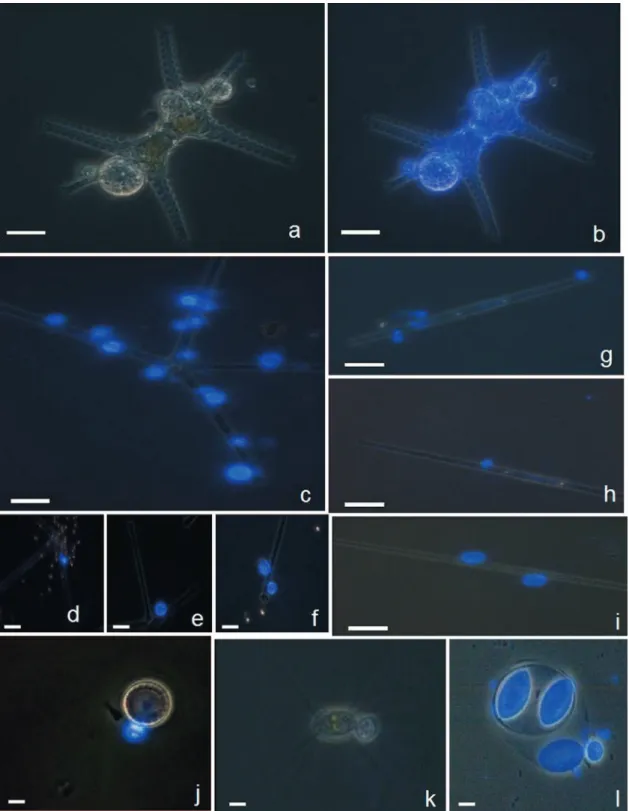
FIG. 2. Examples of microscopic micrographs of phytoplankton eukaryotes with CFW-stained chytrid parasites, obtained via the fractionated
community approach. Typical morphological taxonomic keys are visible under white light for host cell (i.e., the chlorophyte Staurastrum sp.) (a)
and under UV light for the specific parasitic chytrids (b). Under UV light, chytrid epidemics were diagnosed for a diversified host populations,
including both large size (e.g., the chlorophyte Staurastrum sp. [a and b] and the diatoms A. formosa [c to f] and Synedra sp. [g to i]) and small size
(e.g., the diatom Cyclotella sp. [j] and the chlorophytes C. ciliata [k] and O. lacustris [l]) hosts. Multiple infectious chytrids are visible in most
micrographs and different development stages as well, e.g., young sporangia with visible rhizoidal system (d and g), mature sporangia containing
zoospores (e and h), a mature sporangium discharging its zoospore contents (f), and empty sporangia with chitinaceous wall visible (i). Scale bar,
10
m.
DTTP Millipore filters to the black Whatman filters (data not
shown). However, none of the membrane-retaining
ap-proaches yielded satisfactory images of morphological and
cel-lular features of the host cells, e.g., the presence of chloroplast
or viability of the host cell. In addition, almost all of the
infected phytoplankton individuals observed with the total
community approach appeared to be large diatoms.
Accord-ingly, procedures from the total community approach were
FIG. 3. Examples of microscopic micrographs of phytoplankton prokaryotes (cyanobacteria) with CFW-stained chytrid parasites, obtained using the
fractionated community approach. Typical morphological taxonomic characteristics are visible under white light for host cells (micrographs not shown)
and under UV light for parasitic chytrids on their hosts identified as colonial Gomphosphaeria sp. (a) and Microcystis sp. (b) and as the filamentous
Anabaena flosaquae (c to h). The branched rhizoidal system of the parasite is visible on Microcystis sp. (b). On A. flosaquae, young endobiotic thalli and
encysted zoospores attached by long slender penetration tubes to the host are visible (c and d). In addition, tubular vegetative structure (e), mature sporangia
of irregular pyriformic shape (f), and mature sporangia with protruding papilla for discharging zoospores (g and h) are also visible. Scale bar, 10
m.
excluded from comparisons. We will thereafter focus on the
size-fractionation approach using a 1% (vol/vol) CFW final
concentration (from the stock solution) which substantially
enhanced the observational results. We consider this protocol
to be optimal for the diagnosis and quantitative assessment of
phytoplanktonic chytrid infections in natural samples.
Indeed, the latter procedure yielded the best images for
identification and quantitative assessment of both
phytoplank-ton host cells present in the two size classes (
⬎25 m and ⬍ 25
m) under white light illumination, and their fungal parasites
after switching light to UV excitation. Illustrations from
sam-ples collected in 2006 in Lake Pavin and in 2007 in Lakes Pavin
and Aydat are provided on Fig. 2 and 3. Under white light,
diverse phytoplankton host cells were identified based on
phe-notypic features and on their viability through the integrity of
cell wall and the presence of chloroplasts. Under UV
excita-tion, the wall of chytrid sporangia is well visible because of the
presence of chitin (19, 20), allowing the assessment of
pheno-typic keys for identification. These keys include the shape of
thallus (e.g., Fig. 3c,d), the rhizoidal system (e.g., Fig. 3e to h)
and, at times, different development stages of parasites (e.g.,
Fig. 2d to f and 3e to f) (10, 13, 44). Furthermore, the
size-fractionation approach allowed the diagnosis of fungal
phyto-plankton infections not only for large hosts such as diatoms
and colonial and filamentous cyanobacteria in the
⬎25-m size
fractions but also for small nanophytoplankton cells such as
Cyclotella sp., Chodatella ciliata, or Oocystis lacustris (e.g., Fig.
2j to l) in the
⬍25-m size fractions.
Comparative data from the optimal protocol proposed are
presented in the following sections only for the field survey,
i.e., samples collected in 2007 in both lakes, mainly as a “proof
of concept” rather than a valid comparison of sites versus
seasons.
Preliminary data on the natural dynamics of hosts versus
parasites using the proposed protocol. (i) Host community
composition.
Our size-fractionation approach resulted in an
apparent increase in the diversity of infected phytoplankton
populations, compared to previous studies. With this
ap-proach, a total of nine phytoplankton species belonging to
Cyanobacteria, Chrysophyceae, and Chlorophyceae were found
with fungal parasites (Fig. 2 and 3). The major hosts were the
diatoms A. formosa, Synedra spp., Fragilaria crotonensis, and
Cyclotella sp., and the Chlorophyceae Staurastrum paradoxum,
Staurodesmus incus, and C. ciliata (see Table 2). For a long
time, diatoms, primarily Asterionella spp., have been described
from light microscopy as the preferred hosts for chytrid
infec-tions in several lakes: English Lake District (12) and
Shearwa-ter Lake (39), United Kingdom; Lake Leman, Switzerland
(37); Lake Maarsseveen, The Netherlands (48); and two lakes
in Colorado (29). Reports on the chytrid epidemic on green
algae are more episodic, including host species O. lacustris in
Lake Walensee, Switzerland (22), and the desmids Staurastrum
spp. in Lake Windermere (11) and Shearwater Lake (40),
United Kingdom. Our finding of chytrid infections among
pro-karyotes is new. This occurred only in summer in the eutrophic
Lake Aydat and affected three species of cyanobacteria:
Anabaena flosaquae, Gomphosphaeria sp., and Microcystis sp.
(Fig. 3). The latter two species were detected once in one of
the replicate samples from Lake Aydat. This corroborates our
recent suggestion that chytrid infections and the related
bio-geochemical processes in aquatic systems, primarily through
parasitisms and saprophytism, may represent ecologically
im-portant driving forces in the food web dynamics (17, 31–33).
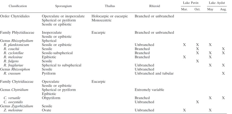
(ii) Parasite community composition.
Based on
morpholog-ical features, the majority of fungal parasites were tentatively
identified as monocentric (i.e., with one center of growth and
development) and eucarpic (using part of the thallus for the
fruit-body and with a specialized rhizoidal system). This is
characteristic of the order Chytridiales, with two families, four
genera, and about ten different species (cf. Table 1 and the
identification keys) recorded in our natural samples. The
fam-ily Phlyctidiaceae contains two genera: Rhizosiphon, which
comprised typical parasites of cyanobacteria and normally
har-bors tubular rhizoids that radiate from the bodies of sporangia
(Fig. 3c to h), and Rhizophidium, which is the largest and most
complex genus of chytrids parasitizing diatoms and
chloro-phytes, especially desmids (10, 44). The second family (i.e.,
Chytridiaceae) was represented by two genera (Chytridium and
Zygorhizidium) (Table 1) with species known mainly as
para-sites of diatoms and green algae, which have the ability to
maintain the wall of their sporangia after zoospore discharge,
as does Rhizophidium (e.g., Fig. 2i) (10, 44).
Based on the limited number of samples analyzed, few
dif-ferences in the occurrence of parasites were noted between the
two lakes sampled, where the more common species were in
the genera Rhizophidium and Chytridium (Table 1). The
Rhi-zosiphon species (i.e., R. crassum) was observed only in the
eutrophic Lake Aydat as a parasite of the cyanobacteria A.
flosaquae (Fig. 3c to h), while Rhizophidium fulgens and
Chytridium oocystidis were observed only in Lake Pavin (Table
1) as a parasite of the small chlorophyta O. lacustris and C.
ciliata (Fig. 2k and l). Only one species in Lake Pavin (R.
planktonicum) and two in Lake Aydat (Rhizophidium cyclotella
and C. versatile) were present during the two sampling times
(Table 1). In our samples, one parasite species often was found
on one host species. An exception was R. planktonicum, which
infected different species of diatoms (A. formosa and Synedra
spp). In contrast, the diatom F. crotonensis was found infected
by two different parasite species in Lake Aydat, i.e.,
Rhizo-phidium fragilaria and C. versatile. These findings corroborate
the complexity of parasitic ecology (2, 5, 15, 17) and lifestyles
in chytrids, which can be facultative parasitic (1),
hyperpara-sitic (28, 41), promiscuous (18), symbiotic (45), and/or
multi-specific within a genus (4) but species multi-specific in the majority of
cases (24, 27).
(iii) Quantitative data.
In Lake Aydat, the total abundance
of phytoplankton increased from 4.5 to 18.5
⫻ 10
6cells liter
⫺1between May and August and were higher than those recorded
in Lake Pavin in March (2.5
⫻ 10
6cells liter
⫺1) and October
(3
⫻ 10
6cells liter
⫺1) (Fig. 4a). In samples collected during
spring, phytoplankton communities were dominated by
di-atoms in both lakes, accounting for ca. 75 and 65% of the total
abundance in Lakes Pavin and Aydat, respectively (Fig. 4b).
The main diatom species were A. formosa (32% of total
phy-toplankton abundance) and Synedra spp. (24%) in Lake Pavin
and Melosira italica (57%) in Lake Aydat. In the summer and
autumn, diatoms were replaced by cyanobacteria, the species
A. flosaquae in Lake Aydat and Cylindrospermum maius (i.e.,
with no infection noted) in Lake Pavin, with an increasing
relative abundance from ca. 65% (of the total abundance) in
Lake Pavin to 95% in Lake Aydat. In addition, chlorophytes
were also quantitatively important in Lake Pavin in autumn
(27% of the total abundance), with O. lacustris (23% of total
abundance) as the major species.
The total abundance of fungal sporangia increased from the
oligotrophic Lake Pavin to the eutrophic Lake Aydat, similar
to the total abundance of phytoplankton. This may represent a
universal pattern, although few quantitative seasonal studies
on fungal parasites are sufficiently complete to permit
gener-alization (21). Indeed, the abundance of sporangia was higher
in spring than in summer-autumn. The counts were 32.9
(March) and 3.9 (October)
⫻ 10
3sporangia liter
⫺1for Lake
Pavin and 10.7 (May) and 1.1 (August)
⫻ 10
3sporangia liter
⫺1for Lake Aydat. The fluctuation in the number of sporangia
thus seems to increase with the trophic status of the lake but
may instead be related to the seasonal changes in the host
FIG. 4. Variations in the numerical (a and c) and relative (b and d) abundances of phytoplankton and their chytrid parasite (i.e., sporangia)
communities. Samples were collected in triplicate (i.e., from three independent sampling operations for each sampling date) on two occasions
during two different seasons in the eutrophic Lake Aydat and in the oligotrophic Lake Pavin. The variability between replicates was low, with a
coefficient of variation that was always
⬍10%.
TABLE 1. Morphological features and occurrence of phytoplankton chytrid parasites in the euphotic layer of Lakes Pavin and Aydat sampled
each on two occasions during different seasons
aClassification Sporangium Thallus Rhizoid
Lake Pavin Lake Aydat Mar. Oct. May Aug.
Order Chytridiales
Operculate or inoperculate
Holocarpic or eucarpic
Branched or unbranched
Spherical or pyriform
Monocentric
Sessile or epibiotic
Family Phlyctidiaceae
Inoperculate
Eucarpic
Branched or unbranched
Sessile or epibiotic
Genus Rhizophydium
Spherical
R. planktonicum
Sessile or epibiotic
Unbranched
X
X
X
R. couchii
Sessile
Branched
X
X
R. cyclotellae
Sessile-subspherical
Branched
X
X
X
R. melosirae
Epibiotic
Branched
X
X
R. fulgens
Sessile
X
R. fragilariae
Spherical to subspherical
Unbranched
X
X
Genus Rhizosiphon
Sessile
Unbranched
R. crassum
Pyriform
Unbranched and tubular
X
Family Chytridiaceae
Operculate
Eucarpic
Sessile or epibiotic
Genus Chytridium
Spherical or pyriform
Extremely variable
Epibiotic
C. versatile
Obpyriform
Branched
X
X
C. oocystidis
Unbranched
X
Genus Zygorhizidium
Sessile
Z. melosirae
Ovate
Unbranched
X
X
aTentative identifications are based on phenotypic keys (see the text).
community composition. Indeed, the numerical abundance of
sporangia appeared to increase with the increasing relative
importance of diatoms within the phytoplankton communities
due to the infection from Rhizophidium spp., Zygorhizidium
melosirae, and C. versatile (Fig. 4b to d). Diatoms are well
known as preferred hosts for chytrid epidemics in the plankton
(24, 27), likely because of the large cell size and capacity of
diatoms to form blooms, thereby increasing the probability
for fungal propagule attachment and development (39).
This may help explain why, in contrast to their abundances,
the host community was less diverse in Lake Pavin, where
fewer species dominated host communities than in Lake
Aydat (Fig. 4b and d).
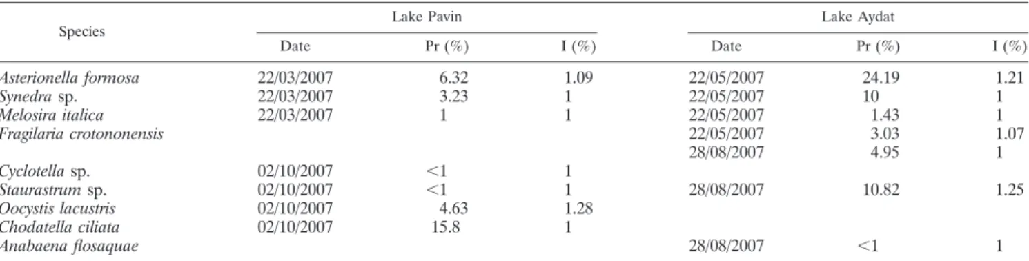
The data on the prevalence and intensity of chytrid infection
show that the prevalence increased with increasing trophic
status and ranged from
⬍1 to 16% in Lake Pavin and from 1
to 24% in Lake Aydat. The more vulnerable populations were
the chlorophyte C. ciliata recorded in October in Pavin and the
diatom A. formosa recorded in May in Aydat. Other highly
exposed host populations (i.e., prevalence
⬎ 5%) included the
A. formosa diatom in Lake Pavin and the Synedra spp. diatoms
and Staurastrum spp. chlorophytes in Lake Aydat (Table 2),
confirming the importance of diatoms and other
bloom-form-ing phytoplankton as preferred hosts for chytrid epidemics (10,
24, 27, 39, 44). The calculation of the intensity of infection
revealed the occurrence of multiple infections (i.e.,
⬎1
para-sites per host cell) for A. formosa in both lakes, O. lacustris in
Lake Pavin, and F. crotonensis and Staurastrum spp. in Lake
Aydat (Table 2 and Fig. 2).
Conclusions.
The present study describes a routine
size-fractionation, CFW staining approach for diagnosing,
identi-fying, and counting phytoplankton chytrid parasites in pelagic
samples. The approach is based on the concentration of large
initial volumes and size partitioning of samples, a step that we
judged necessary in order to yield good analytic images of
infectious sporangia for an accurate diagnosing and
identifica-tion of parasites. In addiidentifica-tion, our approach yields
freeze-con-served particulate DNA samples for quantifying the propagule
stages (i.e., zoospores) of chytrids via FISH targeting of a
specific rRNA oligonucleotide probe that we have recently
designed (M. Jobard et al., unpublished data). Our protocol
can therefore be combined with modern molecular biology
protocols such as fluorescence in situ hybridization-targeting
or cloning/sequencing. Applied to field samples, the approach
provides quantitative preliminary data on infectious sporangia
within phytoplankton communities in two contrasted lake
en-vironments, which were consistent with ecological
consider-ations known from pelagic habitats and host versus parasite
populations. When the samples are analyzed immediately, the
approach does not require toxic fixatives and the related
dis-advantages such as losses during sample storage.
ACKNOWLEDGMENTS
S.R. and M.J. were supported by Ph.D. fellowships from the French
Ministe
`re de la Recherche et de la Technologie and from the Grand
Duche
´ du Luxembourg (Ministry of Culture, High School, and
Re-search), respectively. This study was supported by a grant from the
French ANR Programme Blanc DREP (Diversite
´ et Ro
ˆles des
Eumy-ce
`tes dans le Pe
´lagos) (T.S.-N.).
We thank C. Portelli and D. Sargos for their logistic, technical, and
field assistance.
REFERENCES
1. Alster, A., and T. Zohary. 2007. Interactions between the bloom-forming dinoflagellate Peridinium gatunense and the chytrid fungus Phlyctochytrium sp. Hydrobiologia 578:131–139.
2. Amon, J. P., and R. D. Arthur. 1981. Nutritional studies of a marine Phlyc-tochytrium sp. Mycologia 73:1049–1055.
3. Barr, D. J. S. 2001. Chytridiomycota, p. 93–112. In D. J. McLaughlin, E. G. McLaughlin, and P. A. Lemke (ed.), The mycota, vol. VII, part A. Springer-Verlag, New York, NY.
4. Barr, D. J. S., and C. J. Hickman. 1967. Chytrids and algae I: host-substrate range, and morphological variation of species of Rhizophydium. Can. J. Bot. 45:423–430.
5. Booth, T. 1971. Distribution of certain soil inhabiting chytrid and chytridi-aceous species related to some physical and chemical factors. Can. J. Bot. 49:1743–1755.
6. Bourelly, P. 1970. Les algues d’eau douce. N. Boube´e & Cie, Paris, France. 7. Bush, A. O., K. D. Lafferty, J. M. Lotz, and A.W. Shostak. 1997. Parasitology meets ecology on its own terms: Margolis et al. revisited. J. Parasitol. 83: 575–583.
8. Canter, H. M. 1946. Studies on British chytrids. I. Dangeardia mammillata Schro¨dor. Trans. Br. Mycol. Soc. 29:128–134.
9. Canter, H. M. 1947. Studies on British chytrids. II. Some new monocentric chytrids. Trans. Br. Mycol. Soc. 31:94–105.
10. Canter, H. M. 1950. Fungal parasites of the phytoplankton. I. Studies on British chytrids X. Ann. Bot. 14:263–289.
11. Canter, H. M. 1968. Studies on British chytrids XXVII. Rhizophydium fugax sp. nov. a parasite of planktonic cryptomonads with additional notes and records of planktonic fungi. Trans. Br. Mycol. Soc. 51:699.
12. Canter, H. M., and J. W. G. Lund. 1948. Studies on plankton parasites. I. Fluctuations in the numbers of Asterionella formosa Hass in relation to fungal epidemics. New Phytol. 47:238–261.
13. Canter, H. M., and J. W. G. Lund. 1951. Studies on plankton parasites. III. Examples of the interaction between parasitism and other factors determin-ing the growth of diatoms. Ann. Bot. 15:359–371.
TABLE 2. Prevalence and intensity of chytrid infection for different phytoplanktonic populations
aSpecies
Lake Pavin Lake Aydat
Date Pr (%) I (%) Date Pr (%) I (%)
Asterionella formosa
22/03/2007
6.32
1.09
22/05/2007
24.19
1.21
Synedra sp.
22/03/2007
3.23
1
22/05/2007
10
1
Melosira italica
22/03/2007
1
1
22/05/2007
1.43
1
Fragilaria crotononensis
22/05/2007
3.03
1.07
28/08/2007
4.95
1
Cyclotella sp.
02/10/2007
⬍1
1
Staurastrum sp.
02/10/2007
⬍1
1
28/08/2007
10.82
1.25
Oocystis lacustris
02/10/2007
4.63
1.28
Chodatella ciliata
02/10/2007
15.8
1
Anabaena flosaquae
28/08/2007
⬍1
1
aSamples were collected on two occasions during different seasons in the euphotic layer of Lake Pavin and Lake Aydat, and the percent prevalence (Pr) and intensity (I) values were determined. Dates are expressed in the format day/month/year.
14. De Bruin, A. 2006. The potential for coevolution in aquatic host-parasite system. Ph.D. thesis. Netherlands Institute of Ecology of the Royal Academy of Arts and Sciences, Amsterdam, The Netherlands.
15. Dix, N. J., and J. Webster. 1995. Fungal ecology. Chapman & Hall, London, United Kingdom.
16. Gleason, F. H., and D. Macarthur. 2008. The chytrid epidemic revisited. Inoculum 59:1–3.
17. Gleason, F. H., M. Kagami, E. Lefe`vre, and T. Sime-Ngando. 2008. The ecology of chytrids in aquatic ecosystems: roles in food web dynamics. Fungal Biol. Rev. 22:17–25.
18. Gromov, B. V., A. V. Plujusch, and K. A. Mamkaeva. 1999. Morphology and possible host range of Rhizophydium algavorum sp. nov. (Chytridiales): an obligate parasite of algae. Protistology 1:61–65.
19. Hageage, G. J., and B. J. Harrington. 1984. Use of calcofluor white in clinical mycology. Lab. Med. 15:109–112.
20. Hageage, G. J., and B. J. Harrington. 2003. Calcofluor white: a review of its uses and application in clinical mycology and parasitology. Lab. Med. 34: 361–367.
21. Holfeld, H. 1998. Fungal infections of the phytoplankton: seasonality, min-imum host density, and specificity in a mesotrophic lake. New Phytol. 138: 507–517.
22. Huber-Pestalozzi, G. 1944. Chytridium oocystidis (spec. nova?) ein Parasit auf Oocystis lacustris Chodat. Aquat. Sci. 10:117–120.
23. Huber-Pestalozzi, G. 1983. Das Phytoplankton des Su¨sswassers. Schweizer-bart, Stuttgart, Germany.
24. Ibelings, B. W., A. de Bruin, M. Kagami, M. Rijkeboer, M. Brehm, and E. van Donk.2004. Host parasite interactions between freshwater phytoplank-ton and chytrid fungi (Chytridiomycota). J. Phycol. 40:437–453.
25. James, T. Y., F. Kauff, C. L. Schoch, P. B. Matheny, V. Hofstetter, C. J. Cox, G. Celio, C. Gueidan, E. Fraker, J. Miadlikowska, and H. T. Lumbsch.2006. Reconstructing the early evolution of Fungi using a six-gene phylogeny. Nature 443:818–822.
26. James, T. Y., D. Porter, C. A. Leander, R. Vilgalys, and J. E. Longcore. 2000. Molecular phylogenetics of the Chytridiomycota supports the utility of ul-trastructural data in chytrid systematics. Can. J. Bot. 78:336–350. 27. Kagami, M., A. de Bruin, B. W. Ibelings, and E. van Donk. 2007. Parasitic
chytrids: their effects on phytoplankton communities and food-web dynam-ics. Hydrobiologia 578:113–129.
28. Karling, J. S. 1942. Parasitism among the chytrids. Am. J. Bot. 29:24–35. 29. Koob, D. B. 1966. Parasitism of Asterionella formosa Hass by a chytrid in two
lakes of Rawah wild area of Colorado. J. Phycol. 2:41–45.
30. Kudoh, S., and M. Takahashi. 1990. Fungal control of population-changes of the planktonic diatom Asterionella formosa in a shallow eutrophic lake. J. Phycol. 26:239–244.
31. Lefe`vre, E. 2007. Taxonomic and functional diversity of heterotrophic flagel-lates in lakes: molecular approaches. Ph.D. thesis. Universite´ Blaise Pascal, Clermont-Ferrand, France.
32. Lefe`vre, E., B. Roussel, C. Amblard, and T. Sime-Ngando. 2008. The
mo-lecular diversity of freshwater picoeukaryotes reveals high occurrence of putative parasitoids in the plankton. PLoS ONE 3:e2324. doi:10.1371/journal. pone.0002324.
33. Lefe`vre, E., C. Bardot, C. Noel, J. F. Carrias, E. Viscogliosi, C. Amblard, and T. Sime-Ngando.2007. Unveiling fungal zooflagellates as members of fresh-water picoeukaryotes: evidence from a molecular diversity study in a deep meromictic lake. Environ. Microbiol. 9:61–71.
34. Luna, B. S., B. K. Stewart, D. L. Bergeron, C. R. Crausen, J. J. Plorde, and T. R. Fritsche.1995. Use of the fluorochrome calcofluor white in the screen-ing of stool specimens for spores of microsporidia. Am. J. Clin. Pathol. 103:656–659.
35. Monheit, J. G., G. Brown, M. M. Kott, W. A. Schmidt, and D. G. Moore. 1986. Calcofluor white detection of fungi in cytopathology. Am. J. Clin. Pathol. 85:222–225.
36. Mu¨ller, U., and P. Sengbusch.1983. Visualization of aquatic fungi (Chytridi-ales) parasitizing on algae by means of induced fluorescence. Arch. Hydro-biol. 97:471–485.
37. Pongratz, E. 1966. De quelques champignons parasites d’organismes planc-toniques du Le´man. Aquat. Sci. 28:104–132.
38. Prescott, G. W. 1961. Algae of the western great lakes area. W. M. C. Company Publishers, Dubuque, IA.
39. Sen, B. 1987. Fungal parasitism of planktonic algae in Shearwater. I. Occur-rence of Zygorhizidium affluens Canter on Asterionella formosa Hass in rela-tion to the seasonal periodicity of the alga. Arch. Hydrobiol. 76:101–127. 40. Sen, B. 1988. Fungal parasitism of planktonic algae in Shearwater. V. Fungal
parasites of the green algae. Arch. Hydrobiol. Suppl. 79:185–205. 41. Seymour, R. L. 1971. Studies on mycoparasitic chytrids. I. The genus
Septo-sperma. Mycologia 63:83–93.
42. Sigee, D. C. 2005. Freshwater microbiology. John Wiley & Sons, Ltd., Chich-ester, England.
43. Sime-Ngando, T., and H. J. Hartmann. 1991. Short-term variations of the abundance and biomass of planktonic ciliates in a eutrophic lake. Eur. J. Protistol. 27:249–263.
44. Sparrow, F. K. 1960. Aquatic phycomycetes, 2nd ed. University of Michigan Press, Ann Arbor.
45. Trinci, A. P. J., D. R. Davies, K. Gull, M. I. Lawrence, B. B. Nielsen, A. Rickers, and M. K. Theodorou.1994. Anaerobic fungi in herbivorous ani-mals. Mycol. Res. 98:129–152.
46. Tsui, C. K. M., and K. D. Hyde. 2003. Freshwater mycology. Fungal Diversity Press, Hong Kong, China.
47. Utermo¨hl, H.1958. Zur Vervollkommung der quantitative Phytoplankton Methodik. Mitt. Int. Verein. Limnol. 9:1–38.
48. Van Donk, E., and J. Ringelberg. 1983. The effect of fungal parasitism on the succession of diatoms in Lake Maarsseveen I (The Netherlands). Freshw. Biol. 13:241–251.
49. Wilhelmus, K. R., M. S. Osato, R. L. Font, et al. 1986. Rapid diagnosis of Acanthamoeba keratitis using calcofluor white. Arch. Ophthalmol. 104:1309– 1312.