HAL Id: hal-01635507
https://hal.archives-ouvertes.fr/hal-01635507
Submitted on 15 Nov 2017
HAL is a multi-disciplinary open access
archive for the deposit and dissemination of
sci-entific research documents, whether they are
pub-lished or not. The documents may come from
teaching and research institutions in France or
abroad, or from public or private research centers.
L’archive ouverte pluridisciplinaire HAL, est
destinée au dépôt et à la diffusion de documents
scientifiques de niveau recherche, publiés ou non,
émanant des établissements d’enseignement et de
recherche français ou étrangers, des laboratoires
publics ou privés.
Distributed under a Creative Commons Attribution - NonCommercial - NoDerivatives| 4.0
International License
Oxidative stress and the amyloid beta peptide in
Alzheimer’s disease
Clemence Cheignon, M. Tomas, D. Bonnefont-Rousselot, Peter Faller,
Christelle Hureau, Fabrice Collin
To cite this version:
Clemence Cheignon, M. Tomas, D. Bonnefont-Rousselot, Peter Faller, Christelle Hureau, et al..
Ox-idative stress and the amyloid beta peptide in Alzheimer’s disease. Redox Biology, Elsevier, 2018, 14,
pp.450-464. �10.1016/j.redox.2017.10.014�. �hal-01635507�
Contents lists available atScienceDirect
Redox Biology
journal homepage:www.elsevier.com/locate/redox
Review article
Oxidative stress and the amyloid beta peptide in Alzheimer
’s disease
C. Cheignon
a,b, M. Tomas
a,b, D. Bonnefont-Rousselot
c,d,e, P. Faller
f, C. Hureau
a,b, F. Collin
a,b,⁎aLCC (Laboratoire de Chimie de Coordination), CNRS UPR 8241, 205 route de Narbonne, 31062 Toulouse Cedex 09, France bUniversité de Toulouse; UPS, INPT, 31077 Toulouse, France
cDepartment of Metabolic Biochemistry, La Pitié Salpêtrière-Charles Foix University Hospital (AP-HP), Paris, France dDepartment of Biochemistry, Faculty of Pharmacy, Paris Descartes University, Paris, France
eCNRS UMR8258 - INSERM U1022, Faculty of Pharmacy, Paris Descartes University, Paris, France
fBiometals and Biology Chemistry, Institut de Chimie (CNRS UMR 7177), University of Strasbourg, 4 rue B. Pascal, 67081 Strasbourg Cedex, France
A R T I C L E I N F O
Keywords: Oxidative stress Amyloid beta peptide Metal-ions
Reactive oxygen species Oxidative damages
A B S T R A C T
Oxidative stress is known to play an important role in the pathogenesis of a number of diseases. In particular, it is linked to the etiology of Alzheimer’s disease (AD), an age-related neurodegenerative disease and the most common cause of dementia in the elderly. Histopathological hallmarks of AD are intracellular neurofibrillary tangles and extracellular formation of senile plaques composed of the amyloid-beta peptide (Aβ) in aggregated form along with metal-ions such as copper, iron or zinc. Redox active metal ions, as for example copper, can catalyze the production of Reactive Oxygen Species (ROS) when bound to the amyloid-β (Aβ). The ROS thus produced, in particular the hydroxyl radical which is the most reactive one, may contribute to oxidative damage on both the Aβ peptide itself and on surrounding molecule (proteins, lipids, …). This review highlights the existing link between oxidative stress and AD, and the consequences towards the Aβ peptide and surrounding molecules in terms of oxidative damage. In addition, the implication of metal ions in AD, their interaction with the Aβ peptide and redox properties leading to ROS production are discussed, along with both in vitro and in vivo oxidation of the Aβ peptide, at the molecular level.
1. Introduction
Energy conversion is one of the very fundamental process of life. Energy conversion is there since the origin of life and the basic me-chanism, i.e. the use of movement of ions across a semipermeable membrane (chemiosmosis), is present in all living organisms. Also, the overall design of the enzyme that converts the ion gradient into che-mical energy in form of ATP is the same throughout the living beings
[1].
Electron transfer reactions are used to form the ion gradient across a membrane. In other words, these are redox reactions, in which elec-trons are passed in a chain from afirst donor via several intermediates to afinal acceptor. For humans, animals and a lot of other beings, the final electron acceptor is dioxygen. An advantage of this final acceptor is its high redox potential and hence the high energy in the reaction:
O2+ 4 e-+ 4H+→ 2H2O (1)
The electron donors are in principle the food we take up. Thus the energy we need for living stems from a redox reaction between food (and its transformed products) and O2. In reaction(1), O2accepts four
electrons and four protons to produce two molecules of water. In re-action(2), a partial O2reduction produces the superoxide anion (O2•–),
hydrogen peroxide (H2O2) and the hydroxyl radical (HO•).
⟶+ ⋅−⎯+⎯⎯⎯⎯⎯⎯⎯⎯⎯+ → ⟶+ ⋅ + −
− − + −
O2 e O2 e H O e HO HO
2H
2 2 (2)
These intermediates are potentially dangerous, because they are either very reactive, and hence difficult to control (like HO•), or they are
precursors that easily form very reactive and uncontrollable species (like O2•–+ NO→ peroxynitrite). While thermodynamically favored,
http://dx.doi.org/10.1016/j.redox.2017.10.014
Received 26 September 2017; Received in revised form 14 October 2017; Accepted 17 October 2017
⁎Corresponding author at: LCC (Laboratoire de Chimie de Coordination), CNRS UPR 8241, BP 44099, 205 route de Narbonne, 31077 Toulouse Cedex 4, France.
E-mail address:fabrice.collin@univ-tlse3.fr(F. Collin).
Abbreviations: 4-HNE, 4-HydroxyNonenal; AD, Alzheimer’s Disease; AICD, Amino-terminal APP Intra Cellular Domain; ApoE, Apolipoprotein E; APP, Amyloid Precursor Protein; ATP, Adenosine TriPhosphate; Aβ, Amyloid beta peptide; AβDP, Aβ-Degrading Proteases; CNS, Central Nervous System; CSF, CerebroSpinal Fluid; CTF, CarboxyTerminal Fragment; CYP27A1, sterol-27-hydroxylase (cytochrome P450); CYP46A1, cholesterol-24-hydroxylase (cytochrome P450); DNA, DeoxyriboNucleic Acid; ENDOR, Electron Nuclear Double Resonance; ESI-MS, ElectroSpray Ionisation Mass Spectrometry; GlcNAc, N-acetyl-D-glucosamine; HYSCORE, Hyperfine Sublevel Correlation; ITC, IsoThermal Calorimetry; LRP1, Low density lipoprotein receptor-related protein 1; MALDI-TOF, Matrix-Assisted Laser Desorption Ionisation– Time Of Flight; MCO, Metal-Catalyzed Oxidation; MS/MS, tandem Mass Spectrometry; NMR, Nuclear Magnetic Resonance; PSEN1, PSEN2, genes encoding for Presenilin-1 and -2; RNA, RiboNucleic Acid; ROS, Reactive Oxygen Species; SH-SY5Y, neuroblastoma cell line; SOD, SuperOxide Dismutase; XAS, X-ray Absorption Spectroscopy
Available online 18 October 2017
2213-2317/ © 2017 The Authors. Published by Elsevier B.V. This is an open access article under the CC BY-NC-ND license (http://creativecommons.org/licenses/BY-NC-ND/4.0/).
O2reaction with organic electron donors are kinetically prevented by
the triplet ground state of O2. Thus the reaction(1)can be well
con-trolled as such that little partial reduction (reaction (2)) occurs. The partially reduced oxygen species O2•–, H2O2, HO•belong to the family of
compounds called reactive oxygen species (ROS). ROS are broadly de-fined as oxygen-containing chemicals with reactive properties[2]. The life in aerobic environment and with O2 as afinal electron acceptor
results in a constant production of ROS in our body. ROS are produced enzymatically (for instance in macrophages to kill invaders) or non-enzymatically, as a side reaction. Latter is the case in the respiratory chain, where the overall physiological reaction is(1), but“unwanted” side reactions leak ROS. Due to the importance of energy conversion, most ROS produced in the body come from the respiratory chain and are hence potentially dangerous. Thus several enzymes and small compounds exist to control the levels of ROS. Generally, ROS are kept at a low level but not fully eliminated. As they have messenger function, their total suppression is detrimental. Accumulation of too high levels of ROS is dangerous and defined as oxidative stress. ROS accumulation can occur either by an overproduction or an insufficient elimination of ROS.
Elimination can occur by different mechanisms and is performed by an antioxidant compound. By definition, an antioxidant compound is an endogenous or exogenous molecule that "when present in low con-centrations compared to that of an oxidizable substrate significantly delays or inhibits the oxidation of the substrate"[3]. Diverse mechan-isms are possible like i) scavenging of ROS, ii) quenching of ROS sources and iii) regeneration of endogenous antioxidants[4].
Considering the central role of oxygen, the various systems of pro-duction and elimination of ROS and their regulations, it is not aston-ishing that oxidative stress has been observed in a multitude of diseases. Moreover, oxidative stress can enter into a vicious cycle, as the pro-duced ROS can destroy biomolecules, which may lead to higher ROS accumulation. For instance, when ROS attack metalloproteins, it can lead to the release of redox-competent metal ions with a subsequent increase of ROS production (see below).
In neurodegenerative diseases like Alzheimer’s and Parkinson’s, the brains show oxidative damage and oxidative stress often seem to be implicated in many of them. The brain might be particularly sensitive to oxidative damage upon oxidative stress due to the very high dioxygen consumption of the brain (20% of the total body consumption). But not only that, Halliwell listed 13 points called“problems of the brain”, that could explain the high sensitivity, including the somehow surprising modest antioxidant defense of the brain[5]. Although the occurrence of oxidative stress in several neurodegenerative diseases is relatively well established, the question of“cause or consequence” is much more dif-ficult to answer. The question is important as the time point when oxidative stress occurs in the etiology is key for the validity/efficiency as a therapeutic target.
2. Linking oxidative stress and AD
2.1. Definition of AD and hallmarks
In 1907, Aloïs Alzheimer related in the article “Über eine eigen-artige Erkankung der Hirnrinde” (“On an unusual Illness of the Cerebral Cortex”) the uncommon case of a 51-year-old patient who was suffering from memory loss, disorientation, hallucinations and cognitive im-pairment. After the death of the patient, post-mortem examination showed an atrophic brain with “striking changes of the neurofibrils” and“minute military foci” caused by the “deposition of a special sub-stance in the cortex” [6]. One century later, this “unusual illness” named Alzheimer’s Disease (AD) has become the most widespread neurodegenerative disease whose etiology is still unknown [7]. Ac-cording to the World Alzheimer Report[8], 46.8 million people were suffering from dementia worldwide in 2015; this number is expected to almost double every 20 years. Approximately 5–8% of individuals over
age 65, 15–20% over age 75, and 25–50% over age 85 are affected by dementia[9]. The major prevalence is in Asia (22.9 million people) while Europe and the Americas account for 10.5 and 9.4 million people, respectively. AD is the most common form of dementia, accounting for 50–75% of all dementias[9].
AD is characterized by a progressive deterioration of cognitive functions that can be linked to a significant reduction of the volume of the brain in AD patients as compared to healthy patients[10]. The atrophy results from the degeneration of synapses and the death of neurons, in particular in hippocampus[11], the brain region playing a role in memory and spatial orientation. The age is the highest risk factor for AD, the risk of developing the disease reaching 50% for individuals beyond age 85[9]. Women are more susceptible than men to suffer from AD, because of their higher life expectancy, and because the de-crease in estrogen levels due to menopause could inde-crease the risk of developing AD[12].
Apart from the global reduction in the brain volume, one of the hallmarks of AD is the presence of amyloid plaques in brain, caused by the“deposition of a special substance in the cortex”, as firstly described by Aloïs Alzheimer. These plaques, also named senile plaques, are found in the extracellular space of AD brain and are particularly present in the hippocampus region. They are mainly composed of a peptide, named Amyloid-β (Aβ), that is aggregated and forms mostly β-sheet richfibrils [13]. Another hallmark of the disease is the presence of intracellular neurofibrillary tangles in the brain[14], also observed in Parkinson’s disease (PD)[15]and composed of hyperphosphorylated Tau protein[16]. This microtubule-associated protein normally inter-acts with tubulin to stabilize microtubules. In AD and PD, Aβ would cause an activation of p38 MAPK in cell that leads to the abnormal phosphorylation of Tau[17]. This latter induces accumulation as paired helicalfilaments that aggregate inside neurons in neurofibrillary tan-gles, making the microtubules unstable and causing the loss of neuron functionality.
2.2. Aβ and the amyloid plaques formation
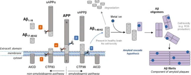
The Aβ peptide is a 38- to 43- amino acid residue peptide whose 1-letter code sequence is DAEFRHDSGYEVHHQKLVFFAEDVGSNKGAIIGL MVGGVVIAT. It is generated after enzymatic cleavage byβ- and γ-se-cretases of APP, the Amyloid Precursor Protein, a type-1 trans-mem-brane protein expressed in various tissues, especially in the central nervous system (CNS) [18]. Its major neuronal isoform encompasses 695 amino acid residues[19]. Although its physiological function is still unclear, APP would play an important role in brain development, memory and synaptic plasticity[19]. The metabolism of APP can follow two different pathways (Fig. 1). In the non-amyloidogenic one (pre-dominant), APP isfirst cleaved by α-secretase and then by γ-secretase to form truncated Aβ17–40/42(P3) peptides or byβ-secretase to lead to the
formation of the truncated Aβ1–16peptide. In the amyloidogenic one,
which occurs to a minor extent, APP is cleaved consecutively byβ- and γ-secretases leading to the formation of full-length Aβ peptides (mainly Aβ1–40/42). Both pathways also lead first to the formation of
amino-terminal fragments (secreted APP (sAPP)α or β) and carboxyterminal fragments (CTF83 or CTF99) and then to the formation of the amino-terminal APP intracellular domain (AICD)[20]. The latter one is in-volved in nuclear signalization.[19]Depending on the exact location of the cleavage byγ-secretase, several lengths of peptide can be released, from Aβ1–38to Aβ1–43. However, the most abundant species produced in
the brain are Aβ1–40and to a lesser extent Aβ1–42. A third way of APP
cleavage has been recently discovered[21]. It involvesη-secretase that cleaves APP at amino acids 504–505 and leads to the generation of the higher molecular mass carboxy-terminal fragments Aη-α and Aη-β, after second cleavage byα- and β-secretase, respectively. The first one, Aη-α, contains the Aβ1–16 peptide in its sequence and was reported to be
neurotoxic.
metabolism[22]. They are mainly produced intracellularly in vesicles like endosomes and released in the extracellular space of healthy brain during neuronal activity, without leading necessarily to Alzheimer’s pathology. Aβ is subject to a proteolytic degradation by Aβ-degrading proteases (AβDPs), which regulates Aβ levels in the brain [23]. Its function in the brain is still unknown, although Aβ could play a role in synaptic plasticity and memory[24].
There are two major forms of AD: the sporadic or late-onset form, the most common one, and the familial or early-onset form, re-presenting less than 5% of the cases [25]. Individuals living with Down’s syndrome (also called trisomy 21) have an increased risk of early-onset AD because they carry an extra copy of chromosome 21 in which is located the gene responsible for APP formation[26]. Muta-tions of several genes (including PSEN1 and PSEN2) coding for APP, Presenilin 1 and Presenilin 2 (two sub-units ofγ-secretase), identified as causative genes, have been found to cause mainly early-onset AD, while ApoE (involved in Aβ clearance) is considered as being the most common high genetic risk factor for late-onset AD[25,27]. The mu-tations on both PSEN1 and PSEN2 lead to a higher Aβ production, PSEN1 mutations specifically conducting to an increased Aβ1–42
for-mation[25]. Sixty-five mutations of APP are indexed in the Alzheimer
Disease & Frontotemporal Dementia Mutation Database, with only 15 being non-pathogenic [28]. As APP mutations can occur in the Aβ
domain, APP proteolysis by bothβ- and γ-secretases can lead to the formation of mutated Aβ peptides (the most frequent ones are pre-sented inFig. 2). The mutations are divided in three categories: mu-tations at the β-secretase cleavage site (N-term), at the γ-secretase cleavage site (C-term) and in the mid-domain amyloid-β region[29]. The mutations at theγ-secretase cleavage site can alter the cleavage position and lead to an increase of the Aβ1–42/Aβ1–40ratio. The
mu-tations at theβ-secretase cleavage site increase the rate of APP pro-teolysis by the β-secretase. The mutations in the mid-domain of Aβ region in APP alters Aβ assembly by increasing the propensity of Aβ to form oligomers andfibrils[30].
AD is a multifactorial disease and the multiple mechanisms related to the disease are unclear. However, since Aβ has been found in healthy brain in soluble form but in aggregated form in AD patient brain[13], a hypothesis has been proposed to explain the formation of the senile plaques. The amyloid cascade hypothesis (Fig. 1) formulated in the early 1990s[31–34]has become a dominant model for AD pathogenesis
[35], although still controversial[36,37]. The hypothesis proposed that an abnormal extracellular increase of Aβ levels in brain could lead to Aβ aggregation into β-sheet rich structures [38]. Aggregation starts with the formation of oligomers species that are reorganized into
protofibrils and fibrils, found in amyloid plaques. Oligomers accumu-lated in AD patient brains [39]are suggested to be the more toxic species for cells[40,41]as they can in particular permeabilize cellular membranes, thus initiating a series of events leading to cell dysfunction and death[42]. According to this hypothesis, other events such as the intracellular formation of neurofibrillary tangles and the disruption of synaptic functions would result from this early and key event. Factors influencing this cascade are modulators and can have an important impact. Regarding oxidative stress, metal ions such as zinc, iron and copper are such modulators and they have been found in amyloid plaques[43]. Cu and Zn are excreted within the synaptic cleft of some neurons. They are supposed to play an important role in aggregation according to the amyloid cascade hypothesis[44], as they can bind Aβ
and thus modulate the aggregation process. They act either on the ki-netics or on the thermodynamics by impacting the morphology of the formed aggregates[45]. Furthermore, amyloid aggregates (low mole-cular weight) with entrapped redox-active metal ions such as copper ions are considered more toxic since they can produce ROS, deleterious for the Aβ peptide itself and for the surrounding biomolecules[46]. 2.3. Oxidation of surrounding molecules
Oxidation of biomolecules in the context of AD is mainly related to neuronal membrane biomolecules and to a disruption of membrane integrity. It involves oxidation of lipids (among them, cholesterol), proteins and nucleic acids, and impairment of Aβ clearance by the low density lipoprotein receptor-related protein (LRP1) due to its oxidation. After a brief reminder of the existence of oxidative stress in AD, the consequences of the oxidation of biomolecules on membrane integrity and protein functionality will be addressed, in relation with AD pa-thogenesis.
Fig. 1. A schematic view of APP proteolytic cleavage. In the non-amyloidogenic pathway, APP isfirst cleaved by α-secretase and then by γ-secretase to form truncated Aβ17–40/42peptides or byβ-secretase leading to the formation of the truncated Aβ1–16. In the amyloidogenic pathway, APP is cleaved consecutively by theβ- and γ-secretases leading to the formation of
full-length Aβ1–40/42peptides. According to the amyloid cascade hypothesis, the Aβ peptide would be further able to interact with metal ions present in the brain and form oligomers and then
fibrils, found in the senile plaques in vivo.
Fig. 2. Most frequent familial AD mutations occurring on Aβ1–43. The amino acid residues
mutated and the names of the mutations are colored. (1-letter code). (For interpretation of the references to color in thisfigure legend, the reader is referred to the web version of this article.).[25].
2.3.1. Evidence of brain oxidative/nitrosative stress in AD
Several pieces of evidence suggest that oxidative stress and ni-trosative stress play a key role in the pathogenesis of AD[47]. Oxidative stress occurs early in the course of AD, which would support its role in AD pathogenesis [48], in relation with the presence of Aβ. Indeed, elevated levels of Aβ1–40and Aβ1–42 have been reported to be
asso-ciated with increased levels of oxidation products from proteins, lipids and nucleic acids in AD hippocampus and cortex (Fig. 3) [49]. By contrast, brain regions with low Aβ levels (e.g., cerebellum) did not present high concentrations of oxidative stress markers[50–52]. More recently, it has been confirmed that protein and lipid oxidation was observed in brain regions rich in Aβ, where redox proteomics allowed identification of oxidized proteins in early stages of the disease[53]. In addition to ROS production by Aβ peptides in the presence of metal ions (see the section“Aβ peptide and ROS production” below), mitochon-dria dysfunction has also been involved in AD pathogenesis, via mi-tochondrial ROS generation[54,55]. Biomarkers of oxidative stress in the AD brain have been well documented, with markers of protein, lipid, DNA and RNA oxidation[56]. Thus, protein oxidation has been classically evidenced by increased levels of carbonylated proteins, especially in the hippocampus and parietal cortex, i.e. in the brain areas the most involved in AD[51]. In human brain, membrane proteins were more oxidatively damaged than cytoplasmic proteins [57]. Protein modification also occurred by indirect oxidation due to reaction with 4-hydroxynonenal (4-HNE), a lipid peroxidation product, and by nitra-tion. The latter process leads to a nitrosative stress due to reaction of proteins with peroxinitrite (ONOO–, that results from reaction of su-peroxide radicals with nitric oxide), and increases the susceptibility of brain proteins to proteosomal degradation[58]. Regarding lipid oxi-dation, increased concentrations of 4-HNE have been reported in the brain regions showing the typical histopathologic alterations of AD (i.e., hippocampus)[59]. Oxidative modification of lipoic acid by 4-HNE was
detected in AD brain [60], and 4-HNE-lysine adducts were increased not only in neurons containing neurofibrillary tangles but also in “ap-parently” normal pyramidal neurons located in the hippocampal tissue sections [61]. Oxidation of nuclear and mitochondrial DNA has also been reported in AD, with increased levels of oxidized bases (i.e., 8-oxo-2-dehydroguanine, 8-hydroxyadenine, 5-hydroxyuracil) in temporal, parietal and frontal lobes[62,63]. Increased levels of 8-hydroxyguanine have even been detected in the hippocampus of patients with a pre-clinical stage of AD[64]. This oxidative stress, especially oxidative DNA
damage, has been detected not only associated with the most vulner-able regions, but also in peripheral AD blood cells[65]. RNA oxidation also occurred, especially mRNA oxidation in the frontal cortex[66].
2.3.2. Consequences of the oxidation of biomolecules on membrane integrity and protein functionality
Alteration of functional integrity of neuronal membranes in AD could result from interactions between amyloid-forming proteins and membranes, leading to membrane permeabilization via several hypo-thetic mechanisms such as transmembrane oligomeric pore structures
[67]. Besides this process, oxidative stress by itself could be responsible for a disruption of membrane integrity. As an example, lipid perox-idation could be involved in a loss of phospholipid asymmetry in sy-naptosomal membranes[68]. Indeed, this asymmetry is maintained by the ATP-dependent enzyme aminophospholipid-translocase orflippase, whose activity depends on at least one critical cysteine residue, possibly oxidized by 4-HNE. This lipid peroxidation product can conjugate with several membrane proteins, resulting in alterations of their structure and function, with a consequent neurotoxicity in AD brain[69]. Pro-teins involved in glycolysis and ATP production could thus become dysfunctional, and this impairment of brain energy metabolism, sec-ondary to oxidative stress, seems to be a key event in AD[70]. Re-ciprocally, decreased ATP levels could result in electron leakage and increased mitochondrial ROS production, thereby generating another source of oxidative stress in AD[71]. Several proteins directly involved in glucose metabolism and ATP synthesis have been reported to be inactivated by oxidation in AD brain (e.g., fructose biphosphate aldo-lase, triose phosphate isomerase, glyceraldehyde phosphate dehy-drogenase, phosphoglucose mutase, enolase, pyruvate kinase) [72]. ATP synthase itself could be oxidatively modified and consequently inactivated in AD brain, theα subunit of the enzyme being a target for oxidative damage at the very early stages of AD[73,74]. In advanced stages of AD, ATP synthase activity was also decreased in AD brain
[75]. This decreased activity would result from a direct binding be-tween Aβ and ATP synthase and from inhibition of O-GlcNAcylation of the Thr432 residue on the ATP synthase subunit α[76]. Oxidation-induced impairment of enzymes involved in ATP production could be related with transportation abnormalities and dysfunction of in-tracellular glucose catabolism in AD[77]. Interestingly, the alterations of metabolic disorders could be supported by the link between AD and diabetes [78]. Accordingly, it has been recently shown that mTOR
Fig. 3. Induced oxidative stress in cell of AD brain regions of high Aβ levels, where Aβ-metals is one of the production source for ROS. 4-HNE = 4-hydro-xynonenal; 8-oxo-dG = 8-oxo-dehydroguanine. Orange star indicates oxidative damages. (For inter-pretation of the references to color in thisfigure le-gend, the reader is referred to the web version of this article.).
(whose signaling pathway plays a key role in regulating cell growth as well as lipid and glucose metabolism), aberrantly activated in AD from early stages, would be involved in AD neurodegeneration, via an in-hibition of both insulin signaling and alteration of protein homeostasis
[79]. Similarly, in the triple transgenic mouse model of AD (3xTg-AD) that develops both Aβ and Tau pathologies in an age-dependent manner, oxidative and nitrosative stresses have been suggested to contribute to impairment of insulin signaling in AD brain[80].
Oxidative stress could be involved in the clearance of Aβ. It has thus been hypothesized that Aβ would oxidize LRP1, leading to accumula-tion of the neurotoxic peptide Aβ in the brain. Indeed, LRP1 is a mul-tifunctional protein that is notably in charge of the efflux of Aβ from the brain to the blood, across the blood-brain barrier [81,82], and LRP1 activity is decreased in AD[83]. Thus, Aβ, by oxidizing LRP1, would
lead to disruption of its own clearance[84]. LRP1 oxidation has been evidenced by the presence of 4-HNE-LRP1 adducts in AD hippocampus. Such alteration of Aβ clearance would lead to an increased Aβ accu-mulation in the brain, which could be a determinant factor in AD pa-thogenesis.
Protein Tau also constitutes a target for oxidative stress in AD. As an example, 4-HNE is able to induce modifications of protein Tau con-formation, which supports the involvement of oxidative stress (notably induced by Aβ) in the pathogenesis of AD, by favoring neurofibrillary tangles formation[85]. Nitration of protein Tau could also promote a conformation change that may favorfibril assembly. It constitutes an early event in AD, since the appearance of nitrated Tau in neuro fi-brillary tangles appears essentially before the maturation of Tau in-clusions[86]. Moreover, due to the role of protein Tau both in micro-tubule dynamics and in the protection of neuronal genomic DNA and of cytoplasmic and nuclear RNA towards ROS-induced damage, Tau al-teration would lead to increased DNA and RNA oxidation[87]. Some authors have suggested that the DNA repair proteins might be in-activated by oxidative modifications, which could result in impaired DNA repair capabilities via the base excision repair pathway[88,89]. It is noteworthy that oxidation of DNA can result, in addition to base oxidation, in DNA strand breaks, which could contribute to neurode-generation by favoring the formation of neurofibrillary tangles[90].
Cholesterol in cell membranes, more specifically in microdomains rich in cholesterol named lipid rafts, is able to bind to APP, thereby promoting its insertion into the phospholipid monolayers; this induces the activity of the β-secretase, thus favoring the amyloidogenic pathway, by accumulation of Aβ1–42peptide[91]. More precisely,
es-terified cholesterol (and not free cholesterol) enhanced Aβ formation
[92], so that the balance between free and esterified cholesterol
con-stitutes a modulator of amyloidogenesis. Cholesterol can be oxidized in vivo, to form oxysterols that represent a way to eliminate excess cho-lesterol from the brain; this way prevent chocho-lesterol accumulation, since the brain cannot degrade cholesterol. Oxysterols can thus equili-brate the local synthesis of sterols in brain[93]. It is noteworthy that oxysterols could modify specific sites on Aβ, e.g. at Lys16, which could increase Aβ aggregation and neurotoxicity. Among oxysterols, 3β-hy-droxy-5-oxo-5,6-secocholestan-6-al, that can be converted into its aldol form, can bind to an amine of Aβ to lead to a Schiff base[94]. This covalent modification of Aβ increases its amyloidogenicity [95], by decreasing the aggregation critical concentration and favoring the for-mation of spherical aggregates[96]that are neurotoxic[97]. Another oxysterol, named 24-hydroxycholesterol, is produced in the brain by action of the cholesterol 24-hydroxylase (CYP46A1) and can cross the blood brain barrier[98]. This oxysterol is involved in the regulation of cholesterol homeostasis in the brain, by inducing apoE-mediated efflux of cholesterol in astrocytes via a liver X receptor (LXR)-controlled pathway, which may be a process involved in the pathogenesis of AD
[99]. It has been observed that another oxysterol, 27-hydro-xycholesterol, produced in the brain by CYP27A1, mostly goes from the circulation to the brain by crossing the blood brain barrier [100]. Consequently, two main oppositefluxes of oxysterols coexist, i.e.
24-hydroxycholesterol from the brain and 27-24-hydroxycholesterol into the brain, so that the balance between 24-hydroxy- and 27-hydroxy-cho-lesterol would be of importance for amyloidogenesis [101]. The in-creased ratio of 27-hydroxycholesterol to 24-hydroxycholesterol ob-served in AD brains supports this hypothesis[102]. In SH-SY5Y cells (i.e., a human neuroblastoma cell line and a classical model for AD pathology), it has been suggested that 24-hydroxycholesterol would favor the processing of APP to the non-amyloidogenic pathway[103]. Nevertheless, a comprehensive in vitro analysis of APP andα-, β- and γ-secretases has been performed by Gamba et al. [104] in a human neuroblastoma cell line (SK-N-BE) treated with 1 µM of 24-hydroxy- or 27-hydroxycholesterol after differentiation into neuron-like cells. Under these conditions, both oxysterols induced an overexpression of APP and an increasedβ-secretase activity, leading to amyloidogenesis. The contradictory results obtained by Gamba et al. and Prasanthi et al.
[103]could be related to the differences in the oxysterol concentrations tested (1 µM vs. 5–25 µM, respectively) and to the cell treatment with oxysterols (after retinoic acid-driven differentiation to a neuron-like phenotype vs. a direct challenging, respectively). The conditions with 1 µM oxysterols seem much closer to the actual amounts recovered from normal and AD brains and thus more patho-physiologically levant. Interestingly, plasma level of 24-hydroxycholesterol, via its re-lation to the mass of metabolically active neuronal cells, could be used as a marker of brain atrophy in AD patients[105]. In addition to 24-hydr and 27-24-hydrcholesterol of enzymatic origin, other oxy-sterols (including 7-ketocholesterol, 7 α-hydroxycholesterol,4β-hydro-xycholesterol, 5α,6α-epoα-hydroxycholesterol,4β-hydro-xycholesterol, and 5β,6β-epoxycholesterol) deriving from cholesterol autooxidation were detected in post-mortem human AD brain and the change of their levels was associated with AD progression[106].
Finally, it is noteworthy that the genotype of apoE, the main cho-lesterol-carrier protein in brain, impacts oxidative stress, since plasma from AD apoε4 carriers was more oxidized than plasma from AD non-apoε4 carriers [107,108]. This genotype would influence cholesterol
metabolism and formation of oxysterols[109]. Of note, apoE structure could play a role since apoE2 has two Cys residues, whereas apoE3 has only one Cys and apoE4 has no free thiol group; therefore, the lower number of Cys residues in apo E4 would lead to a lesser protection against oxidative stress. Oxidation of apoE, evidenced by analysis of oxidative stress-related modifications of the cerebrospinal fluid (CSF) proteome, could thus affect thiol-mediated antioxidant activity, which would allow excess oxidative damage to the lipoprotein particles and promote Aβ protein aggregation[110].
3. Metal implication in AD
3.1. Role of metals in brain
Like other tissues, the brain contains several essential d-block metal ions, such as Fe, Zn, Cu, Mn, Mo, Cr, Co and non-essential metals. In general, the brain belongs to the organs with the highest d-block metal content per weight. The content of the most abundant d-block ions Fe, Zn and Cu are 0.3 g, 0.1 g and 0.004 g per kg brain, respectively[111]. These three metal ions seem to be the most relevant regarding Aβ and/ or oxidative stress. Fe, Cu and Zn ions are generally bound to proteins, in order to control their reactivity. They have most often the role in metalloproteins of catalytic center, electron transfer site or structural component. Only Zn occurs at higher concentration in non-protein bound forms at certain places, where it seems to play the role of a messenger. The metabolism of these ions is tightly controlled by a machinery that is able to sense the metal concentration, to perform metal transport in the blood or through the membranes, to provide the metal ion during protein folding and maturation, and to stock metal ions. It is well documented that conditions leading to too high or too little metal ions content can be lethal. This is the case for Wilson’s and Menkes’ diseases. First is a Cu-overload, second a Cu deficiency genetic
disease. In either of these diseases, the brain is highly affected, in line with its high metal content[112,113].
3.2. Misregulation in AD
There is a large body of evidence for metal ion misregulation in AD, in particular for Cu, Zn and Fe[114,115]. If the misregulation is an early or late event in AD is still a matter of research. A well-established fact is the accumulation of Cu, Zn and Fe in the amyloid plaques, a hallmark of AD[116]. Interestingly, human amyloid plaques accumu-late much higher metal concentrations than plaques in AD model mice
[117]. APP is also implicated in metal metabolism, as it can promote iron efflux of neurons[118], under the control of an iron-responsible element[119]. Moreover, its transcription was reported as being pro-moted by Cu. Zn and Cu-binding sites where reported in vitro
[120–122]. There are also a multitude of metalloproteins and trans-porters affected in AD, as well as metal concentrations, metal reparti-tion and homeostasis (for recent review see [114]). Importantly, an increase in loosely bound Cu in human AD brains compared to healthy subjects was reported [123]. It is well established that such loosely bound Cu and Fe can promote oxidative stress[5].
3.3. Metals and oxidative stress
Metal ions, in particular Cu, Fe and Mn, play a central role in oxi-dative stress. They are implicated in the production and defense of oxidative stress. Free or loosely bound Cu and Fe are very efficient catalysts of ROS production. They can be reduced to Cu(I) or Fe(II) by physiological relevant reducing agents (like glutathione or ascorbate) and can then react with dioxygen or hydrogen peroxide to form su-peroxide and hydroxyl radicals, respectively[5]. On the other hand, the same metal ions are also present in the catalytic center of antioxidant enzymes, like Cu in SOD1 or Fe in catalase, where they destroy the superoxide anion and H2O2, respectively. This clearly shows the
im-portance of the coordination chemistry. Depending on the coordination site, Cu and Fe can be pro-oxidants or antioxidants. Hence it becomes clear how important the control of these metal ions metabolism is, in terms of concentration, transport, storage and incorporation into active sites. In case of failure of Fe and Cu homeostasis, free or loosely bound Fe and Cu concentrations can increase, which are often competent to catalyze the production of ROS[111]. Such Cu and Fe can also bind to off-target biomolecules and disturb their function, which could also contribute to increased oxidative stress.
4. Aβ peptide and ROS production 4.1. Coordination of Aβ with metal ions
As described above, metal ions such as zinc, iron and copper are present in the brain. They are necessary and required to regulate the neuronal activity in the synapses and are involved in biological func-tions of metallo-proteins. In several diseases such as AD, the metal ion homeostasis is disrupted and the concentration and distribution are far from the physiological ones. In particular, Cu and Zn levels can reach up to three times the normal levels observed in healthy brains[124]. Moreover, high content of these metal ions is found in amyloid plaques extracted from AD brains[43]. As they can bind to Aβ under
physio-logical concentrations, their coordination modes are of interest to un-derstand their role in AD.
4.1.1. Zn(II) coordination to the Aβ peptide
Zn ion exists only as Zn(II) and its coordination to Aβ is still not well-established.[125–127]Although it is consensual that a complex 1:1 is formed[126], the nature of the amino acid residues involved in the coordination sphere is still under debate. A novel binding model has recently been proposed, based on Nuclear Magnetic Resonance (NMR) and X-ray Absorption Spectroscopy (XAS) studies of Zn coordination with mutated and N-terminal acetylated peptides[128]. Zn(II) would be bound by imidazole rings of His6 and either His13 or His14 residues, the carboxylate group of Glu11 and the carboxylate group of Asp1, Glu3 or Asp7 (Fig. 4). Zn(II) affinity for Aβ has been investigated by iso-thermal calorimetry (ITC) and competition studies, leading to an affi-nity constant in the 105M-1range, which would permit Zn-Aβ in vivo interaction[129,130].
4.1.2. Cu(II) coordination to the Aβ peptide
Copper is a redox-active ion, physiologically occurring mainly in two redox states: Cu(I) and Cu(II). The Cu(II) coordination to Aβ has been widely studied for years and was challenging as several species are formed depending on the pH. Numerous studies have been realized in the past decade and the results have been recently reviewed
[125,131–133], leading to a consensual model with different Cu(II) binding modes depending on the pH. The two major binding modes, called components I and II, observed around physiological pH, are shown inFig. 4. For components I, it is now well-established that Cu(II) is bound to the NH2terminus, to the adjacent CO function from
Asp1-Ala2 and to imidazole rings of His6 and either His13 or His14
[134–138]. For component II, two distinct models have been proposed. In thefirst one, Cu(II) is bound to the carbonyl function from Ala2-Glu3
and to the imidazole rings of the three His residues[136,138]. In the second one, Cu(II) is bound to the N-terminal amine of Asp1, to the amidyl function of Asp1-Ala2, to the carbonyl group of Ala2 and to the imidazole ring of one His residue[134,135,139]. Although reminiscent to the structure of Cu in the Cu,Zn-SOD, thefirst model does not explain the effect of pH on the coordination as all the residues involved in Cu (II) coordination that can undergo deprotonation are already deproto-nated. The second model explains the change of Cu(II) binding mode that occurs around pH 7.8 with the deprotonation of the Asp1-Ala2 amide function, leading to its coordination. Furthermore, Electron Nuclear Double Resonance (ENDOR), Hyperfine Sublevel Correlation (HYSCORE) and NMR studies highlight the involvement of both the NH2terminus of Asp1 and the deprotonated Asp1-Ala2 amide bond,
favoring the second model (illustrated inFig. 4)[131]. A carboxylate group has also been proposed to be involved in apical position for several components, coming from Asp1[134–136]or from Glu3, Asp7 and Glu11 carboxylates in equilibrium with Asp1 for component I
[135]. Numerous studies on Cu(II) affinity for Aβ have been reported
(for reviews, see Arena & al.[140]and Zawisza & al.[133]). Depending on the method used, two ranges of affinity constants have been re-ported: 109−1010 M−1 for potentiometry and ITC studies, and 107−108M−1for Tyr10fluorescence studies. This difference has been
explained in a more recent paper, proposing that the affinity constant calculated from Tyr10 fluorescence experiments was underestimated because the inner-filter effect was not correctly taken into account
[141]. Furthermore, a Cu(II) affinity constant in the 109M−1range has
also been evaluated based on competition studies, in line with the af-finity values from potentiometry and ITC[142].
4.1.3. Cu(I) coordination to the Aβ peptide
Cu(I) coordination with Aβ has been investigated more recently than Cu(II) coordination and the involvement of histidine residues is now consensual. Several binding models are suggested, two of them being most populated. The first model proposes a linear binding of histidine residues to Cu(I) with a dynamic exchange between His6, His13 and His14, while the second one involves an equilibrium be-tween the His dyad and the His triad for Cu(I) coordination. NMR studies have shown the implication of the three histidine residues in the Cu(I) coordination with a dynamic exchange, in line with the two proposed models[135]. However, XAS studies[135,143]and a com-parison of synthetized Cu(I) complexes His-His dipeptides and Cu(I) complexes with truncated Aβ6–14and Aβ10–14peptides have validated
the model involving a linear binding mode with 2 histidine residues
[144,145]. In addition, according to tandem mass spectrometry (MS/ MS) studies on the Cu(I)-Aβ structure, the two histidine residues mostly involved in Cu(I) coordination would be His13 and His14[146]. Thus, evidences suggest that Aβ is bound to Cu(I) by histidine residues in a linear fashion with a dynamic exchange between His6, His13 and His14, the major form being His13 and His14 dyad (Fig. 4). This is in line with affinity studies realized on three Cu(I) complexes with one His-Ala mutation on Aβ peptide (named H6A, H13A and H14A)
[147–149]that point out a slightly lower affinity than for the native peptide, H6A having a stronger affinity than the other two mutants. These results indicate that Aβ only needs two histidine residues for binding Cu(I), His13-His14 dyad being the major form. To the best of our knowledge, only three studies have been carried out on Cu(I) affi-nity for Aβ, leading to three very different affinity constants of 1015M-1
[148], 1010.4M-1[149]and 107M-1[147]. The two last values are the
most realistic ones and actually in agreement, the difference coming from the value of the formation constant taken into account by the authors for the competitor used (i.e. ferrozine) for evaluating Cu(I) af-finity for Aβ. More investigations have still to be done to determine more precisely the affinity constant and evaluate the biological re-levance of the Cu(I)-peptide interaction.
4.1.4. Fe(II) coordination to the Aβ peptide
Very few structural studies on iron coordination, mainly as Fe(II) and Fe(III) ion, to Aβ have been reported. Fe(III) coordination to Aβ is not possible at physiological pH because of the formation of the highly stable Fe(III)(OH)3precipitate[150]. For Fe(II)-Aβ coordination, to the
best of our knowledge, only one study has been performed by using1H, 13C and 2D NMR, highlighting the involvement of Asp1, Glu3, the three
His but neither Tyr10 nor Met35 in Fe(II) sphere[151]. A comparison of the NMR data of component I of Cu(II)-Aβ (for which the co-ordination mode is well established) with the NMR data obtained for Fe (II)-Aβ, a preferred coordination mode has been proposed (Fig. 4). Both the terminal amine and the carboxylate group of Asp1, the Asp1-Ala2 and His6-Asp7C˭O peptide bonds, the imidazole ring of His6 as well as the one of either His13 or His14 are proposed to be involved in Fe(II)-Aβ. Further investigations would be needed in order to validate this proposition of binding mode and to evaluate its affinity for Aβ as well as to determine the affinity constant of Fe(II) for Aβ.
4.2. ROS production by Aβ-metals
Redox active metal ions such as copper and iron are known to be involved in ROS production. In the presence of a reducing agent, they can have a catalytic activity, by cycling between two redox states[152]. Cu and Fe can be coordinated to Aβ, as detailed above, and the resulting complex could be directly involved in ROS production, thus estab-lishing a direct link between AD and oxidative stress. ROS production has been mostly studied with Cu-Aβ, because Fe-Aβ has a lower redox activity.[153]Iron is found in the amyloid plaques predominantly in a colloidal form (originating from ferritin), but histochemical studies indicate that it could also be bound to Aβ[154]. The coordination mode of Fe(II) with Aβ has been characterized (Fig. 4)[151]but Fe(III) does not form a stable complex with Aβ because it finally converts into Fe (III)(HO)3and precipitates. Thus, the physiological stable formation of
a binary Fe(III)-Aβ is unlikely. However, ROS production by Fe-Aβ still might be relevant as Fe(II)-Aβ is stable and the Fe(III) complex formed during ROS production might not have time to precipitate. As the in-volvement of iron bound to Aβ in ROS production is still unclear, we focus here only on Cu-Aβ.
In the case of copper, the pro-oxidant role of the Cu-Aβ system is not clearly established because the complex is more active in ROS production than several biological relevant Cu-peptides or Cu-proteins
[155]but less efficient than copper in buffer[4,153,155–158]. How-ever, latter is not very relevant biologically, as all Cu in biology is normally coordinated to a biomolecule. In vitro studies have shown that Cu-Aβ is able to catalyze the formation of H2O2and HO•in the
presence of O2 and a reducing agent such as ascorbate (Fig. 5a)
[153,155,156,159]. Although it was generally proposed that H2O2
production by Cu-Aβ occurs via a two-electron process, a recent study has highlighted the formation of superoxide as an intermediate in the production of H2O2by Cu-Aβ and O2[160].
Copper is redox-active and cycles between the +I and +II oxidative states when bound to Aβ. An electrochemistry study has shown that a preorganization mechanism was needed to allow the electron transfer for the oxidation of Cu(I) or the reduction of Cu(II) since the Cu(II) and Cu(I) coordination spheres are very different (Fig. 5b, top)[161]. The energy required for the rearrangement between the Cu(I) and Cu(II) geometries (linear and square-planar respectively) being very high, the electron transfer would rather proceed via a low-populated redox-competent state in which Cu(I) and Cu(II) binding modes are highly similar, thus inducing a low reorganization energy. This transient state, called here electrochemical in-between state, is in equilibrium with the resting states and represents about 1/1000 of all the species in solution. The electron transfer during the metal-catalyzed ROS production has also been proposed to occur via a similar state, called here catalytic in-between state. The copper environment in that state as well as the reactivity towards the substrates (O2or H2O2) have been investigated
by computational studies[162–165]. The nature of the amino acid re-sidues involved in the catalytic in-between state has been studied with MS/MS by identifying the sites of oxidative damage on the peptide
[166], since the latter is oxidized during the metal-catalyzed ROS production. By comparing the non-specific oxidations detected on Aβ28
after the radiation-induced ROS production with the copper-mediated oxidations of Aβ28, Asp1, His 13 and His14 have been found to be the
metal-specific targeted amino acid residues. Furthermore, kinetic stu-dies of the copper-mediated Aβ28 oxidation have shown that Asp1
would be thefirst amino acid residues damaged. Thus, in this study, the proposed ligands for both Cu(II) and Cu(I) coordination in the catalytic in-between state are Asp1, His 13 and His14. As they have been found to be the main targets for HO•, they are supposed to be the amino acid residues the closest from copper during the metal-catalyzed ROS pro-duction. A similar study performed with the full-length Aβ40peptide
has been reported recently, leading to the same conclusion[167]. Finally, in a recent paper, the evaluation of the ROS production by Cu bound to a wide series of modified peptides by fluorescence and UV–Vis–based methods has led to the proposition of a coordination model of the Cu-Aβ complex in the catalytic in-between state involved in ROS production [168]. The terminal amine and the carboxylate group of Asp1 as well as the imidazole group of one His are proposed to be involved in the coordination sphere of both Cu(I) and Cu(II), leading to the electron transfer with a minimal reorganization energy (Fig. 5b, bottom).
5. Oxidative damages undergone by the Aβ peptide
5.1. In vitro damage on Aβ residues during metal-catalyzed oxidation (MCO)
As discussed above, ROS are radicals and molecules deriving from
the incomplete reduction of molecular oxygen. They are produced in small quantity during the in vivo metabolism of oxygen, through four successive 1-electron reductions of O2leading to H2O formation. They
are necessary to maintain the homeostasis in cells and play an im-portant role in signaling[169]but are also reactive oxidants, able to damage biomolecules. In cells, endogenous enzymes are in charge of the antioxidant defense to prevent the ROS mediated damages.[46]
The superoxide (O2•−) anion, produced by the one-electron reduction of
dioxygen, is capable of inactivating few enzymes,[46]but has a poor reactivity with most of the bio-inorganic substrates due to low rate constant (usually below 102L mol-1s-1).[170,171]Hydrogen peroxide
(H2O2) is the product of the one-electron reduction of superoxide. It can
oxidize proteins with thiol groups and is deleterious in the presence of redox-active metal ions such as iron and copper as it can produce the hydroxyl radical during the Fenton or Haber-Weiss reaction. H2O2is
regulated in vivo by two enzymes (catalase and glutathione peroxidase). The hydroxyl radical (HO•) is the result of the third one-electron re-duction of oxygen, and can be produced in the presence of metal ions from H2O2. HO•has a very short half-life (10-9s) compared with O2•−
(10-6s) and is thus the more reactive and deleterious ROS,[169]being able to oxidize the biomolecules such as proteins, lipids, DNA[172]
because of its very high redox potential (E°' = 2.34 V[173]). To control the quantity of pro-oxidants (ROS) and prevent the damages on the biomolecules, the body has protecting mechanisms including enzymatic and chemical antioxidants. However, in some diseases such as AD
[174], an imbalance may occur between pro-oxidants and antioxidants, due to a higher ROS production or a reduced activity of the enzymes responsible for the ROS degradation, leading to oxidative damages on biomolecules[175].
During the metal-catalyzed ROS production, the Aβ peptide un-dergoes oxidative damages. This is in line with the detection of oxidized Aβ in amyloid plaques in vivo [176]. Studies on single amino acid
Fig. 5. (a) Mechanism of ROS production from a reductant and dioxygen catalyzed by the Cu-Aβ complex. The ROS produced are the superoxide anion (O2• −), hydrogen peroxide (H2O2) and the
hydroxyl radical (HO•). (b) Top: Resting states that are the most populated states of Cu(II)-Aβ (left) and Cu(I)-Aβ (right). The redox reaction between these states is sluggish due to a high re-organization energy. Bottom: proposed Cu(I/II) environment in the catalytic in-between state[168].
residue oxidations could allow a prediction on the residues targeted during the MCO of Aβ[177–179]. The physiological main targets for HO•are the sulfur-containing amino acids (methionine, cysteine), the basic amino acids (arginine, histidine, lysine) and the aromatic amino acids (phenylalanine, tyrosine, tryptophan)[180].Table 1provides the main oxidation products of these amino acid residues. Oxidation of Aβ28by HO•produced byγ-radiolysis has shown that His and Phe
re-sidues are mainly targeted[166], in line with the oxidations reported previously for free amino acid residues. However, in the case of MCO of Aβ, the ROS are produced at the metal center. Thus, the oxidations are site-specific and can differ from the amino acid oxidations usually de-tected without metal ion.
5.1.1. Oxidation of aspartate
The Aβ peptide has 3 aspartate residues at positions 1, 7 and 23. In the literature, only Asp1 has been found to be oxidized during MCO of Aβ. Asp1 is involved in the coordination of Cu(II) in the resting state
[131,132]and its involvement in the Cu sphere during ROS production has also been proposed [166,168]. Thus, it would appear as a pre-ferential target for the hydroxyl radical produced at the metal center. Several oxidative damages have been detected during MCO of Asp1 both in the presence of ascorbate[166,167,181]and of hydrogen per-oxide[182]. Fig. 6a summarizes the oxidative mechanism leading to the formation of either pyruvate, isocyanate or 2-hydroxyaspartate function through the formation of an alkoxyl radical. The formation of a pyruvate function upon Asp1 oxidation was previously detected during MCO of Aβ[166,181,182]and would proceed through theα-amidation pathway[177]. The intermediate alkoxyl radical is generated from the hydroperoxide function by reaction that could involve the hydroperoxyl radical (HO2•), the protonated form of the superoxide anion[177]. Asp1
is also subject to a backbone cleavage on theα-position of the peptide
[141,153], leading to an isocyanate function in a reaction mechanism proceeding through the diamine pathway[177]. Asp1 was also found to convert into 2-hydroxyaspartate upon MCO, which corresponds to the formal addition of an oxygen atom[166].
5.1.2. Oxidation of histidine
The Aβ peptide contains 3 histidine residues located at position 6, 13 and 14. They are involved in both Cu(II) and Cu(I) coordination in the resting states by their imidazole ring, and it has been proposed that they are involved in the Cu sphere during ROS production[166,168]. Histidine residues s have been found oxidized into 2-oxohistidine (Fig. 6b) during MCO of Aβ bound to copper in the presence of
ascor-bate[156,166,167,183,184]or hydrogen peroxide[182]. The reaction mechanism of histidine oxidation by HO•radicals starts with an attack at the C-2 position of the imidazole ring [185,186] (Fig. 6b). The
resulting hydroxyhistidinyl radical generated would be further oxidized into 2-oxohistidine after reaction with Cu(II), that is itself reduced in Cu (I)[185]. His13 and His14 have been found to be more sensitive to oxidation, His6 being not detected on its oxidized form[166,182,184]
or affected after longer oxidation time [183]. His13 and His14 were also found converted into dehydrooxohistidine after catalytic photo-oxygenation, in the absence of a metal ion[187].
5.1.3. Oxidation of phenylalanine
Three phenylalanines are present in the Aβ sequence at positions 4, 19 and 20. None of them is involved in the Cu(II) or Cu(I) coordination, nevertheless Phe19 and Phe20 have been found oxidized during MCO of Aβ in the presence of Cu(II) and ascorbate.[166,167]Phe19 and Phe20 has been detected with the formal addition of an oxygen atom, likely oxidized into hydroxyphenylalanine (Fig. 6c).[177]This oxida-tion seems to occur after the oxidaoxida-tion of Asp1 which is involved in Cu binding[166].
5.1.4. Oxidation of tyrosine
Although the amino acid residues involved in copper coordination are more vulnerable to oxidation, non-coordinating amino acid residues can also be oxidized. It is the case for Tyr10 which is sensitive to oxi-dation and is responsible for the Aβ peptide cross-linking by dityrosine formation (Fig. 6d).[177]The latter process, induced by Cu(II), has been detected for Aβ in the presence of H2O2[188]. MCO of Tyr10 into
dityrosine was found to have an impact on aggregation as Aβ cross-linking was correlated with the formation of covalent oligomers
[189,190]. Furthermore, a study has proposed that Tyr10 acts as a gate that promotes the electron transfer from Met35 to Cu(II) for its reduc-tion in Cu(I)[191]. However, this is in contradiction with the stability of Cu(II)-Aβ reported by several group.
5.1.5. Oxidation of methionine
Methionine is an amino acid residue very sensitive to oxidation. In vivo, the enzyme methionine sulfoxide reductase is responsible for the reduction of the methionine sulfoxide (Fig. 6e), a main oxidized form of the methionine[192]. Methionine can also be converted into sulfuranyl / hydroxysulfuranyl radical cation by a one-electron oxidation[193]. Reviews have reported about oxidation of the methionine of the Aβ peptide located at position 35 and its role in toxicity and oxidative stress[194,195]. Although methionine is very sensitive to oxidation, its conversion into methionine sulfoxide occurs only after the oxidation of His13 and His14 during the in vitro MCO of Aβ in the presence of Cu (II)/ascorbate[183]. This highlights the site-specificity of the amino
acid residue oxidation catalyzed by the bound copper. Met35 has also been found to promote Tyr10 oxidation [196] and to interact with Gly33, inducing its peroxidation by promoting the formation of a carbon-centered radical, leading to a hydroperoxide[158,197]. How-ever, this particular mechanisms would have to be confirmed by further independent studies.Fig. 6f shows the general mechanism of hydro-peroxide formation after H abstraction by the hydroxyl radical. The sulfuranyl radical generated by primary oxidation of Met35 (Fig. 6e) is able to induce a similar H abstraction.
5.1.6. Other cleavages
Other oxidative cleavages have been reported for Aβ bound to Cu (II) in the presence of H2O2such as the cleavage of the peptide bond of
Asp1/Ala2, Ala2/Glu3, Val12/His13 or His13/His14[182].
5.2. In vivo characterization of Aβ oxidation
Purification and characterization of Aβ peptides from in vivo sam-ples is a crucial objective when investigating the etiology of AD. It is essential in order to ascertain the biological relevance of the results obtained in vitro. However, this is a challenging process from its be-ginning. The complexity of the biological samples (cerebrospinalfluid,
Table 1
Main oxidation products of the principal amino acid residues undergoing HO•attack [180].
Amino acid residue 3-letter abbreviation Products of oxidation by HO•
Cysteine Cys Cysteic acid
Cystine
Methionine Met Methionine sulfoxide
Methionine sulfone
Arginine Arg 5-hydroxy-2-amino valeric acid
Histidine His 2-oxohistidine
Lysine Lys 3,4 or 5-hydroxylysine
Phenylalanine Phe 2-hydroxyphenylalanine
Tryptophan Trp N'-Formylkynurenine
Kynurenine
Tyrosine Tyr Dihydroxyphenylalanine (DOPA)
Dityrosine
Several studies have reported the damages undergone by the Aβ peptide during the copper-mediated oxidation. The amino acid residues damaged are summarized inFig. 6 and further described in the following paragraphs.
brain tissue, or serum), their low content in Aβ peptides, the specific physicochemical properties of these peptides (i.e. tendency to aggregate and to bind to other proteins[198]and to adsorb to the surface of the laboratory tubes [199]), among others, make this purpose very ar-duous. Furthermore, it becomes more difficult again when the final aim is the identification of the oxidative damages potentially undergone by the Aβ peptides. First, the amount of oxidized Aβ that can be expected in a biological sample is very low. Second, oxidative modifications can be induced in the proteome as a consequence of the purification and characterization procedures; for instance, oxidation of methionine re-sidue in protein can be the result of such a non-wanted oxidation, as this residue is one of the most sensitive one to oxidation. Thus, many control samples are needed to avoid such artifact.
A soft and selective purification method such as immunoprecipitation combined with gel electrophoresis, immunoassays and/or mass spectro-metry (MS) are commonly used for the purification and characterization of Aβ peptides from biological sources. Regarding the extraction proto-cols, the capture of the target peptides by using antibodies coupled to protein G coated magnetic beads has extensively been used, giving suc-cessful results[200–202]. To this end, the Aβ peptides are typically im-munocaptured by using the antibodies 6E10, 6C3[203](both N-terminal) and 4G8 (whose epitope lies within amino acids 18–22 of the Aβ se-quence, UniProtKB P05067[672-713]). Several extractions with different
antibodies on the same sample are usually performed, making possible to target different fragments of the Aβ sequence. This approach allows not to lose the Aβ peptides which have undergone changes in their primary structure, therefore potentially lacking the sequence specifically re-cognized by a single antibody. As already stated before, the analysis of the recovered peptides is usually made by means of gel-based and/or MS-based techniques, and it was by following the latter strategy that Näslund and coworkers discovered that methionine sulfoxide (MetO) in Aβ1–40is
abundant in senile plaques[204]. They reported the presence of this species in the brain tissue from an individual with sporadic AD by iden-tifying the Aβ peptides with immunoblotting (6E10 antibody directed to amino acids 4–9 of Aβ[205]) and subsequently characterizing them by electrospray ionisation– mass spectrometry (ESI-MS) of the non-digested purified Aβ. Although an unambiguous identification of a modified pep-tide could be more complicated, this work allowed them to assign a species with +16 mass units as the MetO. More recently, and confirming the previous assignation, the MetO at position 35 was detected in CSF by Portelius and collaborators by using the 6E10, 4G8 and 11A50-B10 (re-active to the C-terminus) antibodies in afirst immunoprecipitation step, which was directly followed by a mass spectrometric analysis [206]. MALDI-TOF MS measurements were performed and the data were accu-rately evaluated with an in-house developed software in this study. To-gether with the hypotheses coming from in vitro studies already developed
Fig. 6. Schematic view of the different oxidative modifications (red spheres), cleavages (orange arrows) and interactions (dashed arrows) undergone by the Aβ1–42peptide during the
in previous sections, the role of Met35 oxidation in the neurotoxicity of the Aβ peptides was demonstrated to be critical in J20 mice (a transgenic mouse model for AD) expressing a mutated Aβ1–42where a Leu was in
place of the Met35 residue[207].
The difficulties previously exposed for these kind of analyses and the fact that, for years, the broadly used detection techniques were immunoassays (not suitable for detection and characterization of che-mical modifications on peptides) are probably some of the reasons ex-plaining the low number of papers reporting about oxidized Aβ in vivo. During the last decade there has been a shift from gel-based to MS-based proteomic studies, thus overcoming some of the methodological issues [200,208,209]. Further targeted proteomics studies should be conducted in order to assess the chemical nature of Aβ oxidized species, which will shed light into the comprehension of the consequences as-sociated to the disease.
6. Perspective and future research
An important feature in AD is the presence of oxidative damages in neuronal lipids and proteins in particular, which clearly links oxidative stress to AD. Oxidative stress can be an early event in the etiology of AD, since markers of oxidation appears in mild cognitive impairment brain regions [210,211]. It can have different origins, but the
over-production of ROS is considered as a major contribution. Loosely bound metal ions like copper and iron are very efficient catalysts for the production of ROS and an increase in loosely bound Cu has been de-scribed in AD[123]. Cu ions bound to Aβ might also be contributing to the observed oxidative stress in AD.
A part of recent research is interested in the characterization of the oxidative damages undergone by the Aβ peptide itself, and the way the oxidized peptides coordinate metal ions to further produce ROS
[166,168,212]. First interest lies in the understanding of the con-sequences of ROS attack towards surrounding molecules and the Aβ peptide itself. Regarding the latter, oxidative damages would have consequences on metal ion coordination with further impact on ROS production by the oxidized Aβ peptide and on Aβ aggregation. Oxy-genation of Aβ was previously reported as attenuating the formation of β-sheet rich fibrils for the Aβ1–42peptide[187]. Current efforts in our
laboratory are focusing on the impact of MCO of Aβ regarding the ag-gregation process, and the possibility that oxidation may favor the formation of small oligomeric species, known to be more toxic than fibril ones. The reorganization of Cu binding site upon MCO of Aβ has also been found to promote ROS production [212]. Another way of research relies in the possibility of developing innovative therapeutic strategies tofight against AD, based on the better knowledge about the mechanisms of ROS production associated with AD. Lots of in vitro studies have been devoted to developing chelating molecules able to prevent in particular Cu(II) induced ROS production by Aβ, with no convincing results to date. The question of direct therapeutic chelation of copper ions tofight against AD is thus still in debate[213]. Some of the novel approaches is now including the presence of zinc along with copper in chelating strategies, zinc being present in brain in higher content[214,215]. And in parallel, antibody-based therapeutic strate-giesfighting against Aβ aggregates are now emerging and seem to be promising[216]. In this context, all the efforts for a better knowledge of
the molecular mechanisms involved in AD etiology and for developing novel therapeutic strategies are welcome.
Acknowledgements
The authors acknowledge the French agency for research (ANR, grant ANR-13-BSV5-0016) forfinancial support. The ERC aLzINK grant (ERC-StG-638712) is acknowledged forfinancial support.
References
[1] F.L. Sousa, T. Thiergart, G. Landan, S. Nelson-Sathi, I.A.C. Pereira, J.F. Allen, N. Lane, W.F. Martin, Early bioenergetic evolution, Philos. Trans. R. Soc. B: Biol. Sci. 368 (1622) (2013).
[2] C. Gorrini, I.S. Harris, T.W. Mak, Modulation of oxidative stress as an anticancer strategy, Nat. Rev. Drug Discov. 12 (12) (2013) 931–947.
[3] B. Halliwell, J.M.C. Gutteridge, Free Radicals in Biology and Medicine, Clarendon Press, Oxford, 1989.
[4] S. Chassaing, F. Collin, P. Dorlet, J. Gout, C. Hureau, P. Faller, Copper and heme-mediated abeta toxicity: redox chemistry, abeta oxidations and anti-ROS com-pounds, Curr. Top. Med. Chem. 12 (22) (2012) 2573–2595.
[5] B. Halliwell, Oxidative stress and neurodegeneration: where are we now? J. Neurochem. 97 (6) (2006) 1634–1658.
[6] A. Alzheimer, R.A. Stelzmann, H.N. Schnitzlein, F.R. Murtagh, An English trans-lation of Alzheimer's 1907 paper, "Uber eine eigenartige Erkankung der Hirnrinde", Clin. Anat. 8 (6) (1995) 429–431.
[7] M. Goedert, M.G. Spillantini, A century of Alzheimer's disease, Science 314 (5800) (2006) 777–781.
[8] M. Prince, A. Wimo, M. Guerchet, G. Ali, Y. Wu, M. Prina, World Alzheimer Report 2015. The global impact of dementia. An analysis of prevalence, incidence, cost and trends, Alzheimer’s Disease International, London, 2015.
[9] B. Duthey, Background paper 6.11: Alzheimer disease and other dementias, Priority Medicines for Europe and the World. "A public Health Approach to Innovation", 2004, pp. 1–74.
[10] M.P. Mattson, Pathways towards and away from Alzheimer's disease, Nature 430 (7000) (2004) 631–639.
[11] L.O. Soto-Rojas, F. de la Cruz-López, M.A.O. Torres, A. Viramontes-Pintos, M. del Carmen Cárdenas-Aguayo, M.A. Meraz-Ríos, C. Salinas-Lara, B. Florán-Garduño, J. Luna-Muñoz, Neuroinflammation and alteration of the blood-brain barrier in Alzheimers disease, in: I. Zerr (Ed.), Alzheimer's Disease– Challenges for the Future, InTech, 2015.
[12] S.C. Janicki, N. Schupf, Hormonal influences on cognition and risk for Alzheimer disease, Curr. Neurol. Neurosci. Rep. 10 (5) (2010) 359–366.
[13] G.G. Glenner, C.W. Wong, Alzheimer's disease: initial report of the purification and characterization of a novel cerebrovascular amyloid protein, Biochem. Biophys. Res. Commun. 120 (3) (1984) 885–890.
[14] L. Minati, T. Edginton, M.G. Bruzzone, G. Giaccone, Current concepts in Alzheimer's disease: a multidisciplinary review, Am. J. Alzheimer'S. Dis. Other Dement. 24 (2) (2009) 95–121.
[15] M. Goedert, NEURODEGENERATION. Alzheimer's and Parkinson's diseases: the prion concept in relation to assembled Abeta, tau, and alpha-synuclein, Science 349 (6248) (2015) 1255555.
[16] I. Grundke-Iqbal, K. Iqbal, Y.C. Tung, M. Quinlan, H.M. Wisniewski, L.I. Binder, Abnormal phosphorylation of the microtubule-associated protein tau (tau) in Alzheimer cytoskeletal pathology, Proc. Natl. Acad. Sci. USA 83 (13) (1986) 4913–4917.
[17] E. Giraldo, A. Lloret, T. Fuchsberger, J. Vina, Abeta and tau toxicities in Alzheimer's are linked via oxidative stress-induced p38 activation: protective role of vitamin E, Redox Biol. 2 (2014) 873–877.
[18] R. Jakob‐Roetne, H. Jacobsen, Alzheimer's disease: from pathology to therapeutic approaches, Angew. Chem. Int. Ed. 48 (17) (2009) 3030–3059.
[19] N.N. Nalivaeva, A.J. Turner, The amyloid precursor protein: a biochemical enigma in brain development, function and disease, FEBS Lett. 587 (13) (2013) 2046–2054.
[20] V.W. Chow, M.P. Mattson, P.C. Wong, M. Gleichmann, An overview of APP pro-cessing enzymes and products, NeuroMol. Med. 12 (1) (2010) 1–12.
[21] M. Willem, S. Tahirovic, M.A. Busche, S.V. Ovsepian, M. Chafai, S. Kootar, D. Hornburg, L.D. Evans, S. Moore, A. Daria, H. Hampel, V. Muller, C. Giudici, B. Nuscher, A. Wenninger-Weinzierl, E. Kremmer, M.T. Heneka, D.R. Thal, V. Giedraitis, L. Lannfelt, U. Muller, F.J. Livesey, F. Meissner, J. Herms, A. Konnerth, H. Marie, C. Haass, eta-Secretase processing of APP inhibits neuronal activity in the hippocampus, Nature 526 (7573) (2015) 443–447.
[22] C. Haass, M.G. Schlossmacher, A.Y. Hung, C. Vigo-Pelfrey, A. Mellon, B.L. Ostaszewski, I. Lieberburg, E.H. Koo, D. Schenk, D.B. Teplow, D.J. Selkoe, Amyloidβ-peptide is produced by cultured cells during normal metabolism, Nature 359 (6393) (1992) 322–325.
[23] T. Saido, M.A. Leissring, Proteolytic degradation of amyloid beta-protein, Cold Spring Harb. Perspect. Med. 2 (6) (2012) a006379.
[24] D. Puzzo, O. Arancio, Amyloid-beta peptide: Dr. Jekyll or Mr. Hyde? J. Alzheimers Dis. 33 (Suppl. 1) (2013) S111–S120.
[25] R.C. Barber, The genetics of Alzheimer's disease, Scientifica (Cairo) 2012 (2012) 246210.
[26] F.K. Wiseman, T. Al-Janabi, J. Hardy, A. Karmiloff-Smith, D. Nizetic, V.L. Tybulewicz, E.M. Fisher, A. Strydom, A genetic cause of Alzheimer disease: mechanistic insights from Down syndrome, Nat. Rev. Neurosci. (2015). [27] J. Nasica-Labouze, P.H. Nguyen, F. Sterpone, O. Berthoumieu, N.-V. Buchete,
S. Coté, A. De Simone, A.J. Doig, P. Faller, A. Garcia, Amyloidβ protein and Alzheimer’s disease: when computer simulations complement experimental stu-dies, Chem. Rev. 115 (9) (2015) 3518–3563.
[28] M. Cruts, J. Theuns, C. Van Broeckhoven, Locus-specific mutation databases for neurodegenerative brain diseases, Human. Mutat. 33 (9) (2012) 1340–1344. [29] E. Karran, M. Mercken, B. De Strooper, The amyloid cascade hypothesis for
Alzheimer's disease: an appraisal for the development of therapeutics, Nat. Rev. Drug Discov. 10 (9) (2011) 698–712.