HAL Id: hal-03141810
https://hal.umontpellier.fr/hal-03141810
Submitted on 15 Feb 2021
HAL is a multi-disciplinary open access
archive for the deposit and dissemination of
sci-entific research documents, whether they are
pub-lished or not. The documents may come from
teaching and research institutions in France or
abroad, or from public or private research centers.
L’archive ouverte pluridisciplinaire HAL, est
destinée au dépôt et à la diffusion de documents
scientifiques de niveau recherche, publiés ou non,
émanant des établissements d’enseignement et de
recherche français ou étrangers, des laboratoires
publics ou privés.
Distributed under a Creative Commons Attribution| 4.0 International License
for Resistant Pathogens That Spread to Patients
Lidia Beka, Matthew Fullmer, Sophie Colston, Michael Nelson, Emilie
Talagrand-Reboul, Paul Walker, Bradley Ford, Iain Whitaker, Brigitte Lamy,
Johann Gogarten, et al.
To cite this version:
Lidia Beka, Matthew Fullmer, Sophie Colston, Michael Nelson, Emilie Talagrand-Reboul, et al..
Low-Level Antimicrobials in the Medicinal Leech Select for Resistant Pathogens That Spread to Patients.
mBio, 2018, 9 (4), pp.e01328-18. �10.1128/mbio.01328-18�. �hal-03141810�
Low-Level Antimicrobials in the Medicinal Leech Select for
Resistant Pathogens That Spread to Patients
Lidia Beka,aMatthew S. Fullmer,aSophie M. Colston,a* Michael C. Nelson,a* Emilie Talagrand-Reboul,b,cPaul Walker,d*
Bradley Ford,dIain S. Whitaker,eBrigitte Lamy,b,f,hJohann Peter Gogarten,a,g Joerg Grafa,g
aDepartment of Molecular and Cell Biology, University of Connecticut, Storrs, Connecticut, USA
bÉquipe Pathogènes Hydriques Santé Environnements, UMR 5569 HSM, Université de Montpellier, Montpellier,
France
cDépartement d’Hygiène Hospitalière, CHRU de Montpellier, Montpellier, France dDepartment of Pathology, University of Iowa Hospitals and Clinics, Iowa City, Iowa, USA
eInstitute of Life Sciences, Swansea University College of Medicine, Swansea, Wales, United Kingdom fLaboratoire de Bactériologie, CHRU de Montpellier, Montpellier, France
gInstitute for Systems Genomics, University of Connecticut, Storrs, Connecticut, USA hINSERM U1065, C3M, Team 6, Nice, France
ABSTRACT Fluoroquinolones (FQs) and ciprofloxacin (Cp) are important
antimicro-bials that pollute the environment in trace amounts. Although Cp has been recom-mended as prophylaxis for patients undergoing leech therapy to prevent infections by the leech gut symbiont Aeromonas, a puzzling rise in Cp-resistant (Cpr)
Aeromo-nas infections has been reported. We report on the effects of subtherapeutic FQ
concentrations on bacteria in an environmental reservoir, the medicinal leech, and describe the presence of multiple antibiotic resistance mutations and a gain-of-function resistance gene. We link the rise of Cpr Aeromonas isolates to exposure of
the leech microbiota to very low levels of Cp (0.01 to 0.04 g/ml), ⬍1/100 of the clinical resistance breakpoint for Aeromonas. Using competition experiments and comparative genomics of 37 strains, we determined the mechanisms of resis-tance in clinical and leech-derived Aeromonas isolates, traced their origin, and determined that the presence of merely 0.01 g/ml Cp provides a strong com-petitive advantage for Cpr strains. Deep-sequencing the Cpr-conferring region of
gyrA enabled tracing of the mutation-harboring Aeromonas population in
ar-chived gut samples, and an increase in the frequency of the Cpr-conferring
mu-tation in 2011 coincides with the initial reports of Cpr Aeromonas infections in
patients receiving leech therapy.
IMPORTANCE The role of subtherapeutic antimicrobial contamination in selecting
for resistant strains has received increasing attention and is an important clinical matter. This study describes the relationship of resistant bacteria from the medicinal leech, Hirudo verbana, with patient infections following leech therapy. While our re-sults highlight the need for alternative antibiotic therapies, the rise of Cpr bacteria
demonstrates the importance of restricting the exposure of animals to antibiotics approved for veterinary use. The shift to a more resistant community and the dis-persion of Cpr-conferring mechanisms via mobile elements occurred in a natural
set-ting due to the presence of very low levels of fluoroquinolones, revealing the chal-lenges of controlling the spread of antibiotic-resistant bacteria and highlighting the importance of a holistic approach in the management of antibiotic use.
KEYWORDS Aeromonas, antibiotic resistance, ciprofloxacin, leech therapy, genomics,
microbiome
Received 19 June 2018 Accepted 25 June
2018 Published 24 July 2018
Citation Beka L, Fullmer MS, Colston SM,
Nelson MC, Talagrand-Reboul E, Walker P, Ford B, Whitaker IS, Lamy B, Gogarten JP, Graf J. 2018. Low-level antimicrobials in the medicinal leech select for resistant pathogens that spread to patients. mBio 9:e01328-18.https://doi.org/ 10.1128/mBio.01328-18.
Editor Edward G. Ruby, University of Hawaii at
Manoa
Copyright © 2018 Beka et al. This is an
open-access article distributed under the terms of theCreative Commons Attribution 4.0 International license.
Address correspondence to Joerg Graf, [email protected].
* Present address: Sophie M. Colston, Center for Bio/Molecular Science and Engineering, U.S. Naval Research Laboratory, Washington, DC, USA; Michael C. Nelson, Sema4, Branford, Connecticut, USA; Paul Walker, Department of Otolaryngology Head and Neck Surgery, Loma Linda University, Loma Linda, California, USA. This article is a direct contribution from a Fellow of the American Academy of Microbiology. Solicited external reviewers: Ashok Chopra, UTMB; Jo Handelsman, University of Wisconsin-Madison.
RESEARCH ARTICLE
crossm
®
on February 15, 2021 at MONTPELLIER BIBLIOTHEQUE
http://mbio.asm.org/
A
ntibiotic-resistant pathogens and the clinical infections that they cause are a serious concern for human and animal welfare. Because the overuse of antimicro-bials in humans and livestock fuels the selection of resistant bacteria, the impor-tance of environmental contamination with antibiotics is receiving increasing at-tention (1). Point sources, such as hospital and pharmaceutical industry discharges, can introduce large amounts of antibiotics into the environment. Antimicrobials leach and diffuse into their surrounding environments, resulting in concentration gradients over larger areas (1). While environmental levels of antibiotics may not be sufficient to prevent bacterial growth, these levels can select for and maintain resistant mutants (1). This concept is supported by in vitro studies demonstrating that low-level antibiotics can select for genetic markers that confer resistance and contribute to the spread of antibiotic-resistant bacteria (2–4). Increases in environ-mental antibiotic resistance, especially in food products, are important to the One Health initiative, exemplifying avenues of transmission from environmental bacteria to humans and ones that influence human health (5). While previous work studied the effects of low-level antibiotics in lab-grown bacteria, our knowledge is limited regarding the changes that occur in bacterial populations in their natural setting, for example, the host animal (1). In this study, we investigated these dynamics in the gut of the medicinal leech, Hirudo verbana, and used this natural system to understand the role of low levels of antimicrobials in enabling resistant bacteria to persist among sensitive strains in their environment.Medicinal leeches are administered to patients after tissue reconstructive surgery to increase blood flow by releasing vasodilators and anticoagulants while actively remov-ing blood through the process of bloodlettremov-ing. This treatment for venous congestion promotes tissue salvage and improves surgical outcomes (6–9). In up to 36% of the applications, bacterial infections can occur at the tissue reconstruction site where leeches are administered, reducing the success of the surgery and potentially resulting in serious systemic consequences (6, 9). The suspected cause of these infections originates with the simple microbial community of the H. verbana digestive tract, which includes the human pathogen Aeromonas (10–13). We previously reported culturing exclusively Aeromonas veronii from H. verbana (10), but clinicians have reported pri-marily recovering Aeromonas hydrophila from infected wounds. As infections associated with leech therapy may progress to septicemia (8), it has become best practice to treat patients prophylactically with the widely used fluoroquinolone (FQ) ciprofloxacin (Cp), which dramatically reduces the incidence of these wound infections (9). Since 2011, infections by Cp-resistant (Cpr) A. hydrophila strains were reported in seven patients
from the United States, Canada, and France, contributing to concerns of a widespread increase in severe wound infections that lead to poor surgical outcomes, including tissue loss, amputation, and septicemia (8, 14–19) (see Tables S1 and S2 in the supplemental material). Aeromonas isolates from wounds of patients who received prophylactic ciprofloxacin therapy have been observed to be highly resistant to Cp (20), although the reason for this resistance is unclear.
Sartor et al. (21) raised the possibility that medicinal leeches were exposed to FQs at the farm where they are raised in France by feeding them on blood derived from FQ-treated poultry. Although this practice could explain the occurrence of resistant
Aeromonas isolates causing leech therapy-associated infections, no strain comparisons
or FQ measurements were reported (21). We were interested in determining whether FQs were present in the leech gut and whether the concentrations detected could account for the rise of a CprAeromonas population. Using a combination of
compar-ative genome sequence analysis and high-throughput amplicon sequencing, we de-termined that Cprclinical and leech-derived isolates are linked, carry resistance genes,
and could be detected in the leech digestive tract. We also determined that very low FQ concentrations in the leech digestive tract could select for and maintain naturally occurring symbiotic Aeromonas strains with increased Cpr.
on February 15, 2021 at MONTPELLIER BIBLIOTHEQUE
http://mbio.asm.org/
RESULTS
Establishing a collection of clinical and leech-derived aeromonads. In order to study the magnitude and prevalence of Cpr among aeromonads, we established a
collection of 37 isolates from hirudotherapy wound infections that occurred post-Cp treatment, from leeches obtained in 2012 to 2015 from the FDA-approved supply chain, and from leeches obtained prior to 1999 or from a different supplier (leech control isolates). MIC assays confirmed that wound isolates were Cpr(Fig. 1; also see Table S3
in the supplemental material). MICs of isolates from the FDA-approved supply chain ranged from 0.004 to ⱖ32 g/ml with the majority of isolates (77%) being Cpr
(ⱖ4g/ml) (22). In contrast, the control isolates were all Cp sensitive (Cps), with the
observed MICs being far below the cutoff for intermediate Cp resistance (Cpi; 2g/ml)
(22), ranging from 0.002 to 0.008g/ml. This suggests that microbes cultured from the digestive tract of leeches from an FDA-approved supplier gained Cprafter 1999 (Fig. 1).
Detection of FQs inside the leech digestive tract. Based on the detection of Cpr
bacteria inside the leech digestive tract and the suggestion by Sartor et al. that farmed leeches could have been fed FQ-containing blood (21), we wanted to determine if FQs were present inside the leech gut. We analyzed the leech digestive tract content for the presence of two FQs, Cp and enrofloxacin (Ef). Ef is very similar to Cp and is approved for veterinary treatment on poultry farms (1, 23). The digestive tract contents of 10 leeches received in 2014 from the primary FDA-approved supplier were tested using liquid chromatography-mass spectrometry (LC-MS), and Cp was detected in all 10 animals, ranging from 0.01 to 0.04g/ml Cp with an average concentration of 0.02 ⫾ 0.007g/ml (Table S4). Ef was also detected in most samples (0.01 ⫾ 0.008 g/ml) but always at a lower concentration than Cp. The presence of Cp in leeches could be due to the deethylation of Ef yielding Cp, which has been shown to occur in the livers of chickens and other animals (24, 25). The detection of Cp and Ef is consistent with the hypothesis that leeches were exposed to FQ-contaminated poultry blood. Alternatively, leeches could have been exposed to FQs through contaminated water or have been directly treated with the antibiotics, although there is no direct evidence supporting these possibilities. The concentration of FQs that we detected in the animals is much FIG 1 Resistance of Aeromonas isolates to Cp. Resistance MICs are plotted for the leech control group,
which consists of pre-1999 strains from the supply chain prior to the contamination concern and one strain from a 2012 noncontaminated supplier. MICs are also shown for isolates from the main supply chain and from patients treated with leeches in 2012 to 2015. The Aeromonas strains from the main supply chain were significantly more resistant to Cp, based on the Kruskal-Wallis test using Dunn’s multiple-comparison test of sample mean ranks indicated by different lowercase letters. MICs of isolates from pre-1999 leeches differed significantly from those of leech supplies and of patient isolates in 2012 to 2015 (P⫽ 0.0005 and ⬍0.0001, respectively).
Antimicrobials in Leech Select for Resistant Bacteria ®
on February 15, 2021 at MONTPELLIER BIBLIOTHEQUE
http://mbio.asm.org/
lower than the clinical breakpoint for Cprstrains (MIC,ⱖ4g/ml) (22) and only slightly
higher than the MIC that we determined for our control isolates (0.002 to 0.008g/ml). Competitive growth of Aeromonas isolates with or without Cp. To determine whether FQ concentrations at 1/400 of the resistance breakpoint for Aeromonas were sufficient to provide CprAeromonas spp. with a growth advantage over a sensitive
strain isolated from the leech prior to the concerns regarding FQ contamination, we conducted competition experiments in the leech as previously reported (26) in the presence or absence of Cp. An A. hydrophila strain from a therapy-associated wound infection, CA-13-1, and an A. veronii strain isolated from an FDA-approved medicinal leech, Hv13-B-13b, were highly resistant to Cp (MICs,ⱖ32g/ml) (Table S3). Each strain was competed against the CpsA. veronii leech-derived control strain, Hm21, which is a
well-characterized strain that belongs to the largest phenotypic group of A. veronii leech isolates (10) and competes equally well with other leech isolates in these competition assays (26, 27). The MIC of Hm21 was 0.008g/ml as determined with Etests (0.02 g/ml when grown in broth), and the Cp concentration detected in the leech was as low as 0.01 g/ml. Based on the Cp sensitivity of Hm21 and the Cp concentrations detected in the leeches, the resistant and susceptible strains were competed in leeches fed 0, 0.0025, 0.007, or 0.01g/ml Cp.
When no Cp or 0.0025 or 0.007g/ml Cp was present, the CpsHm21 outcompeted
CA-13-1 approximately 100,000-fold in vivo (Fig. 2). At the same concentrations, Hm21 also outcompeted the Cpr A. veronii strain, although to a lesser extent (~100- to
1,000-fold). The strikingly low colonization ability of the resistant strains in the absence of Cp could indicate a fitness cost of harboring antibiotic resistance markers or that these strains are not as well adapted to the leech digestive tract habitat. Opposite results were obtained in the presence of 0.01g/ml Cp, both in vivo and in vitro. The data confirmed that the Cp concentration detected in the leech digestive tract is sufficient to shift the microbial community toward Cpr. The impact of these low FQ
levels is reflected in the overall increase in the Cp MICs of Aeromonas strains that were cultured from the FDA-approved leeches. Interestingly, only one of 22 isolates had a MIC below 0.1g/ml, and the majority exceeded 4 g/ml. This change in the Cprcould
explain the increase of infections in leech therapy patients.
Notably, we also observed a pronounced difference in the competitive indexes between in vivo and in vitro conditions for one of these strains. The CprA. hydrophila
strain CA-13-1 had a 4-orders-of-magnitude-lower competitive index in vivo than in blood when ⱕ0.007g/ml Cp was present (Fig. 2). These experiments suggest that there are additional colonization barriers that CA-13-1 must overcome in vivo, which dramatically lower its ability to compete against the leech isolate Hm21. In contrast to CA-13-1, the competitive indexes of the CprA. veronii strain Hv13-B-13b were similar
under all four Cp concentrations between the in vitro and in vivo assays (Fig. 2). This further supports the idea that A. veronii, the dominant symbiont in the leech, is better suited to this niche. In fact, we primarily cultured A. veronii from the H. verbana gut in the past and reported evidence for a high level of horizontal gene transfer between organisms of this group, which are specialized for this particular environment within the medicinal leech (10, 26, 28). The presence of 0.01g/ml Cp alleviates the difference between the in vivo and in vitro results, suggesting a role of the native leech gut microbiota in competing against CA-13-1 (Fig. 2). This effect could be indirect through a modification of a host response, which can ultimately increase the stringency of the competition, or direct, as the presence of other microbes can enhance the competition for nutrients. Importantly, these data show that commonly used in vitro fitness assays oversimplify natural conditions and can give dramatically different results. It is likely that the competitive index will be magnified in other host-associated settings and ex
vivo environments where native microbial communities exert additional in situ selective
pressures that are absent in the simplified in vitro systems. A discrepancy in results obtained from experiments performed under laboratory versus natural conditions
on February 15, 2021 at MONTPELLIER BIBLIOTHEQUE
http://mbio.asm.org/
affecting resistant bacterial populations has been previously shown in the soybean rhizosphere (29).
Presence of gyrA mutation in Aeromonas isolates collected over time. The competition assay results suggested that the low Cp levels detected in the leech were sufficient to provide a growth advantage to Cprstrains and allow them to outcompete
a Cp-susceptible member of the native community isolated in 1996. However, the ratio of Cpr to Cps Aeromonas strains inside the leech digestive tract remains unclear.
Because all the leech-derived Cprstrains were isolated using a medium containing Cp,
FIG 2 Competitiveness of Cprclinical (CA-13-1) and leech-derived (Hv13-B-13b) Aeromonas isolates in the presence and
absence of ciprofloxacin. Competitive index (CI) values above 100indicate that the Cprstrain outcompetes the susceptible
pre-1999 leech control isolate, Hm21. (A and C) Leeches from another supplier (in which FQs were not detected) were fed blood meal containing Cp concentrations of 0, 0.0025, 0.007, and 0.01g/ml, and intraluminal fluid (ILF) was sampled 72 h postfeeding. (B and D) The same competition assays done in vitro (Blood) with the respective conditions. The Cpsstrain
outcompetes CA-13-1 and Hv13-B-13b in 0.01-g/ml-Cp-fed leeches and in blood, but this is reversed at lower Cp concentrations. Statistical analyses were performed with the Kruskal-Wallis test using Dunn’s multiple-comparison test, treatment groups which differ significantly from each other (P value of⬍0.05) are indicated with lowercase a and b. Error bars show the median within the interquartile range.
Antimicrobials in Leech Select for Resistant Bacteria ®
on February 15, 2021 at MONTPELLIER BIBLIOTHEQUE
http://mbio.asm.org/
the total Aeromonas population in the leech digestive tract could not be quantified. To estimate the abundance of the Aeromonas population in the leech digestive tract with elevated Cpr, we assessed the frequency of a Cprindicator mutation in DNA gyrase
subunit A, i.e., gyrA (S83I). We developed a novel deep-sequencing approach on an Illumina MiSeq allowing the species identification (30) and detecting the characteristic S83I mutation.
Deep-sequencing assays across three leech shipments from 2013 and 2014 allowed us to classify 80% of the gyrA sequences as originating from A. veronii carrying the Cpr-conferring mutation, S83I. In contrast, A. hydrophila gyrA (S83I) sequences
ac-counted for between 0.02 and 7.5% of the sequences (Fig. 3). Interestingly, in one additional shipment from 2013 the majority of sequences (45.7 to 74.1% across 4 animals) were identified as A. hydrophila gyrA (S83I) (Fig. 3). These data suggest an unexpected variability in the relative abundance of these two species within the leech gut and a disconcerting prevalence of the gyrA (S83I) allele over several years. To determine whether a similarly high frequency of the S83I mutation could be detected in Aeromonas spp. from digestive tract contents from past shipments, we extended this analysis to include six archived samples obtained in 2009 and 2011. The A. hydrophila
gyrA (S83I) allele was detected, although this sequence accounted for less than 1% of
the total sequences. These findings indicate that A. hydrophila strains carrying the Cpr-enabling allele gyrA (S83I) have been present at low abundance in leeches sold by
medical suppliers for many years and that a dramatic change in the abundance of gyrA (S83I)-positive A. veronii occurred since 2011. The relatively low abundance of A.
hy-drophila in leeches and their frequent recovery from leech therapy-associated wound
infections in patient samples raise the interesting possibility that among leech-derived isolates, A. hydrophila strains may be more virulent than A. veronii. However, the identification of these pathogens has been problematic, because A. veronii has been commonly misidentified as A. hydrophila in clinical settings (31).
Using genomics to establish a link between infection and hirudotherapy. The observed colonization defect of the clinical A. hydrophila strain in the absence of Cp (Fig. 2) and the higher abundance of A. veronii gyrA sequences in the leech digestive tract contents (Fig. 3) point to the possibility that pathogenic A. hydrophila is not well adapted to the leech digestive tract. Because A. hydrophila is commonly found in aquatic environments, it may have been present in the hospital or pharmacy aquaria in which leeches are maintained and then transmitted to the patient via a nosocomial route. In fact, the well-recognized link between hirudotherapy and Aeromonas infec-FIG 3 Abundance of the gyrA (S83) mutation in leeches over time. Leech crop content was sampled from
leeches supplied by the main FDA-approved distributor (D) or farm (F) in 2009, 2011, two shipments in 2013, and one in 2014. We determined the percent relative abundance of gyrA (S83I) in total reads (a) and
A. hydrophila-specific reads (b). The mean and standard deviation are shown as error bars. An asterisk
indicates values for individual leech samples for which there were zero sequencing reads of gyrA (S83I).
on February 15, 2021 at MONTPELLIER BIBLIOTHEQUE
http://mbio.asm.org/
tions is based on a few publications in which biochemical methods were used to identify wound and leech isolates as the same species (15, 18, 32). However, more robust source-tracking of these infectious agents has not been performed. Identifica-tions based on biochemical tests have been shown to misidentify Aeromonas species (10, 33). To test the link between hirudotherapy and Aeromonas infections, we per-formed the first genome-based comparison of isolates from hospitals across various geographic locations with those cultured directly from medicinal leeches (Table S1).
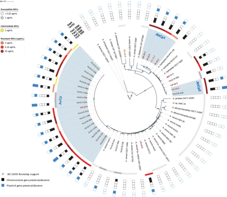
To accurately identify the 32 clinical and leech-derived isolates, their genomes were sequenced and a suite of housekeeping genes (HKG) was compared with those from 27 published genomes for bioinformatic species identification (Table S5) (34). Using this approach, seven clinical isolates were identified as A. hydrophila, two as A. veronii, and one as a likely novel species (Fig. 4), whereas most isolates were misidentified using biochemical methods (Table S6). In all cases, the HKG-based identifications were further
FIG 4 Maximum likelihood reconstruction of 16 single-copy housekeeping genes. Bootstrap support values between 80 and 100% are represented in the tree
by variously sized blue triangles (80%, small triangles, to 100%, large triangles). Resistance levels are indicated by colored boxes: highly susceptible,ⱕ1g/ml (light gray); susceptible, 1g/ml (dark gray); intermediate, 2 g/ml (yellow); resistant, 4 g/ml (orange), 8 to 16 g/ml (red), and 16 to 32 g/ml (dark red). The presence of Cpr-conferring chromosomal mutations is shown by filled black squares, while the presence of resistance plasmid genes is shown by filled blue
squares. For example, MO-11-1 has a black square for ParCS80rto represent that it has an S-80-R mutation. The names of strains derived from clinical isolates
are colored in dark red. Names of clinically associated isolates, such as those from leech aquaria, are colored orange.
Antimicrobials in Leech Select for Resistant Bacteria ®
on February 15, 2021 at MONTPELLIER BIBLIOTHEQUE
http://mbio.asm.org/
supported by the average nucleotide identity (ANI) analysis (35) (Fig. S1), as each strain’s ANI value wasⱖ0.96 compared to the type strain for each respective species (Fig. S1). These results are consistent with previous wet-lab studies that identified the majority of wound isolates as A. hydrophila and leech isolates as A. veronii (10, 15–17). The phylogenetic comparison allowed us to determine the relatedness of clinical and leech isolates by identifying three important clades. Four clinical A. hydrophila strains from the United States and Europe were placed into one clade (AhCp1) and shared identical HKG sequences (29,688 bp) (Fig. 4). The wound isolate, CA-13-1, and leech digestive tract isolate, Hv13-B-10d, were both confirmed to be A. hydrophila and also had identical HKG sequences (AhCp2). Notably, we identified 18 A. veronii strains that had nearly identical HKG sequences and were grouped into a single clade comprising 17 leech isolates and strain LR-14-4, a leech therapy wound isolate. The HKG sequences of these isolates differed by 0 to 8 bp (median, 1 bp) (AvCp). The AvCp clade was further analyzed by performing a whole-genome alignment and by calculating a well-supported phylogeny that grouped the clinical isolate LR-14-4 within a highly supported clade containing two leech-derived isolates (Fig. 5). Although the genetic
FIG 5 Phylogenetic reconstruction of the AvCp group. This cladogram is a maximum likelihood reconstruction generated from whole-genome alignments.
Bootstrap support values are represented by dots: dark green, 90%⫹ bootstraps; light green, 80%⫹; khaki, 70%⫹. Posterior probabilities of 95%⫹ from a Bayesian inference are represented by blue dots, and branch lengths do not carry meaning. Resistance levels are indicated by colored boxes: susceptible, ⱕ1g/ml (gray); intermediate, 2 g/ml (yellow); resistant, 4 g/ml (orange), 8 g/ml (dark orange), 16 g/ml (red), and 32 g/ml (dark red). The presence of Cpr-conferring genes and mutations is shown by filled black squares. The names of taxa derived from clinical isolates are colored in orange.
on February 15, 2021 at MONTPELLIER BIBLIOTHEQUE
http://mbio.asm.org/
content was nearly identical, the Cp MICs of the AvCp group varied greatly: 14 isolates were Cpr, two were Cpi, and two were Cps(Table S7). Nearly isogenic strains that span
the scope of resistance phenotypes have rarely been reported (1) and may help in the elucidation of additional resistance mechanisms.
ANI analysis of the AhCp1, AhCp2, and AvCp clades revealed that the genomes of every strain within each clade were extremely similar to each other (99.89% identity or higher), although none were identical (Fig. S1). In contrast, the intraspecies ANI values for all Cpsleech isolates were much lower (median of 96.24% identity), a finding that
agrees with the results of the HKG phylogeny, in which the Cpsleech isolates did not
form a monophyletic group.
Both the ANI and HKG analyses reveal a very close relationship among the clinical strains and those cultured directly from leeches. The two strains from AhCp2 had ANI values of 99.97%, and LR-14-4 grouped with two leech isolates with which it shared an average ANI value of 99.87%, confirming the leech-to-human transmission of the Cpr
clinical isolates. In these three clades, the high similarity of the genomes of strains with elevated resistance to Cp is consistent with the presence of a strong selection pressure promoting the proliferation of CprAeromonas strains in the leech gut.
Whole genomes provide insight into the resistance mechanisms of the Aero-monas isolates. Further analyses of the genomes allowed us to gain insight into the genetic factors underlying Cp resistance. While the first step to becoming resistant to Cp typically begins with the acquisition of the gyrA (S83I) mutation, greater resistance can be attained by additional point mutations in topoisomerase IV (parC), e.g., parC (E84K) or parC (S80I) (20, 36). The acquisition of resistance genes, such as efflux pumps encoded by qepA, the qnr (quinolone resistance) gene family that protects DNA gyrase, and acetylases such as aac(6=)-Ib-cr, leads to further resistance (20, 37–40), resulting in synergistic increases of the MIC (41). The mutation gyrA (S83I) was present in all Cprand
Cpistrains, as well as in four Cpsstrains (MICs, 0.25, 0.5, 1, and 1g/ml Cp) that had at
least an ~60-fold-higher MIC than the average MIC (0.0044g/ml) of our control strains (Table S7). These results confirm that the gyrA (S83I) mutation can be used as a sentinel marker for bacteria with an elevated MIC, as it was used to estimate the size of the population with elevated resistance in the deep-sequencing experiment (Fig. 3).
We wondered if a similar stepwise pattern of Cpr acquisition occurred in our
Aeromonas strains, and the AvCp clade provided us with the opportunity to address this
question. The stepwise acquisition of resistance genes appears to have independently occurred multiple times within the AvCp clade, since all of the strains have the gyrA (S83I) mutation and carry qnrS2 but the two sensitive strains do not have a mutation in
parC (Fig. 5). We identified two clades (each containing four strains) with significant
bootstrap support that carry the parC (E84K) allele and two clades (each consisting of two strains) that carry the parC (S80I) allele, suggesting that these parC mutations were acquired independently multiple times after the gyrA S83I mutation (Fig. 5). This is evidence that the stepwise acquisition of mutations observed in laboratory experi-ments also occurs in the environment.
The acquisition of plasmids carrying qnr genes and efflux pump-encoding genes is an important factor for further elevating the fluoroquinolone resistance level. The qnr genes have been identified on plasmids of various incompatibility (Inc) groups and sizes that exist within a wide range of hosts (40), and the observations are consistent with our findings. Every CprA. veronii strain carried qnrS2 on an ~34.5-kb conjugative
plasmid belonging to the IncU group. These plasmids are very similar to pAS37, which has been shown to carry qnrS2 in Aeromonas caviae (42). All CprA. hydrophila wound
isolates contained gyrA (S83I), either parC (S80I) or parC (E84K), and qnrS2, except for strain MO-11-1, which lacked qnrS2 (Fig. 4). The predicted QnrS2 proteins were all 218 amino acids (aa) in length and identical across the strains analyzed in our study. CA-13-1 and Hv13-B-13b, which were competed in the leech digestive tract against the sensitive control (Fig. 2), both carry chromosomal mutations and the qnrS2 gene on an IncU plasmid. qnrS2 was carried on a large IncU plasmid, except in A. hydrophila strain Hv13-B-10d, where it was located on a small (~6.8-kb) high-copy-number plasmid,
Antimicrobials in Leech Select for Resistant Bacteria ®
on February 15, 2021 at MONTPELLIER BIBLIOTHEQUE
http://mbio.asm.org/
pHv13-B-10d-C. The spread of qnrS2 was likely due to a transposition event as qnrS2 is flanked by a 22-bp inverted repeat and a 5-bp duplication, suggesting that it is located on a mobilizable element that lacks a transposase gene. This transposition is similar to what has been reported for qnrS2 in A. caviae (42). Whether or not the presence of high-copy-number plasmids leads to elevated QnrS2 levels remains to be evaluated in
Aeromonas, but studies of other genera support the idea that the presence of qnrS2 on
plasmids does facilitate higher levels of Cpr, even if the production of QnrS2 does not
significantly augment the MIC (40). Hv13-B-10d and two Cpr A. veronii strains also
carried qepA, a presumptive quinolone efflux pump, on a small plasmid. One particu-larly interesting strain is A. veronii Hv13-B-11a, which carries qnrS2 on the IncU plasmid, pHv13-B-11a-A, but has no mutation in gyrA or parC. This strain is Cpsbut has a much
higher MIC (1g/ml) than the susceptible control strains. These data indicate that the IncU plasmid carrying qnrS2 is transferred readily between Aeromonas species and that resistance plasmids are maintained even in Cpsstrains that do not harbor chromosomal
mutations known to facilitate Cpr.
DISCUSSION
In this study, we describe the rise of FQ resistance in a natural symbiont of the medicinal leech digestive tract. Our results indicate that an FQ concentration of 0.01g/ml within the leech is sufficient to ensure the long-term persistence of Cpr
Aeromonas spp. and likely promotes the acquisition and spread of antibiotic resistance
genes. The observed rise in the abundance of Aeromonas strains carrying a resistance-conferring gyrA mutation (Fig. 3) and the wide distribution of a plasmid carrying qnrS2 among both A. veronii and A. hydrophila strains (Fig. 4) suggest that FQ concentrations as low as 0.01 g/ml impose a sufficient selective pressure for the maintenance of resistance markers. Our data show that in Aeromonas isolates exposed to low amounts of Cp, the mutation in gyrA likely occurred before a mutation in parC. However, given that the gyrA (S83I) mutation is thought to be a first step toward resistance, it is notable that a single strain that did not have the gyrA or parC mutation, Hv13-B-11a, acquired the qnrS2-carrying plasmid, contrary to the canonical paradigm (20). This observation is of particular importance as conjugatable resistance plasmids can be transferred at very high frequencies, which can surpass mutation rates in a given gene under certain conditions, e.g., the IncU plasmid transfer rate from Aeromonas salmonicida into
Escherichia coli is high compared to an S83L mutation in gyrA in clinical E. coli strains
(43–45).
Our results strongly suggest that the therapeutic use of leeches containing FQs led to nosocomial infections by Cprstrains following leech therapy. For a safe and effective
therapeutic administration of leeches, a monitoring program for FQ levels and Cpr
strains should be implemented by the suppliers, and alternative antibiotic therapies against leech-acquired Aeromonas infections are needed (15, 18). The source of sub-MIC levels of FQs within the medicinal leech must be identified and eliminated, although the threat of resistant Aeromonas from contaminated environments remains. For example, Cpr Aeromonas strains have been cultured from environmental sources,
including lakes, rivers, and sewage treatment facilities (46–49), and environmental strains have been observed to carry IncU plasmids harboring qnrS2 (42). If such strains are present in the water that is used to ship leeches to hospitals or pharmacies, or if these strains colonize the leech surface or digestive tract, they might lead to nosoco-mial infections.
The in vivo fitness experiments in the presence of 0.01g/ml Cp demonstrate the ability of the Cprstrains to outcompete a natural leech symbiont that is representative
of the Aeromonas community prior to the FQ contamination. Interestingly, even susceptible strains derived from leeches between 2012 and 2014 displayed elevated MICs, and this evolution of resistance in the Aeromonas population occurred outside a lab environment. This suggests that very small amounts of Cp in animal digestive tracts and perhaps other environments are sufficient to select for strains with elevated resistance. Our in vivo results are supported by previous experiments conducted in vitro
on February 15, 2021 at MONTPELLIER BIBLIOTHEQUE
http://mbio.asm.org/
that emphasized the importance of sub-MICs of antimicrobials (4), as they induced predictable changes in the microbial community. In chemostats inoculated with human fecal matter containing microbiota exposed to various Cp levels, a concentration as low as 0.43g/ml was enough for enterococci to incur a loss of colonization resistance to a pathogenic Salmonella strain, while lower Cp concentrations did not affect coloniza-tion resistance (50). Sub-MIC Cp levels were observed to promote biofilm formacoloniza-tion of a respiratory tract pathogen, Moraxella catarrhalis, under anaerobic conditions (51). In the early 2000s, the increased occurrence of human infections due to FQr
Campylo-bacter spp. was linked to the ingestion of poultry meat from chickens that were treated
with an FQ and whose meat became contaminated with resistant bacteria (52). More recently, there were significant increases in the number of FQrCampylobacter isolates
from surveyed farms compared to the averages collected between 2004 and 2008 (53). The inclusion of in vitro and in vivo competition assays in our study revealed important differences between laboratory and natural settings and highlights shortcomings of laboratory experiments that cannot be ignored. We hypothesize that the complexity of microbial communities in the environment and the resulting increased competition for resources may intensify the consequences of low-level antimicrobials and explain discrepancies between in vitro and in situ assays that have been observed previously (29).
Naturally occurring isogenic strains isolated from the environment are very uncom-mon (1), and their analysis provides researchers with an advantage in studying evolu-tionary relationships and resistance mechanisms. The AvCp clade strains were obtained over a period of 3 years, and although they are not identical, their high similarity suggests that leech husbandry has indirectly facilitated the rise of very similar strains with elevated resistance to Cp. The 18 AvCp A. veronii strains likely arose from a common ancestor that acquired the key gyrA (S83I) mutation, and the descendants contained an average of 679 single nucleotide polymorphisms (SNPs) (ranging from 352 to 1,163, with a median of 649) across their ~4.8-Mbp genomes. The high degree of similarity of these strains provides a rare opportunity to analyze the factors that lead to increased Cp resistance levels. The results of the whole-genome analysis suggest that the acquisition of resistance markers (mutations in parC and the gain of qepA) occurred independently multiple times in a stepwise manner (Fig. 3). Despite this high level of similarity and possession of qnrS2, gyrA, and parC mutations, this clade ranged from being susceptible to being highly resistant to Cp (MICs of 0.25 to⬎32g/ml Cp) (Fig. 4; see also Table S3 in the supplemental material). Currently, we cannot account for all of the differences in MICs of the individual AvCp isolates. Possible additional factors include differences in expression levels of qnrS2, mutations that affect cell envelope permeability, and yet-to-be-identified resistance mechanisms (38, 54, 55).
Our study also points to the changes that have occurred in the leech digestive tract microbiota over time. The amplicon sequencing analysis of the gyrA gene revealed a significant rise in the relative abundance of Aeromonas species with the Cpr-related
gyrA mutations between 2011 and 2013 (Fig. 5). The competition data suggest that
these elevated-Cpr strains, which appear to dominate the gut, can outcompete the
typical Aeromonas strain (Hm21) only in the presence of FQ contamination. As with Hm21, which represents the Aeromonas community prior to FQ contamination, other
Aeromonas strains isolated prior to this problem also lacked the gyrA and parC
muta-tions and plasmid resistance genes (Fig. 4). It is very likely that these changes to the
Aeromonas community in the leech gut were due to the coincidental exposure of FQ
either as a direct consequence of feeding blood derived from FQ-treated poultry or through other means of FQ contamination at the leech farm. The potency of FQ is alarming, and whatever the source of contamination may be, low-level antimicrobials in a natural environment appear to facilitate the spread of resistance markers (1).
Overprescription and incorrect usage of antimicrobials in agriculture have played a major role in the rise of antibiotic resistance, and recent literature has emphasized that the excessive application of antibiotics on farms can result in the indirect contamina-tion of soil, rivers, and animal food products (1, 56). A large fraccontamina-tion of antibiotics
Antimicrobials in Leech Select for Resistant Bacteria ®
on February 15, 2021 at MONTPELLIER BIBLIOTHEQUE
http://mbio.asm.org/
released into the environment is in an active form, unmetabolized by animal renal or digestive systems (57). Studies on pasture animals observed that FQs are excreted mostly unchanged and thus can be found in farm soil (58). Discharge from pharma-ceutical plant and hospital efflux systems further exacerbates antibiotic pollution of these natural sites (1, 52). Antimicrobials emanate from these point sources and form spatial and temporal concentration gradients that encompass the concentrations used in our competition experiments.
Interestingly, some studies have quantitated the concentrations of FQs in various environments. Several studies have reported the presence of FQs at concentrations similar to those detected in our study, e.g., in animal farm wastewater (up to 0.0075g/ml Cp), river water (up to 0.0059 g/ml Cp), and manure (up to 19 mg/kg Ef) (59, 60). One study investigating the effluent of a sewage treatment plant near several pharmaceutical plants reported Cp levels ranging between 28,000 and 31,000g/liter and in a follow-up study reported contamination of well water with antibiotics in the surrounding area (61, 62). A meta-analysis of the occurrence and sources of FQs in the environment showed that an average of 0.021g/ml Cp occurs in hospital wastewater (63), while in another study a median concentration of 0.163g/ml Cp and values as high as 6 g/ml were also detected in water (64). The detection of Cp in water is especially important because it is resistant to degradation in aqueous environments and can remain biologically active for long periods of time (64). The same study found evidence for the increased occurrence of Cp resistance genes in soil with prolonged exposure to below-therapeutic levels of Cp (64). While concerns have been raised regarding the toxicological risks associated with pollutant antibiotics, the effects on the rise and spread of resistance mechanisms in bacterial communities in natural environ-ments have drawn relatively little attention (1, 63). When considering the environmen-tal impact of FQs in the future, it is important to assess the influence of very low antibiotic levels on the gain, spread, and persistence of antibiotic resistance genes and mutations in environmental bacteria.
Our study demonstrates that very low levels of antibiotics exert a selection pressure on microbial populations and that in vitro experiments can underestimate the effects of antibiotics in the natural environment. Antimicrobial concentrations well below the clinical breakpoint can lead to a dramatic increase in the abundance of strains with elevated resistance and the spread of plasmid-carried resistance genes between dif-ferent species culminating in an ecological disturbance. If FQ pollution is not better controlled, FQrstrains will replace sensitive ones and spread resistance to pathogens,
leading to an adverse effect on the wellbeing of humans, as postulated by the One Health Initiative (65).
MATERIALS AND METHODS
Strains and growth conditions. Clinical strains were provided by the UCLA School of Medicine,
University of Iowa Hospitals and Clinics, Washington University in St. Louis, and the University Hospital of Montpellier (France). Strains were isolated from the following settings: wounds of patients who received leech therapy, surgical instruments used on one of these patients, or aquarium tanks in which the leeches used on patients were housed (see Table S1 in the supplemental material).
The leech-derived strains used in this study were isolated from leeches obtained from various shipments from the main FDA-approved distributor, Leeches USA, Westbury, NY, USA; the FDA-approved leech farm Ricarimpex SAS, Eysines, France; and another distributor and leech farm in Europe, Biebertaler Blutegelzucht, Biebertal, Germany. Leeches from the main FDA-approved distributor were shipped in December 2012, February 2013, April 2013, June 2013, and November 2014. Leeches were dissected as previously described (10). Briefly, leeches were anesthetized in 70% ethanol and dorsally dissected through the crop of the digestive tract. Sterile swabs were used to collect the intraluminal fluid (ILF) from the leech crop and were initially streaked onto LB medium plates with and without Cp-HCl (Santa Cruz Biotechnology Inc., Dallas, TX). Before 2012, control Aeromonas isolates were cultured onto LB medium containing no Cp. For culturing Aeromonas isolates from leeches after 2012, Cp was present in plates at the following concentrations: 0g/ml, 1 g/ml and/or 2 g/ml, and 4 g/ml and/or 6 g/ml. Plates were then incubated at 30°C for approximately 14 h to identify naturally resistant bacterial subpopulations. Strains were subcultured onto plates with the same ciprofloxacin concentrations as plates on which they were first isolated and preserved as frozen stock.
DNA extraction, genome library preparation, and sequencing. Genomic DNA was extracted from
pure bacterial cultures using the Epicentre MasterPure Complete DNA and RNA purification kit (Epicentre,
on February 15, 2021 at MONTPELLIER BIBLIOTHEQUE
http://mbio.asm.org/
Madison, WI) per the manufacturer’s instructions. Genomic DNA was then quantified using a Qubit 2.0 fluorometer (Life Technologies, Inc., Carlsbad, CA) and diluted to 0.2 ng/l for Illumina Nextera XT (FC-131-1096) (Illumina, Inc., San Diego, CA) DNA library preparation. Genomic tagmentation, PCR of tagged DNA, and PCR product cleanup were done according to the manufacturer’s instructions. The Qubit 2.0 fluorometer and the Agilent 2100 Bioanalyzer (Agilent Technologies, Santa Clara, CA) with the high-sensitivity DNA kit were both used for library dilution to 4 nM for loading into an Illumina MiSeq sequencer, which generated 250-bp reads. Demultiplexing was performed as previously described (13). Leech control isolate Hm21 was sequenced in a different study as previously described (10), and leech-derived isolate Hv13-B-10d was sequenced using PacBio RS II.
Quality filtering of reads, genome assembly, and annotation. For Illumina-sequenced strains,
paired MiSeq fastq read 1 and read 2 files were imported into CLC Genomics Workbench software (CLC Bio-Qiagen, Aarhus, Denmark). Reads were trimmed, and those with Q scores ofⱖ15 were kept for the downstream assembly. Genome de novo assemblies were done with scaffolding using the same software; gene prediction and annotation of assembled genomes were performed using Prokka (66) for all strains except for the five leech control isolates, which were annotated using the RAST server (67) (Tables S3 and S5).
Species identification and resistance marker detection by sequencing. Illumina-compatible
amplicon primers (Table S8) were designed that amplify a 383-bp region of DNA gyrase subunit A, gyrA, covering the region of amino acid positions 63 to 176, based on the TruSeq amplicon format previously described by Nelson et al. for the analysis of 16S rRNA amplicons (68). We determined this fragment of
gyrA to be sufficient for Aeromonas species discrimination, and the amplified region includes the
nucleotide positions corresponding to a known point mutation leading to an amino acid substitution (S83I) that is thought to be the first step in acquiring resistance (36). Bioinformatic analysis of these amplicons from multiple leech samples allowed us to determine both relative Aeromonas species abundance and the presence of mutations that can potentially confer resistance to ciprofloxacin and enrofloxacin.
gyrA amplicons for high-throughput sequencing were generated in triplicate PCRs using the fusion
primers (sequences provided in Table S8) with the method described for 16S rRNA gene amplicons described by Nelson et al. (68). The reaction mixtures included 12.5l Phusion high-fidelity PCR master mix with 25l HF buffer, 3 M forward and reverse primer, 20 ng DNA template, and distilled water (dH2O) to final volume. The PCR cycling conditions were 95°C for 5 min, followed by 30 cycles of 95°C
for 30 s, 55°C for 30 s, and then 72°C for 1.5 min followed by a final 72°C for 5 to 10 min before cooling to 4°C. The amplified products were pooled and checked by agarose gel electrophoresis before purification with an 0.65⫻ volume of AMPure XP beads. The purified amplicons were quantified by PicoGreen and sized on an Agilent 2100 Bioanalyzer with the high-sensitivity DNA kit to pool equimolar amounts from each sample to form the final sequencing library.
Sequencing was performed on an Illumina MiSeq using a 2- by 250-bp paired-end protocol. After sequencing, the reads were demultiplexed according to their sample indexes, and read pairs were merged using SeqPrep to form single, high-quality contigs which were then quality trimmed using a Q30
cutoff over a 10-bp sliding window and minimum length cutoff of 375 bp. The primer sequences were removed with Cutadapt (https://cutadapt.readthedocs.io/en/stable/), and the reads were then formatted for analysis using QIIME.
For QIIME analysis, the reads from all samples were clustered by Uclust based on 99.5% sequence identity, which allows for less than a 2-bp difference between reads. Representative sequences were then selected and aligned against a set of reference gyrA sequences selected from Aeromonas strains for which the gyrase A sequence was available in GenBank, including sequences from isolates whose genomes have been fully sequenced. Clusters for which two or fewer sequences were clustered were removed from further analysis to limit the effect of spurious sequences due to PCR and sequencing errors. Species-level taxonomic assignments were made to the representative sequences using BLAST against the known reference sequences. The presence of potential antibiotic resistance-conferring mutations was determined using a custom perl script.
MIC determination of all isolates sequenced in this study. In accordance with the Clinical and
Laboratory Standards Institute (CLSI) guidelines, strains were first grown on blood agar (BA) and incubated for 14 h at 30°C. To prepare the inoculum, isolated colonies from the BA plates were suspended in 0.85% NaCl solution to the equivalent of an 0.5 McFarland turbidity standard. Mueller-Hinton agar (MHA) test plates were inoculated with the cultures, and Etest strips (BioMérieux, SA) with an analytical range from 0.002 to ⱖ32g/ml were applied to MHA to determine MICs. MICs were interpreted after 16 to 18 h of incubation at 35°C per manufacturer’s instructions (document 16246A, BioMérieux) using CLSI interpretation criteria (22) for most of the Aeromonas isolates in Fig. 4 (Table S7). The criteria state that Aeromonas isolates are Cpsif they are inhibited byⱕ1g/ml Cp, are Cpiif the MIC
is 2g/ml Cp, and are Cprif the MIC isⱖ4g/ml.
Competition assays. Competition assays were conducted as described previously (69). Briefly,
leeches were fed heat-inactivated sheep’s blood (Quad 5, Ryegate, MT) inoculated with 500 CFU/ml each of a test strain and a competitor strain to assess the colonization capability of the test strain in vivo. The competitor strain for all assays was Hm21RT, a spontaneous rifampin-resistant mutant containing a trimethoprim cassette inserted into the chromosome via a mini-Tn7 (70). This competitor strain was derived from the leech strain Hm21, which was isolated from the crop of the medicinal leech, Hirudo
verbana (10). Test strains were ciprofloxacin-resistant Aeromonas leech isolates that were also selected for
spontaneous rifampin resistance. Growth rates of all strains were determined in LB at 30°C in order to confirm that the mutants did not exhibit any growth defects in vitro. Hm21RT was also competed against
Antimicrobials in Leech Select for Resistant Bacteria ®
on February 15, 2021 at MONTPELLIER BIBLIOTHEQUE
http://mbio.asm.org/
another commonly used competitor strain, Hm21RS, a spontaneous rifampin- and streptomycin-resistant mutant of Hm21, to verify that Hm21RT’s growth in the leech did not vary from Hm21RS (data not shown). The following final concentrations of ciprofloxacin were also used in the blood meals: 0, 0.0025, 0.007, and 0.01 g/ml. Leeches used were obtained from BBEZ (Bierbertaler Blutegelzucht GmbH, Bierbertal, Germany). At least four animals were used for each competition, kept at 25°C after feeding, and assayed at 72 h. This time point was chosen because growth of A. veronii has already plateaued inside the leech gut, thereby minimizing the effects of small differences in growth rate between the test and competitor strains (27).
Competition indexes (CIs) were calculated as follows: (test strainoutput/competitor strainoutput)/(test
straininput/competitor straininput). A CI of 1 indicated that the test strain colonized to the same levels as
the competitor strain, whereas a CI of⬍1 indicated that the test strain had a colonization defect. The limit of detection was 10 CFU/ml.
Growth in blood. Prior to feeding, an aliquot of each heat-inactivated blood meal inoculated with
the competitor and test strains was removed and incubated for 72 h at 25°C. Samples were serially diluted and plated as described above for the in vivo competition assay.
Statistical analysis. Data were analyzed in GraphPad Prism 6 (GraphPad, San Diego, CA). A
Kruskal-Wallis one-way analysis of variance with Dunn’s post hoc test was used to determine if the CIs differed from one another (P⬍ 0.05). Sample means in Fig. 2 were log transformed due to high variation and analyzed with a one-sample t test to determine if sample means were significantly different (95% confidence interval) from a CI value of 1.
Detection of ciprofloxacin and enrofloxacin using HPLC. Compounds were quantified via
ultrahigh-performance liquid chromatography electrospray ionization mass spectrometry (UHPLC-ESI-MS) with accurate-mass detection. HPLC was performed with a reverse-phase HPLC column: an Agilent PLRP-S PSDVB column with 3.0-m particles and dimensions of 50 mm in length and 1.0 mm in diameter (P/N PL1312-1300) was used with an Agilent 1290 HPLC system. The column was maintained at 50°C with a flow rate of 0.6 ml/min. Chromatography was as follows: solvent consisted of water with 0.1% (vol/vol) formic acid for channel A and acetonitrile with 0.1% formic acid for channel B. Following column equilibration at 5% B, the sample was injected via autosampler, and the column was flushed for 1.0 min. From 1.0 min to the end of the run, the column eluant was directed to the MS source. From 1.0 min to 4.0 min, the gradient was linearly ramped from 5% to 95% solvent B. From 4.0 to 4.8 min, the column was held at 95% B, and from 4.8 to 5.0 min, the column was reequilibrated with 5% solvent B. Ciprofloxacin eluted starting at 2.7 min, and enrofloxacin eluted starting at 2.75 min.
The mass spectrometer used was an Agilent 6538 quadrupole time of flight (QTOF) spectrometer with ESI source; resolution is approximately 20,000 and accuracy is 1 ppm. Source parameters were as follows: drying gas, 8.0 liters/min; drying gas heat at 350°C; nebulizer, 55 lb/in2; capillary voltage, 3,500 V;
capillary exit, 100 V. Spectra were collected in positive mode as appropriate from 50 to 1,700 m/z at a rate of 2 Hz.
Samples were quantified with the Agilent MassHunter Quantitative Analysis package, using centroid data mode and peak definitions of 332.1377 and 360.1687 m/z for ciprofloxacin and enrofloxacin, respectively. Both analytes used a⫾50-ppm window for the m/z definition, which was evaluated for lack of interfering background signals with the samples. The relative standard deviation was determined using error propagation from the curve fit and technical replicates.
Blanks were run between each sample to eliminate the possibility of carryover interferences, and external standard curves were conducted with authentic ciprofloxacin and enrofloxacin standards (Sigma-Aldrich, Poole, United Kingdom).
MLSA reference tree generation. The multilocus sequence analysis (MLSA) reference tree in Fig. 4
was generated using the method described in the work of Colston et al. (34). Sixteen housekeeping genes (atpD, dnaJ, dnaK, dnaX, gltA, groL, gyrA, gyrB, metG, mdh, radA, recA, rpoC, rpoD, tsf, and zipA) were used for the MLSA. The full-length sequence of each gene was initially derived from the previously published genome of A. veronii Hm21, and these sequences served as queries for BLAST searches against the annotated proteins of all 56 genomes. Multiple sequence alignments (MSAs) were generated by aligning the genes using MUSCLE (v3.8.31) (71). In-house scripts created a concatenated alignment of all 16 genes. A model of evolution was determined by using the Akaike information criterion with correction for small sample size (AICc), as implemented in jModelTest 2.1.4 (72). A maximum likelihood (ML) phylogeny was generated from the concatenated MSA, and individual gene phylogenies from the individual gene MSAs were determined by using PhyML (v3.0_360-500M) (73). PhyML parameters consisted of a general time-reversible (GTR) model, estimated proportion of invariable sites (p-invar), 4 substitution rate categories, estimated gamma distribution, and subtree pruning and regrafting enabled with 100 bootstrap replicates.
MLSA and genome alignment distance calculation. The number of differences between the
sequences in the concatenated alignment was calculated using the R package Ape. The dna.dist function was called with the model parameter set to “N” and pairwise.deletion parameter set to “TRUE.”
Average nucleotide identity analysis. Assembled contigs were reconstituted from the
RAST-generated GenBank files for all genomes by using the seqret function of the EMBOSS package (74). All genomes were treated in the same manner to ensure that any biases were consistent across the entire data set. JSpecies1.2.1 (75) was used to analyze these contig sets for the ANI, using default parameters. We report here the averages of the reciprocal comparisons.
Whole-genome phylogenetic reconstruction. A whole-genome alignment of the 18 members of
the AvCp clade was generated from sequence files in GenBank format using the progressiveMauve algorithm of Mauve (76). Hm21, Hv221, Hv571, Hv13-B-10a, and Hv13-B-11a were included to serve as
on February 15, 2021 at MONTPELLIER BIBLIOTHEQUE
http://mbio.asm.org/
outgroups for rooting purposes. The XMFA alignment files were converted into FASTA format using in-house scripts. A model of evolution was determined by using the Akaike information criterion with correction for small sample size (AICc), as implemented in jModelTest 2.1.4 (72). Phylogenies were calculated using both RAxML v8.1.17 (77) under GTR CAT and GTR plus estimated gamma plus invariable sites models (producing identical topologies) and MrBayes v3.2.4 x64 (78) under a GTR model, with estimated gamma.
Data availability. All scripts used for analysis, along with the gyrA reference data sets, are available
fromhttp://github.com/joerggraflab/SARIS.
Accession numbers. All gyrA sequencing data were deposited under BioProjectPRJNA296880to the INSDC SRA. The URL ishttps://www.ebi.ac.uk/ena/data/view/PRJNA296880. All new genome sequencing data presented in this study have been deposited to the European Nucleotide Archive under the study accession numberPRJNA297409, except for one genome which is deposited under the study accession numberPRJEB6940. The URL ishttps://www.ebi.ac.uk/ena/data/view/PRJNA297409. More information regarding sequencing metadata and individual sample accession numbers is shown in Table S5.
SUPPLEMENTAL MATERIAL
Supplemental material for this article may be found athttps://doi.org/10.1128/mBio
.01328-18.
FIG S1, DOCX file, 3.9 MB. TABLE S1, DOCX file, 0.1 MB. TABLE S2, DOCX file, 0.02 MB. TABLE S3, DOCX file, 0.01 MB. TABLE S4, DOCX file, 0.01 MB. TABLE S5, DOCX file, 0.02 MB. TABLE S6, DOCX file, 0.03 MB. TABLE S7, DOCX file, 0.01 MB. TABLE S8, DOCX file, 0.01 MB. ACKNOWLEDGMENTS
We thank Susan Janton for excellent technical assistance; Brian Nussenbaum, Car-men Giltner, and Romney M. Humphries for providing strains; Rudy Rosenberg for arranging a shipment of leeches, and Jeremiah Marden for helpful comments on the manuscript. We acknowledge the Mass Spectrometry Facility at Montana State Univer-sity (Murdock Charitable Trust and NIH 5P20RR02437 of the CoBRE program), the UConn Bioinformatics Facility for providing computing resources, and the Center for Environmental Science and Engineering at the University of Connecticut for analyses. Illumina sequencing was performed at the Microbial Analysis, Resources and Services Facility of the University of Connecticut.
This research was supported by NIH R01 GM095390 to J. Graf, P. Visscher, and H. Morrison. B.L. and E.T.-R. are supported by the Association des Biologistes de L’Ouest and by the Association pour la recherche et le développement en microbiologie & pharmacie (ADEREMPHA).
J. Graf is a leech microbiology consultant for the German leech farm Biebertaler Blutegelzucht GmbH, Biebertal, Germany, and the company does not direct or approve J. Graf’s research and publications.
REFERENCES
1. Andersson DI, Hughes D. 2014. Microbiological effects of sublethal levels of antibiotics. Nat Rev Microbiol 12:465– 478.https://doi.org/10.1038/ nrmicro3270.
2. Jørgensen KM, Wassermann T, Jensen PØ, Hengzuang W, Molin S, Høiby N, Ciofu O. 2013. Sublethal ciprofloxacin treatment leads to rapid de-velopment of high-level ciprofloxacin resistance during long-term ex-perimental evolution of Pseudomonas aeruginosa. Antimicrob Agents Chemother 57:4215– 4221.https://doi.org/10.1128/AAC.00493-13. 3. Blaser MJ. 2016. Antibiotic use and its consequences for the normal
micro-biome. Science 352:544 –545.https://doi.org/10.1126/science.aad9358. 4. Gullberg E, Cao S, Berg OG, Ilbäck C, Sandegren L, Hughes D, Andersson
DI. 2011. Selection of resistant bacteria at very low antibiotic concen-trations. PLoS Pathog 7:e1002158.https://doi.org/10.1371/journal.ppat .1002158.
5. Robinson TP, Bu DP, Carrique-Mas J, Fèvre EM, Gilbert M, Grace D, Hay
SI, Jiwakanon J, Kakkar M, Kariuki S, Laxminarayan R, Lubroth J, Mag-nusson U, Thi Ngoc P, Van Boeckel TP, Woolhouse ME. 2016. Antibiotic resistance is the quintessential One Health issue. Trans R Soc Trop Med Hyg 110:377–380.https://doi.org/10.1093/trstmh/trw048.
6. Whitaker IS, Izadi D, Oliver DW, Monteath G, Butler PE. 2004. Hirudo
medicinalis and the plastic surgeon. Br J Plast Surg 57:348 –353.https:// doi.org/10.1016/j.bjps.2003.12.016.
7. Whitaker IS, Kamya C, Azzopardi EA, Graf J, Kon M, Lineaweaver WC. 2009. Preventing infective complications following leech therapy: is practice keeping pace with current research? Microsurgery 29:619 – 625.
https://doi.org/10.1002/micr.20666.
8. Sartor C, Limouzin-Perotti F, Legré R, Casanova D, Bongrand MC, Sambuc R, Drancourt M. 2002. Nosocomial infections with Aeromonas hydrophila from leeches. Clin Infect Dis 35:E1–E5.https://doi.org/10.1086/340711. 9. Whitaker IS, Josty IC, Hawkins S, Azzopardi E, Naderi N, Graf J, Damaris
Antimicrobials in Leech Select for Resistant Bacteria ®