HAL Id: inserm-00529034
https://www.hal.inserm.fr/inserm-00529034
Submitted on 24 Oct 2010HAL is a multi-disciplinary open access archive for the deposit and dissemination of sci-entific research documents, whether they are pub-lished or not. The documents may come from teaching and research institutions in France or abroad, or from public or private research centers.
L’archive ouverte pluridisciplinaire HAL, est destinée au dépôt et à la diffusion de documents scientifiques de niveau recherche, publiés ou non, émanant des établissements d’enseignement et de recherche français ou étrangers, des laboratoires publics ou privés.
Time-frequency relationships between heart rate and
respiration: A diagnosis tool for late onset sepsis in sick
premature infants
Guy Carrault, Alain Beuchée, Patrick Pladys, Lotfi Senhadji, Alfredo
Hernandez
To cite this version:
Guy Carrault, Alain Beuchée, Patrick Pladys, Lotfi Senhadji, Alfredo Hernandez. Time-frequency relationships between heart rate and respiration: A diagnosis tool for late onset sepsis in sick premature infants. Computers In Cardiology, Sep 2009, Park City, UT, United States. pp.369 - 372. �inserm-00529034�
Time-Frequency Relationships between Heart Rate
and Respiration: A Diagnosis Tool for Late
Onset Sepsis in Sick Premature Infants
G Carrault
1,2,4, A Beuchee
1,2,3,4, P Pladys
1,2,3,4, L Senhadji
1,2, A Hernandez
1,2,41
Inserm, U642, Rennes, F-35000, France
2
Université de Rennes 1, LTSI, Rennes, F-35000, France
3
CHU Rennes, Pôle Médico-Chirurgical de Pédiatrie et de Génétique Clinique, Néonatologie,
Rennes, F-35000, France
4
Inserm CIC-IT804, Rennes, F-35000, France
Abstract
The diagnosis of late onset sepsis in premature infants remains difficult because clinical signs are subtle and non-specific and none of the laboratory tests, including CRP and blood culture, have high predictive accuracy. Heart rate variability (HRV) analysis emerges as a promising diagnostic tool. Entropy and long-range fractal correlation are decreased in premature infants with proven sepsis. Besides this, respiration and its relations to HRV appear to be less. The objective of this study was to determine if analysis of time-frequency correlations between the heart rate and respiration amplitude may help for the diagnosis of infection in premature infants. An estimator of the linear relationship between nonstationary signals, recently introduced, is explored. The tests were performed on a cohort study of 60 premature infants. The results show that the correlation in the low frequency band tended to be higher in the sepsis group.
1. Introduction
Late-onset sepsis, defined as a systemic infection in neonates older than 3 days, occurs in approximately 10% of all neonates and in more than 25% of very low birth weight infants who are hospitalized in neonatal intensive care units. In view of the high morbidity and mortality associated with infection, reliable markers are needed.
Recurrent and severe spontaneous apneas and bradycardias is one of the major clinical early indicators of systemic infection in the premature infant. It requires prompt laboratory investigation so that treatment can start without delay. Various hematological and biochemical markers have been evaluated in this indication but they are invasive procedures that cannot be repeated several times.
Heart rate variability measurements such as a
decreased approximate entropy 1, 2 or sample entropy 3 are significantly associated with sepsis or sepsis-like illness in premature infants. These diagnostic tools are non invasive, easily available and fast.
This work can be viewed as a supplementary work in this direction. The following question is addressed: Could the association between heart rate variability and respiration in a selected population of sick premature infants be used as a diagnosis tool of late onset sepsis?
In this paper, a novel estimator for characterizing the evolution of linear relationship between signals, in the time-frequency domain, is used 4. This estimator is based on the computation of the correlation between Heart Rate Variability (HRV) and respiration signals filtered, in narrow and overlapping frequency bands, using a continuous filter bank 4. The description of the method is proposed in the first section. Then, the protocol -including data collection, statistical tools, inclusion and exclusion criteria-, is described. Results are reported in the third section while interests and perspectives are drawn in the conclusion section.
2. Methods
For the past decades, numerous signal processing approaches have been proposed to measure the degree of association (in a general sense, i.e. interdependency). These methods may be divided into two categories depending on whether or not the nonlinear nature of the relationship is taken into account. Linear methods were developed first. Many estimators based on linear cross-correlation or coherence function were proposed. For the second group, mutual information [5] and on nonlinear regression [6, 7]) were introduced in the EEG field. More recently nonlinear dynamical systems and chaos [8, 9]) were designed.
In a recent previous work, we studied the interrelationship between HRV and respiration and
demonstrated a decrease in the non linear regression coefficient for the sepsis population 10. Unfortunately, if nonlinear methods have the capability to account for the nonlinearity of relationship, they are generally independent from frequency, a key parameter in the heart rate variability and its interrelationship with the respiration. On the opposite, linear methods do not analyze the nonlinear property of relationship but they can characterize its dependence on frequency. However, It is known [11] that the coherence function provided estimators generally with strong bias and variance. To alleviate these difficulties, a new estimator was recently proposed and can be of interest for our purpose 4. It uses a local linear correlation coefficient, computed at the outputs of narrow band-pass filter, as the function of frequency and time, which is maximized for time delay. To our knowledge, the application of this latter method in the cardio-respiratory field has never been reported. Let us briefly recall the method:
We consider two observations, x(t) the HRV signal and y(t) the respiration signal. The problem is to characterize the statistical relationship, simultaneously in the time and frequency domains, between the nonstationary signals x(t) and y(t). The estimation of the local linear correlation coefficient is given by :
Rx2,y(t, f ) mm
max
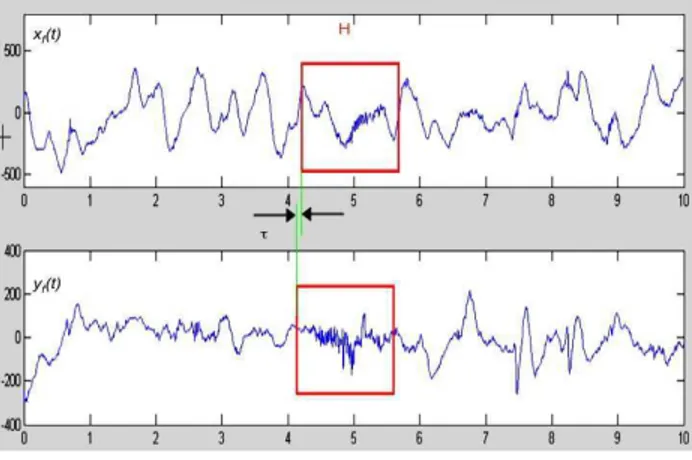
xf(k) yf(k) kH 2 H 2 2 xf2(k) kH 2 H 2 yf2(k) kN 2 N 2 where xf(t) and yf(t) are zero-mean and narrow band
filtered signals over a sliding window of duration H (Figure 1a) of an appropriate filter bank (Figure 1b).
R2x,y(t,f) is computed with different delays between the
two windows. This last one is derived from the Short-Time Fourier Transform (STFT).
3. Results
Data were obtained from a cohort of 60 premature infants (post-menstrual age < 33 weeks and chronological age > 72 hours) hospitalized in the neonatal intensive care unit at the university Hospital of Rennes. The infants were placed in bassinets, positioned on their side, wrapped by a single blanket roll, and loosely covered by another. The monitoring (Powerlab system®, ADInstruments) consisted of two ECG, EOG and EEG
leads, one pulse oxymetry saturation (SaO2), nasal flow and abdominal respiration recordings (sampling rate 400 Hz). Finally, babies were paired in age and weight.
Figure 1a : Selection of the temporal support H for the computation of R2(t,f) with different delays
Figure 1b: filter bank derived from short time Fourier Transform
Inclusion criteria were :
Post-menstrual age less than 33 weeks and postnatal age more than 72 hours,
Unusual and recurrent bradycardias (defined as more than one occurrence per hour and/or a need for bag-and-mask resuscitation and/or the intention of the attending physician to investigate for a suspected infection).
While Exclusion criteria were :
ongoing inflammatory response (CRP >5mg/l prior to the recording),
medication with antibiotics, morphine, cathecolamin, sedative drugs or Doxapram
intra-tracheal respiratory support, intra-cerebral lesion and malformation.
The study was approved by the local ethics committee. Parents were informed and consent was obtained.
A sequence of 4096 successive cardiac cycles, i.e. about half an hour each, were extracted from ECG recordings, and respiration signal and were subjected to analysis of heart rate variability parameters.
Both Sepsis and No-sepsis groups were compared using Mann and Withney or Kruskall-Wallis tests. The level of significance was set at p= 0.05.
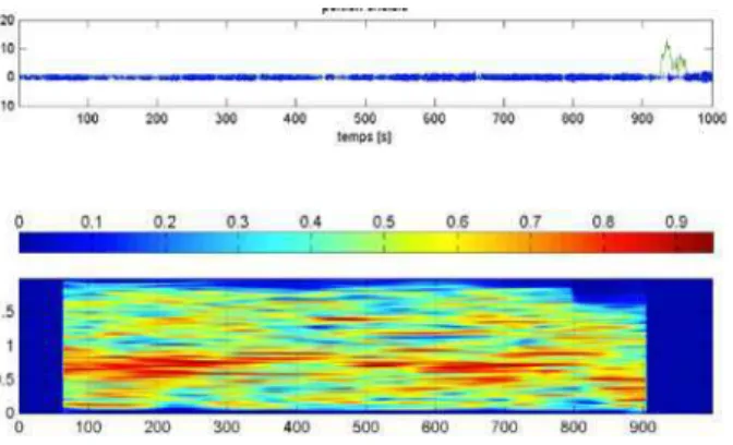
Representative examples (HRV, respiration signal and Time-frequency linear correlation coefficient) in the sepsis and non sepsis group are displayed figure 3 and 4. Both signals are nonstationary.
Figure 2: Typical square modulus of Time-frequency linear correlation coefficient for a sepsis baby. The upper traces represent the HRV and respiration signals.
The analysis reveal two distinct signatures: i) a higher correlation coefficient for the sepsis group in the lower band around 0.2 Hz, ii) a strong relationship in the higher frequency band for the non sepsis group.
Figure 3: Typical square modulus of Time-frequency representation linear correlation coefficient for a non-sepsis baby. The upper traces represent the HRV and respiration signals
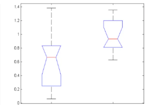
Figure 4 : Distribution of R2(t,f) greater than 0.9 for a) sepsis babies and b) non-sepsis babies
These qualitative findings were statistically verified. Figure 4 depicts the sub-band distribution of the time-frequency correlation coefficient R2(t,f), over a threshold set to 0.9.
We confirm statistically (p<0.05 whatever the statistical tests) that the higher correlation is retrieved in the low frequency band for the sepsis group (0.02<f<0.2
Hz) while an important distribution of high correlation (p<0.04 whatever the statistical tests) is observed in the non sepsis group for the high frequency (1<f<1.4 Hz) (figure 5).
Several threshold (0.6, 07, 0.8, 0.9) were tested and the threshold 0.9 appears the most discriminative between the two groups.
This conclusion is also in agreement with estimation of the respiration frequency of each baby. A significant difference is observed between the sepsis and non sepsis groups (Figure 6).
Sepsis No Sepsis
Band med med Test_u Kw
0-0.02 3.97 0 0.5554 0.5285 .02-0.2 7.47 2.74 0.0483 0.0483 0.2-0.4 6.61 5.65 0.4887 0.4887 0.4-0.6 8.03 8.13 0.8175 0.8175 0.6-0.8 7.39 8.34 0.0956 0.0956 0.8-1.0 5.87 6.44 0.191 0.191 1.0-1.2 4.92 7.84 0.0061 0.0061 1.2-1.4 4.31 7.29 0.0129 0.0129 1.4-1.6 1.68 2.40 0.1178 0.1178 1.6-1.8 0 0 0.8777 0.867 Figure 5 : Distribution of the linear time-frequency correlation coefficient square modulus values greater than 0.9 in different sub-bands. The grey lines indicate significant values.
Figure 6: Frequency Peak estimation of the respiration spectrum: 1) sepsis babies and 2) non sepsis babies
4. Discussion
and
conclusions
In this study, the question about the functional coupling among respiration and heart rate variability is addressed. There are two main results in this study. Differently from our initial work [10], the linear relationship between HRV and respiration signals is computed as a function of time and frequency. Results show that the relationships are circumscribed within a particular region of the time-frequency plane for the sepsis group and a different one for the non-sepsis group (it is worthwhile to be aware that frequency-independent methods can be blind to relationships set up on a narrow frequency band).
In premature infants less than 33 weeks, with unusual and frequent apneas and bradycardias, we observed in the past that approximate entropy can reliably confirm or refute the diagnosis of systemic infection at an early stage. This measure has a good repeatability and good accuracy to test for the diagnosis of infection in this selected population. These findings corroborate the earlier works of Griffin and Pincus et al [12]. Nevertheless, in the sick premature infant, the mechanism is probably not just a change in regularity. The clinical findings here clearly demonstrate that HRV and respiration and their relationships could be efficient diagnosis tools and may help identifying culture-positive sepsis in a population of infants with recurrent bradycardias.
Acknowledgements
The authors would like to express their thanks to Stephanie Sergant for her fruitful help during the data collection process.
References
[1] Pincus SM, Goldberger AL: Physiological time-series analysis: What does regularity quantify? Am J Physiol 1994;266:H1643-1656.
[2] Beuchée A, Carrault G, Bansard JY, Boutaric E,
Bétrémieux P, Pladys P. Uncorrelated randomness of heart rate is associated with sepsis in sick premature infants. Neonatology 2009; 96 (2);109-114.
[3] Lake DE, Richman JS, Griffin MP, Moorman JR: Sample entropy analysis of neonatal heart rate variability. Am J Physiol Regul Integr Comp Physiol 2002;283:R789-797. [4] Ansari-Asl K., Bellanger JJ, Bartolomei F, Wendling F,
Senhadji L. Time-frequency characterization of interdependencies in nonstationary signals: application to epileptic EEG. IEEE Trans Biomed Eng 2005; 52: 1218-26.
[5] Mars N, Lopes da Silva F. Propagation of seizure activity in kindled dogs. Electroencephalography and Clinical Neurophysiology 1984:56,194-209.
[6] Pijn J, Lopes Da Silva F, Propagation of electrical activity: nonlinear associations and time delays between EEG signals in Basic Mechanisms of the EEG. Zschocke and Speckmann, Eds. Boston: Birkauser, 1993.
[7] Wendling F, Bartolomei, F, Bellanger JJ, Chauvel P
Interpretation of interdependencies in epileptic signals using a macroscopic physiological model of the EEG. Clin Neurophysiol 2001; 112:1201-18.
[8] Iasemidis LD. Epileptic seizure prediction and control. IEEE Transactions on Biomedical Engineering 2003;50, 549-558.
[9] Lehnertz K. Non-linear time series analysis of intracranial EEG recordings in patients with epilepsy--an overview. Int J Psychophysiol, 1999;34:45-52.
[10] Loforte R, Carrault G, Mainardi L, Beuchée A. Heart rate and respiration relationships as a diagnostic tool for late onset sepsis in sick preterm infants. Computers in Cardiology 2006; 33;737-40.
[11] Zaveri H, Williams W, Sackellares J, Beydoun A,
Duckrow R, Spencer S. Measuring the coherence of intracranial electroencephalograms. Clin Neurophysiol, 1999;110: 1717-25.
[12] Griffin MP, Lake DE, Moorman JR: Heart rate
characteristics and laboratory tests in neonatal sepsis. Pediatrics 2005;115:937-941.
Address for correspondence Carrault G. LTSI-INSERM U642 Bt22 Campus de Beaulieu Université de Rennes 35042 Rennes Cedex France guy.carrault@univ-rennes1.fr