HAL Id: hal-03035897
https://hal.archives-ouvertes.fr/hal-03035897
Submitted on 3 Dec 2020
HAL is a multi-disciplinary open access
archive for the deposit and dissemination of
sci-entific research documents, whether they are
pub-lished or not. The documents may come from
teaching and research institutions in France or
abroad, or from public or private research centers.
L’archive ouverte pluridisciplinaire HAL, est
destinée au dépôt et à la diffusion de documents
scientifiques de niveau recherche, publiés ou non,
émanant des établissements d’enseignement et de
recherche français ou étrangers, des laboratoires
publics ou privés.
Exploring the chemical space of 1,2,3-triazolyl triclosan
analogs for discovery of new antileishmanial
chemotherapeutic agents
Julia Fernández de Luco, Alejandro Recio-Balsells, Diego Ghiano, Ana
Bortolotti, Juán Manuel Belardinelli, Nina Liu, Pascal Hoffmann, Christian
Lherbet, Peter Tonge, Babu Tekwani, et al.
To cite this version:
Julia Fernández de Luco, Alejandro Recio-Balsells, Diego Ghiano, Ana Bortolotti, Juán Manuel
Be-lardinelli, et al.. Exploring the chemical space of 1,2,3-triazolyl triclosan analogs for discovery of new
antileishmanial chemotherapeutic agents. RSC Medicinal Chemistry, Royal Society of Chemistry, In
press, �10.1039/D0MD00291G�. �hal-03035897�
Exploring the chemical space of 1,2,3-triazolyl triclosan
analogs for discovery of new antileishmanial
chemotherapeutic agents.
Julia Fernández de Luco,a,≠ Alejandro I. Recio-Balsells,a,≠ Diego G. Ghiano,a,≠ Ana Bortolotti,b Juán Manuel Belardinelli,b Nina Liu,c Pascal Hoffmann,d Christian Lherbet,d Peter J. Tonge,c Babu Tekwani,e,† Héctor R. Morbidoni,b,f,* and Guillermo R. Labadiea,g,*
a.
Instituto de Química Rosario, UNR, CONICET, Suipacha 531, S2002LRK, Rosario, Argentina. E-mail: labadie@iquir-conicet.gov.ar; Fax: +54 341 4370477; Tel: +54 341 4370477.
b.Laboratorio de Microbiología Molecular, Facultad de Ciencias Médicas, Universidad Nacional de Rosario, Santa Fe 3100, S2002KTR,
Rosario, Argentina. E-mail: morbiatny@yahoo.com
c.
Institute of Chemical Biology & Drug Discovery, Department of Chemistry, Stony Brook University, Stony Brook, NY 11794, USA
d.
LSPCMIB, UMR-CNRS 5068, Université Paul Sabatier-Toulouse III, 118 Route de Narbonne, 31062 Toulouse Cedex 9, France
e.National Center for Natural Products Research & Department of Biomolecular Sciences, School of Pharmacy, University of Mississippi, MS
38677, USA
f.Consejo de Investigaciones, Universidad Nacional de Rosario g.
Departamento de Química Orgánica, Facultad de Ciencias Bioquímicas y Farmacéuticas, Universidad Nacional de Rosario, Suipacha 531, S2002LRK, Rosario, Argentina
†
Present address: Department of Infectious Diseases, Division of Drug Discovery, Southern Research, Birmingham, AL 35205, USA
≠
These authors contributed equally to this work.
Triclosan and Isoniazid are known antitubercular compounds that have proven to be also active on leishmania parasites. On those grounds, a collection of 37 diverse 1,2,3-triazoles inspired on the antitubercular molecules triclosan and 5-octyl-2-phenoxyphenol (8PP), was designed in search for novel structures with leishmanicidal activity and prepared using different alkynes and azides. The 37 compounds were assayed against Leishmania donovani, the etiological agent of leishmaniasis, yielding some analogs with activity at micro molar concentration and against M. tuberculosis H37Rv resulting in scarce active compounds with MIC of 20 μM. To study the mechanism of action of these catechols, we analyzed the inhibition activity of the library on the M. tuberculosis enoyl-ACP reductase (ENR) InhA , obtaining poor inhibition of the enzyme. The cytotoxicity against Vero cells was also tested, resulting in none of the compounds being cytotoxic at concentrations of up to 20 μM.The derivative 5f could be considered a valuable starting point for future antileishmanial drug development. The validation of a putative leishmanial InhA orthologue as a therapeutic target needs to be further investigated.
1. Introduction
Neglected Tropical Diseases (NTDs) are an ever-growing list of treatable and preventable diseases that proliferate in impoverished environments. One of the most relevant characteristics of NTDs is their global impact being present in every continent. NTDs are usually emergent in regions lacking from adequate hygiene conditions and many of them are vector-borne. Tropical and subtropical areas where the climate conditions are hot and humid are the most affected, due to the combination of the presence of many vectors and poor living conditions.
Leishmaniasis is one of the most concerning vector-borne NTDs, an expanding endemic disease caused by the protozoan parasite
Leishmania spp and transmitted by over 90 phlebotomine sand fly
species. Visceral leishmaniasis (VL) or Kala-azar, is the most severe form of the disease which if not treated, has a mortality rate close to 100%, surpassing 30,000 deaths per year. Unfortunately, there is no vaccine available and the regular treatment includes drugs that
are toxic to the host, so they are used at low doses ending in the generation of drug resistant forms of the parasite.
The World Health Organization (WHO) has reported a rate of 1.3 million cases of leishmaniasis per year and 350 million people exposed to infections.1 This fact has alerted the scientific community and driven it to the urgent search for new effective and less toxic drugs with novel structures and different targets.
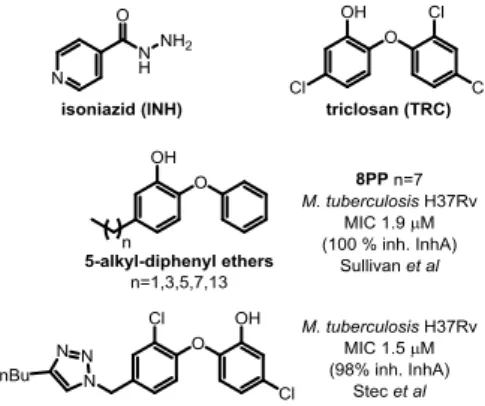
Bacterial fatty acids biosynthesis is a validated target for drug discovery,2 however, it has not been fully exploited. An interesting target to explore for the development of new antileishmanial drugs is the ortholog of the enzyme 2-trans-enoyl fatty acid reductase3 from the Mycobacterium tuberculosis FASII complex. This enzyme, called InhA, was discovered as a target for the drug isoniazid (INH) (Figure 1), which is one of the current drugs for the treatment of tuberculosis.4 InhA catalyzes the reduction step that ends the process of a two carbon extension in the pathway of saturated fatty acid synthesis. Interestingly, antituberculosis drugs, such as triclosan (TRC) -an uncompetitive inhibitor of InhA- (Figure 1), have also shown in vitro activity.5 Kinetoplastid Type II FAS are found in mitochondrial organelles.6 Thus, since parasites have FASII enzymes
they may be targeted by drugs active on bacterial fatty acid synthesis.
The M. tuberculosis FAS II complex is analogous to other bacterial FAS II, but is not capable of de novo fatty acid synthesis. However, FAS II from Mycobaterium is capable of elongating acyl-CoA generated by FAS I producing very long chain α-hydroxy, -alkyl branched fatty acids known as mycolic acids. Even though mutations in the inhA gene are known to confer resistance to INH, the enzyme remains a validated and reliable target for drug discovery.
Triclosan7 has been described as an InhA targeting antitubercular drug which does not require KatG activation, however its poor bioavailability prevented its use.8 In spite of that, TRC was used to inspire the development of other scaffolds such as 5-alkyl-diphenyl-ethers, one of which, 5-octyl-2-phenoxyphenol (8PP, Figure 1)9 has good anti-tubercular activity and higher solubility. Therefore, 8PP and similar molecules are good starting points for structure-based drug discovery
Figure 1. Reported analogs with biological activity against
Mycobacterium tuberculosis.
One approach to enhance the activity of a drug candidate is to incorporate additional heterocycles as building blocks.10A recently introduced and popular strategy is to use 1,2,3-triazoles to link structurally diverse fragments with different pharmacophores.11-13 A straightforward way to prepare 1,2,3-triazoles, is the well-known click chemistry reaction which involves azides and alkynes as starting materials. This kind of approach is a very efficient way to build new compounds libraries as drug candidates.14 Also, when used as linkers, 1,2,3- triazoles are inert to degradation, soluble and prone to bind to biomolecular targets.13
Stec et al., have already taken advantage of this approach by preparing a collection of derivatives exchanging one of TRC phenyl rings by 1,2,3 -triazoles, introducing ketones, amides and isoxazoles.15 One of those analogs with a 1,2,3-triazole on position 4´ and a butyl substituent in position 4 of the triazole, showed micromolar activity against MTB and 98% inhibition of pure InhA (Figure 1).
We have previously reported bioactive 1,2,3-triazolyl derivatives against Leishmania parasite (amino sterols) and M. tuberculosis (fatty acids derivatives).16, 17 In this opportunity, our aim was to develop novel structures inspired on the structures of TRC and 8PP, to find new chemical entities as candidates for antileishmanial drug discovery.The presence of putative enoyl-ACP reductase encoding genes in Leishmania prompted us to prepare and assay a library of 1,4 disubstituted 1,2,3-triazolyl catechols. The collection was
initially assayed against Leishmania donovani promastigotes and M.
tuberculosis H37Rv to determine IC50 and MIC. Cytotoxicity against
Vero cells was also evaluated and afterwards, to understand the mechanism of action of these catechols and test our hypothesis of an InhA ortholog being the target of these molecules, the most promising compounds were tested against purified Mtb InhA.
2. Results and discussion
2.1. Design and Synthesis
The interactions between enoyl acyl ACP reductases (ENRs) and TRC (Figure 1) have been characterized for M. tuberculosis InhA18 as well as for E. coli FabI and P. falciparum (PfENR).19 In the co-crystallized structure, TRC forms a complex with NAD+ and InhA. The interactions of this ternary complex consist in the stacking of the hydroxylated ring with the nicotinamide ring of NAD+. Other relevant interactions are the presence of hydrogen bonds of the hydroxy group of triclosan with the 2-hydroxy group of NAD+ and with InhA Tyr158.20 These interactions have proven to be the same for other ENR orthologs. Based on that knowledge, we hypothesized that a putative inhibitor of Leishmania Fab1 ortholog should have a similar structure.
The search in the TDR Targets database21 for enoyl-[acyl-carrier-protein] reductase (ENR) provided an ORF for an ortholog of an InhA-like present in Leishmania major genome (TDR Targets ID: 27659; GeneDB ID: LmjF.34.0610 ). Moreover, on the analysis of the
L. donovani genome by Ravooru et al22 revealed that the E9B7Z4
encoded ORF is closely associated to enzyme of the class trans-2-enoyl-CoA reductase (1.3.1.38), that also was well conserved along
Leishmania spp. The activity of TRC against the intracellular and free
form of L. donovani have been recently linked to ENR by an in silico modelling based on the GenBank CBZ37546.1 sequence.3 Based on those precedents we performed an multiple alignment of M.
tuberculosis, E. coli ENRs and different Leishmania spp. putative
ENR sequences. The result showed highly conserved residues on the substrate binding and active site loops. Also, as was shown before, putative ENR proteins of different Leishmania species were highly conserved (Figure S1, Supporting Inf.). Those precedents led us to design a collection of compounds inspired on TRC and 8PP that could potentially target leishmanial ENR (Figure 1).
As was previously mentioned, the 1,2,3-triazole motif is widely used in medicinal chemistry, being present in compounds with anticancer, antiinflammatory, antituberculosis, antitrypanosomal, antibacterial and antiviral activity. Most of the bioactive reported 1,2,3-triazoles are exclusively 1,4-disubstituted, with little information on the results of expanding the chemical space by obtaining their 1,5-isomers.23 Particularly, there are only a couple of examples of libraries of 1,4,5-trisubstituted 1,2,3-triazoles which have been assayed for antileishmanial or antituberculosis activity.24 One of those libraries was composed by triazolopyridopyrimidines which have shown a moderate activity against different Leishmania species.24
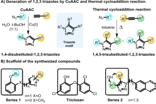
Aiming to expand the SAR studies of the TRC scaffold, a new set of 1,2,3-triazolyl analogs was synthesized using thermal and Cu(I) catalyzed-cycloaddition (CuAAC) reactions. The CuAAC reaction is completely selective being extremely useful for the synthesis of 1,4-disubstituted-1,2,3-triazoles. The thermal cycloaddition provides a mixture of 1,5- and 1,4- regioisomers, being useful to expand our
SAR studies (Scheme 1). Two series of 1,2,3-triazoles analogs of TRC were proposed (Scheme 1, series 1). Due to the critical role of TRC hydroxy group, the designed compounds contained this group in different scaffolds.25 Additionally, the chlorine was removed since it is known that it diminishes the inhibitory activity and interferes with the bioavailability.26 The series 1 scaffold has a phenol connected by a linker to a substituted 1,2,3-triazole that was introduced by a CuAAC reaction. In this series, the linker can be either a methyleneoxy group preserving the catechol moiety of TRC or a methylene group (Scheme 1, series 1). The methyl ether derivatives were also included for the sake of comparison to the free hydroxy derivatives.
Scheme 1. 1,4-disubstituted- and 1,4,5-trisubstituted-1,2,3-triazoles generated by CuAAC reaction and thermal cycloaddition. To increase the diversity of the collection expanding the explored chemical space, 1,4,5-trisubstituted-1,2,3-triazoles were also prepared (Scheme 1, series 2). These 1,4,5-trisubstituted-1,2,3-triazoles were prepared from different azides and internal alkynes by thermal cycloaddition, resulting in mixtures of isomers.
Scheme 2. Synthesis of products starting from catechol 1, 2-methoxyphenol 2 and salicyl alcohol 7.
The synthesis started with the phenol etherification of catechol 1 and guaiacol 2 according to Williamson’s reaction, producing the ether formation with propargyl bromide and potassium carbonate in methanol (Scheme 2). The propargyl-catechol 3 and O-propargyl-2-methoxyphenol 4 were obtained in 46% and 40% yield, respectively. The reaction with catechol 3 also provided the di-propargylated product in 13% yield as by-product. In both reactions, part of the starting material was recovered and could be reused. Once the alkyne intermediates were obtained, the CuAAC reactions was performed with different azides containing aromatic
and carbocycles rings, aliphatic, prenyl and alkyl ethyl ester chains (5a-i, 6a-e, Scheme 2). The average yield for scaffold 5 was 74% and 77% for 6 (Table 1).
The other members of series 1 were prepared with the salicyl azide 8. This key intermediate was prepared from commercially available salicyl alcohol 7 by reaction with sodium azide and triphenylphosphine and in DMF and carbon tetrachloride.27 The reaction CuAAC cycloaddition was performed with commercially available terminal alkynes. The alkynes used were propyne, 1-pentyne, 1-octyne, ethynylbenzene and pent-4-yn-1-ylbenzene (Scheme 2). The average yield of the reaction was of 65% (Table 1).
Table 1. Synthesized 1,2,3-triazoles deriving from catechol, 2-methoxyphenol and salicyilazide.
Comp. Yield (%) R Comp. Yield (%) R 5a 76 CH2Ph 6a 70 CH2Ph 5b 81 (CH2)3Ph 6b 82 (CH2)3Ph 5c 71 Cinnamyl 6c 73 Cyclohexyl 5d 68 Cyclohexyl 6d 79 C8H17 5e 65 C8H17 6e 80 (CH2)COOEt 5f 82 C13H17 9a 76 C3H7 5gZ 70 Neryl 9b 81 C5H11 5gE 70 Geranyl 9c 71 C8H17 5h 79 (CH2)COOEt 9d 68 Ph 5i 78 (CH2)4COOEt 9e 65 (CH2)3Ph
Finally, a thermal cycloaddition in toluene was used to prepare the series 2 with the aim of mimicking the phenol aromatic ring by a hydroxylated triazole motif. Three different azides and three internal alkynes were employed obtaining a mixture of 1,4,5-substituted-1,2,3-triazoles. Benzyl azide 10, 3-phenylpropyl azide 12 and 2-(azidomethyl)naphthalene 14 were combined with the internal alkynes oct-2-yn-1-ol and 3-phenylprop-2-yn-1-ol, hex-3-yn-2-ol to afford the products 11, 13 and 15 (Scheme 3). The phenylpropyl azide 12, with a longer linker is more flexible than benzylazide 10, and the naphthyl derivative is bulkier and has an extended pi-stacking surface.
Yields were calculated including both isolated isomers, which were obtained as a 1:1 mixture of both regioisomers. The ratio was calculated by 1H NMR. The average yield for each scaffold were 61%, 51% and 77% for triazoles 11a-e, 13a-f and 15a-f, respectively (Table 2).
Scheme 3. Synthesis of the products using the thermal cycloaddition methodology.
The collection of 37 triazolyl TRC analogs was characterized using 1H and 13C NMR, and mass spectrometry. The regiochemistry of the series 2 products was determined using nuclear Overhauser experiments (NOE).
Table 2. Synthesized 1,2,3-triazoles using thermal cycloaddition.
Comp R1 R2 Comp R1 R2 Yld
(%)
11a CH2OH C5H11 11b C5H11 CH2OH 52
11c Ph CH2OH 76
11d CH(OH)CH3 C2H5 11e C2H5 CH(OH)CH3 56
13a CH2OH C5H11 13b C5H11 CH2OH 46
13c CH2OH Ph 13d Ph CH2OH 66
13e CH(OH)CH3 C2H5 13f C2H5 CH(OH)CH3 42
15a CH2OH C5H11 15b C5H11 CH2OH 69
15c CH2OH Ph 15d Ph CH2OH 94
15e CH(OH)CH3 C2H5 15f C2H5 CH(OH)CH3 75
For example, for product 11a the signal of the methylene of the methylenehydroxy group at 4.53 ppm showed NOE with the signal of the methylene of the benzyl group at 5.59 ppm and with the methylene from the pentyl group at 2.56 ppm (Figure 2). For the isomer 11b, the irradiation on the same methylene only showed NOE with the methylene of the pentyl chain (Figure 2).
Figure 2. NOE experiments of 11a and 11b. 2.2 Biology
The final obtained compounds were assayed for biological activity against L. donovani promastigotes, against M. tuberculosis H37Rv in
vitro (since the prepared library had been designed based on
compounds with proven antimycobacterial activity) and on mammalian Vero cell lines, for cytotoxicity evaluation (Table 3). Most of the compounds did not present activity against L. donovani promastigotes at 60 µM, the maximum concentration tested. None of the compounds showed cytotoxicity against Vero cells at the maximum concentrations tested of 20 µM. Pentamidine and amphotericin B were used as controls, with IC50 of 4.40 µM and 0.11 µM, respectively. For M. tuberculosis the minimal inhibitory concentration (MIC) was determined by serial microdilution colorimetric assay using MTT as viability indicator.28 In this case TRC was used as positive control, with a MIC99 of 43.0 µM.9
Only four compounds of the series showed IC50 below 60 µM against L. donovani, being 5f the most active compound showing an IC50 of 13.4 µM (Table 3). Regarding the M. tuberculosis assays, the results showed that only analogs 9a, 9b and 9d have MIC below 100 µM. These 3 compounds belong to series 1 and have MIC of 20.0 µM (Table 3).
Having completed the screening stage, the next step was validating the target of the compounds. To do this, the most promising analogs were assayed on the purified mycobacterial InhA.19
Table 3. Results of enzyme inhibition, MICs and cytotoxicity for active compounds against L. donovani and M. tuberculosis.
Comp Ld a IC50 (μM) Mt H37Rvb MIC (μM) Vero IC50 (μM) Mt InhAc inh (%) 5f 13.4 >100 >20 63 5gZ 55.0 >100 >20 85 5gE 51.9 >100 >20 N.I. 9a >60 20.0 >20 92 9b >60 20.0 >20 N.I.d 9d >60 20.0 >20 N.I 15b 31.4 >100 >20 N.I. TRC 43.0 PM 4.40 AMB 0.11
TRC = triclosan, PM = pentamidine, AMB= amphotericin B. a Ld: L.
dononani promastigotes, evaluated by Alamar blue assay. b Mt
H37Rv: Mycobacterium tuberculosis H37Rv, evaluated by serial microdilution colorimetric assay using MTT as viability indicator. c
Mt InhA: remaining M. tuberculosis InhA activity. d N.I.: No
Inhibition.
The initial assay was performed at a fixed compound concentration. For derivatives 5f, 5gZ, 5gE at 40 μM and for derivatives and 9a, 9b,9d and 15b at 50 μM. The reaction velocity in absence (v)0 and presence of the inhibitor (v)I was determined. The results were disappointing since the tested compounds proved to be extremely poor in inhibitory efficacy with only marginal inhibition shown by 3 of the most promising compounds compared to TRC which showed an IC50 of 1.0 μM.9 The remaining enzyme activities were 63, 85 and 92%, for compounds 5f, 5gZ and 9a, respectively (Table 3).
When discussing the most active compounds against L. donovani, the disappointing results could be explained with the fact that they were assayed on the mycobacterial enzyme and not on the actual L.
donovani orthologue. There are substantial differences between Leishmania spp. and M. tuberculosis fatty acid synthesis end
products. While the parasite fatty acids are composed by C14 to C22 fatty acids,29 the mycobacterial FASII could elongate fatty acids up to
40 carbons.30 Those variations would probably justify the sequences differences between the M tuberculosis and Leishmania spp. ENRs that will ultimately redound on the lack of inhibition of InhA. Consequently, we should explore this in detail to obtain a definitive conclusion of whether minor but structurally important differences between the different ENRs may condition the inhibitory activity.
Also, there is low or null inhibition of the most promising compounds against M. tuberculosis. The compounds that display some activity, analogs 5f, 5gZ and 9a, share the critical phenolic hydroxyl on their structure with TRC and 8PP (Table 3), nevertheless they did not show inhibitory effect on InhA. Those results could be explained by different target/s, hypothesis that could be further investigated.
2.3 Structure-activity relationship (SAR)
In the previous section, the results of the biological activities against L. donovani and M. tuberculosis were presented with only few compounds displaying against each pathogen. The inhibition
capacity towards M. tuberculosis InhA of the most attractive collection members was assayed, resulting in surprisingly poor inhibition. As it was previously discussed, there is a lack of overlap between the compounds which show greater potency for L.
donovani and those which show most potency for M. tuberculosis.
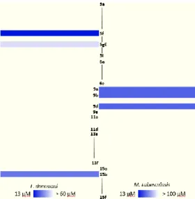
This contrast becomes more visible when analyzing the heatmap in Figure 3 which clearly shows the leads for each pathogen in a brighter tone of blue. Therefore, the need of a more profound analysis of the structure-activity relationship for both, L. donovani and M. tuberculosis separately, became stronger.
Figure 3. Heatmap of active compounds against L. donovani and M.
tuberculosis.
As regards L. donovani promastigotes, two populations of active compounds were identified. The first group comprises analogs from series 1 (5f, 5gZ,5gE; IC50=13.4, 55.0 and 51.9 µM respectively); while the second consists of only compound 15b (IC50=31.4 µM) from series 2 (Figure 4). The active compounds contain long lipophilic substituents on the triazole (pentyl, octyl, geranyl, neryl and tridecyl) and their activity seems to be related to a triazole side chain longer than 5 carbons. The restricted conformation produced by the double-bond on the isoprenyl derivatives seems to be detrimental for the activity. Comparison of analogs 15b and 5f, clearly showed that longer aliphatic saturated chain improved the activity. This structural feature resembles the 5-alkyl-diphenyl ethers moiety present in 8PP where the saturated lipophilic chain plays a critical role in the binding to InhA.9 Three out of the four active compounds have a phenol in their structure, and the fourth compound has a methylenehydroxy group attached to the triazole. None of the methylated phenol derivatives was active, demonstrating the importance of the hydroxy group on the activity. The fact that three active compounds are from series 1 confirms that the incorporation of the ether on the structure mimics TRC and 8PP.
Figure 4. Active compounds against L. donovani promastigotes. On another hand, the results on M. tuberculosis shown that compounds 9a and 9b have a lipophilic saturated aliphatic chain on the 1,2,3-triazole (Figure 5) being like 8PP, but notably less active (8PP MIC99 = 6.4 µM). The other active analog is 9d, which has a phenyl ring decorating the 1,2,3-triazole, fact that shows that the flexible lipophilic chain can be replaced by that non-polar bulkier ring. Surprisingly, the derivative 9c that holds an octyl chain, the derivative structurally most closely related to 8PP, was inactive. The activity vs aliphatic tail relationship for the C5 derivative is less significant than for the C8 when comparing the diphenyl ethers.9 Ethers 5e and 6d, which also resemble 8PP, were also inactive. The rest of the library does not show activity against M. tuberculosis H37Rv with MICs higher than 32 µM. These compounds are considerably less active than the previous collection inspired on 8PP reported by our group, which reached low micromolar MIC on H37Rv.16, 31 Nevertheless, the active members of the collection are twice more active than TRC that has a MIC of 43.0 µM.
A close comparison with the triazolyl-diphenyl ethers reported by Spaugnolo et al32 showed that not only the substitution pattern on the phenol ring, but mostly the presence of the phenoxy moiety, plays a critical role on the antitubercular activity (Figure 5). This is clearly seen for compounds 9a, 9b, and 9d that are 1.8, 5.1 and 9.9 times less active than the diphenyl ethers PT163, PT509 and PT510, respectively (Figure 5).
Figure 5. Active compounds against M. tuberculosis H37Rv (MIC). 2.4 Physicochemical parameters
Finally, the physicochemical parameters were calculated in silico for the active compounds of the library using Data Warrior software33 (Figure S2. Supporting Inf.). The parameters of Lipinski's rule of five were calculated, including the topological polar surface area (TPSA). Lipinski's rule of five is widely used for determination of good pharmacokinetics properties. The parameters considered were the molecular weight (MW<500 Da), the partition coefficient between water and octanol (0<logP<5) and the number of hydrogen bond acceptors (nNHOH<10) and donors (nON<5). If there were two or more violations of these rules, the compounds would present poor oral administration, but there are exceptions to these rules. Another useful parameter is the topological polar surface area (TPSA) that considers the electron density distribution in the molecule and give us an idea of the capacity to penetrate biological membranes. This parameter is commonly used to establish if the compounds could penetrate the blood brain barrier. All the active compounds comply with the rule of five and TPSA parameters. Only compound 5f violates one rule having a logP of 5.67. Interestingly, this compound is the most active of the collection against L.
physicochemical requirements could be taken in a lighter way in order to obtain antiparasitic lead compounds susceptible of further improvement.34
3. Experimental
3.1. Synthesis
General procedure for the synthesis of 1,4-disubstituted 1,2,3-triazoles
The alkyne (1 eq) and the azide (1.5 eq) were suspended in 4 mL of t
BuOH:H2O (1:1). Afterwards, aqueous 1 M CuSO4 (0.025 eq) solution and aqueous 1 M sodium ascorbate (0.1 eq) solution were added. The mixture was stirred overnight at room temperature. Brine was added and the solution was extracted with dichloromethane. Combined organic extracts were dried with sodium sulphate and evaporated. The resulting residue was purified by column chromatography over silica gel using an increasing AcOEt/hexane gradient to afford desired pure products.
Synthesis of 2-((1-tridecyl-1H-1,2,3-triazol-4-yl)methoxy)phenol (5f). Colorless oil. 1H NMR (300 MHz, CDCl3) :7.54 (s, 1H); 7.00 (dd, J1 = 7.8 Hz, J2 = 1.5 Hz, 1H); 6.94 (dd, J1 =7.8 Hz, J2 = 2.1 Hz, 1H); 6.90 (dd, J1 = 7.9 Hz, J2 = 1.5 Hz, 1H); 6.82 (ddd, J1 = 7.8 Hz, J2 = 2.5 Hz, J3 = 2.3 Hz, 1H); 6.11 (s, 1H); 5.26 (s, 2H); 4.35 (t, J = 7.9 Hz, 2H); 1.09 (q, J = 7.6 Hz, 2H); 1.35-1.22 (m, 20H); 0.88 (t, J = 6.7 Hz, 3H). 13C NMR (75 MHz, CDCl3) :146.6 (C); 145.7 (C); 143.5 (C); 122.6 (CH); 122.5 (CH); 120.1 (CH); 115.6 (CH); 113.8 (CH); 63.2 (CH2); 50.5 (CH2); 31.9 (CH2); 30.2 (CH2); 29.6 (CH2); 29.5 (CH2); 29.4 (CH2); 29.3 (CH2); 29.0 (CH2); 26.4 (CH2); 22.7 (CH2); 14.1 (CH3). Synthesis of 4-((2-methoxyphenoxy)methyl)-1-octyl-1H-1,2,3-triazole (6d). Colourless oil. 1H NMR (300 MHz, CDCl3) :7.61 (s, 1H); 7.04 (dd, J1 = 7.0 Hz, J2 = 2.2 Hz, 1H); 6.94 (m, 1H); 6.90 (s, 1H); 6.89 (dd, J1 = 7.7 Hz, J2 = 2.5 Hz, 1H); 5.30 (s, 2H); 4.32 (t, J = 7.3 Hz, 2H); 3.87 (s, 3H); 1.88 (q, J = 6.0 Hz, 2H); 1.37-1.20 (m, 10H); 0.87 (t, J = 6.0 Hz, 3H). 13C NMR (75 MHz, CDCl3) :149.6 (C); 147.6 (C); 144.3 (C); 122.6 (CH); 121.8 (CH); 120.9 (CH); 114.4 (CH); 111.8 (CH); 63.3 (CH2); 55.9 (CH3) 50.4 (CH2); 31.7 (CH2); 30.2 (CH2); 29.0 (CH2); 28.9 (CH2); 26.4 (CH2); 22.6 (CH2); 14.0 (CH3). Synthesis of 2-((4-pentyl-1H-1,2,3-triazol-1-yl)methyl)phenol (9b). White solid. Mp: 88.2-89.2°C.1H NMR (300 MHz, CDCl3) : 7.45 (s, 1H), 7.24 (s, 1H), 7.19 (d, J = 7.8 Hz, 1H), 7.01 (d, J = 7.8 Hz, 1H), 6.86 (t, J = 7.3 Hz, 1H), 5.52 (s, 2H), 2.67 (t, J = 7.7 Hz, 2H), 1.63 (p, J = 7.5 Hz, 2H), 1.36-1.34(m, 4H), 0.85 (t, J = 6.8 Hz, 3H). 13C NMR (75 MHz, CDCl3) : 155.4 (C), 148.4 (C), 130.4 (CH), 130.4 (CH), 121.6 (CH), 121.5 (C), 120.3 (CH), 116.9 (CH), 49.8 (CH2), 31.4 (CH2), 29.0 (CH2), 25.5 (CH2), 22.3 (CH2), 13.9 (CH3). ESI-HRMS Calcd for (M+H+) C14H19N3O 246.1606, found 246.1601.
General procedure for synthesis of 1,2,3–triazoles by thermal cycloaddition
Alkyne (1 eq) and azide (1 eq) were suspended in 2 mL/eq of toluene. The mixture was stirred and heated under reflux for 35 hours. Then, it was allowed to warm to room temperature, brine was added, and the solution was extracted with dichloromethane. Combined organic extracts were dried with sodium sulphate and evaporated. The resulting residue was purified by column
chromatography over silica gel using an increasing AcOEt/hexane gradient to afford desired pure products.
Synthesis of (1-benzyl-4-pentyl-1H-1,2,3-triazol-5-yl)methanol (11a). White solid. MP: 59.3-60.0°C. 1H NMR (300 MHz, CDCl3) : 7.29-7.30 (m, 3H); 7.22-7.19 (m, 2H); 5.59 (s, 2H); 4.53 (d, J = 4,7 Hz, 2H); 2.56 (t, J = 7,7 Hz, 2H); 1.60 (p , J = 7,0 Hz, 2H); 1.27 (m, 4H); 0.85 (t, J = 6,5 Hz, 3H). 13C NMR (75 MHz, CDCl3) : 146.5 (C); 135.2 (C); 131.9 (C); 128.9 (CH); 128.3(CH); 127.5 (CH); 52.3 (CH2); 52.1 (CH2); 31.5 (CH2); 29.6 (CH2); 26.8 (CH2); 22.4 (CH2); 14.0 (CH3). ESI-HRMS Calcd for (M+H+) C15H21N3O 260.1763; found 260.1757.
Synthesis of (4-pentyl-1-(3-phenylpropyl)-1H-1,2,3-triazol-5-yl)methanol (13a). Colorless oil. 1H NMR (300 MHz, CDCl3) : 7.31-7.17 (m, 5H); 4.62 (d, J = 4.3 Hz, 2H); 4.36 (t, J = 7.3 Hz, 2H); 2.69 (t, J = 7.3 Hz, 2H); 2.61 (t, J = 7.6 Hz, 2H); 2.29 (q, J = 7.1 Hz, 2H); 1.68-1.62 (m, 2H); 1.31 (m, 4H, C7-H); 0.88 (t, J = 7.0 Hz, 3H). 13C NMR (75 MHz, CDCl3) : 146.1 (C); 140.6 (C); 131.3 (C); 128.5 (CH); 128.4 (CH); 126.2 (CH); 52.3 (CH2); 47.9 (CH2); 32.7 (CH2); 31.5 (CH2); 31.4 (CH2); 29.7 (CH2); 24.9 (CH2); 22.4 (CH2); 14.0 (CH3).ESI-HRMS Calcd for (M+H+) C17H26N3O 288.2076; found 288.2070. Synthesis of (1-(naphthalen-2-ylmethyl)-5-pentyl-1H-1,2,3-triazol-4-yl)methanol (15b). White solid. MP: 79.2-80.2 °C. 1H NMR (300 MHz, CDCl3) : 7.80 (m, 3H); 7.76 (s, 1H); 7.50-7.47 (m, 2H); 7.27 (m, 1H); 5.64 (s, 2H); 4.72 (d, J = 5.4 Hz, 2H); 2.59 (t, J = 7.5 Hz, 2H); 1.34 (p, J = 7.5 Hz, 2H); 1.14-1.12 (m, 4H); 0.72 (t, J = 7.0 Hz, 3H). 13C NMR (75 MHz, CDCl3) : 144.9 (C); 135.2 (C); 133.2 (C); 133.0 (C); 132.4 (C); 129.0 (CH); 127.8 (CH); 127.8 (CH); 126.6 (CH); 126.5 (CH); 126.2 (CH); 124.7 (CH); 55.9 (CH2); 52.2 (CH2); 31.4 (CH2); 28.5 (CH2); 22.6 (CH2); 22.1 (CH2); 13.7 (CH3). ESI-HRMS Calcd for (M+H+) C19H24N3O 310.1841; found 310.1904.
3.2. Biology
Bacterial strain M. tuberculosis H37Rv
M. tuberculosis strain H37Rv (kindly provided by Dr. L. Barrera
(Instituto Nacional de Microbiología “C.G. Malbrán”, Argentina) was routinely grown at 37 °C under gentle agitation in Middlebrook 7H9 broth (Difco Laboratories, Detroit, MI, USA) supplemented with 1/ 10 v/v of ADS (50 g/L BSA fraction V, 20 g/L dextrose and 8.1 g/L NaCl), glycerol (1% w/v). Tween 80 was added to prevent clumping (0.05% w/v). This medium was designated as 7H9-ADS-Gly for short. When needed, solid media Middlebrook 7H11 supplemented with ADS (1/10 v/v) and glycerol (1% v/v) was used.
In vitro activity against M. tuberculosis H37Rv.
Stock solutions for all the tested compounds were made in DMSO at 40 mM. Working solutions were made by dilution in the above described 7H9 - ADS-G medium at a final concentration of 400 . Antimycobacterial activity was determined by two-fold dilution of the compounds in Middlebrook 7H9-ADS-Gly medium as described previously.16 For this purpose, 96-well plates (Falcon, Cat number 3072, Becton Dickinson, Lincoln Park, NJ) were used. The 96 well-plates received 100 L of Middlebrook 7H9 broth and a serial two-fold dilution of the compounds was made directly on the plate. The initial and final drug concentrations tested were 20 M and 1.25 M, respectively. Compounds were tested in three biological
repetitions each one in technical duplicates. Rifampicin (final concentrations ranging from 2mg/mL to 0.16 mg/mL; stock solution prepared as a 10 mg/mL solution in methanol) was used as control drug. The inoculum was prepared as a 1/25 dilution of a fresh mid-log M. tuberculosis H37Rv suspension (O.D equivalent to Mc Farland 1.0 scale value) made in Middlebrook 7H9-ADS-G. A 100 µL aliquot (containing approximately 106 Colony Forming Units) was used to inoculate the wells. Plates were sealed with Parafilm and incubated at 37°C for five days. After visual inspection of the plates, the turbidity was recorded and the final value for the MIC was assessed by addition of 30 µl of a stock MTT solution as described elsewhere.35 Minimum Inhibitory Concentration (MIC) was defined as the lowest drug concentration preventing mycobacterial growth (yellow color= growth inhibition).
InhA inhibition assay.
InhA Inhibition activity was tested using trans-2-dodecenoyl-Coenzyme A (DD-CoA) and wild-type InhA as described previously.36 Reactions were initiated by the addition of 100 nM InhA to solutions containing 25 μM DD-CoA, 40-50 μM inhibitor, and 250 μM NADH in 30 mM PIPES and 150 mM NaCl, pH 6.8 buffer. Control reactions were carried out with the same conditions as described above but without inhibitor. The inhibitory activity of each derivative was expressed as the percentage inhibition of InhA activity (initial velocity of the reaction) with respect to the control reaction without inhibitor.
In vitro antileishmanial assay.
Leishmania donovani promastigotes of the S1 Sudan strain (2 x 10⁶
cell/mL) were cultured at 26°C in plastic flasks (25 cm2), containing RPMI-1640 medium (without sodium bicarbonate and sodium pyruvate) with 10% FBS. Subculture of L. donovani promastigotes twice a week, with highest cells concentration in the range of 20-25x106 promastigotes/mL. Compounds with appropriate dilution were added to a 96 well microplate with promastigotes (2 × 10⁶ cell/mL) reaching final concentrations of 40, 8 and 1.6 μg/mL. The plates were incubated at 26 °C for 72 h and growth was determined by Alamar blue assay.37 Pentamidine and amphotericin B were used as standard antileishmanial agents. IC50 values were obtained by non-linear regression of dose response logistic functions, using the Microsoft Excel-based plug-in XLfit. All experiments were performed in triplicate.
Cytotoxicity assay
The in vitro cytotoxicity was determined against mammalian kidney fibroblasts (VERO). Vero cells (African green monkey kidney) were cultured in MEM medium supplemented with 10% heat inactivated FCS, 0.15% (w/v) NaHCO3, 100 U/mL penicillin and 100 U/mL streptomycin at 37ºC in 5% CO2 atmosphere.
The assay was performed in 96-well tissue culture-treated plates as described earlier.38 Cells were seeded to the wells of the plate (25,000 cells/well) and incubated for 24 h. Samples were added and plates were again incubated for 48 h. The number of viable cells was determined by neutral red assay. IC50 values were determined from logarithmic graphs of growth inhibition versus concentration. Doxorubicin was used as a positive control (IC50 = 14 mM, Vero cells), while DMSO was used as vehicle control.
4. Conclusions
For this present study, a collection of thirty-seven 1,4-disubstituted and 1,4,5-trisubstituted 1,2,3-triazole analogs of triclosan were designed based on the structure of reported InhA inhibitors. The 1,4-disubstituted products were prepared using CuAAC, while for the 1,4,5-trisubstituted derivatives a thermal cycloaddition was used, in both cases with good yields. The preparation of 1,4,5-trisubstituted 1,2,3-triazoles has not been exploited in library preparation, in part due the lack of selectivity. In our approach this fact proved to be a valuable tool to diversify our collection and expand the chemical space explored. The collection was initially assayed against L. donovani and M. tuberculosis. For L. donovani, four compounds have IC50 below 60 μM. The activity profile showed that a lipophilic substituent in the triazole and an alcohol were required for activity. When the collection was assayed in vitro against M. tuberculosis H37Rv, 3 derivatives displayed a MIC99 of 20.0 μM. To validate the original hypothesis that the compounds have InhA L. donovani ortholog as the target, the most promising compounds of the library were assayed on the mycobacterial enzyme, showing poor inhibition. Those results suggested that InhA may not be the main target for the active compounds against M.
tuberculosis. Nevertheless, to effectively discard the hypothesis for
the active compounds against L. donovani, the compounds should be tested on the leishmanial enzyme.
In summary, compound 5f could be considered a valuable starting point for future antileishmanial drug development, based on its simple preparation and promising activity. The possibility that their activity could be linked to a putative leishmanial InhA orthologue should be a matter of future investigation, together with the exploration of new structural modifications to enhance the activity. For this purpose, the purification of the putative Leishmania ORF and its assay for ENR activity would lead the priority list. That will be followed by testing the analogues against this potential InhA orthologue. Parallelly, the most promising candidates will be assayed in other Leishmania species including the intracellular stage of the parasite.
Conflicts of interest
There are no conflicts to declare.
Acknowledgements
This work was supported in part by grants from National Research Council of Argentina, CONICET (PIP 2012-14/0448); Agencia Nacional de Promoción Científica y Tecnológica, ANPCyT- Argentina (PICT 2011/0589 to GRL and PICT 2005/38198 to HRM); National Institutes of Health, NIH (GM102864 to PJT); Universidad Nacional de Rosario (BIO503 to GRL) and Fundación Josefina Prats. The research leading to these results has, in part, received funding from the UK Research and Innovation via the Global Challenges Research Fund under grant agreement ‘A Global Network for Neglected Tropical Diseases’ grant number MR/P027989/1. GRL is a member of the scientific staff of CONICET-Argentina. HRM is a career member of CIUNR-Argentina. D.G.G., J.F.de L. and A.I.R.B. thanks CONICET for the award of a Fellowship.
1. WHO, Leishmaniasis report, World Health Organization, 2019.
2. J. Yao and C. O. Rock, Biochim. Biophys. Acta -Mol. Cell.
Biol., 2017, 1862, 1300-1309.
3. S. Yadav, H. Mandal, V. Saravanan, P. Das and S. K. Singh,
J. Biomol. Struct. Dyn., 2020, 1-14.
4. C. E. Barry, D. C. Crick and M. R. McNeil, Infect. Disord.
Drug Targets, 2007, 7, 182-202.
5. V. Arango, J. J. Domínguez, W. Cardona, S. M. Robledo, D. L. Muñoz, B. Figadere and J. Sáez, Med. Chem. Res., 2012, 21, 3445-3454.
6. K. S. Paul, C. J. Bacchi and P. T. Englund, Eukarytotic Cell, 2004, 3, 855-861.
7. L. M. McMurry, P. F. McDermott and S. B. Levy,
Antimicrob. Agents Chemother., 1999, 43, 711-713.
8. L. Q. Wang, C. N. Falany and M. O. James, Drug Metab
Dispos, 2004, 32, 1162-1169.
9. T. J. Sullivan, J. J. Truglio, M. E. Boyne, P. Novichenok, X. Zhang, C. F. Stratton, H. J. Li, T. Kaur, A. Amin, F. Johnson, R. A. Slayden, C. Kisker and P. J. Tonge, ACS Chem. Biol., 2006, 1, 43-53.
10. A. N. Bootsma and S. E. Wheeler, ChemMedChem, 2018, 13, 835-841.
11. A. Çapcı, M. M. Lorion, H. Wang, N. Simon, M. Leidenberger, M. C. Borges Silva, D. R. M. Moreira, Y. Zhu, Y. Meng, J. Y. Chen, Y. M. Lee, O. Friedrich, B. Kappes, J. Wang, L. Ackermann and S. B. Tsogoeva, Angew. Chem.
Int. Ed., 2019, 58, 13066-13079.
12. V. B. Sokolov, A. Y. Aksinenko, T. A. Epishina and T. V. Goreva, Russ. Chem. Bull., 2019, 68, 1424-1428.
13. K. Bozorov, J. Zhao and H. A. Aisa, Biorg. Med. Chem., 2019, 27, 3511-3531.
14. P. Thirumurugan, D. Matosiuk and K. Jozwiak, Chem. Rev., 2013, 113, 4905-4979.
15. J. Stec, C. Vilcheze, S. Lun, A. L. Perryman, X. Wang, J. S. Freundlich, W. Bishai, W. R. Jacobs, Jr. and A. P. Kozikowski, ChemMedChem, 2014, 9, 2528-2537.
16. D. G. Ghiano, A. de la Iglesia, N. Liu, P. J. Tonge, H. R. Morbidoni and G. R. Labadie, Eur. J. Med. Chem., 2017, 125, 842-852.
17. E. O. Porta, P. B. Carvalho, M. A. Avery, B. L. Tekwani and G. R. Labadie, Steroids, 2014, 79, 28-36.
18. M. R. Kuo, H. R. Morbidoni, D. Alland, S. F. Sneddon, B. B. Gourlie, M. M. Staveski, M. Leonard, J. S. Gregory, A. D. Janjigian, C. Yee, J. M. Musser, B. Kreiswirth, H. Iwamoto, R. Perozzo, W. R. Jacobs, J. C. Sacchettini and D. A. Fidock,
J. Biol. Chem., 2003, 278, 20851-20859.
19. J. S. Freundlich, F. Wang, C. Vilchèze, G. Gulten, R. Langley, G. A. Schiehser, D. P. Jacobus, W. R. Jacobs Jr. and J. C. Sacchettini, ChemMedChem, 2009, 4, 241-248. 20. T. J. Sullivan, J. J. Truglio, M. E. Boyne, P. Novichenok, X.
Zhang, C. F. Stratton, H.-J. Li, T. Kaur, A. Amin, F. Johnson, R. A. Slayden, C. Kisker and P. J. Tonge, ACS Chem. Biol., 2006, 1, 43-53.
21. L. Urán Landaburu, A. J. Berenstein, S. Videla, P. Maru, D. Shanmugam, A. Chernomoretz and F. Agüero, Nucleic
Acids Res., 2019, 48, D992-D1005.
22. N. Ravooru, S. Ganji, N. Sathyanarayanan and H. G. Nagendra, Front Genet, 2014, 5, 291.
23. D. Dheer, V. Singh and R. Shankar, Bioorg. Chem., 2017, 71, 30-54.
24. R. Adam, P. Bilbao-Ramos, B. Abarca, R. Ballesteros, M. E. González-Rosende, M. A. Dea-Ayuela, F. Estevan and G. Alzuet-Piña, Org. Biomol. Chem., 2015, 13, 4903-4917.
25. A. Chollet, L. Maveyraud, C. Lherbet and V. Bernardes-Génisson, Eur. J. Med. Chem., 2018, 146, 318-343. 26. S. Sivaraman, T. J. Sullivan, F. Johnson, P. Novichenok, G.
Cui, C. Simmerling and P. J. Tonge, J. Med. Chem., 2004, 47, 509-518.
27. Q. Zhang and J. M. Takacs, Org. Lett., 2008, 10, 545-548. 28. L. Caviedes, J. Delgado and R. H. Gilman, J. Clin. Microbiol.,
2002, 40, 1873-1874.
29. H. Bouazizi-Ben Messaoud, M. Guichard, P. Lawton, I. Delton and S. Azzouz-Maache, Lipids, 2017, 52, 433-441. 30. H. Marrakchi, M.-A. Lanéelle and M. Daffé, Chem. Biol.,
2014, 21, 67-85.
31. G. R. Labadie, A. de la Iglesia and H. R. Morbidoni, Mol.
Diver., 2011, 15, 1017-1024.
32. L. A. Spagnuolo, S. Eltschkner, W. Yu, F. Daryaee, S. Davoodi, S. E. Knudson, E. K. Allen, J. Merino, A. Pschibul, B. Moree, N. Thivalapill, J. J. Truglio, J. Salafsky, R. A. Slayden, C. Kisker and P. J. Tonge, J. Am. Chem. Soc., 2017, 139, 3417-3429.
33. T. Sander, J. Freyss, M. von Korff and C. Rufener, J. Chem.
Inf. Model., 2015, 55, 460-473.
34. J. H. McKerrow and C. A. Lipinski, International Journal for
Parasitology: Drugs and Drug Resistant 2017, 7, 248-249.
35. G. Abate, A. Aseffa, A. Selassie, S. Goshu, B. Fekade, D. WoldeMeskal and H. Miorner, J. Clin. Microbiol., 2004, 42, 871-873.
36. L. Bonnac, G. Y. Gao, L. Chen, K. Felczak, E. M. Bennett, H. Xu, T. Kim, N. Liu, H. Oh, P. J. Tonge and K. W. Pankiewicz,
Bioorg. Med. Chem. Lett., 2007, 17, 4588-4591.
37. J. Mikus and D. Steverding, Parasitol. Int., 2000, 48, 265-269.
38. H. Babich and E. Borenfreund, Appl. Environ. Microbiol., 1991, 57, 2101-2103.


