HAL Id: tel-02955510
https://tel.archives-ouvertes.fr/tel-02955510
Submitted on 2 Oct 2020HAL is a multi-disciplinary open access archive for the deposit and dissemination of sci-entific research documents, whether they are pub-lished or not. The documents may come from teaching and research institutions in France or abroad, or from public or private research centers.
L’archive ouverte pluridisciplinaire HAL, est destinée au dépôt et à la diffusion de documents scientifiques de niveau recherche, publiés ou non, émanant des établissements d’enseignement et de recherche français ou étrangers, des laboratoires publics ou privés.
antibiotics in Streptomyces
Celine Aubry
To cite this version:
Celine Aubry. Towards combinatorial biosynthesis of pyrrolamide antibiotics in Streptomyces. Biomolecules [q-bio.BM]. Université Paris Saclay (COmUE), 2019. English. �NNT : 2019SACLS245�. �tel-02955510�
Towards combinatorial
biosynthesis of pyrrolamide
antibiotics in
Streptomyces
Thèse de doctorat de l'Université Paris-Saclay
préparée à l’Université Paris-Sud
École doctorale n°577
Structure et Dynamique des Systèmes Vivants (SDSV)
Spécialité de doctorat : Sciences de la vie et de la SantéThèse présentée et soutenue à Orsay, le 30/09/19, par
Céline AUBRY
Composition du Jury :
Matthieu Jules
Professeur, Agroparistech (MICALIS) Président du Jury
Yanyan Li
Chargée de recherche, MNHN (MCAM) Rapportrice
Stéphane Cociancich
Chercheur, CIRAD (BGPI) Rapporteur
Annick Méjean
Professeure, Université Paris-Diderot (LIED) Examinatrice
Hasna Boubakri
Maitre de conférences, Université Claude Bernard Lyon I
(Ecologie microbienne) Examinatrice
Sylvie Lautru
1
Acknowledgements
J’ai insisté pour rédiger l’ensemble de ma thèse en anglais. La logique voudrait donc que cette section soit écrite en anglais également, mais je n’ai pas pu m’y résoudre. Je vous présente mes excuses pour cette entorse linguistique.
Si le doctorat est l’occasion de creuser un projet principalement réalisé par le doctorant, ce n’est en aucun cas un travail individuel qui s’accomplit seul. De nombreuses personnes, par leur aide sur tous les aspects d’un projet doctoral, qu’ils soient scientifiques, administratifs, sociaux ou émotionnels, m’ont permis de vivre pleinement cette expérience. Ces personnes sont trop nombreuses pour être toutes citées ici, mais sachez que les moments partagés font partie des souvenirs irremplaçables que je garde de ces quatre années dans l’équipe de Microbiologie Moléculaire des Actinomycètes (MMA) à l’Institut de Biologie Intégrative de la Cellule.
Je souhaite exprimer toute ma reconnaissance à Sylvie Lautru, ma directrice de thèse. Tout au long du projet, tu m’as encadrée avec beaucoup de patience et de disponibilité. Tu m’as témoigné une grande confiance concernant la réalisation des expériences et m’as encouragée à gagner en autonomie. Tu as toujours écouté mes suggestions avant de me donner ta vision des choses, puis de discuter ensemble de la suite. Tu as également été présente pour écouter mes doutes et me rassurer dans les moments plus difficiles. Merci de m’avoir guidée tout en me laissant libre de choisir mon chemin et ma manière de faire les choses.
Je remercie les membres de mon jury de thèse, Yanyan Li, Stéphane Cociancich, Annick Méjean, Hasna Boubakri et Matthieu Jules, d’avoir accepté de consacrer du temps à évaluer mes travaux de recherche. Annick Méjean a également fait partie de mes comités de suivi de thèse, avec Muriel Gondry, Christiane Elie et Jean-Luc Pernodet, et je les remercie pour leurs conseils et leur bienveillance concernant mon projet.
Concernant les procédures administratives, j’ai bénéficié de l’aide toujours efficace de Muriel Decraëne et de Catherine Drouet, ainsi que de Marie-Hélène Sarda et de Martine Denis, et de Blandine Champion-Grosjean. Votre expertise des multiples procédures m’a permis de gagner un temps précieux.
Je tiens également à remercier Paolo Clérici, qui a réalisé la synthèse chimique d’un précurseur de l’anthelvencine spécialement pour une de mes expériences, mais aussi Laurent Micouin et son équipe, qui se sont penchés et se penchent encore aujourd’hui sur la caractérisation structurale de l’anthelvencine avec beaucoup de ténacité. Un grand merci également à Zhilai Hong, Yanyan Li et Soizic Prado, qui ont permis l’analyse en spectrométrie de masse à haute résolution des anthelvencines, et m’ont donné les clefs nécessaires à la compréhension des résultats.
2 D’autres petites mains ont directement participé à mon projet, celles de mes stagiaires Jennifer Perrin et Yacine Sellah. Merci beaucoup, d’une part vous m’avez aidée à faire avancer mon projet, et d’autre part, j’ai beaucoup appris en vous encadrant. Jennifer, tu as été ma première stagiaire, et j’ai été impressionnée par ta vivacité d’esprit et ta motivation. Tu t’es aussi très bien intégrée à l’équipe et je me rappelle la période de ton stage comme un moment plein de rires et de bonne humeur. Yacine, ton projet portait sur des parties plus difficiles de ma thèse, mais tu n’as jamais abandonné et ta persévérance a porté ses fruits, merci d’avoir souhaité autant que moi la réussite de ce projet.
Mon implication dans l’encadrement de l’équipe iGEM GO-Paris-Saclay 2018 a également consisté une expérience inestimable. Merci aux encadrants, Stéphanie, Philippe et Mahnaz, j’ai apprécié le résultat de notre coopération ! Merci aussi à tous les étudiants, pour leur enthousiasme et leur créativité, je remercie en particulier les étudiants que j’ai accompagnés à Boston pour présenter les résultats, une aventure pleine de rebondissements et de bons souvenirs.
J’aimerais également remercier tous les membres passés et présents des équipes MES et MMA, nos interactions ont rendu mon séjour ici inestimable. Merci en particulier à Jean-Luc, pour ton oreille attentive et ta connaissance incroyable des évènements qui méritent un apéritif, à Christiane pour les anecdotes invraisemblables et les conseils pragmatiques, à Laetitia pour tous les jeudis aux expériences ratées et ta fidélité presque sans faille aux repas du CESFO, à Luisa pour ton efficacité et pour oser aborder les problèmes sans détours, à Alba pour les soirées au dehors et la complicité en 105, à Jerzy pour les petites astuces de labo et les pauses « café » chez Sylvain, à Laura, pour le partage paisible de l’espace et du projet pyrrolamides, à Sylvain, pour l’accueil dans ta cuisine et les discussions aux sujets divers, à Armel, pour les connaissances sur tout et les bonbons, à Manue, pour ta prévenance à mon égard et la gestion infatigable des soucis de séquençage, à Soumaya, pour les conseils spécial doctorants qui facilitent la vie, à Mathieu, pour les bonjours impromptus et le cactus, à Brittany, pour ta gaieté contagieuse et le voyage à Boston, à Corinne, pour la bonne humeur et la musique, à Stéphanie, pour ton dynamisme et cette incroyable capacité à remotiver les gens, à Hervé, pour ton dévouement à toujours ressusciter « mamie » et ton recul sur la science, à Marc, pour ne pas avoir jeté l’HPLC par la fenêtre malgré la tentation, à Aaron, pour des barbecues mémorables, et à Marie-Joëlle et Michelle, pour avoir toujours eu froid pour moi. Merci aussi à Nelly, pour avoir repris le flambeau en l’absence de Sylvain, et m’avoir permis de poursuivre mes expériences en toute sérénité.
Il a fait bon vivre au bâtiment 400, où les gens sont toujours prêts à apporter leur aide sur un appareil, ou à partager un moment convivial autour d’un repas ou d’un verre. Merci à tous pour cette ambiance des plus agréables ! Merci aussi aux « anciens » de l’équipe MMA qui sont restés dans les environs, Audrey, Florence et Drago, pour leurs conseils avisés.
Un énorme merci à Clara, ma voisine de palier. A nous deux, nous avons égayé le couloir, parfois un peu bruyamment, et acheté les vivres nécessaires aux apéritifs du vendredi soir. Mais bien plus que ça, tu es devenue une véritable amie, et les activités en dehors qui ont commencé par des sessions piscines se sont bien vite diversifiées. Je ne compte plus nos multiples discussions, ni les sorties à l’Opéra ou au cinéma. Ma vie pendant le doctorat n’aurait pas été la même sans toi !
3 Je souhaite adresser un remerciement spécial à Lucile. Sans toi, cette aventure n’aurait jamais commencé. Merci de m’avoir conseillée ce projet que tu avais initié pendant ton stage !
Je souhaite aussi exprimer une reconnaissance toute particulière à Valentin T., un chercheur en génétique et linguistique des populations humaines exceptionnel. Nos discussions sur le milieu de la recherche scientifique m’ont permis de prendre du recul, et d’élargir ma vision du monde. Tes réflexions sur l’éthique et le sens de la vie ont également généré en moi beaucoup de questions, le genre de questions qu’on ne peut pas se permettre d’ignorer. Je continue à chercher mes réponses.
Merci aux amis de tous horizons qui ont manifesté de l’intérêt pour mon projet, et de l’empathie pour mes mésaventures. Merci surtout à ma famille, pour toute sa patience, alors que je ruminais mes problèmes techniques ou partais dans des divagations impossibles à suivre. Merci d’avoir été un soutien sans faille, à tout moment, pendant ces années d’études qui aboutissent maintenant. Ces quelques mots ne sauraient exprimer tout l’amour que j’éprouve pour vous.
4
Index
Acknowledgements ... 1
Index ... 4
List of introduction figures ... 5
List of introduction tables ... 6
List of abbreviations ... 7
Introduction ... 8
1. Natural products and synthetic biology ... 8
1.1. Microbial natural products in human health ... 8
1.2. Strategies to find new natural products ... 13
1.3. Synthetic biology as a tool to produce natural products and expand their scope ... 18
2. Non-ribosomal peptide synthetases (NRPSs), a class of complex modular enzymes ... 22
2.1. NRPS assembly lines and facilitators ... 22
2.2. NRPS domains: structure and substrate specificity ... 26
2.3. Conformational changes and interactions inside NRPS modules ... 31
2.4. NRPS subunit structure ... 36
3. Combinatorial biosynthesis experiments of NRPSs, knowledge from trial and error on the modifications of NRPSs ... 38
3.1. Modifications of A domains ... 38
3.2. Swapping modules or domains to modify NRPS structure ... 41
3.3. Modification of the length of NRPS ... 50
3.5. Directed evolution to restore functionality of the chimeric NRPS ... 53
3.6. Conclusions about points to keep in mind when modifying the NRPSs ... 54
4. The pyrrolamides, a family of metabolites synthesized by NRPSs ... 56
4.1. The pyrrolamides, a family of minor groove binders ... 56
4.2. Congocidine biosynthesis ... 59
4.3. Biosynthesis of distamycin, congocidine and disgocidine in Streptomyces netropsis DSM40846 ... 63
Objectives of the thesis project: ... 66
Chapter I - Revised structure of anthelvencin A and characterization of the anthelvencin biosynthetic gene cluster from Streptomyces venezuelae ATCC 14583……….………...……67
Chapter I - Supplemental Material.………...…….……..………...…...82
Chapter II - Modular and Integrative Vectors for Synthetic Biology Applications in Streptomyces spp. ………..…..…….………..…...99
Chapter II - Supplemental Material.…………...………..….……...……...……..……....127
Chapter III - Refactoring of the congocidine biosynthetic gene cluster: from gene cassettes to gene cluster…...………..……….…...……….…………..……..….…139
Chapter III - Supplemental Material.……….….……..…..…..…...…..…..…...158
General Conclusion ... 181
References ... 184
5
List of introduction figures
Figure 1: Examples of the different classes of specialized metabolites ... 9
Figure 2: All small-molecule approved drugs from 1981 to 2014; n = 1202 (adapted from Newman and Cragg, 2016)...10
Figure 3: Decomposition of biosynthetic gene cluster diversity among all sequenced prokaryotic genomes (Cimermancic et al., 2014) ...11
Figure 4: Structure of specialized metabolites with promising biological activities obtained from recently explored environments ...14
Figure 5: Exemples of DNA assembly methods ...19
Figure 6: Biosynthetic gene cluster refactoring principle ...19
Figure 7: Structures of balhimycin (a) and derivatives (b) (adapted from Winn et al., 2016) ...21
Figure 8: Exchange of tailoring genes to produce novobiocin/clorobiocin analogs (adapted from Pickens et al., 2011) ...22
Figure 9: NRPS biosynthesis model ...23
Figure 10: The different NRPS categories ...24
Figure 11: Model of the position of an MbtH-like protein within an NRPS (Herbst et al., 2013). ...26
Figure 12: Adenylation domain structure (Hur et al., 2012) ...27
Figure 13: Conserved motifs and crystallization of the Phe-adenylation domain PheA (Stachelhaus et al., 1999) ...27
Figure 14: PCP domain structure (Tufar et al., 2014) ...28
Figure 15: X-ray crystal structure of the stand-alone C domain, VibH, from the Vibrio cholerae vibrioactin synthetase (Hur et al., 2012) ...29
Figure 16: Crystal structures of the surfactin thioesterase domain, SrfTE (Hur et al., 2012) ...30
Figure 17: Termination module of SrfA-C (Tanovic et al., 2008) ...31
Figure 18: Four structures of the linear gramicidin synthetase (LgrA) initiation module representing every major conformation of the module in the catalytic cycle (Reimer et al., 2016) ...33
Figure 19: Dynamics of the revised NRPS cycle ...33
Figure 20: Linkers of the domains of the termination module of SrfA-C (Tanovic et al., 2008) ...34
Figure 21: Schematic of a proposed regular helical structure for multi-module NRPS enzymes (Lott and Lee, 2017)...36
Figure 22: Sequence alignment of putative COM domains (Hahn and Stachelhaus, 2004) ...37
Figure 23: Identification of a flavodoxin-like subdomain in GrsA responsible for substrate binding (Kries et al., 2015) ...40
Figure 24: Possibilities of domain substitution in the NRPSs ...41
Figure 25: Structures of daptomycin, A54145 and CDA (Calcium-Dependent Antibiotic), and corresponding NRPSs ...46
Figure 26: Identification of the fusion point used for swapping A-PCP-C tridomains (Bozhüyük et al., 2018) ...47
Figure 27: A-PCP-C (XU) exchange experiments...48
Figure 28: Module or domain deletions of plipastatin ...50
Figure 29: Module insertion in balhimycin NRPS ...51
Figure 30: Evolution of a PCP domain and modification of its role ...54
Figure 31: Chemical structures of the members of the pyrrolamide family and name of their Streptomyces producer ...56
Figure 32: Representation of congocidine binding to DNA (Kopka et al., 1985; Goodsell et al., 1995). ....58
6
Figure 34: Modifications of the pyrrole group to target the four DNA base pairs ...59
Figure 35: S. ambofaciens ATCC 23877 cgc biosynthetic gene cluster and congocidine structure ...60
Figure 36: Biosynthetic pathway of the precursor, 4-acetamidopyrrole-2-carboxylate (Lautru et al., 2012) 60 Figure 37: Biosynthetic pathways of the precursor, 3-amidinopropionamidine and guanidinoacetate ...61
Figure 38: Proposed mechanism for the assembly of congocidine in S. ambofaciens ...62
Figure 39: Biosynthetic gene clusters responsible for the production of distamycin, congocidine and disgocidine in S. netropsis ...63
Figure 40: Biosynthetic pathways proposed for the assembly of distamycin, disgocidine and congocidine 65
List of introduction tables
Table 1: Examples of bioactive molecules produced by Streptomyces ...10Table 2: Examples of approaches activating silent biosynthetic gene clusters ...15
Table 3: Outcomes of the swapping experiments of PvdD ...42
Table 4: Examples of daptomycin combinatorial biosynthesis outcome ...45
Table 5: Members of the pyrrolamide family, producer and biological activity reported...57
Table 6: Effects of the deletion of dst genes on the production of congocidine, distamycin and disgocidine ...64
List of abbreviations
A domain = Adenylation domain AA = amino acid
AMP = Adenosine MonoPhosphate
ANL family = Acyl-CoA synthetase, NRPS adenylation domain, and Luciferase family
ant genes = anthelvencin biosynthetic genes
AntiSMASH = antibiotics and secondary metabolite analysis shell ATP = Adenosine TriPhosphate
ArylCP domain = Aryl Carrier Protein domain BAC = Bacterial Artificial Chromosome BGC = Biosynthetic Gene Cluster bp = base pairs
7 CATCH = Cas9-Assisted Targeting of Chromosome segments
CDA = Calcium Dependent Antibiotic CDPS = CycloDiPeptide Synthase
cgc genes = congocidine biosynthetic genes
CGCL = strain of S. lividans containing part of (or complete) cgc cluster COM domain = Communication Mediating domain
Cy domain = heterocyclisation domain DNA = Desoxyribonucleic Acid
dst genes = distamycin/disgocidine/congocidine biosynthetic genes
DSTL = strain of S. lividans containing part of (or complete) dst cluster E domain = Epimerisation domain
EPR analysis = Electron Paramagnetic Resonance
ESKAPE bacteria = Enterococcus faecium, Staphylococcus aureus, Klebsiella pneumoniae, Acinetobacter baumanii,
Pseudomonas aeruginosa and Enterobacter species
F domain = Formylation domain
FDA = US Food and Drug Administration HDAC = Histone deacetylase
HPLC = High Performance Liquid Chromatography
HR-MSMS = High Resolution Mass Spectrometry with Fragmentations kb = kilobases
LAL family regulator = Large ATP-binding regulators of the LuxR family regulator LCR = Ligase Cycling Reaction
LLHR = Linear to Linear Homologous Recombination MLP = MbtH-like proteins
MS = Mass Spectrometry
NMR = Nuclear Magnetic Resonance NP = Natural Product
NRP = Non Ribosomal Peptide
NRPS = Non Ribosomal Peptide Synthetase
OSMAC approach = “One Strain-MAny Compounds” approach
PCP domain = Peptidyl Carrier Protein domain (or Thiolation (T) domain) PCR = polymerase chain reaction
PKS = polyketide synthase
ppant arm = phosphopantetheinyl arm SAM = (S)-Adenosyl-Methionine
SARP = Streptomyces antibiotic regulatory protein SP = synthetic promoter
R domain = reductase domain RBS = Ribosome Binding Site
RiPP = Ribosomally synthesized and Post translationally modified Peptide RXP = rhabdopeptides and xenortide peptide
SLIC = Sequence- and Ligation-Independent Cloning
T domain = Thiolation domain (or Peptidyl Carrier Protein (PCP) domain) TAR cloning = Tranformation-Associated Recombination cloning
TE domain = Thioesterase domain tRNA = transfer ribonucleic acid WHO = World Health Organisation WT = wild-type
XU = Exchange Unit
8
Introduction
1. Natural products and synthetic biology
1.1. Microbial natural products in human health1.1.1. Historical role of natural products
The simplest definition of “natural product” (NP) as stated in the editorial of Nature Chemical Biology in July 2007 (2007) is “a small molecule that is produced by a biological source”. Natural products consist in chemicals not involved in basal metabolism, and not necessary for growth in a nutrient-rich environment. They may have pharmacological properties or commercial use. The main different groups of natural products are presented briefly in Box 1. In this manuscript, the term “anti-infective” will include antibacterial, antiparasitic, antifungal and antiviral agents, while the term “antibiotic” itself will be used in a stricter sense, only to describe antibacterial compounds.
Natural products have been used in traditional medicine even before the bioactive molecules were identified. A record from 2600 BC listed approximately 1000 plant-derived substances used in Mesopotamia (Cragg and Newman, 2013). Chinese, Egyptian, Greek and Roman civilizations all have documents referring to medicinal plants (Demain, 2009). Even today, a substantial part of the world population relies on plant-derived medicine. One of the most famous recent examples is the antimalarial drug artemisinin (Figure 1). Artemisinin was extracted from
Artemisia annua used in traditional Chinese medicine, and artemisinin analogs are now used to treat
malaria patients.
Box 1: Classes of natural products
Natural products, also called specialized metabolites, are usually classified by their structure or the enzymes directing the biosynthesis (Figure 1). Polyketides are assembly of decarboxylated (alkyl)-malonyl thioesters (Rutledge and Challis, 2015). They are synthesized by polyketide synthases (PKSs), and are usually highly modified and decorated during the biosynthesis or afterwards. For instance, macrolides such as erythromycin are assembled by PKS. Terpenes such as the antimalarial compound artemisinin are constituted of isoprene units assembled by terpene synthases (Gao et al., 2012). Alkaloids, such as caffeine, are specialized metabolites containing nitrogen, very often on a heterocyclic ring, derived from amino acids (Rutledge and Challis, 2015). Peptides, derived from different biosynthetic pathways, can be specialized metabolites. Some of them are ribosomally synthesized and post-translationally modified peptides (RiPPs), such as the thiopeptide thiostrepton (Arnison et al., 2013). Non-ribosomal peptides are made of amino acids, possibly non proteogenic, linked by amide bonds by non-ribosomal peptide synthetases (NRPSs). An example is the molecule of penicillin. Finally, some of the cyclodipeptides are derived from two amino acids joined by cyclodipeptide synthases (CDPS), as is albonoursin (Lautru et al., 2002).
9 Figure 1: Examples of the different classes of specialized metabolites
End of Box 1.
While microbial natural products, also named microbial specialized metabolites, were hardly accessible before the 20th century, they now constitute an important source of
pharmaceuticals. The discovery of the antibiotic penicillin (Figure 1) produced by the fungi
Penicillium is the first example which led to industrial production: by the 1940s, penicillin was in
regular clinical use (Lyddiard et al., 2016). Actinomycin discovery, produced by an Actinomyces species, was soon followed by the discovery of streptomycin in 1943. It marked the beginning of a “Golden era” for anti-infective discovery. For more than 20 years, dozens of classes of compounds were discovered. One half of today’s antibiotics were discovered during that period (Davies, 2006).
1.1.2. Current place of the natural products in the recently approved drugs
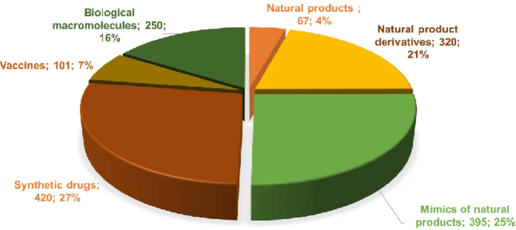
Since the 1970s, the number of natural products reaching the clinical market has slowed down. Newman and Cragg have analyzed the origin of the drugs approved by the US Food and Drug Administration (FDA) from 1981 to 2014, and they showed that still 2/5 of the small molecules approved are natural products or natural product-derived molecules coming from plants and microorganisms (Figure 2) (Newman and Cragg, 2016). To this number can be added the natural product-inspired molecules, which amount to another 25% of all small molecules. Altogether, NP and their derivatives correspond to 45% of the anti-infectives, including 58% of the approved antibacterial drugs. They also correspond to 65% of the anticancer agents approved in the past 30 years (Newman and Cragg, 2016). Natural products and their derivatives are thus still an important source of anti-infective and anticancer agents.
10 Figure 2: All small-molecule approved drugs from 1981 to 2014; n = 1202 (adapted from Newman and Cragg, 2016)
1.1.3. Microbial natural product producers
A minority of microorganisms are responsible for the production of more than 80% of known microbial specialized metabolites. In fact, historically, almost all antibacterial compounds were isolated from actinobacteria and, among this phylum, from bacteria of the Streptomyces genus. Altogether, over 9,000 bioactive compounds were isolated from actinobacteria, and 60 are used in medicine, agriculture or research. 80% of these 60 compounds are from Streptomyces species (Demain, 2009). Nowadays, actinobacterial specialized metabolites represent about 25%, of anti-infective specialized metabolites. Examples of bioactive compounds produced by Streptomyces species are listed in Table 1.
Table 1: Examples of bioactive molecules produced by Streptomyces
Type of compound Producing species Bioactive agent(s) Source or reference
Antibacterial agent producers
Streptomyces venezuelae Chloramphenicol (Ehrlich et al., 1947)
Streptomyces roseosporus Daptomycin (Mchenney et al., 1998)
Streptomyces fradiae Neomycins (Dulmage, 1953)
Streptomyces griseus Streptomycin (Schatz and Waksman, 1944)
Streptomyces aureofaciens Tetracycline (Darken et al., 1960)
Streptomyces clavuligerus Cephalosporin (Brannon et al., 1972)
Antifungal agent producers
Streptomyces noursei Nystatin (Zotchev et al., 2000)
Streptomyces kasugaensis Kasugamycin (Umezawa et al., 1965)
Bioherbicide/
biopesticide producers Streptomyces hygroscopicus Herbimycin (Omura et al., 1979) Antiparasitic
11
Antiviral
agent producers Streptomyces hygroscopicus Hygromycin (González et al., 1978) Immunosuppressant
agent producers Streptomyces hygroscopicus Rapamycin (Chen et al., 1999)
Antitumor agent producers
Streptomyces peucetius Doxorubicin (adriamycin) (Arcamone et al., 2000)
Streptomyces verticillus Bleomycin (Shen et al., 2001)
Streptomyces caespitosus Mitomycine C (Wakaki et al., 1958)
Figure 3: Decomposition of biosynthetic gene cluster diversity among all sequenced prokaryotic genomes (Cimermancic et al., 2014)
The diversity of each node in the phylogenetic tree is represented by the size of the circle (larger circle defines higher degree of diversity).
The biosynthesis of microbial specialized metabolites is most of the time directed by genes physically grouped together in the genome, called Biosynthetic Gene Clusters (BGCs). Cimermancic and co-workers (2014) have analyzed the distribution of BGCs of 1,154 sequenced genomes among the bacterial phylogenetic tree. Figure 3 shows that apart from actinobacteria, confirmed to be remarkably prolific specialized metabolite producers, other important producers
12 can be found in the cyanobacteria, proteobacteria (myxobacteria, Pseudomonas, Burkholderia), and firmicutes (Bacillus) phyla. Among fungi, specialized metabolite producers are in particular found in the ascomycota (Penicillium, Aspergillus) phylum.
1.1.4. Current situation: a crucial need for new pharmaceutical compounds
In the 1950s, geneticists believed that the development of microbial pathogenic strains resistant to antibiotic treatments was highly unlikely (Davies, 2006). And yet, for almost all antibiotic treatments, pathogen bacteria resistant to the antibiotic can be detected only a few years after the introduction of the antibiotic on the clinical market (Davies and Davies, 2010). Resistance to antibiotics arose fast partly because they were used in large quantities irresponsibly, for instance for agricultural applications, and partly because we underestimated microorganisms’ capacity to adapt (Procópio et al., 2012). Antimicrobial resistance is now considered by many organizations (World Health Organisation, European Centre for Disease Prevention and Control …) as a major public health threat (Ferri et al., 2017). In 2014, the Review on Antimicrobial Resistance UK Commission estimated that antimicrobial resistance caused 700,000 deaths worldwide and that this figure was likely to reach 10 million by 2050 (Review on Antimicrobial Resistance). This worrying situation led the World Health Organisation (WHO) to establish a list of bacteria for which new antibiotics are urgently needed in February 2017 (WHO publishes list of bacteria for which new antibiotics are urgently needed, 2017). Bacteria of this list are classified according to three levels of priority, critical, high and medium. In the critical and high levels can be found all the so-called “ESKAPE” bacteria (Enterococcus faecium, Staphylococcus aureus, Klebsiella pneumoniae, Acinetobacter
baumanii, Pseudomonas aeruginosa and Enterobacter species) (Fair and Tor, 2014; Lewis, 2013). A study
commissioned by the Wellcome Trust in 2016 aimed at evaluating alternatives to antimicrobial compounds (Czaplewski et al., 2016). The most advanced approaches were shown to be antibodies, probiotics and vaccines now in Phase II or Phase III trials. However, in the medium term, the commission confirmed that conventional antibiotics would still be needed, as these approaches would mainly serve as adjunctive or preventive therapies.
Meanwhile, the discovery of new microbial natural products with promising antibiotic activity has slowed down. There are three main reasons for this current decline in antibiotic compounds discovery: new compounds are harder to find, industrials have turned away from antibiotic research, and regulation became stricter (Bérdy, 2012). The discovery of new compounds, which seemed to be never ending in the 1960s, slowed down drastically while rediscovery of already known molecules became more and more frequent (Lewis, 2013). Research expenses increased for companies, while the number of leads decreased, and newly discovered antibacterial agents were restricted to last-resort use in hospitals only, which resulted in low profits. This led the big pharmaceutical companies to first turn to synthetic combinatorial chemistry in the 1990s. However, these approaches had very limited success, probably because the “chemical space” of NP and synthetic drugs are different (Harvey et al., 2015). In addition, several drugs approved by the FDA in the past, such as streptomycin, tetracycline, and most aminoglycosides, would not pass the regulation tests today (Bérdy, 2012). For all these reasons, big pharmaceutical companies have now abandoned antibiotic research to join the more profitable chronic disease drug market. Most of the antibiotic drug lead research nowadays is done by start-up companies or academic laboratories.
13 Apart from microbial infections, efficient treatments are still needed for numerous diseases. Cancer was responsible for 9.6 million deaths in 2018 and is representing the second leading cause of deaths worldwide (Cancer, 2108). Even for well-treated cancers, new compounds, as potent but less toxic for the patients, are highly desirable. Parasitic and helminthic infections also remain a worldwide problem, especially in developing regions. Malaria, dengue and leishmaniasis are of particular concern. Soil-transmitted infections affect about 1.5 billion people in the world, and infected children suffer from nutrition and physical impairment (Soil-transmitted helminth infections, 2019). Fungal diseases pose a real threat for people with weakened immune system such as patients with HIV (Human Immunodeficiency Virus) or cancer (Global fungal diseases, 2018). In conclusion, bioactive compounds, whether it is for antibacterial, antifungal, antiparasitic or anticancer therapies, are dearly needed. The next section of this introduction covers the strategies presently employed to discover new natural products with pharmaceutical potential.
1.2. Strategies to find new natural products
1.2.1. Studying new specialized metabolite-producing strains from underexplored environments
Traditionally, scientists isolated microorganisms from the soil, because it was of easy access and relatively easy to reproduce growth conditions. Today, more and more environments are explored, environments that are bound to procure new species of microorganisms, hence maybe new kinds of natural products (Hug et al., 2018). In particular, aquatic environments have attracted increased attention since the 1970s. Oceans contain approximately 87% of life on earth (Bérdy, 2012), they constitute the largest pool of microorganisms. Marine actinomycetes were proven to be remarkable for their specialized metabolite production (Subramani and Aalbersberg, 2012). For instance, the cancer cell cytotoxic salinosporamide A (Figure 4), a proteasome inhibitor, was isolated from Salinispora tropica (Feling et al., 2003).
Extreme environments, such as deserts or polar areas, inhabited by extremophiles including acidophiles, alkalophiles, halophiles, and hyperthermophiles, are also explored. They have led to interesting discoveries (Masand et al., 2018; Tian et al., 2017). Thus, more than 20 new specialized metabolites were identified from Penicillium species isolated from an abandoned copper mine water basin, Berkeley Pit Lake, contaminated with high concentrations of dissolved metal sulfites (Pettit, 2011). Among them are two new polyketide terpenoids berkeleydione and berkeleytrione (Figure 4), with promising activities against cancer and Huntington disease.
Endophytes and symbionts are also a source of specialized metabolites. Bérdy (2012) reported that 80 % of endophytic fungi produce a bioactive compound of some kind, one of the best-known examples being the production of the anti-cancer drug taxol (Paclitaxel) from
14 Figure 4: Structure of specialized metabolites with promising biological activities obtained from recently explored environments
Although there has been an increasing interest in “exotic” environments in the scientific community, a recent study has shown there may be no need to wander so far: the parks of New York contain plethora of yet unknown microorganisms and compounds (Nothias et al., 2016). Altogether, there are still plenty of microorganisms to study, and we will without doubt discover many new natural products by tapping into these resources (Cragg and Newman, 2013; Demain, 2009).
Streptomyces are probably among the best studied bacteria for their specialized metabolism.
They are prolific natural product producers and numerous studies have been carried out to explore their specialized metabolism repertoire. For this reason, the next two sections will be centered on this genus, although the methods that have been used to isolate and characterize Streptomyces metabolites could probably be applied to other genera.
1.2.2. Expressing Streptomyces’ specialized metabolism full potential in the native host
Streptomyces genomes usually contain several dozen biosynthetic gene clusters (BGCs) that
can be predicted by bioinformatics tools such as antiSMASH (antibiotics and secondary metabolite analysis shell) (Blin et al., 2017). For instance, Streptomyces avermitilis genome contains 25 potential BGCs, which correspond to 6% of its genome (Ōmura et al., 2001). Streptomyces ambofaciens genome contains 23 clusters potentially involved in specialized metabolism, and yet, it was known for more than 40 years to produce only spiramycin and congocidine (Aigle et al., 2014). Most of Streptomyces specialized metabolites are not expressed, or not detected, in standard laboratory conditions. The corresponding BGCs are called “cryptic”, or “silent”. Various methods have been employed to
15 activate the expression of these clusters (Rutledge and Challis, 2015), some examples are listed in Table 2.
Table 2: Examples of approaches activating silent biosynthetic gene clusters
Approach Principle Compound
discovered Reference
Variation in growth conditions
Cultivation of Streptomyces armeniacus on
a malt-containing medium armeniaspirols
(Hug et al., 2018)
Co-culturing Cocultivation of S. endus S-522 with Tsukamurella pulmonis TP-B0596 alchivemycin A (Rutledge and Challis, 2015)
Addition of chemical elicitors
Addition of subinhibitory
concentrations of trimethoprim to
Burkholderia thailandensis culture
malleilactone (Hug et al., 2018)
General regulation Induction by an allele of absA1 (from
S. coelicolor) in Streptomyces flavopersicus pulvomycin
(Rutledge and Challis, 2015) Knock out of one
biosynthetic gene cluster
Knocking out the rifA PKS gene responsible for rifampicin biosynthesis from Amycolatopsis mediterranei S699
amexanthomycins A–J (Hug et al., 2018) Pathway specific transcriptional regulation
Inactivation of the repressor gbnR in S.
venezuelae gaburedin A
(Hug et al., 2018)
Heterologous expression
Expression in E. coli of the terpene synthase encoded by the sav76 gene of
S. avermitilis
avermitilol (Rutledge and Challis, 2015)
The empirical approach called “the OSMAC approach” (One Strain-MAny Compounds), is based on the fact that a strain will not express all its spectrum of specialized metabolites in a given condition (Bode et al., 2002). By modifying the culture conditions (nutrient sources, medium components in general, pH, aeration, temperature), different compounds may be produced. The addition of metal ions may also have an effect (Hug et al., 2018; Liu et al., 2013). When in silico data is available to predict the structure or the role of the compound of interest, these modifications may be made rationally. For instance, an iron-depleted medium was used to induce the production of a likely siderophore predicted in Streptomyces coelicolor genome, and this resulted in isolation of coelichelin (Lautru et al., 2005). Another method not requiring any genetic knowledge is the co-cultivation with other species, as interspecies cross talks may induce metabolite production (Liu et al., 2013; Zarins-Tutt et al., 2016). Histone deacetylase (HDAC) inhibitors modulate gene expression by deacetylating histone proteins and they have been especially useful in fungi natural product research. They have also successfully been used for bacteria (Hug et al., 2018; Zarins-Tutt et al., 2016). Finally, chemical elicitors such as sub-inhibitory concentrations of antibiotics may also induce antibiotic production (Rutledge and Challis, 2015; Zarins-Tutt et al., 2016).
16 The methods described above are empirical and do not rely on any knowledge of the mechanisms governing the production of specialized metabolites by the strains. As an alternative to this approach, genetic methods have been developed based on knowledge of specialized metabolism regulation. Specialized metabolites production is under tight regulation in Streptomyces species. Global regulation involves master regulators. It is extremely complex and coordinated with morphological developments (Bibb, 2005; Bibb and Hesketh, 2009). A metabolic switch is observed in fermentors from exponential growth to stationary growth, when most specialized metabolites are produced. During the switch, there are signaling cascades, regulation by small ligands and phosphorylation state (Liu et al., 2013). There are pleiotropic regulators involved in both antibiotic production and aerial hyphae development. It is for example the case of the gene
bldA, which codes for the unique tRNA for the rare leucine codon UUA (van Wezel et al., 2009).
Regulatory genes, specialized metabolite genes and morphology changing-genes containing the rare codon can only be translated when bldA is expressed. There are also pleiotropic regulators of several antibiotic pathways, such as the absA operon, a two-component system used to repress antibiotic production in S. coelicolor and Streptomyces griseus (van Wezel et al., 2009).
In addition to this global level of regulation, the expression of genes directing the biosynthesis of a given metabolite is often controlled locally by transcription regulators located in biosynthetic gene clusters (Hug et al., 2018). The over-expression of pathway-specific activators or deletion of repressors can trigger the production of the expected metabolite. For instance, deleting the tetR repressor encoded in the gene cluster led to the production of kinamycin in S. ambofaciens (Bunet et al., 2011), while stambomycins were only observed after constitutively expressing a Large ATP-binding regulator of the LuxR (LAL) family regulator (Laureti et al., 2011). Pathway-specific
Streptomyces antibiotic regulatory protein (SARP) control the production of many specialized
metabolites. The overexpression of the SARP ccaR allowed for instance to detect clavulanic acid in
Streptomyces clavulagerus (Zarins-Tutt et al., 2016).
Finally, it should be mentioned that knocking down pathways of known metabolites can also be helpful: some compounds may be present in smaller amount, and they will be detected more easily in the absence of the major compounds (Rutledge and Challis, 2015). Knocking down gene clusters may also alleviate competition for common precursors.
The genetic approaches described above rely on the ability to genetically manipulate the strain of interest. When this is not the case, or when no genetic tools have been developed for the strain, another possibility is the heterologous expression of the gene cluster, that is the insertion of the biosynthetic gene cluster in a host strain (Zarins-Tutt et al., 2016).
1.2.3. Producing specialized metabolites by heterologous expression
There are many examples in the literature of Escherichia coli and Saccharomyces cerevisiae used as heterologous hosts because their genetic toolbox is well developed, but they may not be ideal for all actinomycetes natural products (Pickens et al., 2011). Firstly, the high GC-content of actinomycetes genomic DNA often impedes correct translation. Adjusting codon usage requires the synthesis of DNA, which is often problematic in the case of large NRPS or PKS genes. Secondly, there is often a need for chaperone or helper proteins, such as phosphopantetheinyl
17 transferase or MbtH-like proteins, which are encoded in actinomycetes genome, but often not included in the biosynthetic gene cluster of interest (Ongley et al., 2013). Thirdly, precursors from primary metabolism, such as branched-chain acyls, may not be produced in E. coli or Sa. cerevisiae. Historically, Streptomyces albus, and S. coelicolor have been extensively used as heterologous hosts (Baltz, 2010), and they remain among the laboratory favorite pets. Industrial producers have also been used as hosts, such as S. avermitilis or Streptomyces roseosporus (Baltz, 2016). In recent years, various Streptomyces strains have been engineered to constitute good chassis for the production of specialized metabolites. Thus, endogenous gene clusters have been deleted (S. coelicolor, (Gomez‐ Escribano and Bibb, 2011), S. avermitilis (Komatsu et al., 2010), S. albus (Kallifidas et al., 2018)). These strains present a low background noise as they do not produce specialized metabolites anymore. These strains have often been further optimized for the expression of biosynthetic gene clusters, for example by introducing mutations known to be favorable for this expression (in rpoB or rpsL in S. coelicolor), or by increasing the resistance to oxidative stress (deletion of pfk in S. albus).
Most BGCs span from 10 to 120 kilobases (kb). To introduce them in a tractable host imply to be able to manipulate and retrieve DNA fragments of these sizes from the native producer (Ongley et al., 2013; Rutledge and Challis, 2015). The cluster can then be maintained on a stable plasmid or integrated within the host genome. The traditional method to capture a biosynthetic gene cluster is to construct genomic libraries, but the complete process is quite tedious and for very large clusters, it is often difficult to capture the whole cluster on one vector (cosmid, BAC…). It is then necessary to reassemble the cluster from two or three vectors (Perlova et al., 2006). New techniques have been developed recently: Linear to Linear Homologous Recombination (LLHR) allows to bring together two linear pieces of DNA with sequence identity at the extremities in E.
coli (Fu et al., 2012). Another technique of interest is the transformation-associated recombination
(TAR cloning), which is based on yeast natural capacities of recombination. Yamanaka et al. (2014) reported first the use of this method in 2014. They cloned a 67-kb gene cluster directing the biosynthesis of the lipopetide taromycin A in one step, which would have been difficult using a genomic library. Another very recent technique is CATCH (Cas9-Assisted Targeting of Chromosome segments), which combines the use of RNA-guided Cas9 nuclease to cut the cluster from its genome, and the use of Gibson assembly to ligate the cluster to a linear plasmid (Jiang et al., 2015). Using this technique, the authors were able to clone up to 100-kb DNA.
The heterologous expression of a biosynthetic gene cluster is sometimes sufficient to afford the production of a specialized metabolite. This was for example the case of collinone, a polyketide antibiotic that was not detected in the native producer, Streptomyces collinus, but was produced when the biosynthetic gene cluster was transferred in S. coelicolor CH999 (Martin et al., 2001). Yet, the heterologous expression of a gene cluster is often insufficient on its own and further manipulations of the gene cluster, such as the deletion of transcriptional repression (Yamanaka et al., 2014) or the replacement of native promoters by strong and constitutive ones (pathway refactoring, developed in the next section) are often required.
18 1.3. Synthetic biology as a tool to produce natural products and expand their scope
1.3.1. Synthetic biology, a new toolbox for natural product engineering
Synthetic biology has been described as “an engineering approach to improve or completely create systems and organisms with specific or desirable functions” (Guzmán-Trampe et al., 2017). One of the principles of synthetic biology is to rely on fundamental biology, chemistry, and bioinformatics to improve or construct new biological parts, devices, and systems. Engineering can have a role at different scales: protein engineering to modify protein properties, metabolic engineering to implement a biosynthetic pathway, strain engineering to identify and optimize high titer producers (Pickens et al., 2011; Smanski et al., 2016). Synthetic biology permits for instance to control space (from protein scaffold to compartmentalization and bacterium consortia) and time (from allosteric control to regulatory cascades and molecular clock) at different scales in a designed system (Medema et al., 2011). It now plays a prominent role in antibiotic discovery and biosynthetic pathway engineering.
Biological DNA basic parts are small DNA fragments whose sequence confers a specific function. For example, these DNA basic parts include promoters, ribosome binding sites (RBS), coding sequences, and regulators among others. In order to modify a cluster and replace some of its parts, one must have at his disposal libraries containing parts available for replacement. Many libraries of characterized parts are available for Sa. cerevisiae and E. coli (Pickens et al., 2011), and recently, some libraries have been reported for Streptomyces species as well (Smanski et al., 2016). Shao and collaborators (2013) tested several heterologous promoters in Streptomyces lividans when they engineered the spectinabilin pathway. These promoters, however, were not well characterized, limiting their usefulness in other studies. Other libraries were derived from well-characterized promoters, such ermEp1 (Siegl et al., 2013) or kasOp (Bai et al., 2015). In this latter case, the library constructed is based on the already optimized promoter kasOp*. The synthetic promoters derived from kasOp* have a strength varying between 1 to 190% of kasOp*. The authors also characterized 15 native and 174 synthetic RBSs that cover a 200-fold strength range. In contrast, there are not many characterized terminators actually available, though some recent studies aim at filling this gap (Horbal et al., 2018). Some studies have, however, underlined that these DNA parts are characterized in a specific context, including surrounding DNA sequences and the host strain itself, and that their characterization was not systematically transferable outside of this context (Vilanova et al., 2015; Yeung et al., 2017).
In addition to libraries of standard DNA parts, synthetic biology requires performant DNA assembly methods. New DNA assembly technologies have been developed in the past years, and they constitute an extremely useful toolbox for biosynthetic gene cluster capture, (re)assembly and modification (Figure 5)(Kim et al., 2015; Luo et al., 2016; Sands and Brent, 2016). Traditionally, DNA assembly was made by digestion by restriction enzymes and ligation (Sands and Brent, 2016). Since then, more sophisticated methods still based on the use of restriction enzymes have been developed, such as the Biobrick assembly (Knight, 2003), or the Golden Gate assembly (Engler and Marillonnet, 2014). Ligase cycling reaction (LCR) is a technique based on the use of a thermostable ligase and multiple cycle of denaturation-annealing-ligation temperatures (de Kok et al., 2014). Other assembly techniques are based on homologous recombination in vivo, such as DNA assembler (Shao et al., 2009) and Red/ET recombineering (Gust et al., 2004) or in vitro such
19 as Gibson assembly (Gibson et al., 2009), sequence- and ligation-independent cloning (SLIC) (Li and Elledge, 2012), or Gateway system (Sands and Brent, 2016). Many other techniques not described here are available, and allow the assembly of several DNA parts, forming a modified biosynthetic gene cluster (Sands and Brent, 2016).
Figure 5: Exemples of DNA assembly methods
1.3.2. Refactoring of specialized metabolite biosynthetic gene clusters
Refactoring consists in rewriting the DNA sequence without changing its functionality. It may be done to erase all native regulation, to optimize the sequence for heterologous expression or as a first step towards the generation of synthetic pathways within a cell (Figure 6). A pioneering refactoring work is the refactoring of the nitrogen fixating gene cluster (20 genes) from Klebsiella
oxytoca (Temme et al., 2012). In this study, the authors aimed at (i) removing all native regulation
and non-essential genes, (ii) re-organizing the genes into synthetic operons using well-characterized synthetic biological parts (promoters, ribosome binding sites (RBS), terminators) and (iii) randomizing/optimizing codon usage for E. coli expression. Their refactored gene cluster, constituted of 89 genetic parts, was functional, although with a reduced activity.
Figure 6: Biosynthetic gene cluster refactoring principle
20 In addition to modifying transcriptional/translational elements to better control the expression of a set of genes, the refactoring of a gene cluster can also be used to introduce or remove genetic elements that will facilitate the re-assembly of the cluster. Thus, when Osswald and co-workers (2014) refactored the epothilone BGC (56 kb, 7 genes) of Sorangium cellulosum for expression in Myxococcus xanthus, they added unique restriction sites, while subtracting about 700 unwanted restriction sites.
The refactoring of the nitrogen fixating gene cluster and of the epothilone gene cluster involved extensive modifications of the original DNA sequence. This could only be obtained through the synthesis of DNA fragments that were next assembled. Indeed, DNA synthesis is becoming an increasingly attractive option, though still expensive (Kim et al., 2015). However, such an extensive refactoring may not always be required, and there are many examples of simpler refactoring, consisting mainly in replacing native promoters by constitutive or synthetic ones, especially in the case of rather small clusters (Rutledge and Challis, 2015). Such examples include the refactoring of spectinabilin (Shao et al., 2013). The spectinabilin cluster from Streptomyces orinoci remained silent when expressed in S. lividans, even when a gene encoding a transcriptional repressor was deleted. The authors chose nine strong promoters and one inducible promoter to refactor the cluster, and after assembly using DNA assembler method, they observed production of spectinabilin, though with a yield of 10% compared to the production in the WT strain. Using the same assembly method, three novel polycyclic tetramate macrolactams were identified when the BGC refactored with strong promoters was expressed in S. lividans (Luo et al., 2013). Very recently, combining TAR cloning and red/ET recombineering, Moore and colleagues refactored the spz cluster and detected the production of more than a hundred of compounds related to streptophenazine (Bauman et al., 2019).
Once a pathway is refactored, it is usually much easier to replace one part by another one, to refine the knowledge of the biosynthetic pathway (Luo et al., 2013) or to obtain a higher yield when the functions are equivalent (Smanski et al., 2014). It is also possible to obtain a new compound by adding a part with a different function (Smanski et al., 2016). Refactoring thus leads the way to the modification of specialized metabolites to produce new analogs.
1.3.3. Production of non-natural analogs and expansion of the range of specialized metabolites
Once a metabolite of interest has been isolated, it may be interesting to try to improve its properties by generating analogs. Derivatives of natural products can be produced by a number of chemical or biological methods, or by a combination of these methods. Traditionally, microbial natural products were obtained by fermentation and then chemically modified (hemi-synthesis). In the last decades, new methods, based on the metabolic capacities of microorganisms, have been developed. Thus, chemically synthesized precursors analogs can be fed to the producing strain. This method relies on enzymatic substrate promiscuity, but may sometimes be successful, as it was the case for a derivative of balhimycin, bromobalhimycin (Sun et al., 2015). However, the natural metabolite is still produced, as there is a competition between the native substrate and the added one. To avoid such competition, it is possible to resort to genetic engineering to knock out the production of the natural precursor in the strain, prior to the feeding of the precursor analog
21 (mutasynthesis). For instance, new derivatives of balhimycin were obtained when the gene responsible for the synthesis of β-hydroxytyrosine was deleted and the strain fed with fluorinated β-hydroxytyrosine analogs (Figure 7) (Winn et al., 2016).
Figure 7: Structures of balhimycin (a) and derivatives (b) (adapted from Winn et al., 2016)
Another synthetic approach, called combinatorial biosynthesis, consists in combining (subtracting, adding or replacing) biosynthetic genes from various gene clusters. The engineered organism then produces analogs of the original natural product (Goss et al., 2012). For instance, some enzymatic domain exchanges allowed the biosynthesis of ivervectin (22,23-dihydroavermectins), a derivative of the natural product avermectin (Pickens et al., 2011).
Combinatorial biosynthesis can be coupled to mutasynthesis and chemoenzymatic synthesis to increase further the chemical diversity generated. Thus using this combination of methods, Heide (2009) reports the generation of more than a hundred derivatives of the aminocoumarins novobiocin, clorobiocin and coumermycin A1 (Figure 8). Structurally, novobiocin and clorobiocin are similar, except for the group at the C-8 position of the aminocoumarin moiety (methyl or chlorine group) and the 3-OH group of the desoxysugar (a carbamoyl or a methyl-pyrrol-2-carboxyl moiety). All the nine possible hybrids of novobiocin and clorobiocin were tested and it was shown that the better antibiotic activity of clorobiocin was mainly due to the methyl-pyrrol-2-carboxyl moiety attached to the desoxysugar.
Although the refactoring and the genetic engineering of biosynthetic gene clusters have encountered some success, it has often been at the expanse of the yield of the obtained metabolite(s) (Osswald et al., 2014; Shao et al., 2013). This highlights the necessity of a greater understanding of the fundamental biological processes governing the biosynthesis of natural products (Goss et al., 2012; Kim et al., 2015).
22 Figure 8: Exchange of tailoring genes to produce novobiocin/clorobiocin analogs (adapted from Pickens et al., 2011)
The two clusters are shown in parallel, with the genes responsible for the structure differences colored. MePyC = methyl-pyrrol-2-carboxyl.
Combinatorial biosynthesis has been mainly applied to two families of metabolites, non-ribosomal peptides (NRPs) and polyketides. The work carried out on the polyketide biosynthetic systems is out of the scope of this manuscript and will not be addressed here. In the next sections, I will detail our knowledge concerning the non-ribosomal peptide synthetases (NRPSs), and present the combinatorial biosynthetic approaches that were conducted on this family of enzymes.
2. Non-ribosomal peptide synthetases (NRPSs), a class of
complex modular enzymes
The number of non-ribosomal peptides (NRPs) exhibiting anti-infective properties is important. One reason for this lies in the diversity of incorporated monomers: approximately five hundreds, including non-proteogenic amino acids, fatty acids, and sugars (McErlean et al., 2019; Strieker et al., 2010). But this comes with a price: the enzymes synthesizing the NRPs are huge; for instance cyclosporine, an 11-residue peptide, requires an enzyme of about 1.5 mega daltons. An extensive review on NRPS notably describing the incorporated monomers has recently been published (Süssmuth and Mainz, 2017).
2.1. NRPS assembly lines and facilitators
2.1.1. Principle of NRP biosynthesis
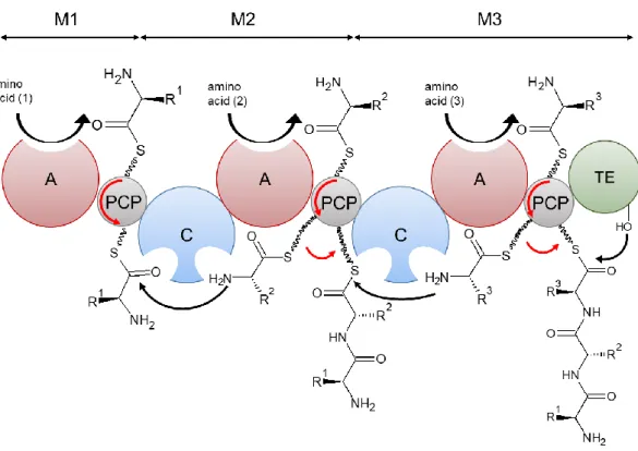
NRPSs are large multi-modular enzymes responsible for the biosynthesis of a non-ribosomal peptide (NRP). Several subunits may be needed, each of them being constituted of modules. The model of assembly is presented on Figure 9. Each module incorporates one monomer to the final peptide. Each module is divided in domains. There are three core domains. The adenylation (A) domain recognizes the amino acid, activates its carboxylate moiety under the
23 form of an amino acid adenylate at the expense of one molecule of ATP, and covalently binds it as a thioester to the 4’-phosphopantetheinyl (ppant) arm of the peptidyl carrier protein (PCP) domain, also called thiolation (T) domain (Keller and Schauwecker, 2003). The PCP domain presents the substrate tethered to its cofactor to the other domains. The condensation (C) domain catalyzes the formation of an amide bond between two amino acids and, thus, the elongation of the peptidyl chain. The initiation module usually only contains A and PCP domains, while the extension modules contain C, A and PCP domains. At the end of the assembly chain, the termination module also usually contains a thioesterase (TE) domain, which releases the product by hydrolyzing the thioester bond, sometimes through intramolecular cyclization. Release of the product can also be catalyzed by a C domain, a reductase (R) domain or even be non-enzymatic (McErlean et al., 2019).
Figure 9: NRPS biosynthesis model
Amino acid substrates are recognized by adenylation domains (A). The aminoacyl-AMP intermediate formed is then loaded on the thiol group of a 4’-phosphopantetheine arm tethered to the peptidyl carrier protein domain (PCP). Condensation domains (C) catalyze successive peptide bond formation. The first module is known as the initiation module (M1) and subsequent modules (M2) are known as elongation modules. The final module (M3) contains an additional thioesterase domain (TE) which catalyses hydrolysis or cyclisation to release the peptide from the NRPS.
In addition to the core domains, optional domains can be included in the modules, such as epimerization domains, methylation domains or cyclization domains (Hur et al., 2012; McErlean et al., 2019; Winn et al., 2016). Epimerization domains catalyze the epimerization of L-amino-acids into their D-form. They are only active on substrates tethered to the PCP domain. The presence of heterocyclic rings in the NRP is explained by the action of the heterocyclization (Cy) domain. Cy domains exhibit a strong specificity, and they produce thiazoline rings from the thiol of cysteine residue, or oxazoline ring from the hydroxyl group of serine or threonine residue. The cycles can be further oxidized or reduced by the corresponding oxidation or reduction domains, which are often stand alone proteins. Methyltransferase domains transfer a methyl group from its cosubstrate
24 (S)-adenosyl methionine (SAM). While N-methyltransferases act in cis during the biosynthesis or in
trans on the complete product, C-methyltransferases tend to methylate precursors before the
assembly of the final molecule. Formylation (F) domains, which add a formyl group, have been little studied until now, except for the F domain of gramicidin NRPS, which exhibits high specificity. Finally, halogenase domains are frequent in NRPSs, and halogen groups play an important role in the antibiotic properties (such as for the antibiotic balhimycin and antifungal syringomycin E). The peptide can also be modified by other tailoring enzymes after being released from the NRPS.
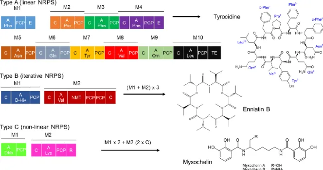
NRPSs are monomeric (Weissman, 2015). An NRPS can be organized as one protein, and then it is called type I NRPS, or as several interacting subunits, which is type II NRPS. Type II NRPS is preponderant in bacteria (Hur et al., 2012). There are three categories of NRPSs (Figure 10). Type A corresponds to linear NRPS: the assembly chain is followed strictly, there are as many monomers as modules, and the order is maintained. This type is often used as a canonical example, and knowing the sequence, one can predict the final NRP. Tyrocidine is synthesized by a type A NRPS. Type B NRPS is called iterative, some of the modules can be reused several times, and the peptide is made of repetitive sequences. Enniatin is an example of type B NRP. Type C is non-linear NRPS, the arrangement of the modules does not correspond to the sequence of amino acids obtained, and one domain, not one module, may be reused. Myxochelin is an example of type C NRP.
Figure 10: The different NRPS categories
NMT= N-methyltransferase domain ; R = reductase cleavage ; D-Hiv = D-2-hydroxyisovaleric acid ; Dhb = dihydroxybenzoyl
2.1.2. PCP domain priming by the PPtases
The attachment of the 4’phosphopantetheinyl (ppant) arm to the PCP domain is done by the Sfp-type phosphopantetheine transferases (PPtases) from a Coenzyme A in a Mg2+-dependant
25 reaction (Hur et al., 2012; Strieker et al., 2010). PCP domain is converted from the inactive apo state to the active holo state. Since there is a large amount of acylated Coenzyme A in the bacteria, the PCP domain is often misprimed with an inactive acylated-ppant. Type II-thioesterases then function as repair enzymes and hydrolyze the acyl group, yielding a functional holo-PCP domain. PPtases and type II-thioesterases are usually not included in a specific biosynthetic gene cluster, they are present on the genome, and play a pleitropic role, priming PCP domains from different BGCs. Sfp was one of the first described PPtases, and it exhibits an important promiscuity. Bunet
et al. (2014) have found a Sfp-type PPtase in S. ambofaciens, associated to no specialized metabolite
cluster, with a pleiotropic role. The deletion of the encoding gene abolished the production of congocidine and coelichelin, synthesized by NRPSs, and of spiramycin, stambomycin and grey-spore pigment, all polyketides synthesized by polyketide synthases. This shows that this PPtase is involved in the priming of the peptidyl carrier and acyl carrier proteins of several of the biosynthetic pathways, and is likely involved with all the NRPS and PKS clusters of the strain.
2.1.3. Role of MbtH-like proteins (MLP) as helpers
MbtH-like proteins (MLP) are small proteins of about 70 amino acids found in some NRP gene clusters (Hur et al., 2012). They were named after the MbtH protein encoded in the BGC of the siderophore mycobactin in Mycobacterium tuberculosis. The function of these proteins is not fully understood yet, but they associate with A domains during NRP biosynthesis and they are considered as chaperones or facilitators. MLP may be needed for the correct solubility and activity of the A domain, or only for its solubility. It may enhance both solubility and activity of an A domain that is functional on its own as well (Schomer and Thomas, 2017). For instance, the purification of Cgc18 involved in congocidine biosynthesis required the MLP partner SAMR23877 to obtain a soluble fraction, and the authors reported many other cases for which solubility and/or activity was impeded in the absence of MLP (Al-Mestarihi et al., 2015). Associated MLP and A domain are bound tightly and copurified, and stoichiometric amounts of 1:1 of MLP:A didomains have been reported (Baltz, 2011).
MLP structure consists of three β-strands, which interact with one adjacent α helix (Miller et al., 2016). There is no obvious catalytic group in MLP structure (Schomer et al., 2018). The structure of SlgN1, the NRPS of streptolydigin, made of MbtH-like domain at the N terminus and adenylating domain, was recently crystallized (Herbst et al., 2013). The MLP interacts with the big N terminal part of the A domain (Figure 11). It is worth noting that MLP has no direct contact to the substrate of the A domain. The full module of EntF containing C-A-PCP-TE domains has also been crystallized bound to its native MLP from E. coli, or to a non-cognate MLP from Pseudomonas
aeruginosa (Miller et al., 2016). The interaction surface is similar to the one reported by Herbst and
collaborators (2013). The presence or absence of the MLP had no visible impact on the structure of the A domain, which suggests that the activation of A domain is not achieved by a conformational change (Miller et al., 2016). However, in the structure of DhbF domain A crystallized with its MLP (required for adenylation activity but not for folding), the A domain adopted a more compact form than its structure in absence of MLP (Tarry et al., 2017). Even the smaller C terminal part of the A domain (Asub), which is not in direct contact with MLP, seemed
26 Not all A domains are dependent on MLP to function correctly. For instance, the A domain CmnO involved in capreomycin biosynthesis is not active without the CmnN MLP, while the A domain CmnF is unaffected by the absence of MLP (Miller et al., 2016). So far, attempts to predict the dependency of A domains to MLPs based on sequence analysis have failed (Miller et al., 2016).
Figure 11: Model of the position of an MbtH-like protein within an NRPS (Herbst et al., 2013).
A) protein structure B) scheme of the domain organization
A domain is separated in two parts, the N terminal core part and the C terminal smaller subdomain. The MbtH-like domain of SlgN1 (dark gray) was crystallized with the core part of the A domain. The remaining domains were positioned by superposing SlfN1 A and SrfA-C structures.
MLPs are usually encoded within the BGC containing the gene encoding their NRPS A domain partner, but a recent study showed that orphan MLPs can be encoded in bacterial genomes (Esquilín-Lebrón et al., 2018). In the case of the orphan and only MLP encoded in M. xanthus DK1622 genome, the authors showed that this MLP interacts with NRPSs from at least seven distinct BGCs. This suggests that MLP are not specific of given A domain or a given cluster. This is indeed confirmed by the observation that MLP can activate non-cognate A domains. It was observed in S. coelicolor, where CdaX can complement the deletion of CchK and restore coelichelin production, and vice versa (Lautru et al., 2007). Schomer and Thomas (2017) have also studied the impact of 7 non-cognate MLPs on EntF activity, involved in enterobactin biosynthesis. EntF native MLP is YbdZ. It copurifies with EntF and improves both its solubility and its affinity for its substrate L-Serine. The authors observed that 5 of the 7 non-cognate MLPs could restore enterobactin production (Schomer and Thomas, 2017). Another study also suggested that the interaction of a MLP with a non-cognate A domain could broaden the A domain substrate promiscuity (Mori et al., 2018).
2.2. NRPS domains: structure and substrate specificity
2.2.1. A domain structure and specificity
A domain is a well-defined globular structure of 550 to 600 amino acids, which consists in two subdomains connected by 5-10 residues: a big N terminal domain of about 450 amino acids (Acore), and a smaller C terminal domain of about 100 amino acids (Asub) (Figure 12). The active site