Université de Montréal
Controlling Senescence by PML and PML Nuclear Bodies
Par
Mariana de la Cruz Acevedo Aquino
Département de biochimie et médicine moléculaire Faculté de Médicine
Thèse présentée
en vue de l’obtention du grade de Philosophiae Doctor (Ph.D.) en biochimie
Février, 2017
Résumé
La sénescence cellulaire est une réponse aux stresses selon laquelle des cellules pouvant proliférer optent pour entrer dans un arrêt du cycle cellulaire en réponse à une variété de stimulations intrinsèques et extrinsèques telle que, par exemple, le raccourcissement des télomères, un stress oxydatif, des dommages à l’ADN ou l’activation constitutive d’oncogènes. Toutes ces stimulations ont en commun le potentiel d’initier ou de promouvoir une transformation néoplasique qui peut dégénérer en cancer. En fait, la sénescence est maintenant acceptée comme un mécanisme cellulaire autonome pour empêcher le développement du cancer et elle est reconnue, particulièrement dans les cellules humaines, pour s’établir et se maintenir à l’aide d’au moins deux voies de suppression tumorale majeures : les voies de p53/p21CIP et de p16INK4A/pRB. Ces deux voies sont capables d’activer et d’augmenter l’expression d’un autre suppresseur tumoral : la protéine PML. En tant que suppresseur de tumeur, PML est suffisant pour induire la sénescence dans des cellules normales, mais il n’arrive généralement pas à activer une réponse complète de sénescence dans les cellules cancéreuses.
Considérant ces faits, le premier objectif de cette thèse était d’étudier le mécanisme de résistance des cellules cancéreuses contre la sénescence induite par PML. Nous avons trouvé que, dans des cellules normales, la surexpression de CDK4 ou de CDK6 (CDK4/6) est suffisante pour contourner la sénescence induite par PML. De même dans les cellules cancéreuses, l’expression de ces kinases, souvent retrouvées augmentées dans de nombreux cancers, prévient probablement l’induction d’une sénescence par PML. Effectivement, grâce à l’inhibition de l’expression et/ou de la fonction kinase des CDK4/6, nous avons réussi à restaurer un programme de sénescence dans des cellules cancéreuses. En fait, l’utilisation de palbociclib, un inhibiteur spécifique de CDK4/6 maintenant en essais cliniques, permet d’augmenter l’habileté de PML à induire un arrêt de croissance plus fort et plus durable dans des cellules en cultures ainsi qu’une meilleure réduction de la progression de tumeurs dans des souris. Cette sénescence plus complète corrèle avec une augmentation de la présence de marqueurs d’autophagie, une meilleure répression des gènes cibles des E2F et une signature d’expression de gènes correspondant à l’inhibition de la méthylation de l’ADN. Ce dernier
point découle du fait que l’inhibition de CDK4/6 par le palbociclib promeut une dégradation par autophagie de la DNA méthyltransférase DNMT1. Nous avons aussi démontré que CDK4 est capable d’interagir avec DNMT1 et de le phosphoryler in vitro. Ces résultats soulignent la valeur potentielle des inhibiteurs de CDK4/6 en tant que modulateurs épigénétiques pour faciliter l’activation de la sénescence dans des cellules cancéreuses.
La sénescence induite par PML est fortement liée aux modifications post-traductionnelles. Parmi ces dernières, la SUMOylation joue un rôle important dans la fonction d’échafaudeur de PML et dans la formation des corps de PML. Les corps de PML sont des structures nucléaires dynamiques stimulées par des stresses, comme l’activation d’oncogènes menant à la sénescence, et dont la formation permet la séquestration de protéines spécifiques pour leur régulation et/ou pour leur modification post-traductionnelle. À travers le recrutement de protéines, les corps de PML régulent de nombreuses fonctions cellulaires telles que la sénescence, l’apoptose, la réponse antivirale, la réponse aux dommages à l’ADN et la régulation de l’expression de gènes. Compte tenu de cela, le deuxième objectif de cette thèse était de caractériser le rôle de la SUMOylation dans la sénescence induite par un oncogène, soit par l’expression de l’oncogène RAS.
À l’aide d’une analyse du protéome de SUMO3 dans les cellules sénescentes versus des cellules en croissances, nous avons pu identifier 25 sites de SUMOylation dans 23 protéines dont l’incidence était significativement régulée par la sénescence. Il est à noter que la plupart de ces protéines (un tiers) sont connues pour être associées au corps de PML. Curieusement, UBC9 (la seule enzyme E2 pour la SUMOylation) a été retrouvée plus SUMOylée dans la sénescence sur sa Lys-49. Des études fonctionnelles d’un mutant d’UBC9 pour la Lys-49 ont démontré une diminution de son association aux corps de PML et la perte de la capacité d’UBC9 surexprimé à retarder la sénescence. De plus, la localisation forcée d’UBC9 dans les corps de PML gêne la sénescence induite par PML ou RAS. Ces résultats nous permettent de proposer des fonctions pro- et anti-sénescence de la SUMOylation des protéines, particulièrement pour UBC9.
Mots-clés : Sénescence, PML, CDK4 et CDK6 (CDK4/6), méthylation de l’ADN,
Abstract
Cellular senescence is a stress response wherein proliferating competent cells undergo a stable cell cycle arrest in response to a variety of intrinsic and extrinsic stimuli, including telomere shortening, oxidative stress, DNA damage or the constitutive activation of oncogenes among others. All these stimuli have in common the potential to initiate or promote neoplastic transformation that can degenerate in cancer. Senescence, particularly in human cells, is established and maintained by at least two major tumor suppressor pathways: the p53/p21CIP and p16INK4A/pRB pathways and is now accepted as a potent cell-autonomous mechanism for suppressing the development of cancer. Both pathways are able to activate and increase the expression of the tumor suppressor protein PML. As a tumor suppressor, PML is sufficient to induce senescence in normal cells; however, upon the same stimuli, cancer cells fail to engage a complete senescence response.
Given this, the first aim of this thesis is to investigate the resistance mechanisms of cancer cells to PML-induced senescence.
We found that overexpression of the CDK4 and CDK6 (which are often up-regulated in cancer) are sufficient to bypass PML-induced senescence in normal cells. In cancer cells the expression of these kinases impairs the PML-induced senescence. By inhibiting the expression and/or function of CDK4/6 we were able to restore the senescence program in cancer cells. Also, the specific CDK4/6 inhibitor palbociclib (currently used in clinical trials) increased the ability of PML to regulate a stronger and more permanent growth inhibition in cell culture and decreased tumor progression in mice. This complete senescence response correlated with an increase in autophagy markers, repression of E2F target genes and a gene expression signature of blocked DNA methylation. Furthermore, CDK4/6 inhibition by palbociclib promotes autophagy-dependant degradation of the DNA methyltransferase DNMT1. More important, we were able to demonstrate that CDK4 directly interacts and phosphorylates DNMT1 in vitro. These results highlight the potential value of CDK4/6 inhibitors as epigenetic modulators to facilitate activation of cellular senescence in cancer cells.
PML-induced senescence is tightly regulated by post-translational modifications (PTMs). Among these PTMs, SUMOylation plays an important role in the scaffold function of
PML and the formation of the PML-NBs (PML-Nuclear Bodies). PML-NBs are dynamic structures triggered by stress such as oncogene-induced senescence, and its formation allows the sequestration of target proteins for their regulation and/or its post-translational modification. By protein recruitment, PML-NBs regulate several cellular functions such as senescence, apoptosis, antiviral response, DNA repair and gene regulation. Given this; the second aim of this thesis is to characterize the role of SUMOylation in oncogene mediated cellular senescence, specifically by the expression of the oncogene RAS.
By a SUMO3 proteome analysis of senescent cells we were able to identify 25 SUMO sites in 23 proteins that were significantly regulated during senescence. Importantly, most of these proteins were PML-NB associated. Interestingly, UBC9 (the only SUMO E2 enzyme), was found more SUMOylated in senescence on its Lys-49. Functional studies of a UBC9 mutant in Lys-49 showed a decreased association to PML-NBs and the loss of UBC9’s ability to delay senescence. Moreover, forced localization of UBC9 into PML-NBs counteracted RAS and PML-induced senescence. These results allowed us to propose a pro- and an anti-senescence function of protein SUMOylation, specifically for UBC9.
Keywords: Senescence, PML, CDK4/6, DNA methylation, palbociclib, PML-NBs,
Table of contents
Résumé ... i
Abstract ... iii
Table of contents ... v
List of tables ... viii
List of figures ... ix
List of abbreviations ... xii
Acknowledgments... xviii
1 Introduction ... 1
1.1 Introduction to cancer ... 2
1.1.1 Origin of cancer ... 2
1.1.2 Cellular transformation ... 3
1.1.3 Characteristics of a tumor cell ... 5
1.2 Cell cycle and Cancer ... 6
1.2.1 General perspective ... 6
1.2.2 The Cyclin Dependant Kinases ... 7
1.2.3 The function of the CDKs during the cell cycle ... 9
1.2.4 Cellular regulation of CDKs by CDK Inhibitors and its role in cancer formation 10 1.2.5 Non- canonical functions of CDK4 and CDK6 ... 13
1.2.6 CDK Inhibitors and its implication in Cancer Treatment ... 18
1.3 Cellular Senescence ... 24
1.3.1 Senescence and Cancer ... 24
1.3.2 Autophagy as a senescent marker ... 32
1.4. The PML protein ... 36
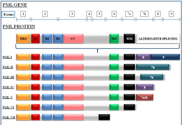
1.4.1 Characteristics of PML ... 36
1.4.3 Role of PML as a tumor suppressor ... 39
1.4.4 Role of PML in Apoptosis ... 41
1.4.5 Role of PML in senescence ... 42
1.5 SUMOylation ... 45
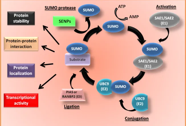
1.5.1 Characteristics of the SUMOylation pathway ... 45
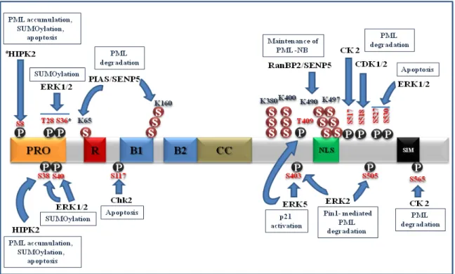
1.5.2 Role of SUMOylation on PML function and regulation ... 47
1.5.3 Role of SUMOylation in senescence ... 51
2 A CDK4/6-dependent epigenetic mechanism protects cancer cells from PML-induced senescence ... 54
2.1 Presentation of article 1. ... 55
2.2 Author’s contribution for article 1 ... 55
2.3 Article 1. ... 56
2.3.1 Abstract ... 57
2.3.2 Introduction ... 58
2.3.3 Materials and methods ... 60
2.3.4 Results ... 63 2.3.5 Discussion ... 69 2.3.6 Acknowledgments... 71 2.3.7 Legends ... 72 2.3.8 Figures... 77 2.3.9 Supplementary data ... 84
2.3.9.1 Supplementary Material and Methods ... 84
2.3.9.2 Supplementary Figure Legends ... 88
3 Quantitative SUMO proteomics reveals the modulation of many PML nuclear body associated proteins and an anti-senescence function of UBC9 ... 99
3.1 Presentation of article 2 ... 100
3.2 Author’s contribution for paper 2 ... 100
3.3 Article 2 ... 101
3.3.1 Abstract ... 102
3.3.3 Materials and methods ... 106 3.3.4 Results ... 114 3.3.5 Discussion ... 121 3.3.6 Acknowledgments... 124 3.3.7 Figure Legends... 127 3.3.9 Supplementary Materials ... 135 4 Discussion... 145 4.1 General discussion ... 146 4.2 Discussion of chapter 2 ... 147
4.2.1 Resume of results in chapter 2 ... 147
4.2.2 Annex 1: Differentially Methylated Regions ... 149
4.2.3 An insight into DNA methylation patterns in normal and in cancer cells ... 150
4.2.4 Discussion Annex 1 ... 155
4.3 Discussion chapter 3 ... 160
4.3.1 Resume of results in chapter 3 ... 160
4.3.2 Annex 2: Overexpression of SUMO-3 stimulate PML-induced senescence bypass by UBC9. ... 162
4.3.3 Role of up-regulated SUMO pathway impairing cellular senescence and promoting cancer cell proliferation ... 163
4.3.4 Discussion Annex 2 ... 166
5 Conclusion ... 169
List of tables
Chapter 1
Table 1.1: Senescence markers ... Erreur ! Signet non défini. Table 2.1: ShRNA mature sequences: ... 84 Table 2.2: Primers used for quantitative real-time PCR are as follow (using both HMBS and TBP as housekeeping genes in all experiments) ... 84 Table 3.1: List of SUMO Sites Regulated by Senescence. ... 125 Supplementary Table S3.1: Statistics on the Identification and Quantification of SUMO Sites... 135 Supplementary Table S3.2: Statistics on the Identification and Quantification of Proteins
List of figures
Chapter 1
Figure 1.2: Functional Diversity of CDKs and the pathways that regulate. ... 8 Figure 1.3: CDKs regulation and phosphorylation levels of pRB during the cell cycle. .. 13 Figure 1.4: Molecular pathways involved in cellular senescence. ... 28 Figure 1.5: The Senescence response and the senescence markers. ... 35 Figure 1.6: The PML-NBs, its interacting proteins and regulating pathways. ... 37 Figure 1.7: Structure of the PML gene and the PML isoforms generated by alternative splicing. ... 40 Figure 1.9: PML post-translational modifications. ... 50 Figure 2.1: CDK4/6 kinases suppress PML-induced senescence... 77 Figure 2.2: CDK inhibitors enhance PML-mediated growth arrest and senescence in tumor cell lines. ... 78 Figure 2.3: CDK inhibitors enhance PML-mediated growth arrest and senescence in xenografts... 79 Figure 2.4: Transcriptome analysis reveals methylation sensitive genes as targets of PML and CDK-inhibitors. ... 80 Figure 2.5: A DNA methylation inhibitor enhances PML-mediated growth arrest and senescence in tumor cell lines. ... 81 Figure 2.6: Palbociclib or 5-Aza-dC confers sensitive to PML- or camptothecin-induced permanent growth arrest. ... 82 Figure 2.7: CDK4 and CDK6 phosphorylate DNMT1 protecting it from autophagy-mediated degradation. ... 83 Supplementary Figure S2.7: Palbociclib decreased DNMT1 levels in a dose dependent manner ... 91 Supplementary Figure S2.1: Disabling RB bypasses PML-induced senescence in normal human fibroblasts. ... 92
Supplementary Figure S2.2: Cyclin-dependent kinase inhibitors p21 and p16INK4A
cooperate with PML to inhibit cancer cell proliferation. ... 93
Supplementary Figure S2.3: CDK inhibitors enhance PML-mediated growth arrest and senescence in tumor cell lines. ... 94
Supplementary Figure S2.4: Gene Set Enrichment Analysis (GSEA) showing homology to our gene expression profiling... 95
Supplementary Figure S2.5: Gene Set Enrichment Analysis (GSEA) showing homology to our gene expression profiling... 96
Supplementary Figure S2.6: Palbociclib or 5-Aza-dC sensitise cells to PML-induced irreversible growth arrest. ... 97
Supplementary Figure S2.7: Palbociclib decreased DNMT1 levels in a dose dependent manner. ... 98
Figure 3.1: Method for SUMO Site Identification. ... 130
Figure 3.2: Western blot validation of SUMO targets regulated in senescence. ... 131
Figure 3.3: SUMOylated Protein Network. ... 132
Figure 3.4: Post-Translational Modifications on UBC9. ... 133
Figure 3.5: UBC9 Can Exhibit Anti-senescent Properties when Forced to PML-NBs. ... 134
Supplementary Figure S3.1: Volcano Plot of Quantified SUMO Sites and Motif Analysis of Identified SUMO sites. ... 138
Supplementary Figure S3.2: Volcano Plot of Quantified Proteins and their Associated GO Terms. ... 139
Supplementary Figure S3.3: SUMOylation of UBC9 Occurs Primarily at Lys-49 and does not affect its Catalytic Activity ... 140
Supplementary Figure S3.4: Immunofluorescence of F-UBC9 WT Co-localizing into PML-NBs More Readily than F-UBC9-K49R Upon RAS Mediated Senescence. ... 141
Supplementary Figure S3.5: Immunofluorescence of F-UBC9 WT Co-localizing into PML-NBs More Readily than F-UBC9-K49R Upon ATO Treatment. ... 142
Supplementary Figure S3.6: PML Fusion Proteins Localize to PML-NBs. ... 143
Supplementary Figure S3.7: In normal primary cells, UBC9 bypasses PML senescence when forced in PML-NBs. ... 144
Figure 4.1: Differentially Methylated Regions. ... 149 Figure 4.2: Model. ... 159 Figure 4.3: Overexpression of SUMO-3 stimulate PML-induced senescence bypass by UBC9. ... 162
List of abbreviations
5-aza-20-CdR 5-Aza-20-deoxycytidine
aa amino acids
CAN Acetonitrile
AGC Automatic gain control
AKT1 V-Akt Murine Thymoma Viral Oncogene Homolog 1
APL Acute Promyelocytic Leukemia
As2O3 Arsenic trioxide
AST Autophagy-Senescence Transition
ATCC American Type Culture Collection
ATG5 Autophagy Related protein 5
ATG7 Autophagy Related protein 7
ATM Ataxia-Telangiectasia-Mutated
ATO Arsenic Trioxide
ATP Adenosine triphosphate
ATR Ataxia Telangiectasia and Rad3-Related Protein
B1 and B2 Cysteine-Rich Domains B-boxes 1 and 2
Baf Bafilomycin A1
BCSCs Breast Cancer Stem Cells
BPH Benign Prostatic Hyperplasia
BrdU 5-Bromo-2’Deoxyuridine
C/EBPbeta1 CCAAT/Enhancer Binding Protein Beta
CAK CDK Activating Kinases
CBP CREB Binding Protein
CC α-Helical Coiled-Coil Domain
CCND1 Cyclin D1 gene
CDC25 Cell Division Cycle protein 25
CDK Cyclin Dependent Kinases
CDKI CDK Inhibitors
CDKN1 Cyclin-Dependent Kinase Inhibitor 1
CDKN2 Cyclin-Dependent Kinase Inhibitor 2
CDT1 Chromatin Licensing and DNA Replication Factor 1
CHEK-2 Checkpoint 2 kinase
CHK1 Checkpoint Kinase 1
CHK2 Checkpoint Kinases 2
CIP/KIP CDK-Interacting Protein/Kinase Inhibitory Protein
CK-1 Casein kinase-1
CKI CDK Inhibitors
CPT Camptothecin
CSC Cancer Stem Cells
Ctrl Control
CYT Cytoplasmic fraction
DAPI 4',6-diamidino-2-phenylindole
DDF DNA Damage Foci
DDR DNA Damage Response
DKO Double Knockout
DMRs Differentially Methylated Regions
DMSO Dimethyl sulfoxide
DNA Deoxyribonucleic acid
DNA-SCARS DNA Segments with Chromatin Alterations Reinforcing Senescence
DNMT1 DNA Methyltransferase 1
DNMTs DNA Cytosine-(C5)-methyltransferases
DSB DNA Double-Strand Break
DTT Dithiothreitol
DYRK Dual-Specificity Tyrosine-Regulated Kinase
EGR1 Early Growth Response Protein 1
EMT Epithelial to Mesenchymal Transition
ER Estrogen Receptor
f-CDKs Flag-CDKs
FDR False Discovery Rate
Fla Flavopiridol
FOXM1 Transcription Factor Forkhead Box Protein M1
G1 Gap 1
G2 Gap 2
GCN5 General Control Non-Repressed Protein 5
GEO Gene Expression Omnibus
Glu Glutamate
GRIP-1 Glutamate Receptor-Interacting Protein 1
GRO Growth-Regulated Oncogene
GSEA Gene Set Enrichment Analysis
GSK3β Glycogen Synthase Kinase-3 beta
H2O2 Hydrogen Peroxide
H3K9me3 Trimethylation at Lysine 9 of Histone 3
HBx Hepatitis B Virus X
HCC Hepatocellular Harcinoma cell lines
HDFs Human Diploid Fibroblast
HER2 Human Epidermal Growth Factor Receptor 2
HIPK-2 Homeodomain-Interacting Protein Kinase-2
HMEC Human Mammary Epithelial Cells
HMGA High Mobility-Group A proteins
HOMER Hypergeometric Optimization of Motif EnRichment
HP-1 Heterochromatin-1
HP1γ Heterochromatin Protein 1 Homologue-γ
HR Hormone Receptor
HSCs Hematopoietic Stem Cells
HSP90 Heat Shock Protein 90
IAP Inhibitors of Apoptosis Proteins
IGF-1 Insulin-like Growth Factor 1
INK4 Inhibitors of CDK4
IP Immunoprecipitation
JARID2 Jumonji and AT-Rich Interaction Domain Containing 2
KO Knock Out
LC-MS/MS Liquid chromatography-tandem mass spectrometry
LMNB1 Lamin Protein B1
LSCs Leukemic Stem Cells
Lys Lysine
M Mitosis
MAPK Mitogen Activated Protein Kinases
Mdm2 Mouse Double Minute 2 Homolog
MEF2 Myocyte Enhancer Factor 2
MEFs Mouse Embryo Fibroblasts
MMP Matrix Metalloproteinase
MMS Methyl methanesulfonate
MRE11 Homolog, Double Strand Break Repair
MS Mass Spectrometry
mTOR Mammalian Target of Rapamycin
MYOD Myogenic Differentiation Factor
NES Nuclear Export Sequence
NES Significance of enrichment values
Ni-NTA Nickel-Nitrilotriacetic Acid
NLS Nuclear Localization Signal
NSCLC Non-Small-Cell-Lung-Carcinoma
NSCs Neural Stem Cells
NUC Nuclear fraction
OIS Oncogen Induced Senescence
PAI-1 Plasminogen Activator Inhibitor 1
Pal Palbociclib
PC3 Human prostate cancer cells
PGC Porous graphite chromatography
PGC1α Peroxisome-Proliferator-Activated Receptor-γ Coactivator-1α
PIAS Protein Inhibitor of Activated STAT
PKC Protein Kinase C
PML Promyelocytic Leukemia Protein
PML-NBs PML Nuclear Bodies
pRB Retinoblastoma protein
PRMT5 Protein Arginine Methyltransferase 5
PTEN Phosphatase and Tensin Homolog
PTMs Post-Translational Modifications
RanBP2 RAN binding protein 2
RARα Reactive Oxygen Species
RMA Retinoic Acid Receptor alpha
ROS Robust Multi-array Average algorithm
RTK Receptor Tyrosine Kinases
RUNX1 Runt Related Transcription Factor 1
RUNX2 Runt Related Transcription Factor 2
S Synthesis
SAE1/ SAE2 SUMO-Activating Enzyme E1 and E2
SAHF Senescence-Associated Heterochromatin Foci
SAPD Senescence-Associated Protein Degradation
SASP Senescence-Associated Secretory Phenotype
SA-β-Gal Senescence-Associated β-Galactosidase
SBE Smad Binding Element
SEM Standard Error of the mean
SENPs Sentrin-Specific Proteases
Ser Serine
shNTC Non-targeting shRNA
shRNA Small hairpin RNA
SILAC Stable Isotope Labeling by Amino acids in Cell culture
SIM SUMO Interacting Motif
Sirt1 Sirtuin-1
Smad Small Mother Against Decapentaplegic
Smad 2 SMAD family member 2
Smad 3 SMAD family member 3
STAT3 Signal Transducer and Activator of Transcription 3
SUMO Small Ubiquitin related Modifier
SUMO1 Small Ubiquitin-like Modifier-1
SUMO2/SUMO3 Small Ubiquitin-like Modifier-2/3
SUMO3m Modified SUMO3
SWING Senescence With Incomplete Growth Arrest
TAF Telomerase-Associated Foci
TCE Total cell extract
TERT Telomerase Reverse Transcriptase
TGF-β Transforming Growth Factor-β
Thr Threonine
TIF Telomere Dysfunction–Induced Foci
TIS Therapy-Induced Senescence
TRIM Tripartite Motif
Tyr Tyrosine
U2OS Human bone osteosarcoma ephitelial cells
UBC9 Ubiquitin-Conjugating 9
UBC9-DN UBC9 Dominant Negative
Ubl Ubiquitin-like protein
UPR Unfolded Protein Response
VEGF Vascular Endothelial Growth Factor
“La humildad y la sencillez son las llaves maestras para abrir todas las puertas del mundo... Donde hay soberbia, habrá ignorancia; mas donde hay humildad habrá sabiduría”
Anónimo
“Imagination is more important than knowledge. Knowledge is limited. Imagination encircles the world.” A. Einstein
« La chance ne sourit qu’aux esprits bien préparés » L. Pasteur
Acknowledgments
To Ferbeyre’s Lab family: My life would not be the same if it weren’t for you guys!!! Dear Gerardo, it has been such an honour working with you. I will be always grateful to you for believing in me. I want to thank your unconditional support, your advice and your friendship. You are the best boss that anyone could possibly ask for. You made me see science in a different way, you are prove that if you love what you do, is always fun. Your passion, your kindness and positivity are an inspiration to anyone that is blessed enough to meet you. You are the best mentor, and thanks to you I realized what I am capable of. It has been an honour to be part of your lab and even more to be part of such a beautiful family. Thank you for made me part of it. I am really going to miss you!!!
Dear Veronique, what can I possibly say? It has been an honour working with you. All that I actually learned and know is thanks to you. Thank you so much for teaching me, for all your patience with me, for your friendship, for your advice and for your love. I am so grateful to you for loving me and loving Lucca the way you do, for your support and for making us fell part of this amazing family. Thank you for encourage me every day and for believe in me. Working with you has been an amazing experience, always fun!!! I had the best experience during my PhD and is all thanks to you. I am going to miss you so much!!! Love you!!!
Dear Emma, thank you so much for your friendship. I will always be grateful for your support. Since the beginning you always made me fell that I could count on you. Thank you so much for always be there for Lucca and for me. I am going to miss you so much!!! I am really going to miss talking to you. I am just hoping that we will continue our friendship forever!!! I wish you all the luck in the world. Love you
Dear Lian, Thank you so much for your positivity, for your friendship, for your support, for receiving me every day with such a big smile. Thank you so much for loving Lucca that much. Your friendship is a treasure for me. I am going to miss you so much; it was so fun to work with you. I really hope we will remain in contact and that we will continue our friendship forever!!!. I wish you all the luck in the world. Love you
Dear Genvieve, the lab is not the same without you. I love your way to see life, I have learned a lot from you. I hope we will keep in touch. Congrats for your beautifull famiy. I want to wish you all the success in your new carrer. Love you!!!
Dear Mathieu, I have missed you so much!!! I will always remember how much fun we used to have working together. I learned so much from you. Thank you for your support.
Dear Fred, I’m honoured to have met you. Thank you so much for everything that you did for us. I am so greatful to you for all your support, for always been patience to teach me. Your passion and your hard work inspired me a lot. Your way to see science is an example to follow. I have no doubt that you will be a great boss and that you will have such a successful career.
Dear Chantalle, I have no words, neither way to say thank you to you. I want to know that without you and without your help and support, I wouldn’t have been able to finish my PhD. I will always be greatful for everything. Thank you so much for loving Lucca that much. I have missed you and always will. Love you!!!
Querida Ana, muchas gracias por tu apoyo, por las risas, por esas salidas juntas tan divertidas. Te voy a extrañar mucho!!! Espero sigamos en contacto. Te deseo todo el exito del mundo. Disfruta mucho el doctorado!!!
Querida Neylen, muchas gracias port u sonrisa, port u apoyo incondicional, por escucharme y siempre brindarme tu hombro para apoyarme en el. Te voy a extrañar mucho!!! Espero puedas finalizar pronto tu doctorado y seguir con tus planes. Nunca te derrotes!!!
Dear Marie-Camille and Stephane, eventhoug we have passed less time working together it has been really fun!!! You both are such an intelligent guys, I see anything but success for both of you. You make a great couple and even a better team. Good luck!!!
Dear Sebastian, thank you so much for always have disponibility to help others. I wish you the best luck in your PhD and in your future career as a scientist. Congratulations for your beautiful family. I hope we will remain in contact. I will miss you!!!
A mi familia Mexico-Canada. Gracias por existir en mi vida. Sin su amor y amistad Lucca y yo no estariamos aqui.
Querida Jaqueline, muchisimas gracias por formar parte de mi vida. Gracias por tomarte el tiempo siempre para mí, por escuchar mis tonterias y por reir conmigo. Gracias por tu apoyo y por echarme tantas porras. Voy a extrañar mucho verte todos los días, pero nuestra amistad se que va a perdurar por siempre. Te deseo la mejor de las suertes en esta nueva etapa de tu vida
Querida Paty, no tengo palabras para agradecer por tu amistad, por tanto apoyo, por tantas largas pláticas, por todos tus consejos, por amar tanto a mi hijo. Eres como una hermana para mí. Gracias por formar parte de mi vida, nada seria lo mismo sin ti. Gracias a ti he adquirido mucha fortaleza para salir adelante. Te amo!!!
Querida Gaby, nena, voy a estar siempre agradecida a la vida que te puso en mi camino. Gracias por echarme tantas porras, por tu apoyo en todo momento, por tu disponibilidad siempre para ayudarnos a Lucca y a mi. Mi vida no seria la misma sin ti. Gracias por creer en mí. Te amo!!!
Querido Omar, quiero agradecerte el que siempre estes ahí para nosotros. Muchas gracias por amar tanto a Lucca. Gracias por todo tu apoyo y ayuda.
A mis sobrinos postizos: Andrea, Aranza y Diego; chicos, no tengo palabras para agradecerles por sus sonrisas, por cuidar de Lucca y jugar con el como si fuera si hermanito. Saben que siempre contaran conmigo en lo que necesiten. Los amo!!!
A mi familia, a mis tíos Miguel Angel y Gloria; muchas gracias tios por todo su apoyo. Ustedes me han acompañado por todos estos años de vida académica, siempre guiándome. Muchas gracias!!! Los amo.
A la familia Espejel-Maruri. Queridos Gabriel, Goly, Erick, Regina y Julieta, muchas gracias por su amor, por sus porras, por sus consejos. Los extraño muchísimo. Los amo!!!
A mi madre María de Lourdes. Mamita hermosa, no sé qué sería de mi vida sin ti, sin tus consejos, sin tu ejemplo, sin tu amor. Gracias mamita por creer en mí, por estar tan al pendiente de nosotros, por todo tu apoyo. Gracias por hacerme la mujer que soy, por educarme y guiarme. Este éxito en mi vida te lo debo a ti, a tu esfuerzo.
A mi hermana Daniela. Hermanita, gracias por apoyarme siempre, por estar siempre ahí para mi, por echarme porras, por tus consejos, por brindarme tanto amor. Estoy tan orgullosa de ti, eres una inspiración para mí. Estos últimos años me has hecho mucha falta, sigo añorando tanto nuestra gran complicidad, pero sé que siempre estas y estarás para mí como yo para ti. Gracias hermana por formar parte de mi vida, por acompañarme aun en la distancia, gracias!!! Te amo.
A mi hijo Lucca. Mi pequeño changuito, eres mi motor de vida. Este trabajo que culmina te lo debo a ti también, de no ser por ti, mamita no hubiera sido capaz de enfrentar tantas adversidades. Tu sonrisa, tus besos, tus abrazos y sobre todo, tu amor me hacen la mujer más fuerte y feliz del mundo. El verte y hacerte feliz es mi misión de vida. Gracias mi niño por tu paciencia, tu tolerancia y por enseñarme lo que es el amor incondicional. Gracias por existir y por escogerme como mama. Te amo con todo mi corazón.
A mi padre Miguel. Papito esta tesis te la dedico a ti!!! Gracias a ti, a todo tu esfuerzo y tu trabajo, estoy yo aquí en Canadá terminando mi doctorado. Gracias a ti y a tu apoyo incondicional soy la mujer que soy. No hay un solo día en que no piense en ti, que no extrañe tu voz, tus besos, tus abrazos y sobre todo tu amor. Tú me enseñaste el valor de la humildad, la honestidad, la lealtad, la perseverancia, la responsabilidad y el trabajo duro. Fuiste y serás toda mi vida mi gran modelo a seguir. Nada llena el vacío de tu ausencia, pero sé que estas muy orgulloso de mí y me enorgullece de sobre manera haber tenido el privilegio de tenerte como padre. Tu ejemplo hace que día a día me esfuerce más para ser un mejor ser humano, para adquirir más fortaleza y transmitir a mi pequeño tus mensajes de amor, vida y sabiduría. Sé que donde quiera que estés estas festejando en grande a tu hija la doctora. Alguna vez me dijiste que mi único trabajo en la vida era ser feliz; gracias papito!!! Soy feliz!!! Te amo.
1.1
Introduction to cancer
1.1.1
Origin of cancer
The adult human is composed of approximately 1015 cells, many of which are required to divide and differentiate in order to repopulate organs and tissues which require cell turnover. This exquisite control over cell multiplicity is achieved by a network of overlapping molecular mechanisms which govern cell proliferation on one hand and cell death on the other; thereby ensuring a homeostasis of cell number and thus maintenance of normal tissue architecture and function (1-3). Any factor which alters this balance between birth and death has the potential if not corrected, to alter the total number of cells in a particular organ or tissue. After many cell generations this increased cellular multiplicity would be clinically detectable as aberrant cell proliferation and cancer (1, 4). Cancer is a leading cause of death worldwide and encompasses more than 100 distinct diseases with diverse risk factors and epidemiology which originate from most of the cell types and organs of the human body and which are characterized by relatively unrestrained proliferation of cells that can invade beyond normal tissue boundaries and metastasize to distant organs (5, 6).
Cancer has been defined in many ways. Starting from Hippocrates observations of angiogenesis, the word cancer itself refers to the thick blood vessels that feed the tumours and that resemble the claws of a crab. Since the time of Laennec, pathologists have viewed cancer as acquiring properties of cells at different developmental stages, but appearing inappropriately in the tumours. In the past century the genetic model of cancer has predominated, beginning with Boveri who first suggested a role for abnormal chromosomes in cancer formation (7). Later on, Peter Nowell proposed the clonal evolution of tumor cell populations to explain how malignant tumors arise over time (8). Tumor progression results from genetic variability within the tumor cell population that allows clonal expansion of more aggressive tumor phenotypes with distinctive karyotypes and biology (9), including genomic instability (10). A common finding of recent studies is that the majority of tumours are genetically heterogeneous, harbouring sub-clonal populations of cells (10). This is a parallel to Darwinian natural selection, with cancer clones as the equivalent of asexually reproducing, unicellular quasi-species. Modern cancer biology and genomics have validated cancer as a
complex, Darwinian, adaptive system raising the fact that the evolution of cancer clones takes place within tissue ecosystem habitats. These habitats have evolved over a billion years to optimize multicellular function but restrain clonal expansion of renegade cells (6).
1.1.2
Cellular transformation
The transformation of a normal cell into a malignant one involves processes by which genes involved in normal homeostatic mechanisms that control proliferation and cell death suffer mutational damage. Mutations can activate genes stimulating proliferation or protecting against cell death, called oncogenes, or can inactivate genes which would normally inhibit proliferation, called tumor suppressor genes (1, 11, 12). The experimental transformation of human cells indicates that the disruption of a limited number of cellular regulatory pathways is sufficient to impart a tumorigenic phenotype to a wide variety of normal cells, which suggest a series of genetic and cellular principles that may govern the formation of most, if not all, types of human cancers (11).
Much of our current understanding of cancer is based on the central dogma that it is a genetic disease, arising as a clone of cells that expands in an unregulated fashion because of mutations that can be either inherited or somatically acquired due to environmental and life-style factors (5, 13). These mutations include point mutations which cause amino acid substitutions; frame-shift mutations or mutations to stop codons which either truncate the protein product or scramble its sequence; chromosomal imbalance or instability resulting in amplification, overexpression or inappropriate expression of a particular gene; loss of a gene or its fusion with another gene as a result of chromosomal breakage and rearrangement resulting in a chimeric protein with altered function. They also often include epigenetic changes that are stably inherited over DNA replication, such as hypomethylation of DNA and hypoacetylation of chromatin, as well as gene specific hypomethylation and hypermethylation that could lead to differential gene expression (1, 7, 14). All these mutations mentioned above usually occur to alter function of oncogenes, tumor suppressor genes or other genes that control, directly or indirectly, cell proliferation (Fig. 1.1).
The products of oncogenes can be classified into six broad groups: transcription factors, chromatin remodelers, growth factors, growth factor receptors, signal transducers, and
apoptosis regulators (15, 16). In contrast to the cellular proliferation stimulating function of oncogenes to drive the cell cycle forward, tumor suppressor genes code for proteins that normally operate to restrict cellular growth and division or even promote programmed cell death (apoptosis). These include inhibitors of cell cycle progression, factors involved in maintenance of cell cycle checkpoints, and proteins required for apoptosis induction (16).
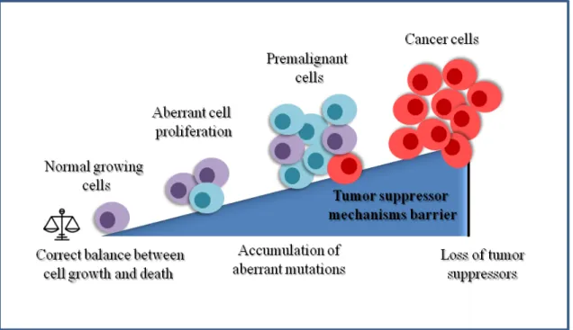
Figure 1.1: Progression to malignant transformation.
Abnormal activation of oncogenes or loss of tumor suppressor function can promote an
unbalance between cell growth and death that can lead to aberrant cell proliferation. This aberrant proliferation induces the activation of tumor suppressor mechanisms that premalignant cells need to bypass in order to become malignant and progress to cancer. Inspired from Narita M & Lowe SW (2005) (17) .
One of the best studied tumor suppressors is a protein known as Retinoblastoma Protein (pRB) and its corresponding gene, RB1, the first tumor suppressor gene to be identified. Since pRB activity stops the expression of genes required for progression into S phase of the cell cycle, its inactivation allows for uncontrolled cell division. In fact, this principle applies to all tumor suppressors: genetic alterations in the gene leading to tumorigenesis prevent the regulatory protein from inhibiting cell proliferation (16). Another tumor suppressor well characterized is p53, which is a transcription factor that activates
programmes such as apoptosis, senescence, and DNA repair in response to a variety of cellular stresses, including DNA damage, hypoxia, and nutrient deprivation (3)
1.1.3
Characteristics of a tumor cell
Cancer cells often show alterations in the signal transduction pathways that lead to proliferation without mitogenic signals or less stringent requirements than those of normal cells (18). Indeed, many growth factors and their receptors, as well as their membrane, cytoplasmic and nuclear downstream effectors give to cancer cells special characteristics to allow them to change their cell physiology with new capabilities that contribute to unscheduled proliferation (11, 19). In addition, most cancer cells acquire genomic instability that leads to additional mutations and chromosomal aberrations as well as chromosomal instability, a defect responsible for numerical changes in chromosomes (20-22). Cancer cells can enable signals that convey in large part by growth factors that bind cell surface receptors, typically containing intracellular tyrosine kinase domains. The latter proceed to emit signals via branched intracellular signaling pathways that regulate progression through the cell cycle as well as cell growth; often these signals influence yet other cell biological properties, such as cell survival and energy metabolism (2). All these attributes include the ability of cancer cells to generate their own mitogenic signals, to resist exogenous growth-inhibitory signals, to evade apoptosis, to proliferate without limits (undergo immortalization), to acquire vasculature (undergo angiogenesis), and in more advanced cancers, to invade and metastasize (11, 23).
Rather than lacking function, cancer cells reproduce at a rate far beyond the normally tightly regulated boundaries of the cell cycle. Cancer can be distinguished from many other human diseases because its root cause is not a lack of, or reduction in, cell function, its roots cause is gain of cell functions that give them the properties to alter a normal biological process: cell division (16).
1.2
Cell cycle and Cancer
1.2.1 General
perspective
Multicellular organisms require an adequate control of cell division and differentiation to coordinate multiple different cell types in functional tissues. The cell division cycle entails the tightly regulated transduction of mitogenic signals to a series of biochemical machineries that control the duplication of DNA and its proper segregation to daughter cells (24). The cell cycle plays a prominent role in development, from egg fertilization to the adult organism. Such key role also holds in pathological conditions, because deregulation of the cell cycle is associated with aberrant cell proliferation and cancer (4). The most critical decision that every proliferating mammalian cell must make is whether to continue another round of cell division or to exit the cell cycle and reach a quiescent state. Likewise, quiescent cells must decide whether to continue in their non proliferative state or to re-enter the cell cycle. All cells have the capacity to enter quiescence and all quiescent cells, except those that have reached a state of terminal differentiation, have the capacity to re-enter the cycle (25).
The phases of the mammalian cell division cycle include two major periods of activity in which the genome is first duplicated (DNA synthesis or S phase) and the two newly replicated genomes are then distributed between the daughter cells (mitosis). Stages of mitosis include prophase, metaphase, anaphase and telophase. Additional gap periods, G1, during which the cell is preparing for DNA synthesis (preceding S phase) and G2, during which the cell prepares for mitosis, are required to coordinate DNA synthesis and segregation with mitogenic signals and to synthesize and assemble the required proteins and cellular structures. Cells in G1 can, before commitment to DNA replication, enter a resting state called G0. Cells in G0 account for the major part of the non growing, non proliferating cells in the human body (24, 26). Thus the cell cycle can be defined as the process by which cells monitor proper conditions for cell division, activate the required biochemical machineries for DNA replication and chromosome segregation, and monitor these steps to generate two healthy genomically stable daughter cells. All these processes are orchestrated by a family of kinases: the Cyclin-Dependent Kinases (CDKs) (20, 24, 27). CDKs serve as focal points for the cells and respond
to proliferative and non proliferative signals and to various forms of genotoxic stress at specific checkpoints, in order to ensure faithful DNA replication and mitosis (28).
1.2.2
The Cyclin Dependant Kinases
Cyclin Dependent Kinases (CDKs) are the catalytic subunits of a large family of heterodimeric serine/threonine kinases whose activity depends on a regulatory subunit: a Cyclin, which control kinase activity and substrate specificity. CDKs play important roles in the control of cell division progression and modulate transcription in response to several extra- and intracellular cues (21, 29, 30). In most cases, the concentration of the kinase subunit is relatively constant; whereas the concentration of the Cyclin subunit oscillates (this oscillation gave their Cyclin name). The kinase is completely inactive without its Cyclin partner, but in addition to the binding of Cyclin, activation of the holoenzyme requires the phosphorylation of a key residue in the activating T-loop of the kinase subunit (31). The CDK family is characterized by a conserved catalytic core made up of an ATP-binding pocket, a PSTAIRE-like Cyclin-binding domain and an activating T-loop motif. Collectively, these features participate in CDK activation, which involves the association with Cyclins via the PSTAIRE helix to: first, displace the T-loop and expose the substrate-binding interface; and second, realign critical residues within the active site thereby priming it for the phospho-transfer reaction. Most CDK family members also possess inhibitory (Thr-14; Tyr-15) and activating (Thr-161) phosphorylation sites. Phosphorylation at Thr-14 and Tyr-15 within the ATP-binding site by inhibitory kinases WEE1 and MYT1 interferes with proper ATP alignment, whereas T-loop phosphorylation at Thr-161 by CDK Activating Kinases (CAKs) improves substrate binding and complex stability to enable full CDK activation (32). The consensus sequence for the phosphorylation site in the amino acid sequence of a CDK substrate is [S/T*]PX[K/R], where S/T* is the phosphorylated serine or threonine, P is proline, X is any amino acid, K is lysine, and R is arginine (33). Based on the sequence of the kinase domain, CDKs belong to the CMGC group of kinases which include also: Mitogen Activated Protein Kinases (MAPKs), Glycogen Synthase Kinase-3 beta (GSK3β) and members of the Dual-Specificity Tyrosine-Regulated Kinase (DYRK) family and CDK-like kinases (34).
Figure 1.2: Functional Diversity of CDKs and the pathways that regulate.
The Cyclin-Dependant Kinases can modulate several cellular processes such as metabolism, hematopoiesis, angiogenesis, proteolysis, epigenetics DNA damage and DNA repair, cell cycle regulation, lipogenesis, neuronal function and transcription. Inspired from Peyressatre, M. et al., 2015 (35).
CDKs were first discovered by genetic and biochemical studies in model organisms such as yeasts and frogs, and from these studies, it has been clearly established the importance of CDKs function as the major eukaryotic protein kinase family involved in the integration of extracellular and intracellular signals to modulate gene transcription and cell division (34). Twenty different CDK exist in mammal cells (24). The evolutionary expansion of the CDK family have led to the division of CDKs into two cell-cycle-related subfamilies: Mitotic CDKs which include CDK1 and Interphase CDKs, which include CDK2, CDK4 and CDK6 and the transcriptional subfamily, which include: CDK7, CDK8, CDK9, CDK11 and CDK20. Unlike the prototypical Cdc28 kinase of budding yeast, most of these CDKs bind one or a few Cyclins, consistent with functional specialization during evolution (29). There are ten Cyclins that belong to four different classes: the A-, B-, D- and E-type Cyclins (19).
Protein kinases mediate most of the signal transduction in eukaryotic cells by modification of substrate activity, and also, control many other cellular processes, including metabolism, transcription, cell cycle progression, cytoskeletal rearrangement and cell movement, apoptosis, and differentiation (Fig. 1.2) (34, 36). Tumour associated mutations frequently deregulate certain CDK/Cyclin complexes, resulting in either continued proliferation or unscheduled re-entry into the cell cycle, two properties characteristic of most human tumour cells (19).
1.2.3
The function of the CDKs during the cell cycle
According to the ‘classical’ model for the mammalian cell cycle, specific CDK/Cyclin complexes are responsible for driving the cell cycle in a sequential and orderly fashion (20) (Fig. 1.3). Extracellular signals, including stimulatory mitogens, inhibitory cytokines, differentiation inducers, cell–cell contacts and growth factors induce the expression of D-type Cyclins (D1, D2 and D3). The three D-type Cyclins are differentially expressed, alone or in combination, in distinct cell lineages, where they assemble with CDK4 and CDK6 to form enzymatically active holoenzyme complexes during G1. CDK4/6 complexes are able to partially phosphorylate and inactivate pRB pocket of proteins (pRB, p107 and p130), which are the only well characterized substrates to date (37, 38). In its phosphorylated state, the pRB pocket proteins represses the transcription of genes that are necessary for cell cycle progression by binding to the transactivation domain of the E2F transcription factor family of proteins. This reduced inhibition of E2F transcription factors initiates a positive feedback loop, where E2F transcription factors promote transcription of the E-type Cyclins, which activate CDK2 and other proteins that are important for initiation of S phase and DNA synthesis. Pocket proteins phosphorylation process is then completed by Cyclin E (E1 and E2)/CDK2 complexes expressed in late G1, leading to their complete inactivation, preventing their binding and complete inactivation of the E2F family of transcription factors (39-41). Given this functions, CDK4/6-Cyclin D and CDK2–cyclin E complexes are essential to drive the G1/S transition. The availability of E-Cyclins during the cell cycle is tightly controlled and limited to the early stages of DNA synthesis. Later during G1, E- Cyclins (Cyclins E1 and E2) become upregulated and activate CDK2 (and, to a lesser extent, CDK1 and CDK3), resulting
in phosphorylation of a broader range of cell cycle-related proteins. CDK1 and CDK2 are subsequently activated by Cyclin A2 during the late stages of DNA replication to drive the transition from S phase to mitosis (G2 phase). Throughout the process of progression through S phase and G2 phase of the cell cycle, the pocket proteins remain hyper-phosphorylated, returning to its hypo-phosphorylated state only following mitosis. The subsequent induction in S phase and the activation of Cyclin B1/ CDK1 at the onset of mitosis, drive the progression of cells through the phosphorylation of a large number of proteins that are involved in DNA replication, as well as in centrosome and chromosome function (42). Following nuclear envelope breakdown, A-type Cyclins are degraded, facilitating the formation of the CDK1/Cyclin B complexes responsible for driving cells into mitosis (30).
1.2.4
Cellular regulation of CDKs by CDK Inhibitors and its role in
cancer formation
Besides the modulation of the CDKs by the Cyclins, the CDK activity is also negatively regulated by small polypeptides, the CDK-Inhibitors (CKIs). Whereas most Cyclins promote CDK activity, CKIs restrain CDK activity, so, close cooperation between this trio is necessary for ensuring orderly progression through the cell cycle. In addition to their well established function in cell cycle control, it is becoming increasingly apparent that mammalian CDKs, Cyclins and CKIs play indispensable roles in many processes such as transcription, epigenetic regulation, metabolism, stem cell self-renewal, neuronal functions, apoptosis and spermatogenesis (32).
In mammals, CKIs are subdivided into two classes based on their structure and CDK specificity.
- The CIP/KIP (CDK-Interacting Protein/Kinase Inhibitory Protein) family which comprises: p21CIP1 (Cdkn1a), p27KIP1 (Cdkn1b), and p57KIP2 (Cdkn1c). This family of CKIs are encoded by the Cyclin-Dependent Kinase Inhibitor 1 (CDKN1) genes and bind to most Cyclin–CDK complexes and prevent their activation.
- The INK4 (Inhibitors of CDK4) family which includes: p16INK4A (Cdkn2a), p15INK4B (Cdkn2b), p18INK4C (Cdkn2c), and p19INK4D (Cdkn2d), encoded by the
Cyclin-Dependent Kinase Inhibitor 2 (CDKN2) gene that primarily target CDK4 and CDK6 and block their association with the D-type Cyclins (25, 32, 43, 44).
Among the INK4 family, p16INK4A is the best known inhibitor of CDK4, and contributes to G1 arrest in two ways. Firstly, to become functional, CDK4 requires cytoplasmic, post-translational folding in a complex involving the Heat Shock Protein 90 (HSP90), an interaction that is disrupted by p16INK4A. In addition, p16INK4A can bind to CDK4 directly and inhibit its catalytic activity. The combination of these two mechanisms results in G1 arrest in cells with functional pRB, but not in pRB-deficient cells (45).
Whereas CIP/KIP proteins act as negative regulators of Cyclin E- and Cyclin A-CDK2 and Cyclin B-CDK1 holoenzymes in a 1:1 stoichiometry, they also act as positive regulators of Cyclin D-CDK4/6 complexes by mediating their assembly early in G1. This results in the titration of p27KIP1 from Cyclin E-CDK2, and the activation of this complex. In turn, p27KIP1 is phosphorylated by Cyclin E-CDK2, targeting it for degradation. Instead, INK4 proteins compete with D-type Cyclins for binding CDK4/6. Enforced expression of INK4 proteins can lead to G1 arrest by promoting the redistribution of CIP/KIP proteins and blocking Cyclin E-CDK2 activity. For example; CDK4 can sequester p27KIP1 and controls its availability for inhibition of CDK2 activity (46). Thus, in cycling cells, there is a reassortment of CIP/KIP proteins between CDK4/6 and CDK2 as cells progress through G1 alternately acting as positive or negative regulators of CDK activity (25, 44, 47).
Owing to their inhibition of CDKs and cell proliferation, CKI are tumour suppressors, and deletion of the CDKN2A–CDKN2B locus, which encodes p16INK4A, p15 INK4B and another protein p14 (ARF; which is involved in the activation of the p53 tumour suppressor), is one of the most frequent mutations in human tumours (42). Intriguingly, increased expression of p16INK4Ais a hallmark of tumours in which pRB function has been lost suggesting a negative feedback regulation (25). Not surprisingly, many components of the cell cycle regulatory machinery, including CDKs, CKI and CDK substrates, are important targets of mutations that lead to human malignancy (37). CDK4 and CDK6 hyperactivity has been well documented in a wide variety of cancers, in fact, genetic alterations in components of the pRB-CDK4/6-Cyclin D-p16INK4A pathway are amongst the most frequently occurring anomalies reported. There are found in more than half of all human tumours being Cyclin D1 gene (CCND1) the second most frequently amplified locus across all human cancers. (35). Together with
amplification and overexpression of Cyclin-D, CDK4 gene amplification has been documented in rhabdomyosarcoma (48) osteosarcoma (49, 50), well-differentiated and de-differentiated liposarcomas (51, 52), and cancer of the uterine cervix (53). Over expression of CDK4 protein has been reported in breast cancer (54), colorectal carcinoma (55), Non-Small-Cell-Lung-Carcinoma (NSCLC) (56), uterine cervical carcinoma (53), glioblastoma (57, 58) and melanoma (59, 60). CDK6 gene amplification and overexpression have been demonstrated in squamous-cell oesophageal carcinoma (61) lymphomas (35), leukemias (62), and gliomas (63). Moreover, CDK6 is overexpressed in medulloblastoma, which is the most common brain tumor in children (64). Also, homozygous deletions of CDKN2A (gene encoding for p16INK4A), are seen in tumours of the pancreas, bladder, breast, prostate and in glioblastoma (65, 66). Conversely, loss of pRB function results in constitutive activation of E2F, Cyclin E1 and CDK2 expression, and therefore loss of reliance on CDK4/6 to initiate the G1/S transition (66-68).
Although the canonical role of Cyclins and CDKs as essential drivers of cell cycle entry and progression has been firmly established, as mentioned before; additional functions of the CDK4 and CDK6 complexes are involve in many other cellular process, and the elucidation of how signal transduction pathways activate CDK4/6 and the substrates that theses kinases are able to regulate in different tumor types should give a rational to create combinatorial therapies to improve therapeutic responses.
From now on, I will refer to CDK4 and CDK6 as an active complex, implying their association with Cyclin D.
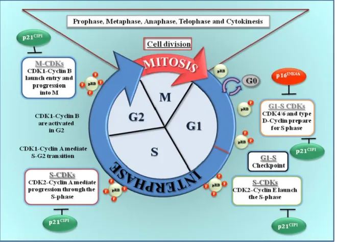
Figure 1.3: CDKs regulation and phosphorylation levels of pRB during the cell cycle.
Cell cycle regulation by CDK/cyclins: CDK1/cyclin B during the mitosis (M), CDK4 and 6/cyclin D for progression through G1 phase, CDK2/cyclin E for G1/S transition, CDK2/cyclin A at the S phase and CDK1/cyclin A for progression through G2 phase.
1.2.5
Non- canonical functions of CDK4 and CDK6
It is clear that tumor proliferation requires an adapted metabolic response of the cells; hence, the regulation of the cell cycle must be linked to metabolic control. The impact that the activities of cell cycle regulators such as Cyclins, CDKs or E2F transcription factor have on cell metabolism are also being uncovered (41, 69). In this segment I will focus only on the non-canonical functions of CDK4 and CDK6.
Role in DNA methylation
DNA methylation plays a central role in the epigenetic regulation of gene expression during development and progression of cancer diseases. The inheritance of specific DNA methylation patterns are acquired in the early embryo and are specifically maintained after cellular replication via the DNA Methyltransferase 1 (DNMT1). Recent studies have suggested that the enzymatic activity of DNMT1 is possibly modulated by phosphorylation of serine/threonine residues located in the N-terminal domain of the enzyme. It has been reported that CDK1, 2 and 5 can phosphorylate Ser-154 of human DNMT1 in vitro controlling its enzymatic activity and protein stability; suggesting that dysregulation of cell cycle via CDKs could induce abnormal phosphorylation of DNMT1 and lead to DNA hypermethylation often observed in cancer cells (70).
Role in transcription
CDK4 and CDK2 complexes phosphorylate Smad2 and Smad3 (Small Mother Against Decapentaplegic 2 and 3), inhibits its transcriptional activities and antiproliferative functions of signalling through the Transforming Growth Factor beta (TGF-β) pathway, thus inhibit cell cycle progression at transition of G1 phase to S. Mutation of the CDK phosphorylation sites of Smad3 increases its transcriptional activity, leading to higher expression of the CKI p15INK4B and increases its ability to downregulate the expression of c-Myc. (71). CDK4 and CDK6 complexes phosphorylate, stabilize and activate the transcription factor FOXM1 (Forkhead Box Protein M1), thereby maintain expression of G1/S phase genes, suppress the levels of Reactive Oxygen Species (ROS), and protect cancer cells from senescence (72). FOXMI regulates the expression of various cell cycle regulators, including proteins that govern the G2/M transition phase (42). Also, it was shown by the group of Alan Diehl that CDK4 phosphorylates and increases the activity of MEP50, a coregulatory factor of the Protein Arginine Methyltransferase 5 (PRMT5), an enzyme associated with histone methylation and transcriptional repression. Increased PRMT5 activity mediates key events associated with Cyclin D1-dependent neoplastic growth, including CUL4 repression, CDT1 overexpression, and DNA re-replication (73).
CDK6 is specifically expressed in proliferating hematopoietic progenitor cells, and physically interact with and inhibit the transcriptional activity of Runt Related Transcription
Factor 1 (RUNX1) resulting in an increase proliferation of myeloid progenitors. Since RUNX1 transcription factors play central roles in hematopoietic, neuronal and osteogenic lineages, this CDK6 function may control terminal differentiation in multiple tissues and cell types (74). CDK6, is part of a transcription complex that induces the expression of p16INK4A and the Vascular Endothelial Growth Factor A (pro-angiogenic factor: VEGFA) through activation of JUN and STAT3 (Signal Transducer and Activator of Transcription 3). In the absence of p16INK4A, CDK6 can exert its full tumor promoting function by enhancing proliferation and stimulating angiogenesis (75). CDK6 can exert its pro-proliferative role only upon silencing of the gene encoding p16INK4A, an event that is frequently seen in human tumours (42, 76). CDK6 can also modulate the activity of the transcription factor EGR1 (Early Growth Response Protein 1) and regulate the balance between quiescence and proliferation in Hematopoietic Stem Cells (HSCs) and Leukemic Stem Cells (LSCs) (76).
Role in DNA damage repair
DNA damage causes stabilization of p53, leading to G1 arrest through induction of p21CIP1. This DNA damage causes an immediate and p53-independent G1 arrest, caused by rapid degradation Cyclin-D1 which leads to a release of p21CIP1 from CDK4/6 to inhibit CDK2 and induce cell cycle arrest. Interference with Cyclin-D1 degradation prevents initiation of G1 arrest and renders cells more susceptible to DNA damage (77). It has been shown that nuclear accumulation of degradation resistant mutants of Cyclin D1/CDK4 complexes triggers DNA re-replication, resulting from CDT1 (Chromatin Licensing and DNA Replication Factor 1) stabilization, which in turn triggers the DNA damage checkpoint and p53-dependent apoptosis. Loss of p53 through mutations or targeted deletion results in increased genomic instability and neoplastic growth and disturbance of critical cell cycle regulatory events will perturb DNA replication fidelity, thereby contributing to neoplastic transformation (78).
Balancing cell proliferation and death
The regulation of the cell cycle is tightly linked to the control of cell death. Indeed, early studies indicated that the pRB/E2F pathway could modulate the expression of multiple pro or anti apoptotic proteins (42). CDK4 interacts with the apoptosis inhibitor survivin, which
is a member of the IAP (Inhibitors of Apoptosis Proteins) family and is specifically expressed during embryogenesis and in tumor cells to suppress cell death signaling. The interaction of Survivin with CDK4 promotes CDK2/Cyclin E activation and pRB phosphorylation. As a result of Survivin/CDK4 complex formation, p21CIP1 is released from its complex with CDK4 and interacts with mitochondrial procaspase 3 to suppress FAS-mediated cell death (79).
In cultured postmitotic neurons, activation of CDKs is a signal for death rather than cell division. In a rat model of stroke, CDK4/Cyclin D1 levels and phosphorylation of pRB increase after the injury, along with deregulated levels of E2F1, which correlates with neuronal death. After administration of a pan-CDK inhibitor (Flavopiridol) into brain ventricles, levels of pRB phosphorylation are blocked and dramatically reduces neuronal death by 80%; suggesting that CDKs could be an important therapeutic target for the treatment of reperfusion injury after ischemia (80).
Control of cell differentiation and migration
In general, cell proliferation and differentiation show an inverse relationship, and are regulated in a coordinated manner during development. Terminal differentiation is usually coupled to permanent exit from the cell cycle. The levels of Cyclins typically decline when cells exit the cell cycle and undergo differentiation. Moreover, induction of the expression of CKIs during cell differentiation prevents activation of CDK complexes in terminally differentiated cells. On the other hand, expression of Cyclin–CDK complexes in proliferating cells inhibits pRB function, thereby promoting proliferation and inhibiting differentiation. An example of this is that pRB binds to and regulates the activity of several cell type-specific transcription factors, including MYOD (Myogenic Differentiation), MEF2 (Myocyte-Specific Enhancer Factor 2) and RUNX2 (Runt Related Transcription Factor 2 ), thereby linking cell cycle arrest and differentiation (42). CDK4 phosphorylate RUNX2 and target it to degradation inhibiting bone differentiation (81). CDK4 also prevent transcriptional activation mediated by MYOD and hence inhibit myoblast differentiation (82). According to this, CDK4 can also suppress the skeletal muscle differentiation program in proliferating myoblasts and inhibit the activity of the MEF2 family of transcriptional regulators (83). Analysis of transgenic mice with mutant JARID2 (Jumonji and AT-Rich Interaction Domain Containing 2), revealed that
CDK4/6 complexes directly phosphorylate and promote degradation of GATA4 inhibiting then differentiation of cardiomyocytes (84).
Cell migration and the actin cytoskeleton are also modulated by Cyclins and CDKs at different levels. CDK6 was shown to localize to the ruffling edges of spreading fibroblasts prior to the formation of filamentous actin and to promote migration in a αvβ3 integrin-dependent manner (85).
Role in metabolism
Multiple cellular functions of CDK4 and CDK6 converge to control the generation of cellular energy and metabolism at both the cellular and organismal levels, a physiological role for cell cycle proteins in metabolism has been documented by the observations that CDK4-deficient mice are viable, but small in organ and body size and infertile. These mice also develop insulin-deficient diabetes due to the loss of pancreatic islet β-cells (86-88). Similarly, CDK6-deficient mice are also viable, but with lower number of cells in the thymus and spleen, and with a small reduction in the abundance of peripheral blood cells (89). These results show that CDK4 and CDK6 are not only involved in controlling proliferation of specific cell types but may play a wider role in establishing homeostatic cell numbers. The lack of phenotypes with more severe consequences for survival in these single knockout (KO) mice is assumed to reflect functional compensation between CDK4 and CDK6. Surprisingly, although CDK4/6 double knockout mice succumbed to anaemia in the late stages of embryonic development, many non haematological cell types from these mice were able to proliferate normally (89). In addition, quiescent KO CDK4/6 fibroblasts exhibit a delay in S phase entry. This cell cycle perturbation by CDK4/6 disruption is associated with an increase binding of p27KIP1 to CDK2 and a lower activation of CDK2 that lead to an impaired pRB phosphorylation (88). CDK4-pRB-E2F1 axis are robustly expressed in non-proliferating β cells, suggesting that besides the control of β-cells, the CDK4 has a role in its function and is involve in glucose homeostasis. Inhibition of CDK4, or inactivation of E2F1, results in a decrease expression of KIR6.2, which impairs insulin secretion and glucose intolerance (90). An independent mechanism linking insulin signalling to CDK4 was provided by the observation that insulin-mediated upregulation of Cyclin D1 (and the subsequent activation of CDK4) in hepatocytes leads to phosphorylation and activation of the histone acetyltransferase GCN5 (General Control
Non-Repressed Protein 5) and suppresses hepatic glucose production independently of cell cycle progression (91).
The functions of Cyclins and CDKs in modulating metabolic pathways contribute to the ability of these proteins to affect differentiation (42). Cell cycle regulators such as E2F1 and pRB play crucial roles in the control of adipogenesis, mostly by controlling the transition between preadipocyte proliferation and adipocyte differentiation (92). Since this pathway is regulated by the CDKs, is not surprise that these kinases have a pivotal role in control adipogenesis. During terminal differentiation of mouse preadipocytes, the level of CDK4 remains constant and Cyclin D3 is the predominant Cyclin partner of CDK4 in mature adipocytes. Knockdown of Cyclin D3 inhibits adipogenesis in vitro (93) and CDK4 inhibition impairs adipocyte differentiation and function. Consistent with this, mice lacking Cyclin D3 or CDK4 are protected from diet-induced obesity, have smaller adipocytes and reduced expression of adipogenesis genes. CDK4 also participates in adipocyte differentiation by directly interacting with and activating PPARγ, the master regulator of adipogenesis (92).
Some of the proposed non-canonical roles may not reflect a normal physiological protein function, but rather a gain of function event that occurs in tumour cells as a consequence of Cyclin and/or CDK overexpression (42).
1.2.6
CDK Inhibitors and its implication in Cancer Treatment
Given the critical role that CDKs play in cell cycle control they have been actively considered as targets for anticancer therapy. However, this strategy has found some practical limitations. For instance, some CDKs may have functions not directly related to cell cycle progression and others, which were believed to be essential for this process, may actually be dispensable. One example to support the latter theory is CDK2. Recent research in animal models suggests that this kinase is not required for mitotic cell division and could be dispensable for cancer cell progression, rendering this CDK not suitable as a treatment target (18, 94). In the same context, emerging evidence suggests that tumour cells may also have specific requirements for individual CDKs. In particular, CDKs that promote transition through the cell cycle are expected to be key therapeutic targets because many tumorigenic events ultimately drive proliferation by impinging CDK4 or CDK6 complexes in the G1 phase