HAL Id: hal-01600989
https://hal.archives-ouvertes.fr/hal-01600989
Submitted on 26 May 2020
HAL is a multi-disciplinary open access
archive for the deposit and dissemination of sci-entific research documents, whether they are pub-lished or not. The documents may come from teaching and research institutions in France or abroad, or from public or private research centers.
L’archive ouverte pluridisciplinaire HAL, est destinée au dépôt et à la diffusion de documents scientifiques de niveau recherche, publiés ou non, émanant des établissements d’enseignement et de recherche français ou étrangers, des laboratoires publics ou privés.
membrane microdomains: entangled relationships
between structural analogies in the membrane and
functional homologies in the cell
Gérald Gaibelet, François Tercé, Sophie Allart, Chantal Lebrun, Xavier
Collet, Nadège Jamin, Stéphane Orlowski
To cite this version:
Gérald Gaibelet, François Tercé, Sophie Allart, Chantal Lebrun, Xavier Collet, et al.. Fluorescent probes for detecting cholesterol-rich ordered membrane microdomains: entangled relationships be-tween structural analogies in the membrane and functional homologies in the cell. AIMS Biophysics, AIMS Press, 2017, 4 (1), pp.121-151. �10.3934/biophy.2017.1.121�. �hal-01600989�
Received: 20 December 2016 Accepted: 15 February 2017 Published: 21 February 2017 http://www.aimspress.com/journal/biophysics
Review
Fluorescent probes for detecting cholesterol-rich ordered membrane
microdomains: entangled relationships between structural analogies in
the membrane and functional homologies in the cell
Gérald Gaibelet 1, François Tercé 2, Sophie Allart 3, Chantal Lebrun 4, Xavier Collet 2, Nadège
Jamin 5 and Stéphane Orlowski 5,*
1 Theranyx SA, Marseille, France
2 Université Toulouse III, UMR 1048 INSERM, Toulouse, France
3 Plateau Technique d’Imagerie Cellulaire, INSERM U1043, Toulouse, France 4 Université Toulouse III, Toxalim UMR 1331 INRA, ENVT, Toulouse, France
5 IBiTecS/SB2SM CEA and I2BC UMR 9198 CNRS, Université Paris-Saclay, Gif-sur-Yvette, France
* Correspondence: Email: stephane.orlowski@cea.fr; Tel: +33-1-69-08-63-99.
Abstract: This review addresses the question of fluorescent detection of ordered membrane (micro)
domains in living (cultured) cells, with a “practical” point of view since the situation is much more complicated than for studying model membranes. We first briefly recall the bases of model membrane structural organization involving liquid-ordered and -disordered phases, and the main features of their counterparts in cell membranes that are the various microdomains. We then emphasize the utility of the fluorescent probes derived from cholesterol, and delineate the respective advantages, limitations and drawbacks of the existing ones. In particular, besides their intra-membrane behavior, their relevant characteristics should integrate their different cellular fates for membrane turn-over, trafficking and metabolism, in order to evaluate and improve their efficiency for in-situ probing membrane microdomains in the cell physiology context. Finally, at the present stage, it appears that Bdp-Chol and Pyr-met-Chol display well complementary properties, allowing to use them in combination to improve the reliability of the current experimental approaches. But the field is still open, and there remains much work to perform in this research area.
Keywords: membrane domains; ordered membranes; lipid rafts; cholesterol; fluorescence;
intracellular trafficking
Abbreviations: ABC, ATP-binding cassette; AFM, atomic force microscopy; ACAT,
acyl-CoA:cholesterol acyl transferase; ApoAI, apolipoprotein AI; ASMase, acid sphingomyelinase; BBM, brush border membrane; Bdp, Bodipy, boron-dipyromethene, difluoro-dimethyl-bora-diaza-indacene; BHK, baby hamster kidney; BSA, bovine serum albumin; Cav-1, caveolin 1; CD, cluster determinent; CHAPS, cholamidopropyl-dimethylammonio-propanesulfonate; CHO, Chinese hamster ovary; Chol, cholesterol; CTE, cholestatrienol; Dansyl, dimethylamino naphtalene sulfonyl; DAT, deep apical tubules; DHE, dehydroergosterol; DiI-C12, didodecyl-tetramethyl-indocarbocyanine; DIM,
phthiocerol dimycocerosates; DMSO, dimethylsulfoxide; DOPC, dioleoylphosphatidylcholine; DRM, detergent-resistant membranes; ER, endoplasmic reticulum; FCS, fluorescence correlation spectroscopy; FCVJ, farnesyl-cyanovinyl julolidine; FLIM, fluorescence lifetime imaging microscopy; FRAP, fluorescence recovery after photobleaching; GPI, glycosylphosphatidylinositol; GPMV, giant plasma membrane vesicles; GUV, giant unilamellar vesicles; HDL, high-density lipoprotein; LDL, low-density lipoprotein; Ld, liquid-disordered; Lo, liquid-ordered; LUV, large unilamellar vesicles; MBCD, methyl-β-cyclodextrin; MDCK, Madin-Darby canine kidney; met, methyl; NBD, nitrobenzoxadiazole; NPC1, Niemann-Pick type C protein 1; PALM, photoactivation localization microscopy; PC, phosphatidylcholine; PEG, polyethylene glycol; PlAP, placental alkaline phosphatase; PS, phosphatidylserine; Pyr, pyrene; Pyr-met-Chol, 21-methylpyrenyl-cholesterol; SIMS, secondary ion mass spectrometry; SM, sphingomyelin; STED, stimulated emission depletion; STORM, stochastic optical reconstruction microscopy; SR-BI, scavenger receptor class B type I; SUV, small unilamellar vesicles; TX100, Triton X-100.
1. Introduction
In cell biology, plasma and internal membranes are not only physical frontiers for the cell and its organelles, but they are endowed with a number of important functions, such as regulation of solutes influx and efflux, signal transduction, ionic conduction and membrane excitation, lipid metabolism and trafficking, bioenergetics metabolism, and even interaction with pathogenic agents, among many other ones. Analysis of their structural and functional characteristics is thus pivotal, and it is now well recognized that they display a typical molecular organization that is underlying to their various biological activities. In particular, they present local structural heterogeneities, essentially of two orders that are (i) transversal heterogeneity meaning a difference of (lipid and protein) composition of the two leaflets of the bilayer, and (ii) lateral heterogeneity (“domains”) meaning that along their 2D space they are far from a monotonous lipid continuum (as historically illustrated by the Singer & Nicolson’s model). We will focus here on the second point, and address the question of their practical experimental approach in cells, based on the background already acquired on model membranes.
Model membranes include monolayer and bilayer lipid membranes, the last ones being obviously more relevant for biological membranes, and they can be either supported on an extrinsic material or self-forming vesicles, of possible various sizes (e.g. SUV, LUV, GUV). They are
composed of pure lipids of well-defined compositions, and they can be submitted to well-controlled physico-chemical conditions. But the advantage to use such a defined experimental system is also a limitation and a concern, as regards the complexity of the situation within a living cell. Indeed, a biological membrane is made of a variety of hundreds of different lipid species, combining the different head-groups, skeletons and the various acyl chains of different length and unsaturation level, in which are associated, either intrinsically (i.e. integral proteins) or extrinsically (i.e. anchored and adsorbed proteins), a number of membrane proteins. The transposition of data obtained on model membranes are hence not at all straightforward to membranes in cells, and they have actually raised a huge literature showing debates on many aspects of the so-called membrane (micro)domains, their various characteristics and even their existence. Section 1 will be devoted to describing the rather large diversity of the various reported membrane domains, all more or less enriched in cholesterol.
In order to efficiently detect and characterize membrane domains, availability of powerful and reliable tools is required. Investigations on in-vivo cell membranes require indirect and if possible non-invasive methods, and imaging techniques are actually the major approach. Besides the techniques of electron microscopy, AFM and SIMS, which allow to reach high resolution, fluorescence remains the widest technique used, with many possible variations, such as wide-field and confocal conventional microscopy, multi-photon excitation, FLIM, and the various recently developed super-resolution microscopy modalities (STED, PALM, STORM), as well as methods giving access to kinetic phenomena (FRAP, FCS). In particular, fluorescence also allows for being combined with other assays, as quantification after cell fractionation, and taking benefit of the molecular information given by a spectroscopic approach (as with any other optical or magnetic spectroscopies). However, this is always under the dependence of having available the most convenient fluorescent probe well suited for the considered study.
In Section 2, we will briefly present the different possible strategies using fluorescence techniques for addressing membrane domain analysis. They rely essentially on two families of probes, so-called “environment-sensitive” and “phase-selective” [1]. In the second case, schematically, fluorescent probes are either extrinsic (exogenous compounds), designed for specifically detecting a key lipid component, or intrinsic, i.e. molecules closely related to the endogenous components of the considered membrane domains. It appears that the exogenous lipophilic dyes are very useful on model membranes, but are generally less suited for a use on living cells because of potential non-physiological handling by the cell or even cytotoxicity. Otherwise, among the lipid analogs as intrinsic probes, phospholipid derivatives have been widely used, but they have shown globally that their intracellular trafficking strongly depends on their nature, and they even can be metabolically instable. We have thus focused this mini-review on fluorescent cholesterol derivatives, in particular considering Bdp-Chol and Pyr-met-Chol, both developed during the last decade, which are worth being presented in detail and compared because they appear performing and promising, as exposed in Section 3. But we also emphasize that they both need to be further functionally characterized in depth when incorporated into various cell types.
2. Diversity of Ordered Membrane Microdomains in the Cell: Importance of Cholesterol
In various bilayer model membranes, of well-defined lipid composition, it has been long ago observed in the presence of two (or more) components that there can co-exist three segregated lipid phases, according to a well-defined phase diagram linking temperature and lipid composition [2,3]. The three main phases, namely solid (or gel), liquid-ordered (Lo) and liquid-disordered (Ld), essentially differ by their translational and rotational diffusion coefficients (“microviscosity”) and local molecular order. In ternary systems including cholesterol and phospholipids, cholesterol, thanks to its rigid and planar structure, displays a typical “packing effect” (both condensing and ordering) leading to a Lo phase when combined with saturated long chain glycerophospholipids, according to the template mechanism [4,5,6], while unsaturated chains remain in Ld phase [3,7]. However, in the presence of SM (bearing longer and more saturated chains than PC), or other sphingolipids, cholesterol induces an even closer specific association, establishing H-bonds (and charge pairs) at the polar interface [8]. This pattern leads to more stable [3], compact and ordered domains, which are thicker than the Ld phase enriched in unsaturated chains [9,10]. The extent of this packing effect (or “ordering capacity”) depends on the precise structure of the sterol that can replace cholesterol [8,11–15]. The structural specificities that appear especially important for such property encompass both hydrophobic and steric constraints, with in particular the presence of the OH group at C3 position and the preservation of the isooctyl side chain.
2.2. The more complex case in cell membranes (theoretically and experimentally)
A largely debated question is how information about model membranes organization can be extrapolated to biological membranes. Indeed, membranes in cells are characterized by some important properties distinguishing them from model membranes. First, they are made of a very complex mixture of a great number of lipids of different chemical structures, whereas a four-component model membrane was hard to be well-enough investigated in-vitro [7]. Second, the presence of a large number of proteins inserted in or associated with these membranes deeply contributes to make these systems highly complicated by the mutual relationships existing between these proteins and the surrounding lipids [16]. Third, a cell membrane, in contrast to model membranes, is well out of thermodynamic equilibrium, since it is involved (and contributes to) the whole cell energy-dependent metabolism, including lipid translocation, molecular sorting and membrane turn-over (endocytosis and exocytosis) [9]. Thus, biological membranes represent a situation very far from the “simple case” of model membranes that are experimentally amenable for being described by defined phase diagrams.
Regarding the in-cellulo experimental approach, studying biological membranes must cope with some specific practical constraints, such as use of solvents (to deliver hydrophobic reagents), limitations of the concentration (and incubation period) of these reagents to be compatible with the cells health, cell culture conditions that must be consistent with the experimental setup chosen for the investigation, and so on. It should be stressed that, illustrating the importance of cell membranes in the cell physiology, any (structural and functional) perturbation of these membranes possibly leads to cell suffering, and conversely any cytotoxic stress possibly induces some membrane alterations (in particular considering the apoptosis cascade).
Historically based on the experimental observation of lipid and protein sorting at the level of trans-Golgi before their integration in the plasma membrane, it has been speculated that membrane microdomains exist and provide local platforms for lateral molecular segregation [17]. Taking into account physico-chemical insights from model membranes studies, it has been postulated that these microdomains harbor a specific lipid composition, displaying specific interactions between cholesterol and phospholipids (SM and PC) with long saturated chains, and presenting a compact and ordered local lipid environment suited for a set of typical membrane proteins. These microdomains co-exist with a more fluid and disordered (and slightly thinner) lipid environment preferentially gathering shorter and more unsaturated hydrophobic chains. The schematic image of packed microdomains floating on a surrounding “fluid sea” raised the so-called “lipid rafts” [18]. This innovative concept has received much attention because of its underlying role in various aspects of cell biology, including signal transduction dependent on receptors recruitment, cellular trafficking and sorting for proteins and lipids, membrane enzymes and transporters regulation, and even interaction with pathogenic agents [19,20–25]. Among a very large body of literature dealing with this important and dynamic aspect of membrane biology and biophysics, we can recommend a recent and fairly comprehensive review [26], in addition to some other interesting ones [16,24,27–30]. Beyond this initial description, some additional features have further emerged for characterizing these raft-type membrane domains.
A seminal property of lipid rafts has been a relative resistance to solubilization by soft detergents under certain conditions (cold), but such procedure has been subsequently rather controversial, which has led to give much interest in non-invasive, biophysical approaches, and in particular fluorescence-based techniques [31]. However, the crucial point is that these membrane microdomains have a size smaller than the optical microscopy resolution limit, precluding direct observation using conventional microscopy imaging, which is a major distinction with ordered lipid phases in model membranes. Anyway, indirect arguments favoring such microdomains have come from the observation, using FCS or FRAP, that some membrane proteins can laterally diffuse only within small confining compartments [32,33,34]. Some significant technical improvements, as well as new emerging techniques, are now available for the experimentalists [24,26,35]. As a reconciliation of the various experimental observations, it is envisioned that these lipid rafts present a periphery with intermediate molecular parameters (in particular different detergent-resistances), with a size possibly fluctuating within a certain range (~ 20 to ~ 200 Å, of course depending on the experimental mean to evaluate it), but overall with a highly dynamic behavior, and possible (reversible) coalescence [26,29,30,36]. It should be mentioned that a still open question is the local nature of the cytosolic leaflet underneath the raft, since cholesterol and SM are rather known to be enriched in the exoplasmic leaflet [37]. Otherwise, there is also a debate regarding the involvement of the “raft proteins”, wondering whether they are to be considered as passively targeted to these (preformed) lipid microdomains [38] (in particular minimizing hydrophobic mismatch for integral proteins [39], and inserting thanks to specific anchors, as GPI or palmitoyl, for extrinsic proteins [40]), or, alternatively, they contribute by themselves to form and stabilize these microdomains (thanks to a kind of lipid-ordering driving force, with thickness adaptation, according to the “mattress model”) [41,42,43]. As an intermediate solution to this debate, the “lipid shell model” has been proposed, according to which a raft protein is basically surrounded by few associated molecules of cholesterol and sphingolipid that display some affinity for it, forming small
independent units highly mobile in the membrane plane (so-called “nanodomains”), which can, in a more or less regulated manner, clusterize to give raft-type microdomains [41]. Finally, such a sub-compartmentalization of the membrane proteins that sequesters some of them in these microdomains can be influenced by interactions with underlying cytoskeleton (e.g. actin and spectrin), but there is also a debate whether this could be an additional regulatory mechanism or an intrinsic process that is primarily responsible for strict limitations of lateral diffusion of specific membrane proteins (and associated lipids), according to the “picket and fence” model [16,24,33,44].
2.4. Some variations for membrane domains in animal cells
Beyond these inherent heterogeneities within the molecular description of the lipid rafts, literature has shown some reports of membrane domains that are significantly different from these paradigmatic membrane rafts, in particular when considering their sizes and/or their lipid and protein compositions [26].
At the level of the apical brush border membrane (BBM) of enterocytes, it has been shown that a detergent-resistant fraction can be isolated, using TX100 at physiological temperature, which presents only a fraction, with typically galectin-4 and GPI-linked alkaline phosphatase, of the classical “raft proteins” isolated at low temperature, and which is furthermore independent of the presence of cholesterol (even if it is present in it), but relies on a high content of glycolipids [45,46]. Corresponding membrane domains were thus coined as “super-rafts”, as a distinction to the “normal raft” membrane fraction, containing aminopeptidase N, caveolin-1 and annexin A2, and essentially present in the deep apical tubules (DAT) [45,47]. This distinction is likely linked to their respective local function, DAT being a membrane with a high endocytic activity while BBM must be resistant to the deleterous effect of high local concentrations of bile salts.
In the same context, the apical membrane of polarized epithelial cells is likely presenting specific local biophysical properties, since in MDCK and Caco-2 cells, but not fibroblasts, it has been shown, using FRAP experiments, that raft proteins appear confined in domains or not, depending on temperature [48]. This was taken as an indication that, at least under a temperature threshold, the raft domains can percolate, hence becoming bicontinuous and thus forming the major membrane environment, the non-raft membrane regions being in contrast limited in separated “domains”. This situation could probably be linked with a specific local lipid composition revealing a kind of “instability” leading to raft clustering, as subsequently evidenced on isolated membranes [49].
In the epithelioid HeLa cells, PALM technique using in parallel fluorescent probes of cholesterol and SM, allowed to respectively visualize cholesterol- and SM-enriched domains of comparable sizes (~ 120–150 nm), both depending on cholesterol presence, whereas the cholesterol-rich domains were independent on SM presence [50]. In another specific cell context, i.e. erythrocytes, rather large, submicrometric membrane domains (~ 0.5 µm), have also been evidenced using a cholesterol-labelling procedure [51]. These domains depend on SM, but only partially colocalized with fluorescently-labelled SM, which were also shown to form submicrometric domains depending on cholesterol [52,53].
In fibroblasts, the capacity of SM to form membrane domains has also been pointed, using high-resolution SIMS (detecting 15N-sphingolipids and 18O-cholesterol), and showing that cholesterol,
even if present in these domains and regulating their abundance, unexpectedly distributed uniformly in the whole plasma membrane [54,55].
In macrophages activated for phagocytosis, using the environment-sensitivity of Laurdan (see below), the phagosomal membranes were shown to display an increased lipid order, while they were shown to be rather poor in cholesterol, but enriched in ceramide as the consequence of a local production by acid sphingomyelinase (ASMase) that was correlated with the phagocytic activity. In contrast, varying cholesterol concentration was without effect on phagocytosis rate, although it influenced the phagosomal membrane order. These data gave the indication of the functional importance of a membrane order induced by the specific association SM/ceramide instead of SM/cholesterol [56].
Actually, ceramide has been recently revealed to be the basis for specific membrane domains in various cell systems, so-called “ceramide-rich platforms” [57]. Their typical properties are their lipid order, in consistency with data obtained on model membranes, and their large, micrometer range size. They are involved in various biological functions, notably including apoptosis [58] but also immunological response, relying on local changes of the biophysical properties of the plasma membrane that allow receptors clustering, leading eventually to signal transduction. Indeed, regulated modulations of the enzymatic activity of ASMase can induce regulations of membrane signaling in minutes time course. Finally, cholesterol has been shown to strongly increase ASMase enzyme activity [59], but the relationships between these ceramide-rich platforms and the classical lipid rafts remain to be clarified.
Among the various types of membrane domains that are characterized by the presence of marker proteins, caveolae are the most widely described for a long time. Historically, they were initially described from their morphological specificity that is plasma membrane invaginations, flask- or bulb-shaped, observed by electron microscopy. They are found in only some cell types, typically including endothelial cells, smooth muscular cells and adipocytes [60–63]. As a protein marker, they display caveolin (3 isoforms in mammalian cells, Cav-1 being the most ubiquitous), an unusual hemi-integral membrane protein that binds cholesterol, with a complex intracellular trafficking [64]. Caveolae also typically contain cavins, a family of membrane-associated soluble proteins that stabilize caveolae, and flotillins [65]. Being characterized by specific protein and lipid components, with an enrichment in cholesterol and SM, they are to be considered as actual membrane domains (and not only membrane invaginations). However, caveolae have been shown to present some heterogeneity: they could be separated, using density gradient ultracentrifugation, between 3 subclasses presenting increasing cholesterol content (ratio to phospholipids from 0.4 to 0.9), i.e. very-high density, high-density (specifically containing fatty acid transport proteins), and low-density (specifically containing SR-BI) [66]. Otherwise, caveolae have been involved in various cellular processes, such as clathrin-independent endocytosis, transcytosis, cholesterol trafficking, lipid homeostasis, signal transduction, cell adhesion, and even mechanical sensing and mechanoprotection [60,61,63,67].
Prominin is a pentaspan membrane protein typically found in the microvilli of epithelial cells, but also in highly curved regions of the plasma membrane of non-epithelial cells, i.e. evaginations, protrusions and microspikes. Such high membrane curvature is generally due to a local marked asymmetry between the two membrane leaflets, leading to a specific composition differing from the rest of the membrane, and these lateral heterogeneities are hence consistent with actual membrane
domains not being only membrane evaginations. In polarized MDCK cells, prominin has been found to present a behavior distinct from PlAP, a GPI-anchored membrane protein revealing “classical rafts”, when submitted to various experimental conditions using different detergent-resistance procedures (soluble in TX100 but insoluble in Lubrol WX) and manipulating local cholesterol concentration, which led to the proposition of the existence of membrane microdomains physically distinct from the classical membrane rafts [68].
Tetraspanins form a rather large family of generally ubiquitous membrane proteins (including CD81 and CD151), although sometimes more cell-specific (e.g. CD63 in prostate cells), presenting a large extracellular domain and 4 transmembrane helices, often bearing palmitoylations. They interact with cholesterol [69] and gangliosides, and promote the organization of a new type of signaling platform, so-called “tetraspanin-enriched microdomains” [70,71]. They are believed to provide the driving force for local protein recruitment by being at the core of homo- and hetero-oligomerization processes, with a stabilizing role of palmitoylated units, and hence forming local specific membrane domains. Functionally, they are involved in regulating various signaling kinases, but also, by interacting with integrins, they contribute to cell adhesion process, and they are involved in some pathogens infections, as well as they regulate the formation of various tubular structures of the cell membrane [70,71,72]. However, these tetraspanin-enriched microdomains appeared distinct from the classical lipid rafts, based on a weak detergent-resistance (soluble in TX100 but insoluble in Brij-99 or CHAPS), and they are, at least partially, resistant to cholesterol depletion [70,71]. Finally, they are well distinct from caveolae by their morphology and their dynamic behavior, and from “GPI-linked protein nanodomains” because they do not contain GPI-anchored proteins, and conversely tetraspanins are not found in these nanodomains, which were in particular characterized by their small size (10 nm range) allowing them to laterally diffuse rather rapidly [27,41]. It remains to evaluate the relationships of these tetraspanin-enriched domains with the paradigmatic lipid rafts [71]. As an illustration of the variety of biologically-relevant membrane systems presenting a specific lipid composition, ordered cholesterol-rich membranes are also found out of cells. Indeed, in-vitro cultured as well as in-vivo cells have the capacity to secrete small membrane objects, generally so-called “extracellular vesicles”, and essentially composed of (at least) two populations of different origins, that are (i) the exosomes, of endosomal nature and coming from exocytosis of the intra-luminal vesicles present within the multivesicular bodies, and (ii) the microvesicles or microparticles, coming from budding of the plasma membrane in various cell types [73,74,75]. The exosomes are characterized by high levels of cholesterol and sphingolipids, as well as a number of membrane proteins, including some GPI-anchored proteins, flotillins and tetraspanins, which are described to associate with lipid domains. The microparticles lipid composition appears to reflect more closely that of the plasma membrane, although without leaflet asymmetry. These extracellular vesicles are involved in various biological processes, such as blood thrombosis, immunological response, and even cancer progression [73,74,75]. Among various other extracellular membranes otherwise reported, such as ectosomes, argosomes and tolerosomes, the prostasomes are endowed with various physiological functions in the reproductive tract [76]. They appear to somehow gather exosomes and microvesicles, but they happened to be released by secretory lysosomes, under the dependence of cell cholesterol trafficking [77]. They globally present high contents of cholesterol and SM, consistent with ordered membranes [78]. In addition, they contain the protein components of raft-type cell membrane domains, Cav-1 and annexin 2, the tetraspanin CD63, and the GPI-anchored
protein CD59 [79]. However, all the extracellular vesicles are better characterized for their biochemical than for their membrane biophysical properties [80], and their structural organization in (micro)domains still remains eluded.
2.5. Membrane domains in various other biological systems
We will further briefly mention some more cases of membrane domains in various biological systems, including yeasts, plant cells and bacteria, in order to illustrate the ubiquity of these membrane characteristics.
In yeasts, the major sterol present in the membranes is ergosterol, replacing cholesterol. However, there are many functional similarities with mammalian cells regarding the sorting mechanisms of various membrane proteins when addressed, after their biosynthesis, toward the plasma membrane (in contrast to those targeted to the vacuole), in particular the dependence on the simultaneous presence of sphingolipids and ergosterol, as well as the presence of these proteins in detergent-resistant membranes (DRM) [81,82,83]. Indeed, the two plasma membrane proteins, Pma1p and Can1p, have been visualized in different, non-overlapping raft domains; Can1p-rich domains had an estimated size of ≈ 300 nm [84]. These data have been extended to some other membrane proteins [85], and finally, at variance with mammalian cells, a number of plasma membrane proteins have been observed to co-segregate and form many stable submicrometer, more or less overlapping, domains (or “patches”), which were mainly dependent on the presence of ergosterol and PS [86]. Also specific to yeasts, DRM domains are present in the endoplasmic reticulum, and their disruption leads to a protein misrouting to the vacuole [87]. In addition, these membrane domains are thought to contribute to the protection against environmental stresses, as well as cell polarization at the level of mating projection by clustering of preformed domains [87], in both the budding yeast Saccharomyces cerevisiae and the fission yeast Schizosaccharomyces pombe. However, S pombe shows polarized sterol-rich domains at its interphase growing tips as well as at its medial septum during mitosis, whereas S cerevisiae displays apparently uniformly distributed sterols during its vegetative life cycle [88]. These membrane behaviors are indeed indicative of the presence of membrane domains based on a close association between ergosterol and sphingolipids. Actually, this has been directly demonstrated on model membranes (GUV) prepared either from total lipid extracts from yeasts, or with defined composition including ergosterol and inositolphosphorylceramide (the yeast counterpart of ceramide), which showed in both cases coexistence of Ld and Lo phases [89], confirming the previous report that ergosterol displays a lipid ordering capacity comparable to that of cholesterol [14]. Recently, however, plasma membrane domains composed of sphingolipids and some GPI-anchored proteins, but devoid of ergosterol, and characterized by a specific solid-ordered structure (“gel phase”), have been evidenced, with a functional significance that remains to be clarified [90].
In plant cells, membrane lipids have different structures, with cholesterol being replaced by phytosterols that are often glycosylated, and sphingolipids presenting a huge structural diversity. However, DRM fractions have been well evidenced, and were characterized by their protein content, which included remorin as a typical marker, and some GPI-anchored proteins, as well as the ABC transporter PGP19 [91]. The facts that this protein pattern is disorganized by a treatment with methyl-β-cyclodextrin (MBCD), and that these proteins are generally involved in cell signaling
showing the same dependence on sterols, indicate their possible involvement in raft-like membrane (micro)domains, aimed at allowing their regulated clustering in a functional context [91]. Further data have shown that these membrane domains also contain flotillin and some members of the B and G subfamilies of ABC proteins, extending similarities with animal cells [92]. Indeed, direct experimental approaches, using powerful imaging and kinetics investigation techniques, have recently provided clear support to the existence of these raft-type domains [92]. Globally, data indicated that the wall-harboring cells, plant as well as yeast, exhibit membrane domains which appeared much more stable than those in animal cells, but their molecular organization remains to be further characterized [93].
In bacteria, membranes generally do not contain cholesterol (or other sterols) in the membranes, and display a lipid composition very different from eukaryotic cell membranes, with high amounts of cardiolipin and presence of some poly-isoprenoids and hopanoids, which are cyclic derivatives of squalene. DRM fractions have been evidenced from various bacteria, with the characteristic presence of a flotillin-1 homolog [94]. The presence of various co-segregated signaling proteins indicated the possibility of being clustered in membrane domains, so-called “functional membrane microdomains” [95]. The existence of such domains is reinforced by the fact that the associated lipids, poly-isoprenoids and hopanoids, display a high potency of lipid ordering, comparable to that of cholesterol [96]. Finally, some pathogenic bacteria get sterols from their growth environment, and they can form raft-type membrane microdomains [97]. Detailed molecular characterizations of these various membrane domains in prokaryotes await further studies.
Thus, despite a relative diversity in the various membrane domains reported, sterols (cholesterol in animal cells) are most often a key constituent of these domains, which are all characterized by a specific marked local lipid order as compared to the other regions of the surrounding membrane. As a consequence, availability and use of suitable fluorescent probes, based either on cholesterol detection or on insertion of cholesterol derivatives, and able to sense molecular order, should be valuable for further investigations of their biophysical and biological properties. Alternatively, some environment-sensitive probes are able to indirectly sense cholesterol content in membranes thanks to their specific fluorescent signals, and especially their different responses when inserted in a Lo compared to a Ld membrane domain. Thus, various fluorescence strategies can be considered for detecting and analyzing cholesterol-rich ordered membrane domains.
3. Required Characteristics for a Fluorescent Marker of Ordered Membrane Microdomains
Basically, a marker of ordered membrane microdomains must present either qualitative or quantitative distinguishing characteristics when interacting with them, to be able to evidence them [1]. In the case of fluorescent labelling, it can reveal such membrane heterogeneities by displaying fluorescence emission properties that are changed when the fluorophore inserts within such microdomains, taking benefit of their specific lipid composition. Witnessing environment sensitivity, this can be an altered emission intensity, a shift of the peak wavelength, or a modification of the emission spectrum with appearance of new peaks, i.e. “solvatochromic probe”. Alternatively, it can be a “phase-selective probe”, and simply detect these microdomains by either a specific detection of their constituents or a preferential partition in them [1]. Indeed, there are either extrinsic (“lipid reagents”) or intrinsic probes, which can be “lipid mimics”, such as long chains hydrocarbons
and polycyclic aromatic hydrocarbons, and “lipid analogs” that are aimed at selectively (or at least preferentially) insert within these domains.
3.1. Environment-sensitive probes
Solvatochromic, or environment-sensitive probes, which include polarity-sensitive, the typical example being Laurdan, and viscosity-sensitive probes (e.g. FCVJ, farnesyl-cyanovinyl julolidine [98]), can sense membrane water permeability and molecular motion, respectively. Thus, they are theoretically able to detect ordered lipid-packing phases (liquid-ordered Lo) in model membranes and (micro)domains in cell membranes (lipid rafts) because of their local order characteristics leading to a decrease of these two parameters [99]. Actually, they are very powerful to study ordered domains in model membranes, where they are present in large homogenous “phases”, allowing rather easy imaging studies, as exemplified using Laurdan on GUV [100]. In contrast, when the same imaging experiments have been conducted on various cell types, some limitations appeared, since, in particular, locally high membrane curvature can have an influence on the measured fluorescence [101]. In addition, in cell membranes, these microdomains are much smaller in size than in model membranes, and generally are only a minor proportion of the whole bulk membrane (except specific cases such as macrophages [102]), and it should also be taken into account that an exogenous dye, such as Laurdan and various derivatives, can be either sequestered in the plasma membrane or massively internalized [103].
In order to better target the plasma membrane, and especially its exoplasmic leaflet, NR12S, a zwitterionic derivative of the solvatochromic fluorophore Nile Red harboring an anchoring C12 aliphatic chain, was synthesized [104]. With the drawback of a limited photostability, it is actually sequestered in the outer leaflet with a negligible flip-flop rate, and it easily distinguishes between Lo and Ld phases in GUV. However, in living cells plasma membrane, the probe gives a signal that is intermediate between that of Lo and Ld phases, witnessing the much higher complexity of the lipid structural organization in biological than in model membranes. Furthermore, in living cells, it presents the valuable property of having fluorescence emission correlated with membrane cholesterol content, which thus could be indirectly monitored by this probe [104].
Hence, especially in the context of labelling living cell membranes, it should thus rather be privileged to use a phase-selective probe to expect a more convenient and reliable signal. Obviously, a probe that cumulates both environment-sensitivity and phase-selectivity, as for certain polycyclic aromatic hydrocarbons (e.g. naphthopyrene [105], and Pyr-met-Chol, see below), is expectedly valuable, in particular allowing for both imaging and spectroscopic analyses.
3.2. Extrinsic probes
The well-known fluorescent cholesterol assay using filipin, a cytotoxic polyene antibiotic, presents the great drawback to in-situ chelate cholesterol and hence to disrupt the raft-type domains [31,106]. Thus, membrane labelling using filipin requires cell fixation, and it only allows to detect all the membrane (unesterified) cholesterol molecules, but independently of its lateral distribution heterogeneities [107].
Another class of extrinsic cholesterol probes is provided by cholesterol-dependent cytolysins of bacterial origin, the most often used being perfringolysin O (or theta-toxin). Interestingly, isolated D4 domain loses its cytotoxicity while keeping its ability to chelate cholesterol, and fluorescent labelling of this toxin fragment provides a convenient cholesterol probe [50,108,109]. As compared to filipin, it appeared to display a lower affinity for cholesterol but a higher selectivity for cholesterol-rich membranes, although not allowing for quantitative cholesterol determination [107]. A recent sophistication of this approach has been to engineer various mutants of this D4 domain to progressively increase cholesterol affinity, hence giving access to a large dynamic range of local membrane cholesterol concentrations [110]. However, the drawback of such protein probe is that it remains at the cell surface, and thus cannot have access to and label the cholesterol-containing domains in internal membranes (unless using micro-injection).
Finally, these various data lead to prefer intrinsically fluorescent probes that can “passively” insert in these membrane domains.
3.3. Intrinsic probes
The phase-selective intrinsic probe case reveals a favorable energetic situation underlying its close neighboring with the lipids constituting the considered ordered membrane microdomains. This situation can be realized with few exogenous lipophilic dyes, either long chain hydrocarbons or polycyclic aromatic hydrocarbons [105]. However, due to the compactness of these domains, the marker should specifically interact with their lipid components, to better accommodate them than those of the more fluid and disordered lipid environment. This will lead to consider lipid derivatives as preferential probes. In particular, such a marker will preserve the structure of the targeted ordered membrane microdomains, even at rather high amounts, and in the case of the lipid “partner” is not in limiting concentration in the membrane, it will display the capability of promoting the further formation of such microdomains, as a “lipid raftogen” ability, which may extend the properties of lipid condensing and lipid ordering often studied in model membranes.
Regarding fluorescent labelling of phospholipids, in particular including SM, the fluorophore dye can be grafted either on their polar head-groups or on their hydrophobic chains, the last case being much more stringent on the ability of the probe to insert in ordered membrane domains [1]. The fluorophores NBD, Bodipy and pyrene have been used, both on model membranes and on cultured cells [1,111,112,113]. In all cases, however, membrane distribution of the probe appeared to strongly depend on the length and the nature (i.e. unsaturation level) of the acyl chains. In addition, metabolic stability can be questioned, at least for head-group labelled SM.
All these considerations lead to the indication that the most suited fluorescent probes of ordered biological membrane domains should be cholesterol derivatives. This prompts us to review their respective properties and practical usability on cultured cells.
4. Compared Membrane and Cell Properties of Various Fluorescent Cholesterol Derivatives for Membrane Labelling
Some excellent and extensive literature reviews have been previously published on many aspects of the fluorescent labelling of cell cholesterol [31,106], with the general assumption that a
bad analog of cholesterol for membrane insertion (e.g. NBD-Chol, see below) will likely not give a reliable cholesterol tracer for its cellular handling, including metabolization and trafficking (i.e. influx, intra-cellular distribution, and efflux), because it will not be expected to correctly participate to intracellular membrane sorting. However, we will adopt here a rather different focus, that is we emphasize that a fluorescent cholesterol-based probe, initially designed for and tested on model membranes, will be submitted to the cell physiology during its use in-situ, which includes all the protein-mediated processes (transport and metabolization) that are involved within the cell in addition to the “simple” physico-chemical behavior in the membranes. Actually, this cell handling will lead to constraints (and also possibly opportunities) to be a reliable marker of ordered membrane microdomains in cells when regarding its intracellular distribution (in particular uptake kinetics and steady-state incorporation). Thus, this requires taking into account the intracellular fate of the considered fluorescent dye, including the membrane compartments satisfactorily reached, as well as those where artifactual accumulation may happen. However, even if it is not a genuine “cell cholesterol mimic”, it can be anyway valuable for microdomains analysis since it is not aimed at precisely tracing cell cholesterol pool. Indeed, for this purpose of membrane domains investigation, it should be well characterized regarding the membrane compartments it will reach and label, in connection with the chosen cell delivery route. This is necessary in order to delimit its experimental use within living cells, and possibly optimize its use on cultured cells, in particular by improving the incubation conditions.
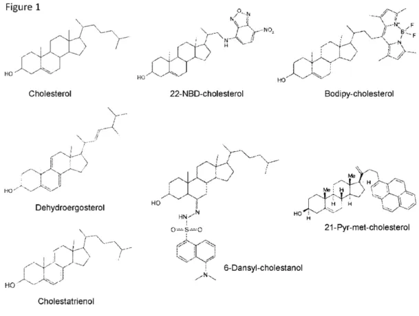
In this Section, we will compare the various fluorescent cholesterol-based probes that have been tested on both model and biological membranes, and which are classified as either intrinsically fluorescent sterols or fluorescent derivatives formed by a fluorophore-grafted cholesterol molecule (see the different chemical structures in Figure 1). The main informations are gathered in a synthetic form in Table 1.
4.1. DHE & CTE
Dehydroergosterol (DHE) and cholestatrienol (CTE) are two close derivatives of cholesterol, which are intrinsically fluorescent due their three conjugated double bonds in the sterol nucleus, CTE being even structurally closer to cholesterol because it bears the same side chain. In model membranes, they both display lipid condensing and ordering effects rather comparable to cholesterol, CTE being even better than DHE [114,115], and a similar extraction by cyclodextrin [116]. In GUV made of a ternary lipid mixture SM/DOPC/Chol, CTE showed a preferential partition in Lo phase [105]. They both display very similar behavior in cells; however, DHE has been much more often used for some practical reasons [31]. DHE can be delivered to cultured cells by means of a short incubation (1 min) with a cyclodextrin complex, allowing to visualize plasma membrane of CHO cells, hepatocytes, and J774 macrophages [117,118,119]. Subsequently, varying chase periods allowed to follow the internal DHE trafficking reaching the endocytic recycling compartment in non-polarized cells and the subapical recycling compartment in non-polarized HepG2 cells, suspected to be a sorting platform for intracellular sterols. In the CHO cells, DHE distribution was indeed found similar to that of [3H]cholesterol [117]. In addition, DHE could be delivered using DHE-labelled HDL as vectors to L-cells fibroblasts [120] and to polarized HepG2 cells, DHE cell uptake being mediated in that case by SR-BI [121]. In macrophages, DHE could be uptaken after labelling
modified LDL, targeting cytosolic lipid droplets [119]. Indeed, DHE has been found to be well esterified in L-cells, even more than cholesterol [120], although it has been otherwise shown to be a poor substrate for purified ACAT1 [122]. However, DHE displays a low quantum yield and suffers from a high photosensitivity, which renders difficult its use within living cells (especially for time-lapse experiments), and requires some instrumental sophistications, such as high quality detection [123] or multiphoton excitation [120]. Globally, DHE has been more efficiently used in living cells for studying intracellular sterol trafficking than for imaging membrane domains, even if it has been shown to display the same membrane compartment distribution than cholesterol.
All the other fluorescent cholesterol derivatives harbor an extrinsic, grafted fluorophore, whose chemical structure will obviously largely influence their physico-chemical as well as biological properties. As an extreme, illustrating case, fluoPEG-Chol bears a very large and hydrophilic reporter group [124], which renders it membrane impermeant, and makes it hardly incorporated in the cells from the exoplasmic leaflet of plasma membrane [31].
Figure 1. Chemical structures of the main fluorescent cholesterol derivatives.
4.2. NBD-Chol
Nitrobenzoxadiazole (NBD) group is an environment-sensitive fluorophore often used to label various lipids, including different phospholipids, and sensing the polar headgroups region of the membrane [125]. It has been grafted on the side chain of cholesterol, either at position 22 or 25. For
both of them, it has been shown that they preferentially partition in model membranes in the Ld phases, in supported planar bilayers [10], as well as in LUV [114,126], in GUV [105], and in giant plasma membrane vesicles (GPMV) [127]. Consistenly, they have been demonstrated to display a disordering effect on phospholipids in model membranes, which could be attributed, as a consequence of the polarity of the charged NBD moiety, to an energetically favored “looping back” conformation of the molecule placing NBD at the membrane interface, leading to an increased transbilayer mobility, and even the possibility of an up-side-down position [114]. It should thus be concluded that NBD-Chol is not an adequate cholesterol derivative that could mimic its intramembrane behavior. Actually, when incorporated into CHO cells, NBD-Chol showed an atypical accumulation in mitochondria, an organelle not known for its cholesterol abundance (but with an important membrane potential) [123]. Also, likely due to its amphiphilic character, NBD-Chol could be delivered easily and rapidly from the cell culture medium without any specific vehicle [120,128]. Finally, NBD-Chol is readily esterified in cells, comparably to cholesterol [120], and it has been widely used for efficiently labelling cytosolic lipid droplets/bodies, thanks to its fluorescence enhancement when surrounded by an hydophobic environment [128,129,130].
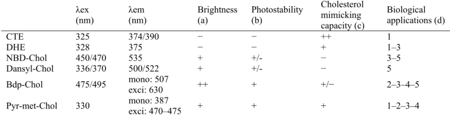
Table 1. Photochemical and biophysical properties of the main fluorescent cholesterol
derivatives, and their typical biological applications.
λex
(nm) λem (nm) Brightness (a) Photostability (b)
Cholesterol mimicking capacity (c) Biological applications (d) CTE 325 374/390 − − ++ 1 DHE 328 375 − − + 1–3 NBD-Chol 450/470 535 + +/- − 3–5 Dansyl-Chol 336/370 500/522 + +/- − 5
Bdp-Chol 475/495 mono: 507 exci: 630 ++ + +/− 2–3–4–5
Pyr-met-Chol 330 mono: 387 exci: 470–475 + + + 1–2–3–4
λex: main excitation wavelength; λem: main emission wavelength; mono: monomers; exci: excimers. Two wavelength values are given when the various bibliographic references provide significantly different values. (a): −, low brightness; +, good brightness, ++, very good brightness. (b): −, highly photosensitive; +/−, prone to photoinactivation; +, fairly photostable. (c): −, total discrepancy with membrane cholesterol behavior; +/−, partial agreement with membrane cholesterol behavior (depending on the lipid environment); +, fair agreement with membrane cholesterol behavior; ++, good agreement with membrane cholesterol behavior. (d): 1, ordered domains detection in model membranes; 2, ordered domains detection in biological membranes; 3, cell imaging by fluorescence microscopy; 4, cellular trafficking of ordered membrane analysis; 5, intracellular esterification analysis and lipid bodies staining.
4.3. Dansyl-Chol
Dansyl (dimethylamino naphtalene sulfonyl) fluorophore is known to be sensitive to the polarity of its environment, and it has a good quantum yield, but suffers from photobleaching. It has nevertheless being grafted onto the sterol nucleus, but on the C6 position saturating the double bound, and thus actually leading to a 6-dansyl-cholestanol [131]. Analysis of membrane penetration depth
has shown that the dansyl group resides in the middle of each lipid layer [132], and this is consistent with the observation of a FRET with DHE in plasma membranes [133]. However, the presence of the rather bulky fluorescent moiety, directly protruding from the sterol nucleus, may explain the observation of its preferential partition into the Ld phase in model membranes (GUV and GPMV) [127]. In addition, when tested on cultured cells, it happened to little localize in cholesterol-rich compartments [127]. However, it was reported in CHO cells (deficient in NPC1) to be esterified similarly to cholesterol [131]. Finally, regarding its bad membrane insertion compared to cholesterol, but nevertheless its good intracellular esterification, Dansyl-Chol appears rather reminiscent of NBD-Chol.
4.4. Bdp-Chol
Cholesterol labelled with the fluorophore group boron-dipyromethene (difluoro-dimethyl-bora-diaza-indacene, Bodipy), grafted on C24, has been reported in various experimental systems for about a decade since the first description of its synthesis (the initially so-called “compound 2”) [134]. Bodipy group is promising because of its high fluorescence quantum yield and its good photostability. In addition, it exhibits the property of excimer formation [135], even if to our knowledge this has not yet been considered for the various experimental protocols using Bdp-Chol. First, on supported planar bilayer membranes, Bdp-Chol incorporation into membrane domains has been correlated with the thickness of the membrane as measured by AFM [10]. It was observed to preferentially partition into the thickest lipid domains, presumably of liquid-ordered phase (Lo), in the presence of brain SM. However, this distribution appeared under the dependence of the nature (length and saturation) of the hydrophobic chains of SM constituting the bilayer membrane, with no preferential partition with SM(18:0), and even a preferential partition in the thinnest domains, presumably of Ld phase, with SM(16:0). In contrast, two derivatives, either made with an oxygenated linker between the sterol nucleus and the fluorophore moiety (Bdp-O-Chol), or from a saturated, hence not flat, sterol nucleus (Bdp-coprostanol), partitioned into the Ld phase, showing the rather specific behavior of Bdp-Chol [10]. Furthermore, desorption rate of Bdp-Chol from mixed monolayers containing SM was similar to that of cholesterol [136].
When tested, by fluorescence imaging, on GUV membranes made of lipid compositions allowing the co-existence of Lo and Ld phases (labelled by DiI-C12), Bdp-Chol was found to
somehow preferentially partition into the Lo phase, but with an enrichment factor of only about 2 [137]. This is in line with an equipartitioning observed in GUV, but a slight Lo preference in GPMV [127], and such partitioning experiments in GUV have shown a clearly lower selectivity for Lo domains when compared to DHE [138]. In addition, fluorescence anisotropy measurements and FRET analysis with DiI-C12 have indicated that Bdp-Chol is inserted in the hydrophobic core of the
membrane, and well oriented (long axis perpendicular to the phospholipid acyl chains). This was in agreement with further studies on GUV and GPMV, and at variance with another close derivative (Bdp-P-Chol) where the fluorophore moiety was differently grafted (via its pentaatomic cycle) onto the sterol nucleus, and found to be oriented parallel to the bilayer normal [139]. Finally, FCS, by evaluation of the translational diffusion coefficients, has indicated that Bdp-Chol forms small complexes with phospholipids in the membrane [137].
It may thus be concluded that Bdp-Chol is a fair fluorescent probe able to provide an interesting molecular tool for studying the structural organization of model membranes, and which hence deserves to be tested on living cell membranes.
When tested on cultured CHO cells, Bdp-Chol has shown essentially four main characteristics for its cell incorporation [140]. (i) It can be incorporated by incubating during some hours (“overnight”) the cells cultured in the absence of serum and using DMSO as a vehicle, as well as using complexes with MBCD for much shorter periods of few minutes. (ii) Its incorporated amounts were comparable with those of radiolabelled [3H]cholesterol in the various membrane fractions separated by sucrose density gradient centrifugation. (iii) It was preferentially found in DRM fractions (as assayed with cold TX100 treatment) in a similar manner than cholesterol, but at variance with the above-mentioned related derivative presenting an oxygenated linker (Bdp-O-Chol). (iv) Very short incubation periods (2 min), using MBCD-complexed Bdp-Chol, led to a prominent plasma membrane labelling, while after a subsequent chase period (for ~ 0.5–2 h), as well as after a much longer incubation (several hours) using DMSO as a vehicle, intracellular punctate structures, likely from the endosomal compartment, were labelled. We will present below (§4.6) the different reports of cell labelling patterns depending on the cell uptake pathway considered.
Globally, these data show that Bdp-Chol behavior in cell membranes is consistent with what was shown on model membranes, that is it somehow relates with that of membrane cholesterol, but it remains to determine the physiological relevancy of its cellular handling, and eventually control it for efficiently in-situ labelling ordered membrane (micro)domains in order to finally achieve a reliable approach for imaging such membrane domains in living cells.
4.5. Pyr-met-Chol
Pyrene fluorophore has been used long ago as a probe for sensing polarity of its environment (I1
peak) in membranes and micelles. In addition, it displays the remarkable capacity to form excimers (“excited dimers”) that give a specific emission peak at longer wavelengths than monomers (IE peak),
and this fluorescent property is useful for reporting the local probe distribution [141]. Pyrene group has been grafted onto the side chain of cholesterol, which led to a molecule with a hydrophobicity comparable to cholesterol, 21-methylpyrenyl-cholesterol [142]. First, calorimetry studies have shown that Pyr-met-Chol behaves like cholesterol for compared solubility in gel and fluid phases in model membranes. In addition, using LUV made of phospholipids with various acyl chains, it has been shown that Pyr-met-Chol prefered saturated chains in Lo phases (as indirectly evaluated by auto-association in the Ld phases). Comparison with Pyr-labelled phospholipids showed that the pyrene moiety does not significantly contribute to the stacking property of the probe. Qualitatively similar data were previously obtained with another Pyr-labelled cholesterol that presented an oxygenated linker (Pyr-O-Chol) [143], but this compound was not enough hydrophobic to be pertinent for further membrane labelling in cells. Finally, using the same model membranes, Pyr-met-Chol has been shown to be able to detect Lo phases according to both a decreased local polarity (due to lipids less permeable to water molecules in this phase), as sensed by the normalized peak I1/I3 (I3 being
proportional to total probe concentration), and an increased local probe concentration, as sensed by the normalized peak IE/I3 [142]. Although performed on model membranes made of phospholipids
membrane (micro)domains, both by preferential lateral distribution and specific environment sensing. As an indirect additional experimental evidence, Pyr-met-Chol, but not NBD-Chol, has been shown on model lipid monolayers to form stable lipid domains that required the presence of SM [144]. We were thus prompted to further test Pyr-met-Chol on living cells (see below).
As a first demonstration of feasibility, Pyr-met-Chol could be incorporated in cultured CHO cells by a short-period (2 min) incubation using a complex with MBCD [142]. Fluorescence imaging allowed to observe a prominent labelling of the plasma membrane, and simultaneous fluorescence emission spectrum could be recorded, showing somehow comparable peaks I1 and IE to those
measured in LUV. However, this comparison revealed a significantly higher value of I1/I3 for plasma
membrane cells, consistent with slightly higher water permeability in biological membranes than in model membranes (likely due to their marked chemical diversity, including membrane proteins). Indeed, when THP-1 macrophages were labelled by quite the same Pyr-met-Chol incubation (5 min), and then treated by the mycobacterium lipid, phthiocerol dimycocerosates (DIM, delivered by the bacteria in contact with the cells for 30 min), the I1/I3 parameter clearly decreased (to a value
comparable to LUV, on which the same effect could actually be directly measured) [145]. This observation was well consistent with a marked global rigidifying effect on the plasma membrane that mediates the deleterious effects of this pathogenic bacteria on these cells (i.e. receptor-mediated phagocytosis and phagosome acidification). Otherwise, the excimer peak IE/I3, when observed on
labelled CHO cells, showed a time-dependent decrease, likely due to continuous membrane turn-over by endocytosis/exocytosis [142]: this possibly precluded the observation of an effect on this peak of the treatment of THP-1 cells by DIM because of the 30 min of contact period with the bacteria (while it could be directly measured in-vitro on LUV) [145]. These data show that taking benefit of the powerful properties of pyrene excimers harbored by Pyr-met-Chol requires to well controlling its cellular incorporation. Indeed, using different cell delivery routes for Pyr-met-Chol, it has been shown that ordered membranes can be visualized thanks to the pyrene excimer fluorescence emission (see below) [128].
4.6. Respective differences and practical complementarity of Bdp-Chol and Pyr-met-Chol regarding their cell incorporation and handling
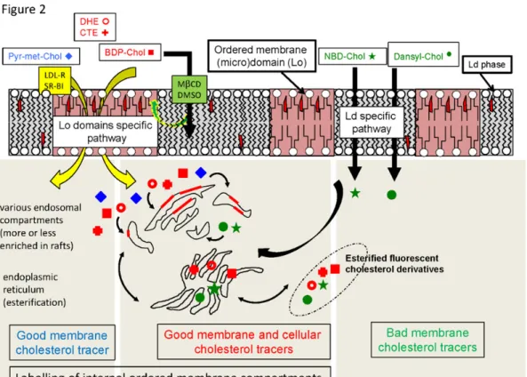
Cellular handling of a xenobiotic, such as a fluorescent probe, relies on the various aspects of its homeostasis, including influx, metabolization, and efflux. In the case of hydrophobic compounds, influx and efflux are strongly dependent on the presence of a donor and an acceptor, respectively. In the case of a sterol compound, it is generally intracellularly stored as an esterified derivative. For a very oversimplified, illustrating scheme, see Figure 2.
For Bdp-Chol, non-specific cell delivery could be obtained using either DMSO as a vehicle or MBCD complexes, with very different influx kinetics [140]. When using MBCD-mediated delivery, incorporation in BHK cells was observed to be very similar to that of DHE regarding its kinetics and its ATP- and clathrin-dependence, and led to labelling of the endocytic recycling compartment [138]. A specific HDL-mediated uptake has also been reported in HepG2 cells, and in that case, intracellular trafficking led to observe Bdp-Chol localized in some internal membrane compartments (tubular endosomes, multivesicular bodies, Golgi and trans-Golgi network) that are known to be relatively enriched in cholesterol [146]. Recently, Bdp-Chol has also been delivered to human
primary fibroblasts via the LDL route, showing a much more rapid (~ 2–4 h) lysosome labelling than from solvent-containing culture medium (~ 18–23 h) [147]. Cellular efflux of Bdp-Chol has been measured from CHO cells, and it was found more readily released than [3H]cholesterol, both in the absence and in the presence of an acceptor (either BSA or ApoAI) [140], possibly as a consequence of its slightly less hydrophobic character. Lastly, esterification capacity of Raw264.7 macrophages was evidenced for Bdp-Chol to be quite similar to [3H]cholesterol, at least in the basal metabolism state [140]. However, in HeLa cells, lipid droplets were more markedly targeted by Bdp-Chol than by DHE [138].
For Pyr-met-Chol, cellular incorporation has been evidenced both under non-specific incubation conditions, using ethanol as vehicle, and specific conditions, either using bile salt micelles to allow an uptake in Caco-2 enterocytes mediated by SR-BI, or taking benefit of the presence of lipoproteins in the cell culture medium [128]. Indeed, it has been previously demonstrated that Pyr-met-Chol could be specifically and stably associated with purified lipoproteins, HDL and LDL, before using these vectors to deliver the probe to target cells harboring the corresponding lipoprotein receptors [148]. The point is that these various cell uptake pathways led to different cellular labelling patterns, with different cell incorporation kinetics, consistently with the two distinct pathways described for HDL, i.e. via SR-BI that is a raft/caveolae-dependent receptor, and LDL, i.e. receptor-mediated endocytosis via clathrin-coated pits [22,149]. However, HDL- and LDL-receptor-mediated cell incorporation finally gave comparable localizations after long incubation periods (72 h) for PC-3 cells. In all cases, cell labelling was only very faint at the level of plasma membrane, and distributed between a diffuse cytoplasmic staining (presumably ER) and intracellular punctate structures, more or less intensely stained. Colocalization studies with various internal compartment protein markers indicated that Pyr-met-Chol preferentially labelled membranes known to be physiologically enriched in cholesterol, essentially lysosomes (Lamp1), caveolae (Cav-1) and prostasome-precursor vesicles (CD63) [128]. In addition, taking benefit of the pyrene fluorophore property to possibly form excimers, emitting at longer wavelengths than the monomers, simultaneous fluorescence cell imaging in two channels corresponding to monomer and excimer emissions allowed to observe that most of (but not all) the punctate structures intensely stained by monomers were emitting in the excimer channel, while this was not the case for the diffuse cytoplasmic staining. This indicated that at least some of the internal membranes labelled by the monomers displayed an ordered structure, consistent with an enrichment in cholesterol, which could be revealed by the excimer formation reliably tracing a high local probe concentration (hence with an improved imaging contrast), while monomer imaging can be observed, within the optical microscope resolution, in the various non-specifically labelled membrane compartments (Figure 3). Furthermore, at the level of cell periphery, some membrane formations attached to the cultured cells, so-called “pericellular structures”, could also be observed, and they presented a clear excimer staining. This was reminiscent with the fact that PC-3 cells, a prostate cancer cell line, has the physiological property to secrete prostasomes, which are small membrane vesicles characterized by their high abundance in cholesterol. Indeed, a membrane fraction collected from the culture medium 96 h after Pyr-met-Chol labelling of the cells showed a clear incorporation of the fluorescent probe, with a significant fraction of excimers. This showed in addition that Pyr-met-Chol was not sequestered in the cultured cells [128]. Regarding its intracellular metabolization, Pyr-met-Chol did not appear to be significantly esterified, as evaluated by thin layer chromatography of cell extracts, as well as by the fact that it did not colocalize with the markers of cytosolic lipid bodies, Bodipy493 and NBD-Chol [128].
Figure 2. Oversimplified schematic presentation of the respective fates of the various
fluorescent cholesterol derivatives considered for their membrane and cellular behaviors, as deduced from the reviewed literature. A good membrane cholesterol tracer is able to fairly mimic cholesterol behavior for membrane insertion, preferring the ordered raft-type (micro)domains. A good cell cholesterol tracer is able to fairly mimic cholesterol cellular trafficking, including various membrane transporters handling and intracellular non-vesicular transport, as well as esterification in the endoplasmic reticulum (ER). Cellular handling of Pyr-met-Chol, BDP-Chol, DHE and CTE results either from a passive diffusion through Lo domains (small green and yellow arrow) or a specific uptake pathway via specific receptors (yellow arrows). In both cases, this leads for these probes to a good insertion in internal ordered membrane compartments. DHE, CTE and BDP-Chol (red symbols) are esterified, and will be considered as good cellular cholesterol tracers since they report well enough the intracellular cholesterol trafficking. NBD-Chol and Dansyl-Chol (green symbols) are also esterified, but they preferentially partition into Ld membranes, resulting in a non-physiological cell trafficking and hence are to be considered as bad cellular and membrane cholesterol tracers. Alternatively, even if Pyr-met-Chol (blue symbols) is not esterified, it can be considered as a good membrane cholesterol tracer. Small red trapezoids within phospholipids depict cholesterol molecules; red rectangular plaques in the endosomal compartments depict raft-type (micro)domains.
As a whole, the comparison of these two mostly relevant fluorescent derivatives of cholesterol (and in connection with the other ones) aimed at detecting ordered membrane microdomains in cells shows some important points: (i) the various possible cell uptake pathways, either non-specific (i.e. with a solvent as a vehicle) or receptor-specific (i.e. using a molecular vector, such as bile salt micelles or lipoproteins), lead to clearly different cell staining patterns; (ii) the cellular incorporation kinetics may vary within very different relevant time ranges (labelling time periods are reported from 2 min to 96 h); (iii) the nature of the considered target cell, which may present very different physiological fates regarding influx and efflux routes, trafficking and metabolization, has a great influence on the observed membrane labelling.
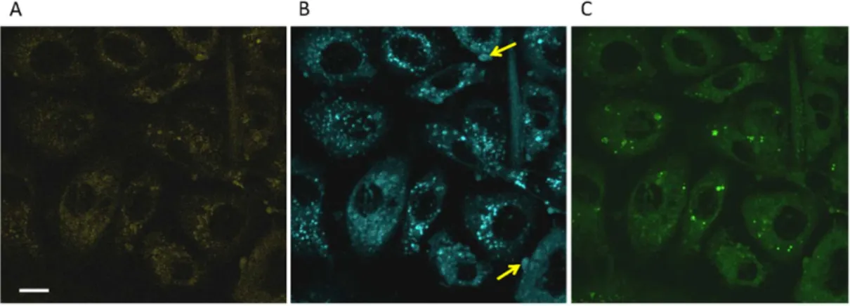
Figure 3. Multichannel fluorescent staining of PC-3 cells using Pyr-met-Chol and
NBD-Chol. Two-photon excitation microscopy imaging of fixed PC-3 cells was performed after a 48 h incubation of 5 µM Pyr-met-Chol and 5 µM NBD-Chol in the presence of 10% fetal calf serum. Pyr-met-Chol was excited at 710 nm, NBD-Chol was excited at 940 nm. Fluorescence emission was simultaneously observed for pyrene monomers (370–409 nm band-pass filter) (Panel A), pyrene excimers (443–507 nm band-pass filter) (Panel B), and NBD (500–550 nm band-pass filter) (Panel C). Scale bar corresponds to 10 µm. Yellow arrows point some of the visible pericellular structures. Comparison of Panels A and B shows that only a (major) fraction of the intracellular punctate structures highly stained by Pyr-met-Chol monomers is well-stained by Pyr-met-Chol excimers. NBD-Chol labelling is shown for the sake of comparison, to show that it gives a clearly different cell staining.
As regards metabolization, even if we only consider fluorescent probes that are biochemically stable, i.e. with no risk of cleavage or alteration of the fluorophore moiety, the sterol moiety is always potentially subject to esterification, which renders it even more hydrophobic by adding an acyl chain in place of the hydroxyl group, and hence leads to an altered intracellular distribution, essentially being sequestered in the cytosolic lipid bodies [150]. Indeed, intracellular esterification is the major difference between Bdp-Chol and Pyr-met-Chol, and this could be relied on the rather high staining of ER by Pyr-met-Chol (as compared to its known low cholesterol content), indicating that it is well accessible to, but not handled by ACAT. Finally, we would like to stress that although