S Y M P O S I U M : A B J S C A R L T . B R I G H T O N W O R K S H O P O N H I P P R E S E R V A T I O N S U R G E R Y
Hip Capsule Dimensions in Patients With Femoroacetabular
Impingement: A Pilot Study
Jan Weidner MD, Lorenz Bu¨chler MD, Martin Beck MD
Published online: 19 July 2012
Ó The Association of Bone and Joint Surgeons1 2012
Abstract
Background Joint-preserving hip surgery, either arthro-scopic or open, increasingly is used for the treatment of symptomatic femoroacetabular impingement (FAI). As a consequence of surgery, thickening of the joint capsule and intraarticular adhesions between the labrum and joint capsule and between the femoral neck and the joint capsule have been observed. These alterations are believed to cause persistent pain and reduced range of motion. Because the diagnosis is made with MR arthrography, knowledge of the normal capsular anatomy and thickness on MRI in patients is important. To date there is no such information available. Questions/Purposes The purpose of this study was to establish thickness, length of the hip capsule, and the size of the perilabral recess in patients with FAI.
Methods We reviewed the preoperative MR arthrography of 30 patients (15 men) with clinical symptoms of FAI. We measured capsular thickness and made observations on the perilabral recess.
Results The joint capsule was thickest (6 mm) anterosu-periorly between 1 and 2 o’clock. The average length from the femoral head-neck junction to the femoral insertion of the capsule ranged from 19 to 33 mm. A perilabral recess was present circumferentially, even across the acetabular notch, where the labrum is supported by the transverse acetabular ligament. The shortest recess occurred superiorly. Conclusions Knowledge of the capsular anatomy in patients with FAI before surgery is important to judge the postoper-ative changes and to plan potential further therapy including arthroscopic treatment of intraarticular adhesions.
Introduction
Femoroacetabular impingement (FAI) is an accepted cause for osteoarthritis of the hip [1, 2, 9]. Both types of impingement, the cam deformity on the femoral side and the pincer impingement on the acetabular side, lead to degeneration of the labrum and cartilage damage [2]. Besides the open approach through surgical dislocation of the hip [3,8,9,13, 14], hip arthroscopy has become the most frequent technique for the treatment of FAI [1,10–12, 16–19,22].
Postoperative soft tissue changes after open or arthro-scopic treatment are reportedly responsible for continuing pain [1] and the arthroscopic resection of these changes leads to reduced pain and increased hip function [1,4,12, 16]. Changes most commonly seen are adhesions between the labrum and joint capsule obliterating the perilabral recess [1] and adhesions between the joint capsule and the
Each author certifies that he or she, or a member of their immediate family, has no commercial associations (eg, consultancies, stock ownership, equity interest, patent/licensing arrangements, etc) that might pose a conflict of interest in connection with the submitted article.
All ICMJE Conflict of Interest Forms for authors and Clinical Orthopaedics and Related Research editors and board members are on file with the publication and can be viewed on request. Each author certifies that his or her institution has approved the human protocol for this investigation and that all investigations were conducted in conformity with ethical principles of research. This work was performed at the University of Berne, Berne, Switzerland, and Kantonsspital Luzern, Lucerne, Switzerland. J. Weidner (&), L. Bu¨chler
Department for Orthopaedic Surgery, Inselspital, University of Berne, 3010 Berne, Switzerland e-mail: [email protected]; [email protected] M. Beck
Department of Orthopaedics, Canton Hospital Lucerne, Lucerne, Switzerland
Clin Orthop Relat Res (2012) 470:3306–3312
DOI 10.1007/s11999-012-2485-2
and Related Research
®bony femoral neck that may limit motion [5] and thick-ening of the joint capsule [12]. Arthroscopic removal of the adhesions leads to a reduction of pain [1]. One previous study [12] reported an average increase of the Merle d’Aubigne´-Postel score from 13 to 16 points with a reduction of pain in 80% of patients. However, it is difficult to judge the impact of intraarticular scar formation and capsule thickening after previous hip surgery for FAI without quantitatively knowing the normal anatomy. Such data are important to understand postoperative changes and how they affect the patient.
In patients with FAI we determined: (1) the thickness of the joint capsule; (2) the distance from the head-neck junction to the capsular insertion on the femur; (3) the depth of the perilabral recess; and (4) whether these measures differed in men and women.
Patients and Methods
From a CD collection of approximately 350 arthro-MR images of hips of patients treated for FAI at our institution between 2005 and 2009, we manually selected 15 men and 15 women. Inclusion criteria were: (1) clinical symptoms of FAI; (2) radiologic findings typical for FAI; and (3) no previous hip surgery. All patients were treated by hip arthroscopy or surgical dislocation of the hip. The mean age of all patients was 35 years (age range, 19–52 years); there were 14 left and 16 right hips. The mean age of the men was 34 years (range, 23–52 years); there were seven left and eight right hips. Mean age of the women was 36 years (range, 19–49 years); there were also seven left and eight right hips in the female group. Of the 30 patients included in the study, 19 had a coxa profunda and one showed acetabular protrusion. Two patients had a pure pincer impingement and nine patients a cam impingement; the majority showed a mixed type of impingement (n = 19). Regarding the acetabular version, we found anteversion in 11 cases and cranial retroversion in 19 cases. The lateral center-edge angle ranged from 18° to 53° (mean, 35.9°). We measured all other parameters on radial MR slices using our standard MR arthrography technique [20]. In patients with suspected FAI, MR arthrography is routinely performed in addition to conventional radio-graphs including an AP and a crosstable view in internal rotation.
We obtained all MR arthrograms on a 1.5-T system (Siemens, Erlangen, Germany) at Klinik Sonnenhof, Berne, Switzerland. Ten to 20 mL gadolinium DTPA (Dotarem 1:200; Guebert AG, Paris, France) was injected into the hip under fluoroscopic guidance before the MR examination. The varying amount depends on the volume of the hip and
the resistance felt by the radiographer during injection. In a large hip, more gadolinium can be injected until the resistance rises. A standard examination includes axial and sagittal T1- and T2-weighted sequences plus a radial pro-ton density-weighted sequence [4]. The radial sequence consists of 14 slices arranged clockwise around the femoral neck. The 12 o’clock position is strictly superior, 6 o’clock inferior, 3 o’clock is anterior, and 9 o’clock posterior.
On the radial MR images around the axis of the femoral neck, we measured the thickness of the hip capsule, the distance of its insertion at the femoral neck as a marker for the capsular length, and the depth of the capsular recess. Length of the joint capsule was measured from the head-neck junction to the point of capsular insertion on the femoral neck. This is to have reproducible and easy-to-identify reference points when using fluoroscopy during the hip arthroscopy. For determining the depth of the per-ilabral recess, we decided to define its depth relative to the adjacent labrum rather than giving total values, believing that relative values are more helpful for understanding the morphology of the perilabral recess than absolute values. The MR sequences are those routinely used for diagnosis and preoperative planning in patients with FAI. The advantage of radial sequences is that every slice of the radial sequence is perpendicular to the joint capsule and labrum. Oblique slicing of the capsule is avoided and the capsular thickness can precisely be measured. The intra-articular contrast fluid facilitates identification of the presence and size of the capsular recess and the insertion of the joint capsule to the femoral neck and identification of intraarticular adhesions.
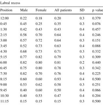
For all patients, one observer (JW) performed the fol-lowing measurements based on the radial MR slices and assigned positions according to the clockwise orientation of the slices (Fig.1): (1) joint capsule thickness: thickness of the joint capsule was measured at the thickest part of the capsule; (2) joint capsule length: we measured the distance from the head-neck junction to the capsular insertion at the femoral neck as an indirect indicator of the joint capsule length; and (3) depth of the perilabral recess: if present, the depth of the recess between the joint capsule and the adjacent labrum was determined and classified in 25% steps relative to the size of the labrum. On the inferior part of the acetabulum, approximately between 4 and 7 o’clock in a right hip, the labrum runs across the acetabular notch supported by the transverse ligament [15, 20]. The com-bined structure of the labrum and transverse ligament is easily identified on the MRI (Fig. 2) and the perilabral recess can be measured like at any other point around the acetabulum. Data of left hips were converted to right hips. We separately analyzed our results for thickness and length of the joint capsule and depth of the perilabral recess for
male and female patients and compared them using a univariate t-test. All calculations and statistics were done using Microsoft Excel (Microsoft Inc, Redmond, WA, USA).
Results
The average capsule thickness at the different locations varied from 2 mm to 6 mm (Table1). The maximum thickness of 6 mm occurred between 1 and 2 o’clock corresponding to the anterosuperior part of the femoral neck. Between 3 and 6 o’clock, the thickness continuously decreased from 5 to 3 mm. The thinnest part of the capsule with approximately 2 mm thickness was found between 8 and 9 o’clock. Between 9 and 12 o’clock, capsule thickness increased to 4 mm (Fig.3).
The length of the joint capsule ranged from 19 to 33 mm (Table 2). The greatest distance was on the posterosuperior side between 9 and 11 o’clock. Between 3 and 6 o’clock, the distance was shortest and measured between 21 and 18 mm (Fig. 4). Anteriorly, the capsule inserts at the intertrochanteric line, a prominent landmark. Inferiorly and superiorly the capsule has a less well-defined insertion to the bone, but the insertion was well visible for the mea-surements. Posteriorly and posterosuperiorly, the insertion of the capsule was difficult to define because of the pres-ence of the zona orbicularis, which can be quite prominent in this area. Therefore, an overestimation of the length of the capsule in the posterior area is possible.
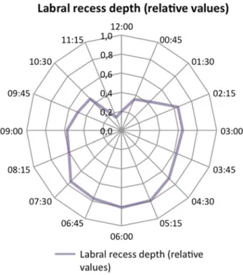
A perilabral recess was present circumferentially. The depth of the capsular recess relative to the labrum varied from 15% to 93% (Table3). The shortest recess occurred superiorly, where it ranged between 20% and 40% of the
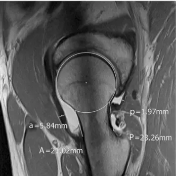
Fig. 1 This figure shows the principle measurements that were obtained on a radial slice along the femoral neck: anterior capsule thickness on this slice is a = 5.84 mm, posterior thickness p = 1.97 mm. Distance of the anterior capsular insertion from the femoral head-neck junction is A = 21.02 mm; the posterior distance is P = 23.26 mm.
Fig. 2 MR arthrography through the 12 and 6 o‘clock positions. (1) Perilabral recess superiorly, the joint capsule is attached to the labrum. (2) Zona orbicularis. (3) Femoral insertion of the joint capsule at the superior base of the neck. (4) Inferior perilabral recess. (5) Labrum, supported by the transverse acetabular ligament. (6) Inferior femoral insertion of joint capsule. (7) Teardrop.
Table 1. Average values for capsular thickness in millimeters Position Male Female All patients SD p value 12:00 4.4 4.1 4.2 1.2 0.245 0:45 4.9 4.5 4.7 1.2 0.212 1:30 6.5 6.0 6.2 1.6 0.185 2:15 6.8 5.5 6.2 1.8 0.012 3:00 5.6 4.3 4.9 1.5 0.002 3:45 4.4 2.9 3.7 1.7 0.005 4:30 3.2 2.3 2.8 1.3 0.028 5:15 3.0 2.4 2.7 1.1 0.081 6:00 2.8 2.3 2.6 1.0 0.067 6:45 3.1 2.8 3.0 0.8 0.118 7:30 2.7 2.2 2.4 0.8 0.092 8:15 1.8 1.9 1.8 0.7 0.340 9:00 1.9 2.2 2.0 0.7 0.222 9:45 2.1 2.4 2.3 1.1 0.246 10:30 2.9 3.3 3.1 1.3 0.225 11:15 4.1 3.7 3.9 1.1 0.202 Results are listed for male and female patients separately and for all patients together. Mean values and SD are given. There was a sta-tistically significant difference between male and female patients; p value of the univariate t-test is \ 0.05.
size of the labrum. Between 12 and 3 o’clock, the depth increased to an average of 64%. On the inferior part of the femoral neck between 3 and 9 o’clock, the depth of the capsular recess ranged from 63% to 93%. On the superior part between 9 and 3 o’clock, depth was between 15% and 64% relative to the labrum (Fig.5).
The comparison of thickness of the joint capsule in men and women showed a thicker capsule for men in all posi-tions except at the posterior capsule. Capsular thickness
Fig. 3 This diagram shows the average capsular thickness along the circumference of the femoral neck.
Table 2. Hip capsule length (mm)
Position Male Female All patients SD p value 12:00 27.7 25.1 26.4 4.8 0.008 12:45 27.1 24.5 25.8 4.7 0.003 1:30 26.9 23.1 25.0 5.0 0.004 2:15 25.3 21.6 23.5 5.4 0.021 3:00 22.5 20.5 21.5 4.8 0.031 3:45 22.0 19.6 20.8 5.8 0.019 4:30 20.6 18.3 19.4 5.0 0.006 5:15 22.1 15.6 18.9 5.9 0.000 6:00 23.2 18.9 21.1 4.9 0.000 6:45 26.9 22.5 24.7 5.6 0.003 7:30 31.6 24.4 28.0 6.7 0.001 8:15 31.2 25.5 28.4 7.2 0.001 9:00 33.4 28.3 30.8 7.2 0.000 9:45 35.6 31.0 33.3 8.7 0.006 10:30 31.7 32.6 32.2 4.6 0.035 11:15 29.2 28.3 28.7 3.2 0.001
Fig. 4 On this diagram, the average length of the joint capsule is shown. The length was measured from the head-neck junction to the distal insertion at the femoral neck.
Table 3. Depth of the capsular recess relative to the adjacent labrum Labral recess
Position Male Female All patients SD p value 12:00 0.22 0.18 0.20 0.3 0.379 0:45 0.45 0.25 0.35 0.3 0.076 1:30 0.42 0.43 0.43 0.4 0.457 2:15 0.58 0.70 0.64 0.4 0.246 3:00 0.57 0.72 0.64 0.3 0.151 3:45 0.52 0.73 0.63 0.4 0.088 4:30 0.68 0.73 0.71 0.3 0.332 5:15 0.77 0.82 0.79 0.3 0.309 6:00 0.82 0.80 0.81 0.2 0.440 6:45 0.75 0.80 0.78 0.3 0.342 7:30 0.82 0.70 0.76 0.4 0.225 8:15 0.60 0.60 0.93 0.4 0.500 9:00 0.53 0.62 0.58 0.3 0.251 9:45 0.40 0.60 0.50 0.4 0.066 10:30 0.40 0.53 0.47 0.4 0.204 11:15 0.15 0.15 0.15 0.3 0.500 Mean values and SD for depth of the capsular recess in relation to the labrum are given. The difference between male and female hips does not show a statistically significant difference (p = 0.07).
between male and female hips differed only for the anterior capsule. Overall, men had a longer joint capsule at any point around the hip owing to the larger size of the hip in men. In general, there was no difference between male and female hips, except anteriorly between 4:30 and 5:15 and posteriorly between 7:30 and 10:30.
Discussion
The intraarticular abnormalities in patients with FAI can be treated with hip arthroscopy or surgical hip dislocation [6–9]. However, some patients report ongoing pain or show a limitation of ROM after impingement surgery. Previous studies have found postoperative changes like thickening of the capsule or the formation of adhesions between the capsule and the femur or between capsule and labrum as a possible cause for pain after hip surgery [1,5,12]. These scars can be removed with hip arthroscopy, which leads to a reduction of pain and increases function of the hip [1,12]. MR arthrography with radial slices along the femoral neck is the standard imaging technique for detection of these postoperative changes [5, 21]. When evaluating a MR study of a patient with continuing pain or limited ROM after hip surgery, the surgeon usually looks for intraartic-ular adhesions or capsintraartic-ular thickening. To correctly detect these MRI findings, it is important to know the normal anatomy of the hip joint including the dimensions of the
hip capsule in patients with FAI before surgery. Currently, this information is not available. With our study, we present answers to the following questions: (1) What is the average thickness of the hip capsule in patients with FAI before surgery? (2) What is the average thickness of the joint capsule in these patients? (3) What is the average depth of the perilabral recess about the entire circumfer-ence of the labrum and adjacent to the transverse ligament as well? (4) Is there a relevant difference between men and women regarding these measurements?
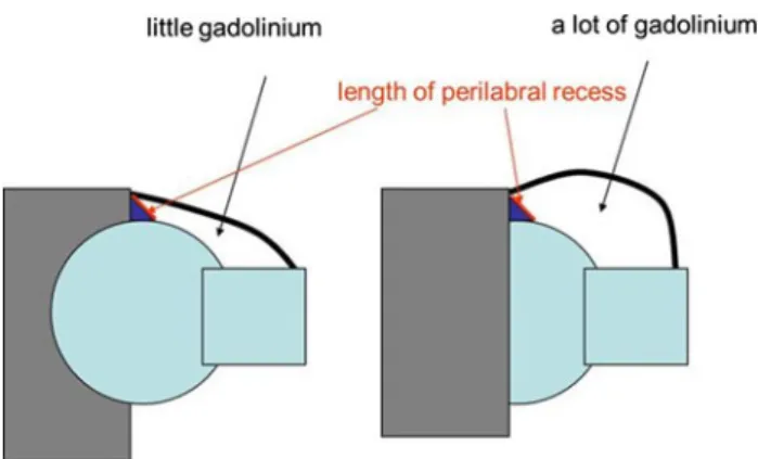
There are some limitations to this study. First, all patients included in this study had a symptomatic FAI. Although we provide a description of the hip capsule morphology in patients with FAI, we cannot say if these values represent the normal capsular anatomy of healthy, asymptomatic patients. Second, all findings are based on MR images on which we were able to measure different values describing the hip capsule morphology. We did not confirm the accuracy of these measures against anatomic specimens. Third, although we determined the length and thickness of the joint capsule and the size of the perilabral recess, our measurements provide no information about how loose or firm the capsule is attached to the joint. Furthermore, rotation of the leg and the degree of hip flexion during the MR examination may have an influence on how close the capsule appears to the joint. Fourth, there are considerable differences among patients regarding the stiffness or looseness of the joint capsule, which we cannot simply measure on MR images. Fifth, although visualiza-tion of the capsular recess is improved by sufficient injection of contrast into the joint, the capsule may be stretched. This may lead to an underestimation of capsular thickness. On the other hand, good filling improved the visualization of the recess on MR images in all cases. The amount of joint distension does not change the length of the perilabral recess, because the capsule is firmly attached to the bone and cannot move (Fig. 6). Sixth, unfortunately operative findings relating to the joint capsule and recess were not taken during surgery. Therefore, we cannot ana-lyze whether the injury pattern correlated with capsular anatomy.
As expected, we found the thickest part of the hip capsule on the anterosuperior portion of the femoral neck along the iliofemoral ligament. This is the area in which the cam deformity usually is most prominent. It is also thicker in male hips, which may be related to the larger size of the hip, but could also reflect a reaction to the underlying cam deformity, which is more common in male hips. The design of the study unfortunately does not allow us to answer this question. It may be the task of another study involving a large number of hips. A very thick joint capsule between 12 and 3 o’clock in patients with a large cam deformity can limit the space between the femur and the joint capsule;
Fig. 5 This diagram shows the depth of the perilabral recess relative to the adjacent labrum.
thus, depending on the technique, it can be more difficult to access and remove the femoral bump.
The length of the capsule, measured from the head-neck junction to the insertion, was between 18 and 33 mm. The shortest distance was anteriorly and the longest posteriorly in the trochanteric fossa. However, the presence of a strong zona orbicularis in this area made the measurement diffi-cult and may have led to an overestimation of the length. To have comparable results, the most peripheral accumu-lation of gadolinium was taken as a reference point. Anteriorly the capsule inserts at the intertrochanteric line and superiorly it attaches at the base of the greater tro-chanter. These are the areas where adhesions and scar formation most likely occur [12].
The relative depth of the perilabral recess is greatest inferiorly between 3 and 9 o’clock. On the superior aspect between 10 and 2 o’clock, the perilabral recess is much smaller (Fig.5). This is the result of the fact that the capsule attaches in this area to the labrum and also because the labrum is larger in this area and the relative length gets shorter. The difference between male and female hips varied around the acetabulum although these were not significant. Inferiorly in the area of the transverse acetab-ular ligament (TAL), which acts as a support for the labrum that runs uninterrupted around the acetabulum [15,20], we identified a recess. Because the TAL/labrum complex is small compared with the superior labrum, the relative length of the recess exceeds the one of the recess of the superior acetabulum (Fig.2).
We found men had a thicker capsule than women anterosuperiorly and about the capsule circumferentially, likely a consequence of men being generally larger than women. The increased thickness may also result from differences in the shape of the hip in men and women with FAI. In women, pincer FAI with a narrow femoral neck is more common than in men [4]. Men more often present with a cam FAI, in which the maximum deformity
generally is between the 12 and 3 o’clock positions and the cam deformity might trigger joint capsule thickening. We found no difference for depth of the perilabral recess.
With the baseline values given in this study, it is pos-sible to better classify scar formation in the hip and thickening of the capsule after surgery. The hip capsule dimensions of patients with FAI before surgery can be compared with the findings in MR studies of patients with ongoing pain or limited hip function after previous hip surgery. Knowledge of the capsular anatomy in patients with FAI before surgery makes it easier to judge the postoperative changes and plan potential further therapy including arthroscopic removal of intraarticular adhesions.
References
1. Beck M. Groin pain after open FAI surgery: the role of intraar-ticular adhesions. Clin Orthop Relat Res. 2009;467:769–774. 2. Beck M, Kalhor M, Leunig M, Ganz R. Hip morphology
influ-ences the pattern of damage to the acetabular cartilage: femoroacetabular impingement as a cause of early osteoarthritis of the hip. J Bone Joint Surg Br. 2005;87:1012–1018.
3. Beck M, Leunig M, Parvizi J, Boutier V, Wyss D, Ganz R. Anterior femoroacetabular impingement: part II. Midterm results of surgical treatment. Clin Orthop Relat Res. 2004;418:67–73. 4. Dudda M, Albers C, Mamisch TC, Werlen S, Beck M. Do normal
radiographs exclude asphericity of the femoral head-neck junc-tion? Clin Orthop Relat Res. 2009;467:651–659.
5. Dudda M, Mamisch TC, Krueger A, Werlen S, Siebenrock KA, Beck M. Hip arthroscopy after surgical hip dislocation: is pre-dictive imaging possible? Arthroscopy. 2011;27:486–492. 6. Espinosa N, Beck M, Rothenfluh DA, Ganz R, Leunig M.
Treatment of femoro-acetabular impingement: preliminary results of labral refixation. Surgical technique. J Bone Joint Surg Am. 2007;89(Suppl 2):36–53.
7. Espinosa N, Rothenfluh DA, Beck M, Ganz R, Leunig M. Treatment of femoro-acetabular impingement: preliminary results of labral refixation. J Bone Joint Surg Am. 2006;88:925–935. 8. Ganz R, Gill TJ, Gautier E, Ganz K, Kru¨gel N, Berlemann U.
Surgical dislocation of the adult hip. A technique with full access to the femoral head and acetabulum without the risk of avascular necrosis. J Bone Joint Surg Br. 2001;83:1119–1124.
9. Ganz R, Parvizi J, Beck M, Leunig M, No¨tzli H, Siebenrock KA. Femoroacetabular impingement: a cause for osteoarthritis of the hip. Clin Orthop Relat Res. 2003;417:112–120.
10. Haupt U, Vo¨lkle D, Waldherr C, Beck M. Intra- and retroperi-toneal irrigation liquid after arthroscopy of the hip joint. Arthroscopy. 2008;24:966–968.
11. Kelly BT, Williams RJ 3rd, Philippon MJ. Hip arthroscopy: current indications, treatment options, and management issues. Am J Sports Med. 2003;31:1020–1037.
12. Krueger A, Leunig M, Siebenrock KA, Beck M. Hip arthroscopy after previous surgical hip dislocation for femoroacetabular impingement. Arthroscopy. 2007;23:1285–1289.
13. Lavigne M, Parvizi J, Beck M, Siebenrock KA, Ganz R, Leunig M. Anterior femoroacetabular impingement: part I. Techniques of joint preserving surgery. Clin Orthop Relat Res. 2004;418:61–66. 14. Leunig M, Beck M, Dora C, Ganz R. [Femoroacetabular impingement: trigger for the development of coxarthrosis] [in German]. Orthopade. 2006;35:77–84.
Fig. 6 Schematic showing the influence of joint distention. The fixed origin and insertion of the capsule remain unchanged independent of the amount of applied intraarticular fluid. The length of the perilabral recess remains unchanged.
15. Petersen W, Petersen F, Tillmann B. Structure and vasculariza-tion of the acetabular labrum with regard to the pathogenesis and healing of labral lesions. Arch Orthop Trauma Surg. 2003;123: 283–288.
16. Philippon MJ. New frontiers in hip arthroscopy: the role of arthroscopic hip labral repair and capsulorrhaphy in the treatment of hip disorders. Instr Course Lect. 2006;55:309–316.
17. Philippon MJ, Briggs KK, Yen YM, Kuppersmith DA. Outcomes following hip arthroscopy for femoroacetabular impingement with associated chondrolabral dysfunction: minimum two-year follow-up. J Bone Joint Surg Br. 2009;91:16–23.
18. Philippon MJ, Weiss DR, Kuppersmith DA, Briggs KK, Hay CJ. Arthroscopic labral repair and treatment of femoroacetabular
impingement in professional hockey players. Am J Sports Med. 2010;38:99–104.
19. Philippon MJ, Yen YM, Briggs KK, Kuppersmith DA, Maxwell RB. Early outcomes after hip arthroscopy for femoroacetabular impingement in the athletic adolescent patient: a preliminary report. J Pediatr Orthop. 2008;28:705–710.
20. Putz R, Schrank C. [Anatomy of the labro-capsular complex] [in German]. Orthopade. 1998;27:675–680.
21. Werlen S, Leunig M, Ganz R. Magnetic resonance arthrography of the hip in femoroacetabular impingement: technique and findings. Oper Tech Orthop. 2005;15:191–203.
22. Wettstein M, Dienst M. [Hip arthroscopy for femoroacetabular impingement] [in German]. Orthopade. 2006;35:85–93.