HAL Id: hal-02563078
https://hal-normandie-univ.archives-ouvertes.fr/hal-02563078
Submitted on 17 Nov 2020HAL is a multi-disciplinary open access archive for the deposit and dissemination of sci-entific research documents, whether they are pub-lished or not. The documents may come from teaching and research institutions in France or abroad, or from public or private research centers.
L’archive ouverte pluridisciplinaire HAL, est destinée au dépôt et à la diffusion de documents scientifiques de niveau recherche, publiés ou non, émanant des établissements d’enseignement et de recherche français ou étrangers, des laboratoires publics ou privés.
Non-auxetic/auxetic transitions inducing modifications
of the magnetic anisotropy in CoFe2O4 thin films
E. Martin, F. Roulland, Stéphane Grenier, F. Appert, J. Juraszek, M. Trassin,
C. Bouillet, E. Chikoidze, C. Arnold, B. Berini, et al.
To cite this version:
E. Martin, F. Roulland, Stéphane Grenier, F. Appert, J. Juraszek, et al.. Non-auxetic/auxetic transi-tions inducing modificatransi-tions of the magnetic anisotropy in CoFe2O4 thin films. Journal of Alloys and Compounds, Elsevier, 2020, 836, pp.155425. �10.1016/j.jallcom.2020.155425�. �hal-02563078�
Non-auxetic/auxetic transitions inducing modifications of the magnetic
anisotropy in CoFe
2O
4thin films
E. Martin a, F. Roulland a, S. Grenier b, F. Appert c, J. Juraszek c, M. Trassin d, C. Bouillet a, E.
Chikoidze e, C. Arnold e, B. Berini e, Y. Dumont e, S. Colis a, S. Barre a, G. Versini a, D. Preziosi a,
C. Leuvrey a, N. Blanc b, N. Boudet b, G. Pourroy a, N. Viart a, C. Lefèvre a
a. Institut de Physique et Chimie des Matériaux de Strasbourg, UMR Unistra-CNRS 7504, 23 rue du Lœss, 67034 Strasbourg Cedex 2, France
b. Institut Neel, UPR 2940, F-38042 Grenoble, France
c. Normandie Univ, UNIROUEN, INSA Rouen, CNRS, GPM, 76000 Rouen, France d. Department of Materials, ETH Zurich, Vladimir-Prelog-Weg 4, 8093 Zurich, Switzerland e. Groupe d’Etude de la Matière Condensée (GEMaC), UMR 8635, Versailles, France
ABSTRACT
An auxetic behaviour is evidenced in CoFe2O4 thin films grown by pulsed laser deposition on (100)
MgO substrates under various O2/N2 pressures. This rare behaviour for an intrinsic material is
observed for intermediate oxidation conditions, in between two non auxetic domains delimited by high (>0.05 mbar) and low (<0.03 mbar) O2/N2 deposition pressures. Combining X-ray resonant
diffraction and Mössbauer spectroscopy, we experimentally prove that the auxetic behaviour is related to the presence of cobalt ions in the tetrahedral sites. The impact of the structural modifications caused by the various oxidation conditions on the electronic and magnetic properties are studied for the various oxidation domains. Variations as important as a transition from a p-type semiconducting to an insulating behaviour, or from an in-plane to an out-of-plane magnetization are observed when spanning from the lowest (0.01 mbar) to the highest (1 mbar) studied oxidation pressures.
Graphical abstract
Schematic representation of both crystallographic and magnetic properties of CoFe2O4 thin films as a function of
1. Introduction
Auxetics are peculiar materials that have a negative Poisson’s ratio and expand (resp. shrink) under uniaxial tension (resp. compression) in the direction transverse to the load [1]. Such materials have gained considerable interest from the scientific community over the last decade thanks to their po-tential applications as stents [2], orthopedic bone plates [3] or skin grafts [4] in medicine, as energy absorbers in sports [5] or crashworthiness situations [6,7], as high sensitivity sensors [8,9], or as highly tunable molecular sieves [10]. The archetype of the auxetic structure is the re-entrant honey-comb network, thanks to its peculiarly designed porosity [6,11–13]. Most of the known auxetic ma-terials are artificially designed metamama-terials [14] or composites which can be patterned through 3d printing or weaving [3,15–18]. Few materials are intrinsically auxetic at the molecular level, be-cause the structure has to feature interior empty spaces to behave in an auxetic way [19–22]. Fur-thermore, as shown in the recent review of R. Peng et al., 2D auxetic materials are rather rare and the huge majority are non-oxides [23]. Recently, such intriguing auxetic behaviours have been ob-served in perovskites [24]or spinels thin films. Hoppe et al. have for example obob-served the coexist-ence of enhanced saturation magnetization with an auxetic behaviour in NiFe2O4 ultrathin films
[25]. Epitaxial CoFe2O4 (CFO) thin films grown on SrTiO3 (STO) substrates displayed an auxetic
behaviour accounted for by the hinge-like 3D honeycomb network formed by the oxygen sublattice [26]. This observation was however questioned by Foerster et al. who observed no auxetic beha-viour, but simply an anomalously low Poisson’s ratio (ν) for thin films of thicknesses ranging from 4 up to 150 nm, grown on STO or on MgAl2O4 (MAO) [27]. This subject has received intensive
consideration because the elastic properties are in close connection with the electronic and magnetic properties and the source of new opportunities to access to new materials properties. CFO is a well-known magnetic material involved in spintronic devices with a high Néel temperature (TN = 793K)
[28], an important saturation magnetization (Ms = 200 emu/cm3), a large magnetic anisotropy (K =
200 kJ/m3) [29], and good corrosion resistance and mechanical stability [30,31]. Recently,
Ferreiro-Vilas et al. reported tuning the Poisson’s ratio in CFO through the deposition method (pulsed laser deposition (PLD) vs. polymer assisted deposition (PAD)) [32]. While the samples grown by PLD show positive ν values, samples grown by PAD have negative Poisson’s ratios. The authors pro-posed a theoretical model stating that the auxetic behaviour of CFO is related to the distribution of Co2+ ions among the octahedral (Oh) and tetrahedral (Td) sites of the spinel structure, as a result of
its accommodation of the epitaxial stress.
In this paper we explore the structural, electronic and magnetic behaviours in cobalt ferrite (CoFe2O4) spinel thin films according to the O2/N2 deposition pressure. We bring new information
mechanical behaviour of the films from auxetic to non-auxetic, the electronic properties from insu-lating to semiconducting, and the magnetic anisotropy form in-plane to out-of-plane, by tuning the oxidizing deposition atmosphere in a pulsed laser deposition experiment. We present here the ex-perimental proof of the cationic redistribution in the spinel structure when the unusual auxetic beha-viour is observed. While both the low and high pressure elaborated samples have a structure close to an inverse spinel, ca. ~1/4 of the tetrahedral sites are occupied by Co2+ for samples elaborated in
the intermediate pressure range, between 0.03 and 0.05 mbar.
2. Materials and methods
2.1. Sample preparation
CFO thin films of 60 nm in thickness were elaborated by pulsed laser deposition (PLD) using a KrF excimer laser with a wavelength of 248 nm, a repetition rate of 10 Hz and an energy density on the target of 1 J/cm2. The distance between the target and the substrate was fixed to 5 cm and the
substrate temperature to 400°C. After deposition, the films were left to cool down to room temperature under the deposition pressure. The films were deposited on MgO (100) substrates (Crystal GmbH) from a ceramic CFO target in the range of [0.02 – 1] mbar of O2:N2. CFO (a=8.39
Å) has a cell parameter approximately double that of MgO (a=4.21 Å), and the growth is expected to be cube-on-cube with a mismatch of 0.4 %. The CFO target was synthesized using a stoichiometric mixture of Fe2O3 (Strem Chemicals 99.8%) and Co3O4 (obtained in the laboratory by
calcination of cobalt carbonate CoCO3, xH2O, Aldrich). The mixture of precursors was ball milled
in a teflon jar using 1 mm diameter zircon balls in an NH3 aqueous solution. The powder was
finally compacted into a 26 mm diameter pellet, and sintered in air, at 1200°C for 20 h in a platinum crucible.
2.2. Experimental methods
The Fe/Co = 2 ratio was checked by energy dispersive X-ray spectrometry (EDX). The structural (θ-2θ and reciprocal space mapping (RSM)) characterization of the thin films was carried out using a Rigaku Smart Lab diffractometer operating with a cupper rotating anode (Kα1 = 0.154056 nm).
The surface and the composition of the films were studied by scanning electron microscopy (SEM) using a JEOL 6700F microscope equipped with EDX analysis. The iron to cobalt ratio in all thin films was equal to 2. Atomic force microscopy (AFM) was performed in order to have a quantitative estimate of the roughness of the sample surface. Scans of 5x5 µm2 were performed on
different locations on each sample in order to check the sample homogeneity. Lamellae of the thin films cross sections were prepared by focused ion beam (FIB) micromachining. The observations were performed using a transmission electron microscope (TEM) JEOL 2100 FCs corrected for
spherical aberration at the condenser level, with a 0.2 nn point to point resolution in TEM and 0.12 nm in scanning TEM mode (STEM). The electrical measurements were performed by using a KEITHLEY 4200-SCS semiconductor characterization system. Four electrical contacts in Ti/Au were RF sputtered on square shaped samples and two points I-V curves were recorded. A special option called “TORNADO” was developed by coupling a PPMS environment with a KEITHLEY 4200-SCS. The range of current was extended down to 10-10 A, the voltage up to 200 V.
“TORNADO” gives the possibility to measure the resistivity, and the Hall Effect in the Van der Pauw configuration for samples with resistance up to 1010 Ω. Magnetic isotherm hysteresis loops
were recorded at room temperature with a SQUID apparatus operating up to 7 T. DANES (Diffraction Anomalous Near Edge Structure) measurements were performed at both the Fe and Co edges (7.11 and 7.71 keV, respectively). The synchrotron experiments were carried out on the collaborating research group (CRG) D2AM beamline at the European Synchrotron Radiation Facility. Samples were mounted on a seven-circle diffractometer equipped with various photodiode detectors, allowing recording the intensity of the incident beam I0, the intensities of the diffraction
peaks and the fluorescence of the samples. The rotation matrix was preliminary determined with the help of different in-plane and out-of-plane reflections. Anomalous factors f’ and f’’ were extracted from the fluorescence data. Knowing f0, f’ and f’’ then allows the simulation of the scans for a given
set of structural parameters. The refinement of the experimental spectra was performed by minimizing the sum of all reliability factors for all reflections and edges through a stochastic basin-hopping algorithm (FitREXS program [33]). Finally, 57Fe Mössbauer spectra were measured using
the conversion electron Mössbauer spectroscopy technique (CEMS) on ~100% 57Fe isotope
enriched CFO samples. The spectra were recorded under normal incidence using a gas-flow proportional counter mounted inside a closed cycle Janis cryostat. A mixture of He-5% CH4 gas
was used at RT, whereas pure He gas was used at 80 K. The radioactive source was 57Co in a Rh
matrix displaced in constant acceleration mode, with an activity of about 1,8 GBq. The spectra were fitted with a least-square technique using the histogram method (MOSFIT program). Isomer shifts are given with respect to -Fe at 300 K.
3. Results and discussion
3.1. Structural, magnetic and electrical characterizations of the CFO thin films
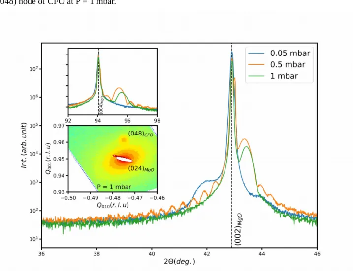
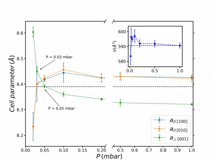
The oxygen-pressure-dependence of the structural properties of CFO films grown on MgO (100) were investigated by X-ray diffraction experiments. θ-2θ scans with a scattering vector parallel to the normal of the surface were used to analyse the crystalline structure of the films (Fig. 1). All scans show the expected (002) and (004) reflections of the (100)-oriented MgO substrate, as well as two other peaks corresponding to the (004) and (008) reflections of CFO. Since no other diffraction
peak is observed, we conclude to a (00l) oriented growth of the cobalt ferrite. (Fig. 1 and its top inset). The 2θ values corresponding to (004) and (008) CFO peaks increase when the pressure is increased, as shown in Fig. 1 for P=0.05, 0.5 and 1 mbar. Combination of both θ-2θ and RSM (reciprocal space mappings) measurements allow a complete determination of cell parameters as a function of the deposition pressure (Fig. 2). The in-plane lattice parameters were determined by RSM (Fig. 1). The zone was chosen in order to observe both the MgO (024) and CFO (048) nodes that present a four-fold symmetry. The proximity between the (024) node of the substrate and the
(048) node of CFO allows an easy observation of the in-plane CFO lattice strain. The thin film and
the MgO substrate display a perfect epitaxy at high pressure (Fig. 1 bottom inset), unlike the films grown at low pressure. The two in-plane cell parameters are identical for all deposition pressures and will be labeled a// in the following.
The variation of the out-of-plane lattice parameter, labeled a┴ in the following, is displayed in Fig.
2. In the [0.05-1] mbar range, it takes lower values than the bulk one as already observed in other oxides, e.g. Y3Fe5O12 or Ba0.5Sr0.5TiO3 [34,35]. For pressure below P=0.05 mbar (Fig. 2), the
out-of-plane parameter is higher than the bulk one and increases strongly up to 8.62 Å for P=0.01 mbar. The cell parameters a// and a┴ are characterized by a monotonic but opposite evolution with
pressure. The CFO unit cell undergoes a tetragonal distortion for all deposition pressures, except for the critical pressure Pc ~ 0.04 mbar, for which it is perfectly cubic. Above 0.05 mbar, the in-plane
parameter a// is equal to 8.42 Å that is twice the lattice parameter of the substrate (aMgO = 4.21 Å).
The film perfectly matches onto the substrate as illustrated by the RSM in Fig. 1. The cell expands laterally due to the in-plane substrate induced tensile strain. For P ≤ 0.03 mbar, the films no longer adopt the parameters of the substrate and the ratio a┴/a// is greater than 1. Finally, in the intermediate
pressure range [0.03-0.05 mbar], a┴ and a// are both higher than the bulk CFO lattice parameter, and
despite the in-plane tensile strain of the films, the cell parameter expands in the out-of-plane direction. The variation of the films cell parameters with the deposition pressure thus allows to delimit three domains of pressures: a low pressure one, P ≤ 0.03 mbar, labelled LP in the following, an intermediate one, between 0.03 and 0.05 mbar, labelled IP and a high pressure one P ≥ 0.05 mbar, labelled HP. The Poisson’s coefficient ν illustrates the unusual behaviour of IP samples. For thin films under 2D in-plane stress, ν is expressed by:
v = abulk−a⊥
2 a∥−a⊥−abulk
where abulk=8.392 Å (JCPDS 22-1086). While both LP and HP samples show a positive ν value, the
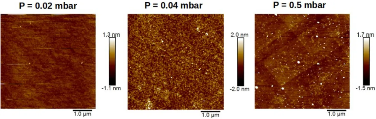
Microscopy techniques provide insights on the surface topography and on the effect of the tensile strain on the CFO thin film structure. AFM observations of the surface are shown in Fig. 3. The root mean square (RMS) surface roughness of the 0.02 mbar sample (LP) is equal to 0.1 nm indicating a low roughness related to the small mismatch between MgO and CFO. The AFM images of the HP sample display fracture lines, possibly assigned to a relaxation phenomenon along the crystallographic planes. Since this sample shows a perfect matching with the substrate, it therefore endures a 0.4% tensile strain (aCFO bulk = 8.392 Å, aCFO films = 8.43 Å), and some local relaxation
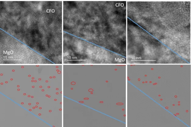
may occur periodically through the fracture lines. HRTEM observations of cross sections have pointed out dislocations within the thin film. Red circles indicate the observed dislocations on the Fourier transform of the HRTEM observation given in Fig. 4. While both IP and HP samples show a quite similar density of dislocation, the LP thin film exhibits a value two to three times higher, that could be ascribed to the high degree of distortion of the unit cell (Table 1).
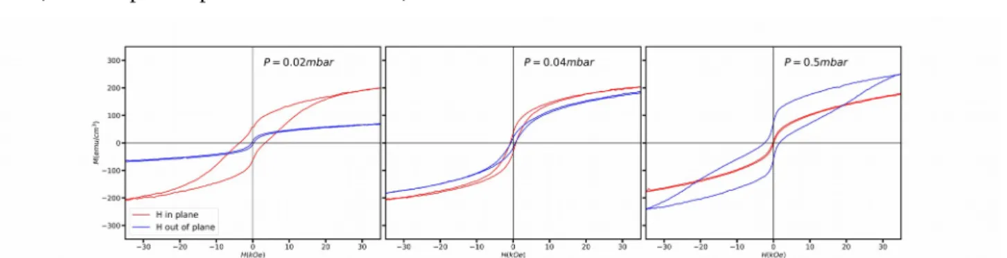
Some information on the magnetic properties of the CFO films may be determined from the room temperature hysteresis loops performed for the three pressure domains (Fig. 5). The LP samples show an in-plane easy magnetization axis, with coercive field values of about 3500 Oe and 300 Oe for the and out-of-plane measurements, respectively. The IP sample shows no preference for in-or out-of-plane easy magnetization direction, with comparable coercive field values fin-or the two configurations. Finally, the HP sample exhibits an out-of-plane easy magnetization axis. The anisotropy constants, i.e. the magnetoelastic anisotropy (Kme) arising from the coupling between the
magnetic moments and the lattice and the shape anisotropy (Kshape) have been calculated (see
Supplementary information = SI). According to Lisfi et al [36], the contribution of the surface anisotropy (Ksurf) is negative, since the thicknesses are lower than 300 nm. All the anisotropy
constants are negative for low pressure-prepared thin films (LP), leading to negative Ktot value
(Table 1). High pressure-prepared samples (HP) have a positive magnetoelastic anisotropy (Kme).
Although the other contributions are negative, the total anisotropy is positive (see SI and Table 1). These results are in agreement with the in-plane and out-of-plane easy magnetization direction deduced from the magnetic hysteresis loops for LP and HP samples respectively. Finally, the magnetoelastic constant of IP sample is positive and close to zero, much smaller than the HP sample in agreement with the quasi-overlay of out-of-plane and in-plane curves.
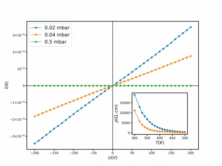
Room-temperature I-V curves obtained for characteristic samples of the three pressure domains are displayed in Fig. 6 and the deduced resistivity is given in Table 1. The HP sample shows a very high resistance, exceeding the range of measurable resistances, R> 1010 Ω. The thermal evolution of
effect measurements recorded on the LP sample at 300 K and 60 kOe (not shown here) evidence positive charge carriers with concentration and mobility equal to p = 1.3×1014 cm-3 and µ = 5
cm2/Vs, respectively. Such a p-type conductivity has already been reported for spinel-type thin
films and can be ascribed to the presence of vacancies in the structure [37–39].
3.2. Resonant X-ray diffraction at Fe and Co edges
Resonant X-ray diffraction was performed at both the Fe and Co edges in order to determine the cationic occupation of the tetrahedral and octahedral sites. This technique has been shown to be very sensitive to the cationic distribution [33]. Fig. 7 shows the simulated spectra for the 224 reflection of (CoxFe1−x)[Co1− xFe1+ x]O4 as a function of the inversion parameter computed
using the FitREXS software [33]. The experimental data recorded on LP, IP and HP samples at the D2AM beamline are given in the insets for both Fe and Co edges. According to the simulated spectra, one can notice that the 224 node is very sensitive to the Td site. It is especially the case at
the Co edge, for which no transition is expected in the case of purely inverse spinel structure (no Co in the Td sites). Conversely, simulations show that the transition at the Co edge is all the more
pronounced when the cobalt content increases in the Td site. The experimental data of the three
samples characteristic of the LP, IP and HP domains show a different behaviour at the Co edge. While no transition is visible for the LP sample, a small transition is observed for the HP sample and an important one is evidenced for the IP sample. The lack of transition at the Co edge for LP sample clearly indicates that the Td site is only occupied by Fe atoms. For the two other samples,
refinements of spectra recorded for different nodes of the reciprocal space, have been performed using the FitREXS software [33]. Results are shown in supplementary information and Table S1. The cobalt content of the Td site for the HP sample (0.07) is not zero but much lower than that of the
IP sample (0.24). The structures of the IP and HP samples are therefore not perfect inverse spinels, and this is even more pronounced for the IP sample. This result brings additional experimental confirmation to the model exposed by Fereiro-Vila concerning the mechanism for auxetic behaviour in CFO thin films: the auxetic behaviour appears when the degree of inversion is not complete, i.e. when the structure is intermediate between the direct and the inverse spinel structures [32]. The critical cationic distribution above which the auxetic behaviour appears is in the range [0.07 – 0.24].
3.3. Mössbauer spectrometry
The elongation of the CFO cell perpendicularly to the substrate observed for the low deposition pressures could have been assigned to the presence of Fe2+, stemming from the reduction of Fe3+
under too mild oxidizing conditions. Goodenough has indeed pointed out the Jahn Teller effect that is possibly associated to Fe2+ [40]. The Mössbauer spectrometry is the ideal method to evidence the
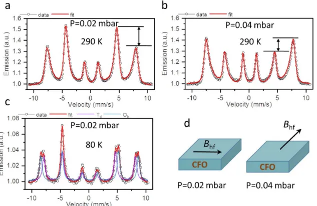
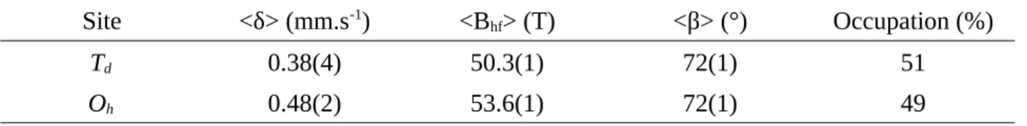
valence of iron. Room temperature (RT) Mössbauer spectra of the LP and IP (as also representative of the HP samples) samples are depicted in Fig. 8 (a) and (b). All spectra exhibit a magnetic sextet with broad lines indicating a distribution of the Fe magnetic environments. They have been fitted with a distribution of hyperfine fields leading to average values of the isomer shift (δ) equal to) equal to 0.30(1) mm/s and 0.31(9) mm/s for LP and IP samples, respectively (Table 2). Those values unambiguously point out to the presence of only the high valence state of iron, Fe3+. At low
temperature [Fig. 8(c)], the LP spectrum is resolved enough to take into consideration two contributions of refined values of δ) equal to equal to 0.38(4) mm/s and 0.48(2) mm/s (Table 3). The refined hyperfine parameters indicate that 51,6 % of the probed iron is located in Td site. This value is close
to what observed for the bulk CFO ( FeOh/FeTd≈1 ) [41]. Another important feature that can be
clearly seen on the RT Mössbauer spectra is the lines intensity ratio of the magnetic sextet that is different for LP and IP samples. Information about the direction of the magnetic moments can be obtained from the relative intensities of the Mössbauer absorption lines, which is given by 3:x:1:1:x:3 for pure magnetic hyperfine splitting, where x= 4 sin
2
⟨β⟩ 1+cos2
⟨β⟩ and <β> is the average "cone-angle" between the incident -ray direction and the direction of the magnetic hyperfine field. Complete in-plane spin orientation is indicated by ratios 3:4:1:1:4:3 or <β>=90°, whereas complete perpendicular orientation is present for ratios 3:0:1:1:0:3 or <β>=0°. For the LP sample, a value of <β> close to 90° is obtained from the fitting, confirming the in-plane magnetic anisotropy already deduced from the hysteresis loops measurements. A cone-angle value <β> = 48.9° is observed for the IP sample, evidencing the out-of-plane orientation of some of the Fe spins [Fig. 8(d)], in agreement with the hysteresis data.
3.4. Discussion about the chemical formula at low pressure
Assuming both cationic vacancies in the octahedral sites as encountered in maghemite (γ-Fe2O3)
and anionic vacancies, the general formula of the CFO thin film is written
(
FeaCo1−a)
Td[
FebCoc⊡2−b−c]
OhO4 −δ,where a and b represent the iron content in tetrahedral and octahedral sites, respectively, c the cobalt content in octahedral sites and δ) equal to the amount of anionic vacancies per formula unit. The EDX analysis showed that Fe to Co ratio is equal to 2, i.e. a+b
1−a+c=2. As shown above, the Mössbauer analysis leads to a
b=
51.6
48.4=1.07. The general chemical formula of the low pressure-prepared sample is thus
(
FeaCo1−a)
[
Fe0.94 aCo1.97 a−1⊡3 −2.91 a]
O4−δTaking into account the results of resonant X-ray diffraction at the Co edge of the (224) node (Fig. 7), i.e. the lack of transition at the Co edge for LP sample, we conclude that the Td site is fully
occupied by Fe atoms. According to Prieto et al. [42], the presence of some Co+III within the
structure can no longer be ruled out. Therefore, depending on the oxidation degree of Co atoms and taking into account the electroneutrality, the anionic vacancies content δ) equal to ranges between 0.00 and 0.24. It results in two extreme cases: 1) no Co+III, which leads to δ) equal to = 0.24 :
(Fe+III)
[
Fe+III0.94Co+II0,97⊡0.09]
O3.76 , and 2) 0.24 Co+III, which leads to the absence of anionic vacancies : (Fe+III)
[
Fe+III0.94Co+II0.73Co+III0.24⊡0.09]
O4 . The formula for the LP sample is in thedomain bounded by these extreme values. The p-type semiconductor behavior can therefore be clearly explained by the presence of cationic and anionic vacancies. The presence of Fe2+ being
ruled out, the increase of the out-of-plane cell parameter observed for these LP samples however remains a puzzle. We are currently undertaking theoretical calculations to determine the optimal structure for CFO in the presence of such an amount of cationic (and anionic if no Co3+ is present)
vacancies.
4. Conclusions
We have studied the influence of the oxidizing O2:N2 pressure on the growth of pulsed laser
deposited CFO thin films on MgO(100) substrates. Increasing this pressure from 0.01 mbar up to 1 mbar, we showed that the structural, magnetic and electrical properties of CFO thin films can significantly be varied, from a p-type semiconductor behaviour with in-plane magnetization to an insulating behaviour with out-of-plane magnetization. Another important result of this study is the demonstration of the possibility to stabilize an auxetic behaviour for the intrinsic spinel CFO compound. A decrease in the O2:N2 oxidizing pressure is shown to induce an increase of the
out-of-plane cell parameter. This increase together with the tensile stress imposed to the films by the MgO (001) substrates tend to increase the volume of the CFO cell, until the relaxation of the substrate-induced stress when the films do not adopt the parameter imposed by the substrate any more. But before that, for intermediate pressures, we could observe a situation for which the cell parameters were increased in all three directions with respect to their bulk value. In these conditions, the CFO film was shown to have a negative Poisson’s ratio, i.e. to be auxetic. We have experimentally determined the cationic distribution in our films by resonant X ray scattering and demonstrated that, as suggested by Fereiro-Vila et al. [32], the auxetic behaviour was related to a high occupation of the tetrahedral sites by cobalt ions (25 %). Such a high amount of Co2+Td is abnormal for the spinel
structure of CFO, which should be inverse. We have even determined the limit quantity of Co2+Td
below which CFO still adopts a non-auxetic behaviour.
An another remarkable result is the evidence of anionic and possible cationic vacancies at low pressures resulting to unexpected magnetic and electrical behaviours. The LP sample is characterized by a symmetry breaking which is not induced by the substrate. The crystal structure solution and the mechanism of distortion along the c-axis is the key point for the understanding of its properties. Theoretical developments are now in progress to better understand the involved mechanism.
Acknowledgments
This work was done with the financial support from the CNRS, the Ministère de l’Enseignement Supérieur et de la Recherche, and the laboratory of excellence Nanostructures in Interaction with their Environment (LabEx NIE 11- LABX-0058-NIE) and from Ile de France region equipment funding (“NOVATECS” C’Nano IdF Project No. IF-08-1453/R). We would like also to acknowledge support from Region of Normandy and the European Regional Development Fund of Normandy (ERDF) in the frame of the MAGMA project. We thank the IPCMS X-ray diffraction and MEB-CRO platforms.
References
[1] K.K. Saxena, R. Das, E.P. Calius, Three Decades of Auxetics Research − Materials with Negative Poisson’s Ratio: A Review, Adv. Eng. Mater. 18 (2016) 1847–1870.
[2] R. Hamzehei, S. Rezaei, J. Kadkhodapour, A.P. Anaraki, A. Mahmoudi, 2D triangular anti-trichiral structures and auxetic stents with symmetric shrinkage behavior and high energy absorption, Mech. Mater. 142 (2020) 103291
[3] S. Vijayavenkataraman, A. Gopinath, W.F. Lu. (2020). A new design of 3D-printed orthopedic bone plates with auxetic structures to mitigate stress shielding and improve intra-operative bending, Bio-Des. Manuf., https://doi.org/10.1007/s42242-020-00066-8
[4] P. Mardling, A. Alderson, N. Jordan-Mahy, C.L.L. Maitre, The use of auxetic materials in tissue engineering, Biomater. Sci. 8 (2020) 2074–2083.
[5] M. Sanami, N. Ravirala, K. Alderson, A. Alderson, Auxetic Materials for Sports Applications, Procedia Eng. 72 (2014) 453–458.
[6] X. Zhang, C. An, Z. Shen, H. Wu, W. Yang, J. Bai, Dynamic crushing responses of bio-inspired re-entrant auxetic honeycombs under in-plane impact loading, Mater. Today Commun. 23 (2020) 100918.
[7] Y. Guo, J. Zhang, L. Chen, B. Du, H. Liu, L. Chen, W. Li, Y. Liu, Deformation behaviors and energy absorption of auxetic lattice cylindrical structures under axial crushing load, Aerosp. Sci. Technol. 98 (2020) 105662.
[8] B. Taherkhani, M.B. Azizkhani, J. Kadkhodapour, A.P. Anaraki, S. Rastgordani, Highly sensitive, piezoresistive, silicone/carbon fiber-based auxetic sensor for low strain values, Sens. Actuators Phys. 305 (2020) 111939.
[9] X. Rong, Y. Li, S. Han, P. Cao, Y. Zeng, W. Xu, M. Fang, W. Liu, D. Zhu, Y. Lu. (2020). Electric field modulation in the auxetic effect of BP-analog monolayer As and Sb by first-principles calculations, Phys. Chem. Chem. Phys., https://doi.org/10.1039/C9CP06933J
[10] X. Li, C. Huang, S. Hu, B. Deng, Z. Chen, W. Han, L. Chen, Negative and near-zero Poisson’s ratios in 2D graphene/MoS2 and graphene/h-BN heterostructures, J. Mater. Chem. C. 8 (2020) 4021–4029.
[11] M. Mir, M.N. Ali, J. Sami, U. Ansari, Review of Mechanics and Applications of Auxetic Structures, Adv. Mater. Sci. Eng. 2014 (2014) e753496.
[12] L. Wei, X. Zhao, Q. Yu, G. Zhu, A novel star auxetic honeycomb with enhanced in-plane crushing strength, Thin-Walled Struct. 149 (2020) 106623.
[13] J.S. Hu, B.L. Wang, J.E. Li, K.F. Wang, Thermal shock resistance behavior of auxetic ceramic honeycombs with a central crack or an edge crack, Ceram. Int. 46 (2020) 11835–11845. [14] R. Gatt, L. Mizzi, J.I. Azzopardi, K.M. Azzopardi, D. Attard, A. Casha, J. Briffa, J.N. Grima,
Hierarchical Auxetic Mechanical Metamaterials, Sci. Rep. 5 (2015) 1–6.
[15] F. Agnelli, A. Constantinescu, G. Nika, Design and testing of 3D-printed micro-architectured polymer materials exhibiting a negative Poisson’s ratio, Contin. Mech. Thermodyn. 32 (2020) 433-449.
[16] C. Quan, B. Han, Z. Hou, Q. Zhang, X. Tian, T.J. Lu, 3d printed continuous fiber reinforced composite auxetic honeycomb structures, Compos. Part B Eng. 187 (2020) 107858.
[17] J. Chen, Z. Du, T. Li, Structural design and characterization of highly elastic woven fabric containing helical auxetic yarns:, Text. Res. J. 90 (2019) 809-823.
[18] H. Kamrul, A. Zulifqar, H. Hu, Deformation behavior of auxetic woven fabric based on re-entrant hexagonal geometry in different tensile directions, Text. Res. J. 90 (2020) 410–421. [19] S.C. Han, D.S. Kang, K. Kang, Two nature-mimicking auxetic materials with potential for
high energy absorption, Mater. Today. 26 (2019) 30–39.
[20] A. Yeganeh-Haeri, D.J. Weidner, J.B. Parise, Elasticity of α-Cristobalite: A Silicon Dioxide with a Negative Poisson’s Ratio, Science 257 (1992) 650–652.
[21] C. Huang, L. Chen, Negative Poisson’s Ratio in Modern Functional Materials, Adv. Mater. 28 (2016) 8079–8096.
[22] R. Gatt, L. Mizzi, K.M. Azzopardi, J.N. Grima, A force-field based analysis of the deformation mechanism in α-cristobalite, Phys. Status Solidi B. 252 (2015) 1479–1485.
[23] R. Peng, Y. Ma, Q. Wu, B. Huang, Y. Dai, Two-dimensional materials with intrinsic auxeticity: progress and perspectives, Nanoscale 11 (2019) 11413–11428.
[24] S. Chen, C. Guan, S. Ke, X. Zeng, C. Huang, S. Hu, F. Yen, H. Huang, Y. Lu, L. Chen, Modulation of Abnormal Poisson’s Ratios and Electronic Properties in Mixed-Valence Perovskite Manganite Films, ACS Appl. Mater. Interfaces 10 (2018) 18029–18035.
[25] M. Hoppe, S. Döring, M. Gorgoi, S. Cramm, M. Müller, Enhanced ferrimagnetism in auxetic NiFe2O4 in the crossover to the ultrathin-film limit, Phys. Rev. B. 91 (2015) 054418.
[26] M. Valant, A.-K. Axelsson, F. Aguesse, N.M. Alford, Molecular Auxetic Behavior of Epitaxial Co-Ferrite Spinel Thin Film, Adv. Funct. Mater. 20 (2010) 644–647.
[27] M. Foerster, M. Iliev, N. Dix, X. Martí, M. Barchuk, F. Sánchez, J. Fontcuberta, The Poisson Ratio in CoFe2O4 Spinel Thin Films, Adv. Funct. Mater. 22 (2012) 4344–4351.
[28] J. Teillet, F. Bouree, R. Krishnan, Magnetic structure of CoFe2O4, J. Magn. Magn. Mater. 123
(1993) 93–96.
[29] R. Valenzuela, Magnetic Ceramics, Cambridge University Press, 2005.
[30] P. He, K. Yang, W. Wang, F. Dong, L. Du, Y. Deng, Reduced graphene oxide-CoFe2O4 composites for supercapacitor electrode, Russ. J. Electrochem. 49 (2013) 359–364.
[31] Z. Wang, W. Jia, M. Jiang, C. Chen, Y. Li, One-step accurate synthesis of shell controllable CoFe2O4 hollow microspheres as high-performance electrode materials in supercapacitor,
Nano Res. 9 (2016) 2026–2033.
[32] E. Ferreiro-Vila, L. Iglesias, I. Lucas del Pozo, N. Varela-Dominguez, C.T. Bui, B. Rivas-Murias, J.M. Vila-Fungueiriño, P. Jimenez-Cavero, C. Magen, L. Morellon, V. Pardo, F. Rivadulla, Apparent auxetic to non-auxetic crossover driven by Co2+ redistribution in CoFe
2O4
[33] C. Lefevre, A. Thomasson, F. Roulland, V. Favre-Nicolin, Y. Joly, Y. Wakabayashi, G. Versini, S. Barre, C. Leuvrey, A. Demchenko, N. Boudet, N. Viart, Determination of the cationic distribution in oxidic thin films by resonant X-ray diffraction: the magnetoelectric compound Ga2-xFexO3, J. Appl. Crystallogr. 49 (2016) 1308–1314.
[34] Y. Dumont, N. Keller, E. Popova, D.S. Schmool, M. Tessier, S. Bhattacharya, B. Stahl, R.M.C. Da Silva, M. Guyot, Tuning magnetic properties with off-stoichiometry in oxide thin films: An experiment with yttrium iron garnet as a model system, Phys. Rev. B. 76 (2007) 104413. [35] Can Wang, B.L. Cheng, S.Y. Wang, H.B. Lu, Y.L. Zhou, Z.H. Chen, G.Z. Yang, Effects of
oxygen pressure on lattice parameter, orientation, surface morphology and deposition rate of (Ba0.02Sr0.98)TiO3 thin films grown on MgO substrate by pulsed laser deposition, Thin Solid
Films 485 (2005) 82–89.
[36] A. Lisfi, C.M. Williams, L.T. Nguyen, J.C. Lodder, A. Coleman, H. Corcoran, A. Johnson, P. Chang, A. Kumar, W. Morgan, Reorientation of magnetic anisotropy in epitaxial cobalt ferrite thin films, Phys. Rev. B. 76 (2007) 054405.
[37] T. Katayama, Y. Kurauchi, S. Mo, K. Gu, A. Chikamatsu, L. Galiullina, T. Hasegawa, p-Type Conductivity and Room-Temperature Ferrimagnetism in Spinel MoFe2O4 Epitaxial Thin Film,
Cryst. Growth Des. 19 (2019) 902–906.
[38] A. Roy, J. Ghose, Studies on Some Titanium-Substituted Fe2MoO4Spinel Oxides, J. Solid State
Chem. 140 (1998) 56–61.
[39] M. Seki, H. Tabata, H. Ohta, K. Inaba, S. Kobayashi, Epitaxial thin films of p-type spinel ferrite grown by pulsed laser deposition, Appl. Phys. Lett. 99 (2011) 242504.
[40] J.B. Goodenough, Jahn-Teller distortions induced by tetrahedral-site Fe2+ ions, J. Phys. Chem.
Solids 25 (1964) 151–160.
[41] G.A. Sawatzky, F. Van Der Woude, A.H. Morrish, M\"ossbauer Study of Several Ferrimagnetic Spinels, Phys. Rev. 187 (1969) 747–757.
[42] P. Prieto, J.F. Marco, A. Serrano, M. Manso, J. de la Figuera, Highly oriented (111) CoO and Co3O4 thin films grown by ion beam sputtering, J. Alloys Compd. 810 (2019) 151912.
Figure captions
Fig. 1. Out-of-plane XRD pattern recorded in the 36°-46° 2θ range of CoFe2O4 (CFO) thin films
elaborated at P = 0.05 mbar (blue), P = 0.5 mbar (red) and P = 1 mbar (green), respectively. Insets are (top): a magnitude around 2θ = 94°, and (bottom) the reciprocal space mapping (rsm) of the (048) node of CFO at P = 1 mbar.
Fig. 2. Evolution of the cell parameters as a function of the pressure deposition. The dashed black
line corresponds to the cell parameter of bulk cubic spinel CFO (JCPDS 22-1086). Inset shows the evolution of the volume with the partial pressure.
Fig. 3. AFM images of 5x5 µm² areas of CoFe2O4 thin films elaborated at P = 0.02 mbar (LP), P =
Fig. 4. TEM observations (top) and their Fourier transforms (bottom) of CoFe2O4 thin films
elaborated at P = 0.02 mbar (left), P = 0.04 mbar (middle) and P = 0.5 mbar (right). Red ellipses indicate dislocations.
Fig. 5. Room temperature magnetic hysteresis loops measured with a maximum applied field of 70
Fig. 6. Room temperature I-V curves for thin films deposited at P = 0.02, P = 0.04 and P = 0.5 mbar.
Fig. 7. Simulated resonant X-ray scattering
(
CoxFe1− x)
[
Co1−xFe1+ x]
O4 thin films as a function ofthe cobalt content (x) in Td site for the 224 node computed with the FitREXS software[21]. The
experimental spectra recorded at the D2AM beamline of ESRF on low pressure (LP), intermediate (IP) and high pressure (HP) samples at both Fe and Co edges are displayed in the insets.
Fig. 8. (a) Room temperature Mössbauer spectra of CFO thin film deposited at P = 0.02 mbar and
(b) at P = 0.04 mbar. (c) CEMS spectra at 80K of CFO thin film deposited at P = 0.02 mbar and (d) sketch of magnetic anisotropy deduced from the fit of the spectrum.
Tables
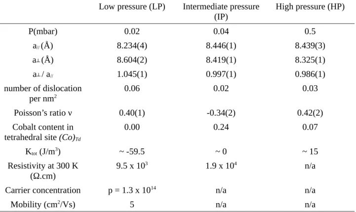
Table 1. Characteristic parameters of CFO thin films deposited at P = 0.02 mbar, P = 0.04 mbar and
P = 0.5 mbar: out of plane (a┴) and in-plane cell parameters (a//), ratio of the cell parameters (a┴ /
a//), density of dislocation deduced from TEM observation, Poisson’s ratio ν, cobalt content in in
tetrahedral site (Co)Td deduced from resonant X-ray diffraction spectra, anisotropy constants Ktot
(see SI), resistivity at room temperature, carrier concentration and mobility.
Low pressure (LP) Intermediate pressure
(IP) High pressure (HP)
P(mbar) 0.02 0.04 0.5 a// (Å) 8.234(4) 8.446(1) 8.439(3) a┴ (Å) 8.604(2) 8.419(1) 8.325(1) a┴ / a// 1.045(1) 0.997(1) 0.986(1) number of dislocation per nm2 0.06 0.02 0.03 Poisson’s ratio ν 0.40(1) -0.34(2) 0.42(2) Cobalt content in
tetrahedral site (Co)Td
0.00 0.24 0.07
Ktot (J/m3) ~ -59.5 ~ 0 ~ 15
Resistivity at 300 K (Ω.cm)
9.5 x 10³ 1.9 x 10⁴ n/a
Carrier concentration p = 1.3 x 10¹⁴ n/a n/a
Table 2. Hyperfine refined parameters deduced from the fit of the CEMS spectra at room
temperature of CFO thin film deposited at P = 0.02 mbar (LP) and P = 0.04 mbar (IP).
Site <δ) equal to> (mm.s-1) <B
hf> (T) <β> (°)
LP 0.30(1) 44(1) 75(2)
IP 0.31(7) 43(1) 46(2)
Table 3. Hyperfine refined parameters deduced from the fit of the CEMS spectra at 80K of CFO
thin film deposited at P = 0.02 mbar (LP).
Site <δ) equal to> (mm.s-1) <B
hf> (T) <β> (°) Occupation (%)
Td 0.38(4) 50.3(1) 72(1) 51
Supplementary information
1. Calculation of the anisotropy constants
The total anisotropy (Ktot) of strained thin film can be written as the sum of the several
contributions. The main one is the magnetoelastic anisotropy (Kme) which describes the coupling
between the magnetic moments and the lattice. The other terms can be considered as correcting factors. The shape anisotropy (Kshape) is correlated to the demagnetizing factor which depends on the
geometry of the system. Finally, the surface anisotropy (KS) takes into account the symmetry
breaking and thus deflects the moment close to the surface. The value of this last term is inversely proportional to the thickness of the samples. The total anisotropy (Ktot) can be thus written as:
Ktot=Kme+Kshape+
2 KS
tc
(1)
where tc is the critical thickness of the film (see text).
• Due to the tetragonal distortion, the magnetoelastic constant can be formulated as [1-3]:
Kme=3
2λ[100]
(
C11−C12) (
a.∥.− a.⊥.)
/aλ[100] is the magnetoelastic constant for a distortion along the [100] direction, C11 and C12 are the
elastic constants.a.∥.and a.⊥. are the in-plane and out-of-plane cell parameters, respectively. For this
work, λ[100], C11 and C12 have been taken from Suzuki’s work [4] , i.e λ[100] = -5.9×10⁻⁴, C11 = 2.7×10¹²
dynes/cm2, C
12 = 1.06×10¹² dynes/cm2. Results of computation are given in Table S1.
• Shape anisotropy is simplified for the thin films due to the fact thatd33=1and 0 elsewhere in the
demagnetizing tensor leading toKshape=− 2 π M
2
where M is the magnetization [5]. Results of computation are given in Table S1.
• Lisfi et al. have estimated that the critical thickness tc for CFO for the spin-reorientation transition
is around 300 nm [1]. The thicknesses of our samples are smaller (~60 nm) such that the first term 2 KS/tc is negative for all our samples. Since IP sample has both out-of-plane and in-plane curves which are almost superimposed, one can assume that Ktot ≈ 0 involving that any external magnetic
field affects the easy direction of magnetization. It results to 2 KS
tc
=− Kme− Kshapefor this sample leading to 2Ks/t ~ -4.5 J/m3. Both LP and HP samples have same thickness as IP one. One can thus
conclude that 2Ks/tc should be the same for all samples. Results of computation are given in Table
S1.
Table S1
Low pressure sample
(LP) Intermediate pressuresample (IP) High pressure sample(HP)
Kme (J/m3) -64.2 4.7 19.7
Kshape (J/m3) -0.2 -0.2 -0.2
2Ks/tc (J/m3) ~ -4.5 ~ -4.5 ~ -4.5
Ktot (J/m3) ~ -59.5 ~ 0 ~ 15
Computation of the magnetoelastic anisotropy (Kme), the shape anisotropy (Kshape), the surface
anisotropy (2Ks/t) and the total anisotropy (Ktot) of samples characteristic of the three evidenced
pressure domains.
[1] A. Lisfi, C.M. Williams, L.T. Nguyen, J.C. Lodder, A. Coleman, H. Corcoran, A. Johnson, P. Chang, A. Kumar, W. Morgan, Reorientation of magnetic anisotropy in epitaxial cobalt ferrite thin films, Phys. Rev. B. 76 (2007) 054405.
[2] R. Thamankar, A. Ostroukhova, F.O. Schumann, Spin-reorientation transition in FexNi1-x alloy
films, Phys. Rev. B. 66 (2002) 134414.
[3] B. Schulz, K. Baberschke, Crossover from in-plane to perpendicular magnetization in ultrathin Ni/Cu(001) films, Phys. Rev. B. 50 (1994) 13467–13471.
[4] Y. Suzuki, G. Hu, R.B. van Dover, R.J. Cava, Magnetic anisotropy of epitaxial cobalt ferrite thin films, J. Magn. Magn. Mater. 191 (1999) 1–8.
2. Resonant X-ray diffraction refinement of the of both IP and HP samples at both Co and Fe edges
The synchrotron experiments were carried out on the Collaborating research group (CRG) D2AM beamline at the European Synchrotron Radiation Facility. Samples were mounted on a seven-circle diffractometer equipped with various photodiode detectors, allowing recording of the intensity of the incident beam I0, the intensities of the diffraction peaks and the fluorescence of the samples. The
rotation matrix was preliminary determined with the help of different in-plane and out-of-plane reflections.
Anomalous factors were first computed using the fluorescence data. The data are first corrected for both background and air absorption. f’’(E) is then extracted by using a step function to match the Sasaki values. f’(E) is then obtained using the Kramers–Kronig transform of f’’(E).
Knowing f0, f’ and f’’ allows thus computation the experimental scans for a given set of structural
parameters. The refinement of the different spectra was performed by minimizing the sum of all reliability factors for each nodes and edges through a stochastic basin-hopping algorithm (FitREXS program) (Figures S1 and S2).
Figure S1: Refinement of IP sample at both Co (left) and Fe (right) edges. Black crosses are experimental data at different hkl reflections and blue curves are the refinement curves.
Figure S2: Refinement of HP sample at both Co (left) and Fe (right) edges. Black crosses are experimental data at different hkl reflections and blue curves are the refinement curves.
![Fig. 7. Simulated resonant X-ray scattering ( Co x Fe 1−x ) [ Co 1−x Fe 1+ x ] O 4 thin films as a function of the cobalt content (x) in T d site for the 224 node computed with the FitREXS software[21]](https://thumb-eu.123doks.com/thumbv2/123doknet/14522064.531613/20.892.87.809.180.931/simulated-resonant-scattering-function-content-computed-fitrexs-software.webp)