HAL Id: cea-01743230
https://hal-cea.archives-ouvertes.fr/cea-01743230
Submitted on 6 Apr 2018
HAL is a multi-disciplinary open access
archive for the deposit and dissemination of sci-entific research documents, whether they are pub-lished or not. The documents may come from teaching and research institutions in France or abroad, or from public or private research centers.
L’archive ouverte pluridisciplinaire HAL, est destinée au dépôt et à la diffusion de documents scientifiques de niveau recherche, publiés ou non, émanant des établissements d’enseignement et de recherche français ou étrangers, des laboratoires publics ou privés.
Uranyl–Organic Coordination Polymers with trans -1,2-,
trans -1,4-, and cis -1,4-Cyclohexanedicarboxylates:
Effects of Bulky PPh_4
+and PPh_3Me
+Counterions
Pierre Thuéry, Youssef Atoini, Jack Harrowfield
To cite this version:
Pierre Thuéry, Youssef Atoini, Jack Harrowfield. Uranyl–Organic Coordination Polymers with trans -1,2-, trans -1,4-, and cis -1,4-Cyclohexanedicarboxylates: Effects of Bulky PPh_4+ and PPh_3Me+
Counterions. Crystal Growth & Design, American Chemical Society, 2018, 18, pp.2609-2619. �10.1021/acs.cgd.8b00250�. �cea-01743230�
1
Uranyl–Organic Coordination Polymers with
trans-1,2-, trans-1,4- and cis-1,4-Cyclohexanedicarboxylates:
Effects of Bulky PPh
4+and PPh
3Me
+Counterions
Pierre Thuéry,*,† Youssef Atoini‡ and Jack Harrowfield*,‡
†NIMBE, CEA, CNRS, Université Paris-Saclay, CEA Saclay, 91191 Gif-sur-Yvette, France ‡ISIS, Université de Strasbourg, 8 allée Gaspard Monge, 67083 Strasbourg, France
ABSTRACT: Three uranyl ion complexes with trans-1,2-cyclohexanedicarboxylic acid (t-1,2-chdcH2) and six with
trans- or cis-1,4-cyclohexanedicarboxylic acid (t- or c-1,4-chdcH2) have been obtained under solvo-hydrothermal
conditions in the presence of PPh4+ or PPh3Me+ counterions. The complex [PPh4][UO2(R-t-1,2-chdc)(HCOO)] (1)
crystallized on use of the pure (1R,2R) enantiomer of the dicarboxylate ligand, while the isomorphous complex [PPh4][UO2(S-t-1,2-chdc)(HCOO)] (2), containing the (1S,2S) enantiomer, resulted from use of the racemic form
through spontaneous resolution. Both contain the rare diaxial (aa) form of the ligand and are one-dimensional (1D) polymers. The complex [PPh3Me][H2NMe2]3[(UO2)4(R-t-1,2-chdc)6]· H2O (3), with the pure enantiomeric, diequatorial
(ee) form of the ligand, is a two-dimensional (2D) species with hnb topology, which derives from the structure of tetranuclear clusters previously reported. Complexes [PPh4][UO2(t-1,4-chdc)(NO3)]·2CH3CN (4) and [PPh4][UO2
(c-1,4-chdc)(NO3)] (5) are 1D polymers, helical in the latter case due to the axial-equatorial (ae) form of the ligand.
[PPh4]2[(UO2)2(t-1,4-chdc)3]·4H2O (6), [PPh3Me]2[(UO2)2(t-1,4-chdc)3]· 2H2O (7), [PPh3Me]2[(UO2)2
(c-1,4-chdc)3]·2H2O (8), and [PPh4]2[(UO2)2(t-1,4-chdc)2(c-1,4-chdc)]·3H2O (9) all crystallize as 2D networks with
honeycomb topology, the shape of the rings and that of the layers varying due to the presence of trans isomers in both the ee and aa forms, and coexistence of cis and trans isomers in 9; in all complexes 6–9, large channels are formed, which contain the counterions. The uranyl emission spectra of compounds 1–4, 6 and 9 in the solid state are in agreement with those usually found for tris-chelated carboxylate complexes, while that of complex 8, containing a mixture of seven- and eight-coordinate uranium atoms, displays a superposition of ill-resolved maxima within a broad envelope. A quantum yield of 0.13 was measured for complex 6.
2
INTRODUCTION
The topology of uranyl–organic coordination polymers,1–5 when they are anionic, can easily be
modulated through modifying the nature of the countercation. The latter is often an additional
metallic species, which can be part of the polymer fabric itself or separate if associated with
coligands such as nitrogen chelators.5 It can also be an organic species, and, in particular, the very
large range in size and geometry available in the family of ammonium cations has been extensively
exploited, as evidenced by the 330 crystal structures of uranyl–containing polymers including such
cations which are reported in the Cambridge Structural Database (CSD, Version 5.38).6 In contrast,
the quite common tetraphenylphosphonium and methyltriphenylphosphonium cations, PPh4+ and
PPh3Me+, have never been used in the synthesis of uranyl–containing coordination polymers, and
the former is only found in a handful of discrete, molecular uranyl complexes. The good solubility
of these cations and their thermal stability make them well adapted to use in (solvo-)hydrothermal
syntheses, and their bulkiness is likely to have a strong effect on the geometry of anionic uranyl–
organic coordination polymers, comparable to that of the equally bulky [ML3]2+ cations, where M
is a d-block metal cation and L is 2,2ʹ-bipyridine or 1,10-phenanthroline.5 Both PPh
4+ and PPh3Me+
have been widely studied, in fact, for their structural influence resulting from “multiple phenyl
embraces”.7–9 In the course of an investigation of the uranyl complexes formed with the different
isomers of cyclohexanedicarboxylate ligands,10–13 we have recently obtained the compound
[NH4][PPh4][(UO2)8(c-1,2-chdc)9(H2O)6]·3H2O (where c-1,2-chdc2– is the dianion of
cis-1,2-cyclohexanedicarboxylic acid), which contains an octanuclear cage of unique geometry, while
crystallization in the absence of possible counterions gives simple two-dimensional (2D) neutral
species.14 In order to characterize more thoroughly the effect of PPh
4+ and PPh3Me+ counterions
3
we have now synthesized a series of complexes, three from trans-1,2-cyclohexanedicarboxylic acid
(t-1,2-chdcH2), either in its racemic or (1R,2R) pure enantiomeric form, and six from
trans-1,4-cyclohexanedicarboxylic acid (t-1,4-chdcH2) or a mixture of the cis and trans isomers
(c,t-1,4-chdcH2). These complexes have been characterized by their crystal structure and, in most cases,
their emission spectrum in the solid state. The structures of the complexes of 1,4-chdc2– in
particular reflect the consequences of the variety of configurations and conformations possible for
cyclohexanedicarboxylates, a feature known to lead to novel properties such as the “breathing” of
3D networks due to the accommodation of guests.15
EXPERIMENTAL SECTION
Syntheses. Caution! Uranium is a radioactive and chemically toxic element, and
uranium-containing samples must be handled with suitable care and protection.
UO2(NO3)2·6H2O (depleted uranium, R. P. Normapur, 99%) was purchased from Prolabo,
rac-trans-1,2-cyclohexanedicarboxylic acid (rac-t-1,2-chdcH2) was from Lancaster,
trans-1,4-cyclohexanedicarboxylic acid (t-1,4-chdcH2) was from Alfa-Aesar, and
1,4-cyclohexanedicarboxylic acid (mixture of cis and trans isomers, c,t-1,4-chdcH2) was from Aldrich.
The (1R,2R) enantiomer of t-1,2-chdcH2, denoted R-t-1,2-chdcH2, was isolated through
crystallization with (R)-1-phenylethylamine as a resolving agent, as in the literature,16 although
both the (1R,2R) and (1S,2S) enantiomers are also available commercially. Elemental analyses
were performed by MEDAC Ltd. at Chobham, UK. For all syntheses, the mixtures in demineralized
water were placed in 10 mL tightly closed glass vessels and heated at 140 °C under autogenous
4
[PPh4][UO2(R-t-1,2-chdc)(HCOO)] (1). R-t-1,2-chdcH2 (17 mg, 0.10 mmol),
UO2(NO3)2·6H2O (35 mg, 0.07 mmol), and PPh4Br (42 mg, 0.10 mmol) were dissolved in water
(0.5 mL) and DMF (0.2 mL). Crystals of complex 1 were obtained overnight (22 mg, 38% yield
based on U). Elemental analysis results indicate the probable presence of about one extra water
molecule. Anal. Calcd for C33H31O8PU + H2O: C, 47.04; H, 3.92. Found: C, 47.20; H, 3.55%.
[PPh4][UO2(S-t-1,2-chdc)(HCOO)] (2). rac-t-1,2-chdcH2 (17 mg, 0.10 mmol),
UO2(NO3)2·6H2O (35 mg, 0.07 mmol), and PPh4Br (42 mg, 0.10 mmol) were dissolved in water
(0.5 mL) and DMF (0.2 mL). Crystals of complex 2 were obtained in low yield within two days.
[PPh3Me][H2NMe2]3[(UO2)4(R-t-1,2-chdc)6]·H2O (3). R-t-1,2-chdcH2 (17 mg, 0.10 mmol),
UO2(NO3)2·6H2O (35 mg, 0.07 mmol), and PPh3MeBr (36 mg, 0.10 mmol) were dissolved in water
(0.5 mL) and DMF (0.2 mL). Crystals of complex 3 were obtained within one week (24 mg, 57%
yield based on the acid). Anal. Calcd for C73H104N3O33PU4: C, 34.59; H, 4.14; N, 1.66. Found: C,
34.89; H, 4.17; N, 2.05%.
[PPh4][UO2(t-1,4-chdc)(NO3)]·2CH3CN (4). t-1,4-chdcH2 (17 mg, 0.10 mmol),
UO2(NO3)2·6H2O (35 mg, 0.07 mmol), and PPh4Br (42 mg, 0.10 mmol) were dissolved in water
(0.5 mL) and acetonitrile (0.2 mL). Crystals of complex 4 were obtained in low yield within one
week.
[PPh4][UO2(c-1,4-chdc)(NO3)] (5). c,t-1,4-chdcH2 (17 mg, 0.10 mmol), UO2(NO3)2·6H2O
(35 mg, 0.07 mmol), and PPh4Br (42 mg, 0.10 mmol) were dissolved in water (0.7 mL) and
N-methyl-2-pyrrolidone (0.3 mL). Crystals of complex 5 were obtained in low yield within one
month.
[PPh4]2[(UO2)2(t-1,4-chdc)3]·4H2O (6). t-1,4-chdcH2 (17 mg, 0.10 mmol), UO2(NO3)2·6H2O
5
(0.2 mL). Crystals of complex 6 were obtained overnight (47 mg, 78% yield based on the acid).
Anal. Calcd for C72H78O20P2U2: C, 48.01; H, 4.36. Found: C, 47.59; H, 3.95%. The same complex
is obtained when DMF is replaced by acetonitrile.
[PPh3Me]2[(UO2)2(t-1,4-chdc)3]·2H2O (7). t-1,4-chdcH2 (17 mg, 0.10 mmol),
UO2(NO3)2·6H2O (35 mg, 0.07 mmol), and PPh3MeBr (36 mg, 0.10 mmol) were dissolved in water
(0.6 mL) and DMF (0.2 mL). Crystals of complex 7 were obtained within four days (7 mg, 13%
yield based on the acid). Anal. Calcd for C62H70O18P2U2: C, 45.37; H, 4.30. Found: C, 45.66; H,
4.34%.
[PPh3Me]2[(UO2)2(c-1,4-chdc)3]·2H2O (8). c,t-1,4-chdcH2 (17 mg, 0.10 mmol),
UO2(NO3)2·6H2O (35 mg, 0.07 mmol), and PPh3MeBr (36 mg, 0.10 mmol) were dissolved in water
(0.6 mL) and DMF (0.2 mL). Crystals of complex 8 were obtained within four days (7 mg, 13%
yield based on the acid). Anal. Calcd for C62H70O18P2U2: C, 45.37; H, 4.30. Found: C, 45.23; H,
4.27%.
[PPh4]2[(UO2)2(t-1,4-chdc)2(c-1,4-chdc)]·3H2O (9). c,t-1,4-chdcH2 (17 mg, 0.10 mmol),
UO2(NO3)2·6H2O (35 mg, 0.07 mmol), and PPh4Br (42 mg, 0.10 mmol) were dissolved in water
(0.6 mL) and DMF (0.2 mL). Crystals of complex 9 were obtained overnight (16 mg, 27% yield
based on the acid). Anal. Calcd for C72H76O19P2U2: C, 48.49; H, 4.30. Found: C, 49.17; H, 3.96%.
Crystallography. The data were collected at 150(2) K on a Nonius Kappa-CCD area
detector diffractometer17 using graphite-monochromated Mo Kα radiation (λ = 0.71073 Å). The
crystals were introduced into glass capillaries with a protective coating of Paratone-N oil (Hampton
Research). The unit cell parameters were determined from ten frames, then refined on all data. The
6
reflections) were processed with HKL2000.18 Absorption effects were corrected empirically with
the program SCALEPACK.18 The structures were solved by intrinsic phasing with SHELXT,19
expanded by subsequent difference Fourier synthesis and refined by full-matrix least-squares on
F2 with SHELXL-2014.20 All non-hydrogen atoms were refined with anisotropic displacement
parameters. The hydrogen atoms bound to oxygen atoms were retrieved from difference Fourier
maps when possible, and the carbon-bound hydrogen atoms were introduced at calculated positions
(as well as the ammonium hydrogen atoms in 3). All hydrogen atoms were treated as riding atoms
with an isotropic displacement parameter equal to 1.2 times that of the parent atom (1.5 for CH3,
with optimized geometry). In compound 3, the dimethylammonium counterion is disordered over
two sites, one of them being itself disordered around a threefold rotation axis, and restraints were
applied for bond lengths, angles and displacement parameters in the disordered parts. One lattice
water molecule in complex 6 is disordered over two positions which have been refined with
occupancy parameters constrained to sum to unity, and the corresponding hydrogen atoms were
not found. In the case of compound 9, restraints on bond lengths and displacement parameters were
applied in several parts of the structure; the highest residual electron density peaks are located near
the uranium atoms, probably as a result of imperfect absorption corrections. The Flack parameter
values are –0.012(8), –0.008(10) and 0.002(10) for the three enantiomerically pure complexes 1–
3, respectively. Crystal data and structure refinement parameters are given in Table 1. The
molecular plots were drawn with ORTEP-3,21 and the polyhedral representations with VESTA.22
7
Table 1. Crystal Data and Structure Refinement Details
1 2 3 4 5
chemical formula C33H31O8PU C33H31O8PU C73H104N3O33PU4 C36H36N3O9PU C32H30NO9PU
M (g mol−1) 824.58 824.58 2534.68 923.68 841.57 cryst syst monoclinic monoclinic trigonal orthorhombic monoclinic
space group P21 P21 R3 Pbcn P21/n a (Å) 7.7964(3) 7.7955(4) 13.6231(3) 20.5334(8) 14.2044(5) b (Å) 14.9394(4) 14.9391(4) 13.6231(3) 14.7720(5) 14.6258(6) c (Å) 13.2101(5) 13.2114(7) 42.3397(12) 23.7294(11) 15.1949(4) α (deg) 90 90 90 90 90 β (deg) 101.558(2) 101.563(3) 90 90 99.312(2) γ (deg) 90 90 120 90 90 V (Å3) 1507.43(9) 1507.35(12) 6805.0(4) 7197.6(5) 3115.15(19) Z 2 2 3 8 4 Dcalcd (g cm−3) 1.817 1.817 1.856 1.705 1.794 µ(Mo Kα) (mm−1) 5.489 5.489 7.214 4.612 5.317 F(000) 800 800 3630 3616 1632 reflns collcd 59027 47434 69249 161672 91403 indep reflns 7755 7762 5744 6823 5909
obsd reflns [I > 2σ(I)] 7017 6232 5668 4459 5116
Rint 0.032 0.041 0.017 0.053 0.030 params refined 389 389 374 454 397 R1 0.031 0.040 0.024 0.037 0.026 wR2 0.078 0.084 0.066 0.085 0.069 S 1.023 1.054 1.075 0.921 1.042 ∆ρmin (e Å−3) −1.30 −1.35 −0.93 −0.92 −1.07 ∆ρmax (e Å−3) 1.15 1.79 1.30 1.34 0.60 6 7 8 9 chemical formula C72H78O20P2U2 C62H70O18P2U2 C62H70O18P2U2 C72H76O19P2U2 M (g mol−1) 1801.34 1641.18 1641.18 1783.32
cryst syst triclinic monoclinic monoclinic monoclinic
space group Pī P21/c P21/c P21/n a (Å) 10.0284(4) 8.7748(4) 28.3178(6) 28.1368(9) b (Å) 13.5216(6) 25.8721(7) 13.1707(3) 19.3550(4) c (Å) 14.0662(6) 13.6877(7) 16.3855(3) 40.1254(12) α (deg) 102.635(3) 90 90 90 β (deg) 95.694(3) 97.150(2) 91.0988(13) 106.988(2) γ (deg) 106.678(3) 90 90 90 V (Å3) 1755.91(14) 3083.3(2) 6110.1(2) 20898.3(10) Z 1 2 4 12 Dcalcd (g cm−3) 1.703 1.768 1.784 1.700 µ(Mo Kα) (mm−1) 4.724 5.368 5.418 4.761 F(000) 884 1600 3200 10488 reflns collcd 99165 74279 192267 383533 indep reflns 6667 5850 11568 39092
obsd reflns [I > 2σ(I)] 6062 4809 10274 23869
Rint 0.042 0.040 0.022 0.071 params refined 443 380 759 2554 R1 0.022 0.030 0.021 0.064 wR2 0.053 0.069 0.049 0.161 S 1.024 1.030 1.020 1.034 ∆ρmin (e Å−3) −1.16 −1.36 −0.79 −3.31 ∆ρmax (e Å−3) 0.49 0.81 0.38 4.92
8
Luminescence Measurements. Emission spectra were recorded on solid samples
using a Horiba-Jobin-Yvon IBH FL-322 Fluorolog 3 spectrometer equipped with a 450 W
xenon arc lamp, double-grating excitation, and emission monochromators (2.1 nm mm−1 of
dispersion; 1200 grooves mm−1) and a TBX-04 single photon-counting detector. The powdered
compound was pressed between two silica plates which were mounted such that the faces were
oriented vertically and at 45° to the incident excitation radiation. An excitation wavelength of
420 nm, a commonly used point although only part of a broad manifold, was used in all cases
and the emission was monitored between 450 and 650 nm. The quantum yield measurement
was performed by using an absolute photoluminescence quantum yield spectrometer
Hamamatsu Quantaurus C11347 and exciting the sample between 300 and 400 nm.
RESULTS AND DISCUSSION
Synthesis. Complexes 1–9, as crystalline solids, were synthesized under
solvo-hydrothermal conditions at a temperature of 140 °C, the organic cosolvent being
N,N-dimethylformamide (DMF) for complexes 1–3 and 6–9, acetonitrile for 4 and
N-methyl-2-pyrrolidone (NMP) for 5. In all cases, the uranium/ligand stoichiometry used was 7:10, in the
expectation that it would favour the formation of an anionic species. All complexes are indeed
anionic, notwithstanding some variations in uranium/chdc2– stoichiometry. This ratio is 1:1 in
1 and 2, in which an extra negative charge is provided by one formate anion formed in situ from
DMF hydrolysis, as previously documented,24 and also in 4 and 5 which include a nitrate anion,
while it assumes the expected value of 2:3 in complexes 3 and 6–9. The PPh4+ or PPh3Me+
counterions are present in all cases (as well as dimethylammonium cations resulting from DMF
hydrolysis in complex 3), indicating that, notwithstanding many other attempts with different
9
these large counterions with uranyl-containing polymers gives systems fairly amenable to
crystallization and to a degree controllable with respect to their stoichiometry. The effect of the
organic cosolvent, other than on the solubility of the initial reaction mixture, is difficult to
predict in cases where it, too, undergoes reaction, the present structures providing examples
where DMF hydrolysis may provide either an anion (formate) or a cation (dimethylammonium)
incorporated in the complex finally obtained. The formation of mixed carboxylato complexes
of uranyl ion, though rarely a direct objective of the syntheses, has proven to be a relatively
common and useful pathway to formation of crystalline coordination polymers, especially
where oxalate is the “unintentional” additional carboxylate.25–27 As is commonly the case with
chiral ligands used in their racemic form, crystallisation can involve the formation of
conglomerate solids where separate crystals contain only one or the other of the enantiomeric
ligand forms and this is illustrated here by complex 2, which has the same structure as complex
1 except for the fact that by chance a crystal was selected containing the (1S,2S) rather than the
(1R,2R) form of the enantiomerically pure ligand used to prepare complex 1.
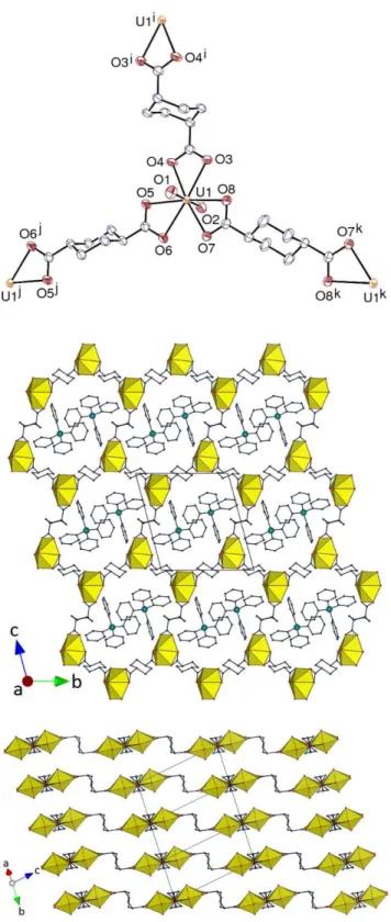
Crystal Structures. The two complexes [PPh4][UO2(R-t-1,2-chdc)(HCOO)] (1) and
[PPh4][UO2(S-t-1,2-chdc)(HCOO)] (2), in which R-t-1,2-chdc2– and S-t-1,2-chdc2– are the
(1R,2R) and (1S,2S) enantiomers of t-1,2-chdc2–, respectively, are isomorphous and crystallize
in the monoclinic Sohncke group P21. While 1 was obtained from the pure (1R,2R) enantiomer
of the ligand, 2 was synthesized from the racemic form, which indicates that resolution occurred
during crystallization, the particular crystal subjected to analysis containing the pure (1S,2S)
enantiomer. The asymmetric unit in both cases contains one uranyl cation chelated by two
carboxylate groups from two dicarboxylate ligands, and one formate anion, the uranium
coordination environment being thus hexagonal bipyramidal, with unexceptional U–O bond
10
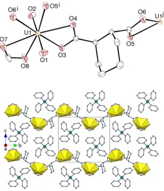
Figure 1. Top: View of compound 1. Displacement ellipsoids are drawn at the 30% probability level. Counterions
and hydrogen atoms are omitted. Symmetry codes: i = 1 – x, y – 1/2, –z; j = 1 – x, y + 1/2, –z. Bottom: Packing with uranium coordination polyhedra colored yellow and hydrogen atoms omitted.
Figure 2. View of compound 2. Displacement ellipsoids are drawn at the 20% probability level. Counterions and
hydrogen atoms are omitted. Symmetry codes: i = 1 – x, y + 1/2, 2 – z; j = 1 – x, y – 1/2, 2 – z. The packing is similar to that in 1.
carboxylate groups axial (aa), so that the cyclohexyl ring is roughly perpendicular to the mean
plane defined by the two carboxylate groups and the attached uranium atoms. In all the uranyl
complexes with t-1,2-chdc2– previously reported,11,12 the ligand assumes the more common
chair conformation with the two carboxylate groups equatorial (ee). A search of the CSD
11
coexistence of the ee and aa forms in the lattice. However, a study of the conformational
preferences of t-1,2-chdcH2 and its mono- and dianion in solution by NMR spectroscopy
revealed that, depending on the medium, the aa population could be significant, representing
as much as 57% for the dianion in DMSO.30 In contrast to the ee form, the aa form is a distinctly
divergent ligand and a zigzag one-dimensional (1D) polymer directed along the b axis is
formed, in which the directions of the cyclohexyl ring and formate ligand alternate from one
unit to the next. Extension of the 1D chains into a 2D network is prevented by the presence of
the terminal formate ligands. It is notable that, when similarly bis-chelating, the ee form of the
ligand readily gives tetrahedral uranyl clusters,11,12 thus showing that its geometry is much
better adapted than that of the aa form to the formation of closed species. The undulating chains
lie parallel to one another in sheets parallel to (0 0 1). The PPh4+ cations lie in undulating sheets
parallel to those of the anionic polymers, forming arrays essentially identical to those
considered to define “multiple phenyl embraces” in a wide variety of simpler derivatives.7–9
PPh4+ cations related to one another by translations along the a axis are involved in fourfold,
“O4PE”,8 embraces [P· ··P distances 7.7964(3) and 7.7955(4) Å in complexes 1 and 2,
respectively]. However, the corresponding interactions are no stronger than dispersion, and no
significant π-stacking or CH···π interaction is present in the lattice. Only some weak CH(cation)·· ·O(carboxylate) hydrogen bonds may be present [C··· O 3.115(10) and 3.280(9) Å
for the shortest ones], which appear to be stronger than dispersion from examination of the
Hirshfeld surfaces31 calculated with CrystalExplorer.32 With a Kitaigorodski packing index
(KPI, estimated with PLATON33) of 0.70, the packing has no solvent-accessible spaces.
Complexes isomorphous to 1 and 2 are obtained when DMF is replaced by acetonitrile or NMP,
in which the formate anions are replaced by nitrate anions, but, in the absence of crystals
12
Changing PPh4+ for PPh3Me+ results in a profound structural modification in the case of
R-t-1,2-chdc2–, which gives the complex [PPh
3Me][H2NMe2]3[(UO2)4(R-t-1,2-chdc)6]· H2O (3).
The same experiment performed with rac-t-1,2-chdcH2 gave the same complex, indicating that
resolution of the two enantiomers occurred during crystallization here also. Instead of including
formate coligands as 1 and 2, 3 contains the other product of DMF hydrolysis,
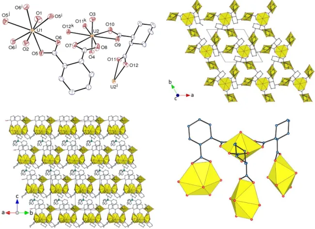
dimethylammonium cations. 3 crystallizes in the trigonal Sohncke group R3; the asymmetric
unit contains two uranyl cations, one of them (U1) located on a threefold rotation axis (Wyckoff
position 3a), and the other (U2) in general position, two R-t-1,2-chdc2– ligands, one highly
disordered H2NMe2+ cation (see Experimental Section), and one PPh3Me+ cation located on a
threefold rotation axis (Figure 3). Both uranium atoms are chelated by three carboxylate groups
from three R-t-1,2-chdc2– ligands and they are thus in hexagonal bipyramidal environments.
The bis-chelating ligands are in the usual chair conformation with both carboxylate groups
equatorial (ee), which results in the formation of a binodal 2D network parallel to (0 0 1). The
point (Schläfli) symbol is {3.92}
3{93} and the topological type is hnb, which, with its nine-node
rings, is an unusual one for nets based on threefold nodes (Figure 4). It is interesting to note
that, if the local environment of the uranyl ion located on the rotation axis is considered, it can
be seen to assume the shape of a half-closed tetranuclear cluster, thus appearing as an
intermediate step (from a geometric, not mechanistic, point of view) toward the formation of
closed, discrete tetrahedral species11,12 (Figure 3). Each of the three tilted uranyl ions attached
to the central one is part of a three-node ring which is also analogous to part of the tetrahedral
cluster. The network thus appears to be derived from the cluster through breaking of two of the
three links around one uranyl group, rotating this group and forming two new links with two
different three-node rings derived from adjacent clusters. While the anionic tetrahedral
13
Figure 3. Top left: View of compound 3. Displacement ellipsoids are drawn at the 30% probability level.
Counterions, solvent molecules and hydrogen atoms are omitted. Symmetry codes: i = 1 – y, x – y + 1, z; j = y – x, 1 – x, z; k = –y, x – y, z; l = y – x, –x, z. Top right: View of the uranyl-based 2D network. Bottom left: Packing with sheets viewed edge-on, and solvent molecules and hydrogen atoms omitted. Bottom right: Local arrangement of uranyl ions and dicarboxylate ligands around the threefold rotation axis.
Figure 4. Simplified side-on and edge-on representations of the uranyl-based 2D network with hnb topology in
14
silver(I) or lead(II) cations (with formation of heterometallic cuboidal clusters with Cs+ and
Rb+), the much bulkier PPh
3Me+ cation promotes a rearrangement which, even if geometrically
minor, yields a 2D coordination polymer. The PPh3Me+ cation is located so that the methyl
group points towards the centre of the three-node ring, and is probably involved in three weak
CH···O hydrogen bonds with three carboxylate oxygen atoms from the ring [C···O 3.349(8) Å,
C–H···O 161°], an interaction clearly apparent on the Hirshfeld surface. Although coulombic
interactions are largely dominant in the packing,34 such weak interactions may play a local role
on the finest details of the arrangement. When viewed sideways, the 2D sheets have a
corrugated aspect, with a thickness of ∼11 Å, one of the faces being lined by the trinuclear rings, while the uranyl cations located on the rotation axes are located on the other face, and the sheets
are separated from one another by layers of PPh3Me+ counterions. Possibly because of the
dominance of the interactions of the methyl groups with carboxylate oxygen atoms, the
counterions are well spaced [shortest P·· ·P distance 13.6231(3) Å] within their sheets and do
not appear to be involved in “phenyl embraces”. As entities with a 3-bladed propeller form,
they are chiral and the chirality of the lattice is reflected in the fact that all define right-handed
helices.
It appears that, among all the coordination modes found up to now in uranyl ion
complexes with the cis and trans isomers of 1,2-chdc2–, which are shown in Scheme 1, the only
one observed in the present complexes with the trans form is the bis-chelating mode. However,
an extra level of variety is introduced by the possible axial or equatorial positioning of the
15
Scheme 1. Coordination modes of c- and t-1,2-chdc2– in uranyl ion complexes.
Complexes [PPh4][UO2(t-1,4-chdc)(NO3)]·2CH3CN (4) and [PPh4][UO2
(c-1,4-chdc)(NO3)] (5), synthesized from t-1,4-chdcH2 and c,t-1,4-chdcH2 (mixture of cis and trans
isomers), respectively, have the same overall formula (except for the solvent), but different
structures. In both cases, the asymmetric unit contains one uranyl cation which is chelated by
two carboxylate groups and one nitrate anion, one 1,4-chdc2– ligand in the chair conformation,
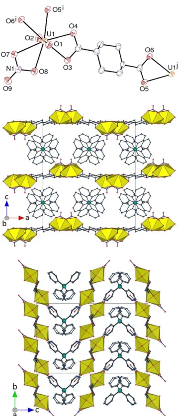
and two PPh4+ cations with twofold rotation symmetry (Figures 5 and 6). The two carboxylate
groups of the trans isomer in 4 are equatorial, and the ligand is thus a divergent, linear linker
which forms a zigzag 1D polymer running along the a axis (a linear chain was previously
observed in [UO2(t-1,4-chdc)(H2O)2], in which the water ligands occupy trans positions in the
uranyl equatorial plane13). These ribbon-like chains are packed in an oblique fashion to form
sheets parallel to (0 0 1), which are separated by large spaces (> 6 Å) containing the counterions
and solvent molecules (KPI 0.69). The counterions are stacked into columns along the b axis,
with an alternation of P··· P distances of 7.253(3) and 7.519(3) Å, in which two
parallel-displaced π-stacking interactions are present within each cation pair [centroid·· ·centroid distances 4.000(3) and 4.181(4) Å, dihedral angles 15.8(3) and 16.2(3)°]. One CH···π
16
Figure 5. Top: View of compound 4. Displacement ellipsoids are drawn at the 50% probability level. Counterions,
solvent molecules and hydrogen atoms are omitted. Symmetry codes: i = x – 1/2, 1/2 – y, 1 – z; j = x + 1/2, 1/2 –
y, 1 – z. Middle and bottom: Packing with chains viewed side-on or end-on, respectively, and solvent molecules
17
interaction involving a proton from the ligand is also possibly present [H··· centroid 2.80 Å, C–
H··· centroid 138°]. Examination of the Hirshfeld surface reveals also several CH· ··O/N
hydrogen bonds between protons of the counterions and solvent molecules and oxo or
carboxylato oxygen atoms, or acetonitrile nitrogen atoms.
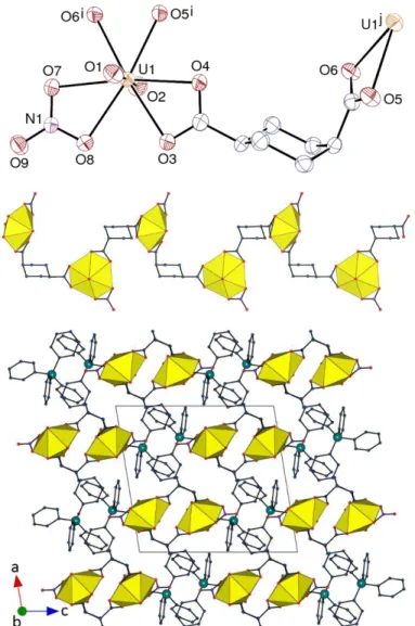
In contrast, one of the two carboxylate groups of the cis isomer of the ligand in 5 is
equatorial and the other is axial, thus conferring a kinked shape to the ligand. A 1D polymeric
chain, directed along the b axis, is formed here also, but it has a distinctly helical shape as a
Figure 6. Top: View of compound 5. Displacement ellipsoids are drawn at the 50% probability level. Counterions
and hydrogen atoms are omitted. Symmetry codes: i = 3/2 – x, y – 1/2, 3/2 – z; j = 3/2 – x, y + 1/2, 3/2 – z. Middle: View of the 1D helical polymer. Bottom: Packing with chains viewed end-on. Hydrogen atoms are omitted in the last two views.
18
result of the ligand geometry and is akin to the chain in the neutral complex [UO2
(c-1,4-chdc)(bipy)], in which bipy is chelating and replaces nitrate.13 The space group being
centrosymmetric, both left- and right-handed helices are present in the lattice. The chains are
arranged so as to form layers parallel to (1 0
ī
), separated from one another by the counterions (KPI 0.68). Centrosymmetric pairs of cations with a P··· P distance of 6.7433(18) Å are formed,and can be considered as a case of sextuple phenyl embrace.7 However, no short contact
indicative of significant π-stacking interaction is present here, but three CH(cyclohexyl)···π interactions are possibly significant [H··· centroid 2.85–2.99 Å, C–H···centroid 151–170°], as
well as one CH···O hydrogen bond involving a proton from the counterion and a carboxylate
oxygen atom [C···O 3.113(5) Å, C–H·· ·O 133°], these contacts being apparent on the Hirshfeld
surfaces.
The two complexes [PPh4]2[(UO2)2(t-1,4-chdc)3]·4H2O (6) and [PPh3Me]2[(UO2)2
(t-1,4-chdc)3]·2H2O (7) differ by the counterion and the number of lattice water molecules, but,
as in complex 3, the absence of a coligand such as formate or nitrate results in the expected
uranium/ligand ratio of 2:3. The asymmetric unit in 6 contains a unique uranium atom chelated
by three carboxylate groups, three centrosymmetric t-1,4-chdc2– ligands and one PPh
4+ cation
(Figure 7). All ligands are in the chair conformation, but two are in the ee and one in the less
usual aa geometry. Examples of coexistence of these two forms are known,28 one of them in a
uranyl–lead(II) complex.13 Uranyl ions are three-fold nodes in the 2D network formed parallel
to (1 –2 1), which has the {63} point symbol of the honeycomb (hcb) topological type. The
same topology was previously encountered in [H2NMe2]2[(UO2)2
(c-1,4-chdc)(t-1,4-chdc)2]⋅2H2O, which crystallizes as a threefold 2D parallel interpenetrated network;13 the
bulkiness of the counterions in 6 may prevent a similar entanglement. Due to the presence of
19
Figure 7. Top: View of compound 6. Displacement ellipsoids are drawn at the 50% probability level. Counterions,
solvent molecules and hydrogen atoms are omitted. Symmetry codes: i = 1 – x, –y, 1 – z; j = 2 – x, 1 – y, 2 – z; k = –x, –y, 2 – z. Middle: View of the uranyl-based 2D network and the counterions. Bottom: Packing of the sheets with counterions and solvent molecules omitted.
20
section. The counterions occupy the channels formed parallel to the a axis, which have a section
of ∼15 × 10 Å2, and within these channels form centrosymmetric pairs involved in sextuple
phenyl embraces with a short P··· P separation of 6.1587(16) Å. One parallel-displaced π -stacking interaction is particularly obvious [centroid···centroid distance 4.027(2) Å, dihedral
angle 0°], and also one CH···π interaction involving a proton from one ligand [H··· centroid 2.69 Å, C–H·· ·centroid 154°] and two CH(counterion)··· O(carboxylate) hydrogen bonds
[C···O 3.091(5) and 3.234(5) Å, C–H·· ·O 124 and 130°].
The asymmetric unit in 7 contains one tris-chelated uranyl cation, one ligand in the ee
conformation, and a second, centrosymmetric one in the aa conformation, and one PPh3Me+
cation (Figure 8). The connectivity is identical to that in 6 and, here also, a 2D network with
hcb topology is formed, parallel to (1 0 –2). The layers have here a square wave-shaped section,
and the counterions are located within the channels (∼15 × 11 Å2) directed along the a axis (KPI
0.68). Once again, the cations in the channels can be considered to contain centrosymmetric
close pairs [P·· ·P 6.680(2) Å] involved in sextuple phenyl embraces, but the Hirshfeld surface
only provides evidence that this embrace involves dispersion interactions and that cation
interactions beyond dispersion are predominantly of the CH··· O type involving oxygen atoms
of the anionic polymer. One CH···π interaction involving a proton from the aa ligand appears to be present [H·· · centroid 2.79 Å, C–H··· centroid 136°] along with four
CH(counterion)···O(oxo/carboxylate) hydrogen bonds, two of them involving the methyl group
21
Figure 8. Top: View of compound 7. Displacement ellipsoids are drawn at the 50% probability level. Counterions,
solvent molecules and hydrogen atoms are omitted. Symmetry codes: i = x + 1, 3/2 – y, z + 1/2; j = x – 1, 3/2 – y,
z – 1/2; k = 2 – x, 2 – y, 1 – z. Middle: View of the uranyl-based 2D network and the counterions. Bottom: Packing
of the sheets with counterions and solvent molecules omitted.
The complex [PPh3Me]2[(UO2)2(c-1,4-chdc)3]·2H2O (8) was synthesized from the
mixture of cis and trans isomers of the ligand, but, as complex 5, it contains only the cis form.
22
in different environments, three ligands in the chair ae conformation and two PPh3Me+ cations
(Figure 9). Atom U1 is chelated by three carboxylate groups, whereas U2 is chelated by two
Figure 9. Top: View of compound 8. Displacement ellipsoids are drawn at the 50% probability level. Counterions,
solvent molecules and hydrogen atoms are omitted. Symmetry codes: i = –x, y – 1/2, 1/2 – z; j = 1 – x, y – 1/2, 1/2 – z; k = –x, y + 1/2, 1/2 – z; l = 1 – x, y + 1/2, 1/2 – z. Middle: View of the uranyl-based 2D network and the counterions. Bottom: Packing of the sheets with counterions and solvent molecules omitted.
23
groups only and bound to one oxygen atom from a third ligand, and it is thus in a pentagonal
bipyramidal environment. This minor difference has no effect on the nature of the polymeric
arrangement, which is here also 2D with the hcb topology, and parallel to (0 0 1). When viewed
sideways, the layers display a triangular wave-shaped or undulating section; the irregular
channels containing the counterions run along the [0 1 1] axis (∼9 × 6 Å2, while the hexagonal
rings have a size of ∼15 × 9 Å2), the overall packing leaving no significant space apart from
that occupied by the water molecules (KPI 0.69). Here, it is the inequivalent cations which
appear to be involved in forming pairs through a phenyl embrace analogous to that found in
complex 4, but that is tighter [P··· P 6.5100(10) Å], and two parallel-displaced π-stacking interactions are possibly present [centroid· ··centroid distances 3.9206(18) and 4.3484(19) Å,
dihedral angles 25.12(15) and 13.60(15)°], along with two CH(cyclohexyl)· ··π interactions [H··· centroid 2.91 and 2.92 Å, C–H·· ·centroid 137 and 123°] and six
CH(counterion)···O(carboxylate/water) hydrogen bonds [C·· ·O 3.260(4)–3.412(4) Å, C–
H···O 143–170°]. The latter interactions involving hydrogen atoms appear prominently on the
Hirshfeld surfaces.
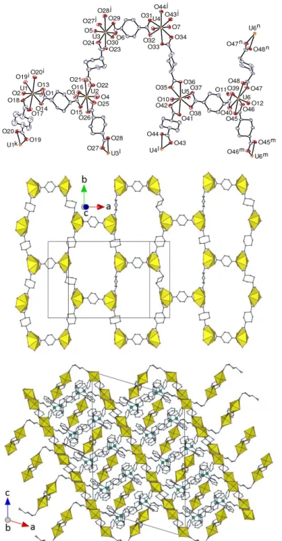
The complex [PPh4]2[(UO2)2(t-1,4-chdc)2(c-1,4-chdc)]·3H2O (9) is the only one in the
present series to include both cis and trans isomers of the 1,4-chdc2– ligand, but other cases,
with varying cis/trans ratios, have previously been described.13 Complex 9 crystallizes in the
monoclinic space group P21/n with a large asymmetric unit containing six tris-chelated uranyl
cations, three c-1,4-chdc2– and seven (two of them centrosymmetric) t-1,4-chdc2– ligands, all in
the chair conformation, and six PPh4+ cations (Figure 10). While all the cis ligands are of the
ae form, five (one of them centrosymmetric) of the trans ligands are of the extended ee form
and two (one of them centrosymmetric) of the kinked aa form. The 2D network formed, parallel
to (1 0 –5), has here also the hcb topology. The layers are strongly corrugated, with a triangular
24
Figure 10. Top: View of compound 9. Displacement ellipsoids are drawn at the 40% probability level.
Counterions, solvent molecules and hydrogen atoms are omitted. Symmetry codes: i = –x – 1/2, y – 1/2, 1/2 – z; j = x, y – 1, z; k = –x – 1/2, y + 1/2, 1/2 – z; l = x, y + 1, z; m = 2 – x, 2 – y, 1 – z; n = 2 – x, 1 – y, 1 – z. Middle: View of the uranyl-based 2D network. Bottom: Packing with sheets viewed edge-on and solvent molecules omitted.
25
along the [1 0
ī
] direction are more elongated and irregular than in 6 and 7 (∼17 × 8 Å2). It ispossible to discern three relatively close cation pairs [P1· ··P4 7.687(4) Å; P2·· ·P5 7.376(4) Å;
P3·· ·P6 7.712(4) Å] involving partial embraces but these only form part of more extended
columnar arrays and the single instance of phenyl ring stacking must be considered rather
distorted [centroid··· centroid distance 4.390(6) Å, dihedral angle 35.6(5)°]. Five
CH(cyclohexyl/phenyl)·· ·π interactions [H··· centroid 2.71–2.99 Å, C–H···centroid 134–151°] are apparent, along with a CH· ··O(water), but no CH···O(carboxylate) hydrogen bond. The
KPI of 0.67 indicates that the packing is no more compact than the former ones.
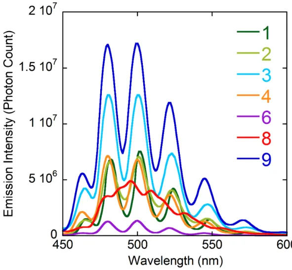
Luminescence properties. The emission spectra of compounds 1–4, 6, 8 and 9 in the
solid state were recorded at room temperature under excitation at a wavelength of 420 nm, a
value suitable for excitation of the uranyl chromophore,35 and they are shown in Figure 11. The
spectra of 1–4, 6 and 9 display the usual series of well-resolved maxima associated with the
vibronic progression corresponding to the S11 → S00 and S10 → S0ν (ν = 0–4) electronic
transitions.36 The maxima positions are very close for these six complexes, with the four main
peaks [S10→ S0ν (ν = 0–3)], at 480–482, 500–501, 521–524 and 545–548 nm. These values are
typical of complexes in which uranyl is chelated by three carboxylate groups,13 and they are
significantly blue-shifted with respect to those generally measured in comparable complexes
with five equatorial donors. It is thus unsurprising that complex 8, in which half the uranyl
centres have five and the other half six equatorial donors, gives a spectrum in which a broad
envelope encompasses several unresolved maxima, the first intense one being at ∼480 nm as expected for the component with six donors, and the second at ∼509 nm, a value at the upper end of the range usual for complexes with five donors.13 A photoluminescence quantum yield
(PLQY) of 0.13 was measured for compound 6, which could be obtained in sufficiently large
26
[NH4][PPh4][(UO2)8(c-1,2-chdc)9(H2O)6]·3H2O.14 This value is relatively low, probably due to
dynamic quenching induced by ambient oxygen, but also static quenching, as already described
for other f-block compounds in their crystalline state.37,38 It is however important to mention
that it is still higher than that for other uranyl-based compounds previously reported.39
Figure 11. Emission spectra of compounds 1–4, 6, 8 and 9 in the solid state, under excitation at a wavelength of
420 nm.
CONCLUSIONS
Substitution on the cyclohexane ring has long been exploited to provide access to multidentate
ligands and these are well-known to form metal ion complexes with properties significantly
dependent upon the conformational preferences of the ring. We report herein a series of uranyl
27
solvo-hydrothermal conditions in the presence of PPh4+ or PPh3Me+ counterions and various
organic cosolvents. The broader spectrum of systems now available with these ligands does
reveal subtle differences in these structures associated with different counterions and also shows
that, in the case of t-1,2-chdc2–, the species obtained from the racemic ligand can display a
degree of selectivity in their isomeric composition.
Although the formation of tetrahedral clusters with t-1,2-chdc2– is frequently observed,
in particular in the presence of NH4+ counterions,12 it is not found with PPh4+ or PPh3Me+
cations. However, the local geometry in the hnb network of compound 3 appears to be derived
from that of the tetrahedral cluster. The cis and trans isomers behave differently in the presence
of the PPh4+ cation since the octanuclear cage which is formed with the former14 is not observed
with the latter. NH4+ cations formed in situ from acetonitrile decomposition probably play an
essential role in the formation of this cage, in which they are included, but experiments
performed with t-1,2-chdc2– in acetonitrile only gave complexes isomorphous to 1 and 2 and
including nitrate ions, and the H2NMe2+ cations formed from DMF hydrolysis and present in
complex 3 are too bulky to act in the same manner as NH4+ (and they cannot form four hydrogen
bonds with oxo groups as the latter). These results suggest that the axial or equatorial
positioning of the carboxylate groups on the cyclohexyl ring in the chair conformation, ee or
aa in the trans isomer (the present results indicate that the difference in energy may be quite
small), and ae in the cis, has a determinant effect on the geometry of the complex. It is notable
that the racemic and pure (1R,2S) enantiomer are no different in the present complexes, due to
resolution of the enantiomers during crystallization in the complexes synthesized from the
racemic form.
The polymeric assemblies formed with bis(chelating) 1,4-chdc2– ligands are either 1D
(helical in one case) or 2D with the hcb topology (finer variations being provided by the
28
trans isomer, linear ee or kinked aa). The prevalence of the hcb topology with both PPh4+ or
PPh3Me+ as counterions, while it has been found only once in previous experiments in the
absence of, or with different counterions,13 points to a particular tendency of these large cations
to induce the formation of species with large rings able to form channels sufficiently spacious
to accommodate them. Probably as a consequence, the entanglement of networks, 2D or 3D,
frequently observed in previous experiments, is absent here. The tendency of PPh4+ and
PPh3Me+ to aggregate in the solid state through phenyl embraces means that they can in fact be
considered to be able to generate exceptionally large cations but the present results show that
this tendency can be disrupted by other weak interactions.
The seven complexes for which the photoluminescence properties were investigated
show uranyl ion emission with the characteristic resolution of vibronic fine structure. The
maxima positions for the six complexes in which the uranyl ion has six equatorial donors match
the values usually found for tris-chelated carboxylate complexes, whereas the spectrum of the
complex in which environments with five and six donors coexist gives a broad envelope
masking a superposition of different series of peaks. A quantum yield of 0.13 was measured for
complex 6, a promising result since this compound is the least emissive in this series.
ASSOCIATED CONTENT Accession Codes
CCDC 1822636−1822644contain the supplementary crystallographic data for this paper. These
data can be obtained free of charge via www.ccdc.cam.ac.uk/data_request/cif, or by emailing
data_request@ccdc.cam.ac.uk, or by contacting The Cambridge Crystallographic Data Centre,
29 AUTHOR INFORMATION Corresponding Authors *E-mail: pierre.thuery@cea.fr. (P.T.) *E-mail: harrowfield@unistra.fr. (J.H.) ORCID Pierre Thuéry: 0000-0003-1683-570X Youssef Atoini: 0000-0003-4851-3713 Jack Harrowfield: 0000-0003-4005-740X Notes
The authors declare no competing financial interest.
REFERENCES
1. Wang, K. X.; Chen, J. S. Extended Structures and Physicochemical Properties of Uranyl–
Organic Compounds. Acc. Chem. Res. 2011, 44, 531–540.
2. Andrews, M. B.; Cahill, C. L. Uranyl Bearing Hybrid Materials: Synthesis, Speciation, and
Solid-State Structures. Chem. Rev. 2013, 113, 1121–1136.
3. Loiseau, T.; Mihalcea, I.; Henry, N.; Volkringer, C. The Crystal Chemistry of Uranium
Carboxylates. Coord. Chem. Rev. 2014, 266–267, 69–109.
4. Su, J.; Chen, J. S. MOFs of Uranium and the Actinides. Struct. Bond. 2015, 163, 265–296.
5. Thuéry, P.; Harrowfield, J. Recent Advances in Structural Studies of Heterometallic
Uranyl-Containing Coordination Polymers and Polynuclear Closed Species. Dalton Trans. 2017, 46,
30
6. Groom, C. R.; Bruno, I. J.; Lightfoot, M. P.; Ward, S. C. The Cambridge Structural Database.
Acta Crystallogr., Sect. B 2016, 72, 171–179.
7. Dance, I.; Scudder, M. Supramolecular Motifs: Concerted Multiple Phenyl Embraces between
Ph4P+ Cations are Attractive and Ubiquitous. Chem. Eur. J. 1996, 2, 481–486.
8. Scudder, M.; Dance, I. Crystal Supramolecular Motifs. Ladders, Layers and Labyrinths of
Ph4P+ Cations Engaged in Fourfold Phenyl Embraces. J. Chem. Soc., Dalton Trans 1998, 3155–
3165.
9. Scudder, M.; Dance, I. Crystal Supramolecular Motifs. Two- and Three-Dimensional Networks
of Ph4P+ Cations Engaged in Sixfold Phenyl Embraces. J. Chem. Soc., Dalton Trans 1998,
3167–3175.
10. Thuéry, P. Two Novel Uranyl–Organic Frameworks with Cyclohexane-1,3-Dicarboxylate
Ligands. CrystEngComm 2009, 11, 232–234.
11. Thuéry, P.; Harrowfield, J. Coordination Polymers and Cage-Containing Frameworks in
Uranyl Ion Complexes with rac- and (1R,2R)-trans-1,2-Cyclohexanedicarboxylates:
Consequences of Chirality. Inorg. Chem. 2017, 56, 1455−1469.
12. Thuéry, P.; Harrowfield, J. Tetrahedral and Cuboidal Clusters in Complexes of Uranyl and
Alkali or Alkaline-Earth Metal Ions with rac- and
(1R,2R)-trans-1,2-Cyclohexanedicarboxylate. Cryst. Growth Des. 2017, 17, 2881–2892.
13. Thuéry, P.; Harrowfield, J. Structural Consequences of 1,4-Cyclohexanedicarboxylate
Cis/Trans Isomerism in Uranyl Ion Complexes: From Molecular Species to 2D and 3D
Entangled Nets. Inorg. Chem. 2017, 56, 13464−13481.
14. Thuéry, P.; Atoini, Y.; Harrowfield, J. Counterion-Controlled Formation of an Octanuclear
Uranyl Cage with cis-1,2-Cyclohexanedicarboxylate Ligands. Inorg. Chem., submitted.
15. Niekiel, F.; Lannoeye, J.; Reinsch, H.; Munn, A. S.; Heerwig, A.; Zizak, I.; Kaskel, S.; Walton,
31
Sorption Properties and Breathing of the Aliphatic Al-MOF [Al(OH)(CDC)]. Inorg. Chem.
2014, 53, 4610−4620.
16. Kimura, K.; Watanabe, Y.; Suda, T.; Senda, H.; Hosoi, S.; Ohta, T.; Kunimoto, K. K. Crystal
Structure of (1R,2R)-trans-1,2-Cyclohexanedicarboxylic Acid-(R)-1-Phenylethylamine Salt.
Anal. Sci. 1999, 15, 609–610.
17. Hooft, R. W. W. COLLECT, Nonius BV: Delft, The Netherlands, 1998.
18. Otwinowski, Z.; Minor, W. Processing of X-Ray Diffraction Data Collected in Oscillation
Mode. Methods Enzymol. 1997, 276, 307–326.
19. Sheldrick, G. M. SHELXT – Integrated Space-Group and Crystal-Structure Determination.
Acta Crystallogr., Sect. A 2015, 71, 3–8.
20. Sheldrick, G. M. Crystal Structure Refinement with SHELXL. Acta Crystallogr., Sect. C 2015,
71, 3–8.
21. Farrugia, L. J. WinGX and ORTEP for Windows: an Update. J. Appl. Crystallogr. 2012, 45,
849–854.
22. Momma, K.; Izumi, F. VESTA: a Three-Dimensional Visualization System for Electronic and
Structural Analysis. J. Appl. Crystallogr. 2008, 41, 653–658.
23. Blatov, V. A. TOPOS, Samara State University, Russia, 2004.
24. Thuéry, P.; Harrowfield, J. Modulation of the Structure and Properties of Uranyl Ion
Coordination Polymers Derived from 1,3,5-Benzenetriacetate by Incorporation of Ag(I) or
Pb(II). Inorg. Chem. 2016, 55, 6799–6816 and references therein.
25. Thuéry, P.; Harrowfield, J. Structural Variations in the Uranyl/4,4ʹ-Biphenyldicarboxylate
System. Rare Examples of 2D → 3D Polycatenated Uranyl–Organic Networks. Inorg. Chem.
2015, 54, 8093–8102 and references therein.
26. Andrews, M. B.; Cahill, C. L. In Situ Oxalate Formation During Hydrothermal Synthesis of
32
27. Knope, K. E.; Kimura, H.; Yasaka, Y.; Nakahara, M.; Andrews, M. B.; Cahill, C. L.
Investigation of in Situ Oxalate Formation from 2,3-Pyrazinedicarboxylate under Hydrothermal
Conditions Using Nuclear Magnetic Resonance Spectroscopy. Inorg. Chem. 2012, 51, 3883–
3890.
28. Hernández-Ahuactzi, I. F.; Höpfl, H.; Barba, V.; Román-Bravo, P.; Zamudio-Rivera, L. S.;
Beltrán, H. I. Pore-Size Tuning in Double-Pillared Metal-Organic Frameworks Containing
Cadmium Clusters. Eur. J. Inorg. Chem. 2008, 2746–2755.
29. Yue, Q.; Huang, Q.; Gao, Y. Y.; Gao, E. Q. Homochiral Coordination Polymers from a Chiral
Dicarboxylic Acid and Dipyridyl Ligands: Structural Diversity, Photoluminescence and
Magnetic Properties. Inorg. Chim. Acta 2016, 443, 110–117.
30. Garza, A. J.; Nag, M.; Carroll, W. R.; Goddard, W. A. III; Roberts, J. D. Conformational
Preferences of trans-1,2- and cis-1,3-Cyclohexanedicarboxylic Acids in Water and Dimethyl
Sulfoxide as a Function of the Ionization State As Determined from NMR Spectroscopy and
Density Functional Theory Quantum Mechanical Calculations. J. Am. Chem. Soc. 2012, 134,
14772–14780.
31. Spackman, M. A.; Jayatilaka, D. Hirshfeld Surface Analysis. CrystEngComm 2009, 11, 19–
32.
32. Wolff, S. K.; Grimwood, D. J.; McKinnon, J. J.; Turner, M. J.; Jayatilaka, D.; Spackman, M.
A. CrystalExplorer, University of Western Australia, 2012.
33. Spek, A. L. Structure Validation in Chemical Crystallography. Acta Crystallogr., Sect. D 2009,
65, 148–155.
34. Gavezzotti, A. The “Sceptical Chymist”: Intermolecular Doubts and Paradoxes.
33
35. Knope, K. E.; de Lill, D. T.; Rowland, C. E.; Cantos, P. M.; de Bettencourt-Dias, A.; Cahill,
C. L. Uranyl Sensitization of Samarium(III) Luminescence in a Two-Dimensional Coordination
Polymer. Inorg. Chem. 2012, 51, 201–206.
36. Brachmann, A.; Geipel, G.; Bernhard, G.; Nitsche, H. Study of Uranyl(VI) Malonate
Complexation by Time Resolved Laser-Induced Fluorescence Spectroscopy (TRLFS).
Radiochim. Acta 2002, 90, 147–153.
37. Tkachuk, A. M.; Razumova, I. K.; Perlin, E. Y.; Joubert, M. F.; Moncorge, R. Luminescence
Self-Quenching in Tm3+: YLF Crystals: II. The Luminescence Decay and Macrorates of Energy
Transfer. Opt. Spectrosc. 2001, 90, 78–88.
38. Tkachuk, A. M.; Ivanova, S. E.; Joubert, M. F. Guyot, Y.; Guy, S. Luminescence
Self-Quenching from 4F
3/2, 2P3/2 and 4D3/2 Neodymium Levels in Double Sodium–Yttrium Fluoride
Crystals. J. Lumin. 2001, 94–95, 343–347.
39. Natrajan, L. S. Developments in the Photophysics and Photochemistry of Actinide Ions and
34
For Table of Contents Use Only
Uranyl–Organic Coordination Polymers with
trans-1,2-, trans-1,4- and cis-1,4-Cyclohexanedicarboxylates:
Effects of Bulky PPh
4+and PPh
3Me
+Counterions
Pierre Thuéry, Youssef Atoini and Jack Harrowfield
One- and two-dimensional uranyl-containing coordination polymers with trans-1,2-, trans-1,4-
and cis-1,4-cyclohexanedicarboxylate ligands result from the use of PPh4+ or PPh3Me+ as
counterions. Both the diequatorial and the unusual diaxial forms are found for the trans isomer