Dichotomous regulation of group 3 innate
lymphoid cells by nongastric Helicobacter species
The MIT Faculty has made this article openly available.
Please share
how this access benefits you. Your story matters.
Citation
Bostick, John W. et al. “Dichotomous regulation of group 3 innate
lymphoid cells by nongastric Helicobacter species.” Proceedings of
the National Academy of Sciences of the United States of America
116 (2019): 24760-24769 © 2019 The Author(s)
As Published
10.1073/pnas.1908128116
Publisher
Proceedings of the National Academy of Sciences
Version
Final published version
Citable link
https://hdl.handle.net/1721.1/124784
Terms of Use
Article is made available in accordance with the publisher's
policy and may be subject to US copyright law. Please refer to the
publisher's site for terms of use.
Dichotomous regulation of group 3 innate lymphoid
cells by nongastric
Helicobacter species
John W. Bosticka,b, Yetao Wangc,1, Zeli Shend, Yong Gea, Jeffrey Browne, Zong-ming E. Chenf, Mansour Mohamadzadeha, James G. Foxd,g, and Liang Zhoua,2
aDepartment of Infectious Diseases and Immunology, College of Veterinary Medicine, University of Florida, Gainesville, FL 32608;bDepartment of Chemical
and Biological Engineering, Northwestern University, Evanston, IL 60208;cDepartment of Microbiology-Immunology, Feinberg School of Medicine,
Northwestern University, Chicago, IL 60611;dDivision of Comparative Medicine, Massachusetts Institute of Technology, Cambridge, MA 02139;eDepartment
of Pediatrics, Division of Gastroenterology, Hepatology and Nutrition, Northwestern Feinberg School of Medicine, Ann & Robert H. Lurie Children’s Hospital
of Chicago, Chicago, IL 60611;fDepartment of Laboratory Medicine and Pathology, Mayo Clinic, Rochester, MN 55905; andgDepartment of Biological
Engineering, Massachusetts Institute of Technology, Cambridge, MA 02139
Edited by David Artis, Weill Cornell Medical College, New York, NY, and accepted by Editorial Board Member Ruslan Medzhitov October 24, 2019 (received for review May 10, 2019)
Intestinal innate lymphoid cells (ILCs) contribute to the protective immunity and homeostasis of the gut, and the microbiota are critically involved in shaping ILC function. However, the role of the gut microbiota in regulating ILC development and maintenance still remains elusive. Here, we identified opposing effects on ILCs by two Helicobacter species, Helicobacter apodemus and Helico-bacter typhlonius, isolated from immunocompromised mice. We demonstrated that the introduction of both Helicobacter species activated ILCs and induced gut inflammation; however, these Helico-bacter species negatively regulated RORγt+group 3 ILCs (ILC3s), es-pecially T-bet+ILC3s, and diminished their proliferative capacity. Thus, these findings underscore a previously unknown dichotomous regu-lation of ILC3s by Helicobacter species, and may serve as a model for further investigations to elucidate the host–microbe interac-tions that critically sustain the maintenance of intestinal ILC3s.
host–microbiome
|
ILC|
Helicobacter|
inflammationI
nnate lymphoid cells (ILCs) are a subset of immune cells that are involved in the protection and homeostasis of mucosal and barrier tissues, but sometimes play pathogenic roles in disease (1, 2). ILCs can be divided into “helper-like” (group 1, 2, and 3 ILCs: ILC1, ILC2, and ILC3) and cytotoxic (natural killer: NK) ILCs. Both helper-like and cytotoxic ILCs express molecules (e.g., cytokines) that promote immunity, but NK cells have the additional capacity to kill other cells through the expression of cytotoxic molecules. Helper-like ILCs are mainly tissue resident at steady state (with the exception of a population of circulating ILC1), but enter circulation under chronic inflammatory condi-tions in both mice and humans (3–6). In contrast, NK cells are readily found in the circulation, but may also establish tissue residency (3). P-selectin glycoprotein ligand-1 (PSGL-1; Selplg) is expressed on the surface of immune cells and plays a key func-tional role in their recruitment from the circulation to areas of inflammation (7) and homing to secondary lymphoid tissues dur-ing steady state (8). The critical signals involved in drivdur-ing the movement of ILCs between tissue residence and circulation re-main to be determined. Specifically, whether PSGL-1 plays an important role in ILC function in the intestine has not been clearly defined.In this study, we sought to determine the role of PSGL-1 in ILC residence in and circulation from the intestine by crossing PSGL-1 knockout mice with Rag1 knockout mice that lack T and B cells but retain ILCs. Strikingly, we found that Rag1‒/‒Selplg‒/‒ double knockout mice had severely reduced ILC3s in the colon. The phenotype was transferable to Rag1‒/‒mice after cohousing and was not dependent on PSGL-1 deficiency. Moreover, the loss of ILC3s was ameliorated by antibiotics treatment, impli-cating the microbiota as the major driving factor for the immune phenotypes. Using selective antibiotics and 16S rRNA gene se-quencing, we identified that the acquisition and overgrowth of
pathobionts, Helicobacter typhlonius and Helicobacter apodemus, contributed to a decrease in the proliferative capacity and sub-sequent loss of ILC3s in the colon.
In human and murine studies, Helicobacter spp. induce path-ogenic responses in their hosts, especially under conditions of compromised immunity (9, 10). Gastric Helicobacter spp., such as Helicobacter pylori, are clinically significant inducers of peptic ulcers, gastric adenocarcinoma, and mucosa-associated lymphoid tissue (MALT) lymphoma (11). Nongastric Helicobacter spp., which populate the intestine rather than the stomach, can induce strong T cell responses and promote the activation and proliferation of both effector and regulatory T cells (12, 13). Previous studies demonstrated that ILCs participate in the pathogenic response to Helicobacter spp., by promoting the production of proinflammatory cytokines (12, 14–17). Here, we demonstrate Helicobacter-induced suppression of ILC3s, uncovering a previously unrecognized neg-ative regulation of ILCs by nongastric Helicobacter spp.
Results
Rag1‒/‒Selplg‒/‒Mice Have Reduced Numbers of ILC3s in the Colon.
To investigate the role of PSGL-1 in ILC maintenance and/or function in the gut, we bred Rag1-deficient mice with Selplg-deficient
Significance
Innate lymphoid cells (ILCs) in the intestine maintain both de-fense against pathogens and homeostasis of intestinal tissue, which is exposed to environmental influences, including mi-crobes and ingested foods. We identified a pair of Helicobacter species that activate ILCs but negatively regulate proliferation of group 3 RORγt+ILCs (ILC3s) that are important for host
im-munity and inflammation. This opens the door for future in-vestigations that explore the molecular factors produced by microbes that may influence the maintenance of ILC3s in the intestine.
Author contributions: J.W.B. and L.Z. designed research; J.W.B., Y.W., Z.S., Y.G., Z.-m.E.C., M.M., and J.G.F. performed research; Z.S., J.B., M.M., and J.G.F. contributed new reagents/ analytic tools; J.W.B., Z.-m.E.C., and L.Z. analyzed data; and J.W.B. and L.Z. wrote the paper.
The authors declare no competing interest.
This article is a PNAS Direct Submission. D.A. is a guest editor invited by the Editorial Board.
Published under thePNAS license.
Data deposition: The data reported in this paper have been deposited in the Gene Ex-pression Omnibus (GEO) database,www.ncbi.nlm.nih.gov/geo(accession no.GSE136171).
1Present address: Program in Molecular Medicine, University of Massachusetts Medical
School, Worcester, MA 01655.
2To whom correspondence may be addressed. Email: liangzhou497@ufl.edu.
This article contains supporting information online athttps://www.pnas.org/lookup/suppl/ doi:10.1073/pnas.1908128116/-/DCSupplemental.
First published November 18, 2019.
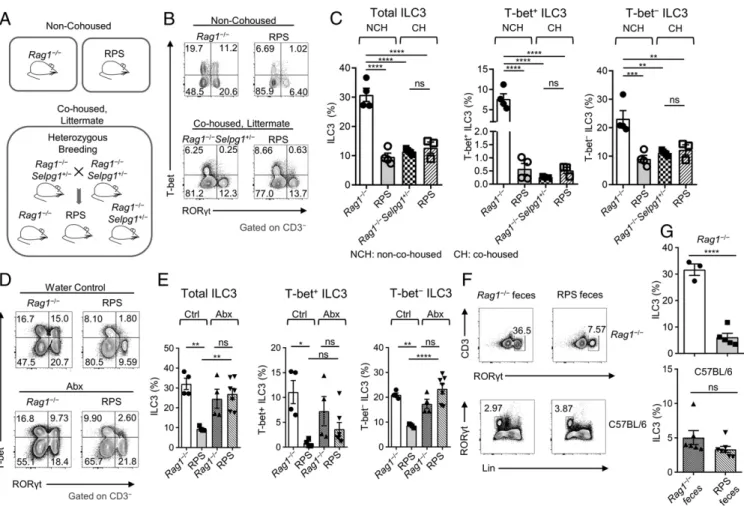
mice (Rag1‒/‒Selplg‒/‒: referred to as RPS), and examined ILC population frequencies among lamina propria lymphocytes (LPLs) in the small and large intestines. We observed a significant de-crease of ILC3 (CD3‒RORγt+) in the large intestine of RPS mice compared to Rag1‒/‒mice (SI Appendix, Fig. S1 A–D), but no difference in the small intestine (SI Appendix, Fig. S1 E and F). PSGL-1 was expressed in ILC3s and deleted in RPS mice (SI Appendix, Fig. S1G). ILC3s are a heterogeneous population of cells, composed of T-bet−adult lymphoid tissue inducer-like cells, which may or may not express CCR6 or CD4, and T-bet+cells, which can up-regulate NKp46 (18–20). Both T-bet+and T-bet−
populations showed decreases in frequency in the large in-testine, with a more marked loss in the T-bet+ILC3 population (SI Appendix, Fig. S1 C and D). T-bet+ILC (ILC1 and NK cell) frequencies were significantly decreased in RPS compared to control Rag1‒/‒ mice (SI Appendix, Fig. S1 A and H). Addi-tionally, we noted an increase in ILC2 frequencies in the large intestine of RPS mice, presumably due to changes in homeostatic balance due to the loss of ILC3s (21) (SI Appendix, Fig. S1 I and J). Together, RPS mice demonstrated significantly reduced ILC3 frequencies in the large intestine with a notable reduction of T-bet+ILC3s.
A Transmissible Microbiota Population Isolated from Rag1‒/‒Selplg‒/‒ Mice Suppresses ILC3s in the Colon.Initial experiments compared RPS mice to nonlittermate Rag1‒/‒control mice. Unexpectedly, when we cohoused Rag1‒/‒mice with RPS mice, Rag1‒/‒ mice had a reduction of ILC3s (SI Appendix, Fig. S1 K and L). Thus, we performed heterozygous breeding to generate cohoused, lit-termate mice (Fig. 1A). We observed no difference in ILC3 frequency between Rag1‒/‒Selplg‒/‒and Rag1‒/‒Selplg+/−controls. Specifically, Rag1‒/‒Selplg+/−and RPS littermate mice both showed reduced ILC3 frequencies compared to nonlittermate, non-cohoused Rag1‒/‒mice (Fig. 1 B and C). T-bet+ILC3s had the most significant decrease in Rag1‒/‒Selplg+/−mice cohoused with RPS mice (Fig. 1C). These data suggested that the ILC3 pheno-type observed in RPS littermate mice was transmissible and might be attributed to microbiota that were introduced at the time of generating the double knockout mice, and not directly caused by the Selplg gene deficiency. Therefore, to clarify the contribution of the microbiota to the phenotype, we treated the RPS mice with a broad-spectrum mixture of antibiotics (ampicillin, vancomycin, metronidazole, neomycin, and gentamicin). After 12 d of treat-ment, we observed that ILC3 percentages in RPS mice had in-creased (Fig. 1 D and E). Notably, the increase in ILC3s was predominantly in the T-bet−ILC3 population, but T-bet+ILC3s
Fig. 1. The microbiota from RPS mice induce a transmissible dysbiois and loss of ILC3s. (A) Illustration of mouse housing and breeding scheme. (B) FACS plots
of RORγt and T-bet staining gated on CD3‒ LPLs from the large intestine (LI) from non-cohoused (Top) and cohoused (Bottom) RPS, Rag1‒/‒, or
Rag1−/−Selpg1+/−mice. (C) Quantification of ILC3 subset frequencies from Rag1‒/‒Selplg+/‒compared to RPS littermate, cohoused (CH) or non-cohoused mice
(NCH). Data are pooled from 2 independent experiments (n= 3 to 4 per group); one-way ANOVA. (D) FACS plots of RORγt and T-bet staining gated on CD3‒LI
LPLs from water-treated (Ctrl; Top) and antibiotics-treated (Abx; Bottom) RPS compared to Rag1‒/‒mice. (E) Frequencies of ILC3 subsets in antibiotics-treated
mice comparing RPS and Rag1‒/‒mice. Data are pooled from 2 independent experiments (n= 4 to 7 per group); one-way ANOVA. (F) FACS plots of RORγt and
CD3‒staining (Top) or Lineage (Lin) and RORγt staining (Bottom) gated on LI LPLs. (G) Quantification of ILC3 frequencies after indicated feces gavage in the LI
of Rag1‒/‒(Top; n= 3 to 5 per group) and C57BL/6 (Bottom; n = 6 to 7 per group) mice. Data are pooled from 2 and 3 independent experiments, respectively.
Error bars indicate SEM. *P< 0.05, **P < 0.01, ***P < 0.001, ****P < 0.0001; ns, not significant.
IMM
UNOLOGY
AND
INFLAM
either did not recover or only partially recovered (Fig. 1 D and E). Next, we performed a bone marrow transfer experiment to test the contribution of the genotype (SI Appendix, Fig. S2A). Rag1‒/‒mice were lethally irradiated, and bone marrow from either Rag1‒/‒or RPS mice was transferred intravenously. After 11 wk, we exam-ined ILC3 frequencies. ILC3 frequencies were lower in mice that were irradiated and received a Rag1‒/‒ bone marrow transfer compared to nonirradiated Rag1‒/‒mice, presumably due to par-tial recovery of ILC3s after bone marrow reconstitution. None-theless, no difference in ILC3 frequencies was observed in mice that received Rag1‒/‒versus RPS bone marrow (SI Appendix, Fig. S2 B and C), suggesting that the Selplg gene deficiency alone could not account for the ILC3 loss. Importantly, gavage of RPS feces into Rag1‒/‒mice reduced ILC3 frequencies compared to Rag1‒/‒ feces or PBS controls in the large intestine (Fig. 1 F and G andSI Appendix, Fig. S2 D, F, H and I), but not the small intestine (SI Appendix, Fig. S2 E and G), indicating that the ILC3 phenotype was potentially induced by the microbiota in RPS feces. Of note, ILC3s were not reduced when wild-type C57BL/6 mice were gavaged with RPS feces, suggesting that adaptive immunity can prevent the loss of ILC3 after RPS feces gavage (Fig. 1 F and G). Feces collected from Rag1‒/‒ mice that were previously gavaged with RPS feces, also known as RPS secondary (RPS 2°)
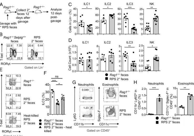
feces, could also reduce ILC3 percentages in the colon of Rag1‒/‒mice after gavage, indicating that an actively growing or self-sustaining agent is responsible for the ILC3 reduction in the colon (Fig. 2 A–D). This was further supported by the result that heat-killed RPS 2° feces could not significantly decrease ILC3s compared to nontreated feces (Fig. 2 E and F). In contrast, gavage of RPS 2° feces into C57BL/6 mice did not reduce ILC3 frequencies in the large intestine, supporting the finding that adaptive immunity can prevent the loss of ILC3s (SI Appendix, Fig. S2J). We examined other ILC populations after RPS 2° feces gavage and identified a decrease in ILC1 (Lineage‒T-bet+RORγt−EOMES−) frequencies and an increase in ILC2 frequencies (Fig. 2C). However, neither ILC1 nor ILC2 cell counts showed significant change after gavage with RPS 2° feces (Fig. 2H). There was a significant increase of NK cells (Lineage‒T-bet+RORγt−EOMES+) after gavage (Fig. 2 C and D). Furthermore, similar to the enhanced gut inflammation ob-served in primary RPS mice (SI Appendix, Fig. S3 A and B), RPS 2° feces gavage resulted in increased inflammation and cell in-filtration, which coincided with the loss of ILC3s (SI Appendix, Fig. S3 C and D). Increased neutrophils and eosinophils were observed in the large intestine 12 d after gavage (Fig. 2 G and H). Together, these data implicated the microbiota as the major
Fig. 2. Serially gavaged RPS feces suppress ILC3 frequencies and are associated with inflammation. (A) Illustration of RPS secondary (2°) feces gavage
ex-periment. (B) FACS plots of RORγt and T-bet gated on Lin−LI LPLs. (C) Quantification of ILC frequencies and (D) counts after gavage with RPS 2° feces or
Rag1‒/‒control feces. Data are pooled from 3 independent experiments (n= 5 to 8 per group). (E) FACS plots of RORγt and T-bet staining gated on Lin−LI
LPLs. (F) Quantification of ILC3 frequencies after gavage with heat-killed or nontreated RPS 2° feces. Data are from 1 experiment (n= 4 to 5 per group);
one-way ANOVA. (G) FACS plots of (neutrophil; CD11b and Ly6g) and (eosinophil; CD11c and Siglec-F) staining gated on CD45+LI LPLs. (H) Quantification of
neutrophil and eosinophil frequencies from Rag1‒/‒mice gavaged with Rag1‒/‒or RPS 2° feces. Data are representative from 1 of 2 independent experiments
(n= 3 per group). Error bars indicate SEM. *P < 0.05, **P < 0.01, ***P < 0.001; ns, not significant.
contributing factor to the loss of ILC3s and inflammation ob-served in RPS mice.
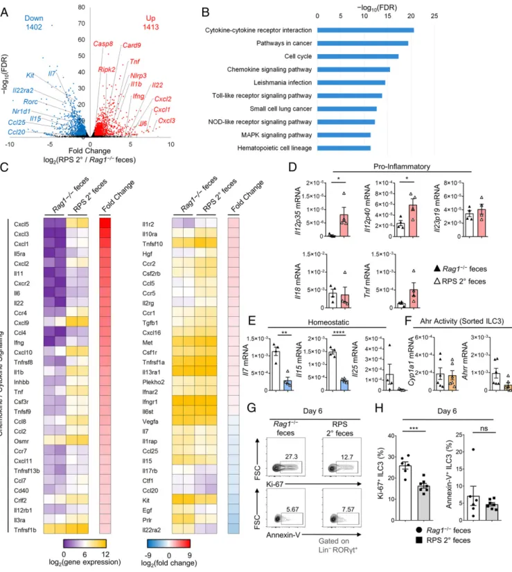
Introduction of RPS Microbiota Changes Gut Transcriptome and Reduces ILC3 Proliferation. Changes initiated by the microbiota in the host colon environment of RPS mice could contribute to the loss of ILC3s. To test this, we examined transcriptional changes in the large intestine of Rag1‒/‒mice 12 d after gavage with RPS 2° feces by RNA sequencing (RNA-seq). Differential gene analysis identified 2,815 significantly changed genes (1,402 down-regulated; 1,413 up-regulated; [q ≤ 0.05; fold change ≥1.5]) in the colon after gavage (Fig. 3A). Many of the increased genes were related to inflammation and response to infection, including Tnf, Ifng, Il1b, Il11, and Il6 (Fig. 3A). Pathway analysis confirmed that cytokine and chemokine signaling was increased, along with Toll-like and NOD-like receptor pathways (Fig. 3B
and SI Appendix, Fig. S4 A and B). Detailed examination of
chemokines and cytokines revealed that genes Cxcl1, Cxcl2, Cxcl3, Cxcl9, and Cxcl10 related to the chemotaxis of inflam-matory cells, such as neutrophils and NK cells, were highly up-regulated, consistent with their increased frequencies, as dem-onstrated by flow cytometry (Figs. 3C and 2 G and H). Notably, genes related to ILC3 homeostasis were significantly decreased, including Il7, Il15, and Kit (Fig. 3 A and C). To confirm the RNA-seq results, we examined the transcription of genes in whole colon tissue that are involved in inflammation and ILC homeostasis by quantitative reverse transcription PCR (qRT-PCR). At day 12 after gavage, we observed no significant dif-ference in proinflammatory cytokine transcripts for Il18 and Il23 (IL-23p19), but a significant increase in Il12a (IL-12p35), Il12b (IL-12p40), and Tnf in the colon (Fig. 3D). Previous studies have shown that IL-12 can contribute to ILC3 plasticity, inducing their conversion into ILC1s (22). We observed increased expression of Il12a and Il12b (Fig. 3D), indicating that ILC3 conversion to ILC1 through plasticity was possible in the intestinal environment after gavage.
Changes in homeostatic molecules were confirmed by qRT-PCR, with reductions in both Il7 and Il15 transcripts (Fig. 3E), both of which have been shown to support ILC3 maintenance in the intestine (23, 24). Similar transcriptional changes of cytokines were observed in the cecum (SI Appendix, Fig. S4C). We examined the expression of other molecules that may influence the main-tenance of ILC3s. Previous studies have shown that increased IL-25 can reduce ILC3 numbers in the intestine (25), but RPS 2° feces-gavaged mice had decreased Il25 expression (Fig. 3E). The aryl hydrocarbon receptor (Ahr) is a key transcription factor in-volved in the maintenance and function of ILC3s (26–28). We examined transcriptional expression in fluorescence-activated cell sorting (FACS)-purified ILC3s from the large intestine of RPS 2° feces-gavaged mice and did not observe any significant difference in the expression of Cyp1a1 or Ahrr (Fig. 3F), indicating no change in Ahr activity. Together, these data indicate that RPS 2° feces gavage can induce a proinflammatory environment with reduced homeostatic molecules.
Reduction of homeostatic cytokines could contribute to re-duced proliferation or survival of ILC3s (23, 24, 28). Thus, we performed a kinetic study of the ILC3 compartment after in-troduction of RPS 2° feces to examine proliferation and survival. Flow cytometry results showed that at day 6 after gavage with RPS 2° feces, ILC3s had a significant reduction in the prolifer-ation marker, Ki-67 (Fig. 3 G and H). We also observed a re-duction in proliferation as early as day 1 and day 3 (SI Appendix, Fig. S4 D–G). However, no increase in apoptosis, as revealed by Annexin V staining, was observed in ILC3s at any time point (Fig. 3 G and H andSI Appendix, Fig. S4 D–G). Additionally, no change in antiapoptotic Bcl2 expression was observed in sorted ILC3s, suggesting that the loss of ILC3s in the large intestine after RPS 2° feces gavage may be mediated by a reduction in
proliferation but not apoptosis (SI Appendix, Fig. S4H). We ex-amined other potential molecules related to the regulation of ILC3 proliferation at early time points after gavage. Ahr and c-Kit (CD117), an Ahr-regulated growth factor receptor, have been shown to control ILC3 proliferation (18, 26–28). Flow cytometry staining for Ahr and c-Kit found no changes in the expression of either molecule in ILC3s from the large intestine (SI Appendix, Fig. S4 I and J). Together, these results suggest that dysbiosis may lead to the early proliferation changes and reduction of ho-meostatic promoting factors that are important for sustaining the ILC3 compartment.
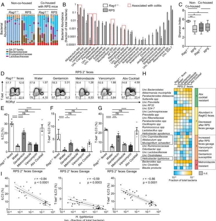
Helicobacter Species Induce Gut Dysbiosis and Are Associated with the Loss of ILC3s in Immunocompromised Mice. To identify the microbiota species that may mediate the change in proliferation and loss of ILC3s, we performed 16S rRNA gene sequencing to com-pare the microbial composition in RPS, Rag1‒/‒, and Rag1‒/‒mice cohoused with RPS mice. Sequencing data revealed that compared to Rag1‒/‒mice, RPS mice had perturbed microbiota, characterized by outgrowth of Lactobacillus, Prevotella, Allobaculum, and Heli-cobacter spp. (Fig. 4 A and B) and decreased bacterial diversity (Shannon Index) (Fig. 4C). Notably, this perturbed microbiota was transmissible to cohoused Rag1‒/‒mice (Fig. 4 A and C).
Prevotella and Helicobacter spp. are known pathobionts in immunocompromised mice and may contribute to inflammation (9, 29). To narrow down possible bacterial agents that contrib-uted to the phenotype, we treated mice with a broad-spectrum antibiotics mix (ampicillin, neomycin, vancomycin, metronida-zole, and gentamicin) or individually with antibiotics that tar-geted specific classes of bacteria (e.g., anaerobes, gram-positive, etc.). We observed that loss of ILC3s was mediated by a pop-ulation of bacteria that were vancomycin and metronidazole resistant but susceptible to gentamicin or a combination of all antibiotics (Fig. 4 D–F andSI Appendix, Fig. S5A). We proceeded to assess the fecal microbial materials from antibiotics-treated mice by 16S rRNA gene sequencing to identify the bacteria that were differentially present among the antibiotics treatment groups. Based on analysis, we identified 4 species that correlated with reduced ILC3s and susceptibility or resistance to the anti-biotics combinations: H. typhlonius, Mucispirillum schaedleri, an unclassified species from the Coprobacillaceae family, and an un-classified Allobaculum spp. (Fig. 4H). Neither M. schaedleri, Allobaculum, nor Coprobacillaceae have been shown to induce inflammation in immunocompetent mice. Rather, they have been associated with outgrowth under inflammatory conditions (30). However, recent work suggests that Nod2- and/or Cybb-deficient mice are susceptible to Mucispirillum-induced colitis (31). We examined M. schaedleri by qPCR and found its low abundance in mice gavaged with RPS 2° feces (SI Appendix, Fig. S5B). Heli-cobacter spp. have been known to initiate inflammation in immune-deficient mice (10, 17). We found in our gavage experi-ments that higher H. typhlonius shed in the feces of mice strongly correlated with lower total large intestinal (Pearson r= −0.84, P value <0.0001), T-bet+, and T-bet− ILC3 frequencies (Fig. 4I); hence, the expansion of H. typhlonius in RPS mice warranted further investigation.
H. typhlonius and H. apodemus Promote the Loss of Colonic ILC3s.H. typhlonius induces murine colitis within an immunocompromised background, such as Il10−/−, Prkdcscid, and Tbx21−/−Rag2−/−mice (17, 32, 33). Thus, we aimed to isolate the Helicobacter spp. that may be responsible for the observed phenotypes (i.e., reduction of ILC3s) from RPS 2° feces. While H. apodemus (Massachusetts Institute of Technology, MIT 18-1095S) was present in both Rag1−/− and RPS 2° feces, H. typhlonius (MIT 18-1095F) was identified only in RPS 2° feces (SI Appendix, Fig. S6A). To elu-cidate the contribution of these specific species to the ILC phenotype observed, we gavaged Rag1−/− mice with either
IMM
UNOLOGY
AND
INFLAM
Fig. 3. The microbiota from RPS mice induces colonic transcriptional changes that result in and reduce ILC3 proliferation. (A) Volcano plot of significantly
up-regulated (up) and down-up-regulated (down) genes in the LI of Rag1‒/‒mice gavaged with RPS 2° or Rag1‒/‒feces. (B) The top pathways identified by pathway
analysis of differentially regulated genes using the Kyoto Encyclopedia of Genes and Genomes database. (C) Heatmap of cytokine or chemokine gene ex-pression and fold change. Data are representative from 1 experiment with 2 biological replicates per group. (D) Gene exex-pression relative to Actin as measured
by qRT-PCR for indicated proinflammatory and (E) ILC homeostatic genes expressed in the proximal colon of mice gavaged with RPS 2° or Rag1‒/‒feces. (F)
Gene expression relative to Actin as measured by qRT-PCR for Ahr target genes in FACS-purified ILC3s from the LI of mice gavaged with RPS 2° or Rag1‒/‒
feces. Data are representative from 1 of 2 independent experiments (n= 4 to 6 per group). (G) FACS plots of Ki-67 (Top) and Annexin V (Bottom) staining
gated on Lin‒RORgt+LI LPLs. (H) Quantification of Ki-67+(Left) and Annexin V+(Right) ILC3 frequencies in LI LPLs at day 6 after gavage with RPS 2° or Rag1‒/‒
feces. Data are pooled from 2 independent experiments (n= 6 to 7 per group). Error bars indicate SEM. *P < 0.05, **P < 0.01, ***P < 0.001, ****P < 0.0001; ns,
not significant.
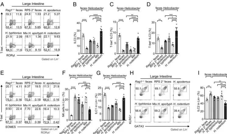
H. apodemus, H. typhlonius, or a mixture of the 2 bacteria to examine ILC populations 12 d later (SI Appendix, Fig. S6B). As a control, we used Helicobacter rodentium, another Helicobacter spp. that colonizes mice but was absent in our colony of Rag1−/−mice (SI Appendix, Fig. S6B). Compared to H. rodentium, gavage with
H. apodemus and/or H. typhlonius (single or combined trans-faunation) reduced large intestinal ILC3s (Fig. 5 A and B), par-ticularly T-bet+RORγt+ILC3 (Fig. 5 C and D andSI Appendix, Fig. S6C), and increased NK cell frequencies and counts (Fig. 5 E and F andSI Appendix, Fig. S6D), consistent with the phenotype observed
Fig. 4. H. typhlonius is associated with gut dysbiosis and loss of ILC3s. (A) Bacterial (family) abundances by 16S rRNA gene sequencing in non-cohoused or
cohoused Rag1‒/‒and RPS mice. (B) Quantification of bacterial abundance as a fraction of total bacteria for significantly changed (P≤ 0.05, Mann–Whitney
test) species; unclassified (Unc.). (C) Shannon Index measuring bacterial diversity of non-cohoused and cohoused Rag1‒/‒and RPS mice. Data are
represen-tative from 1 experiment with 4 to 7 biological replicates per group; one-way ANOVA. (D) FACS plots of RORγt and T-bet staining gated on LI LPLs. (E)
Quantification of total, (F) T-bet+and (G) T-bet−ILC3 frequencies in LI LPLs from RPS 2° feces-gavaged Rag1−/−mice after antibiotics treatment. Data are
pooled from 2 independent experiments (n= 4 per group); one-way ANOVA compared to water control mean. (H) Heatmap indicating the abundance of
bacteria as measured by 16S rRNA gene sequencing after selective antibiotics treatment or untreated controls (n.d., not detected). Data are representative
from 1 experiment with 2 biological replicates per group. (I) Scatterplots indicating H. typhlonius abundance compared to total (Left), T-bet+(Center), or
T-bet−(Right) ILC3 frequencies. Regression lines (solid) and 95% confidence intervals (dashed) are plotted. Pearson correlation (r) and P value are indicated.
Data are pooled from 5 experiments (n= 33). Error bars indicate SEM. *P < 0.05, **P < 0.01, ***P < 0.001, ****P < 0.0001; ns, not significant.
IMM
UNOLOGY
AND
INFLAM
after gavage with RPS 2° feces. In addition, gavage with H. typhlonius reduced the frequencies of Ki-67+T-bet− ILC3s, but not Ki-67+T-bet+ILC3s, in the large intestine, similar to RPS 2° feces (SI Appendix, Fig. S5C). This observation supports the re-duction of T-bet−ILC3s, but cannot account for the loss of T-bet+ ILC3s. Of note, endogenous H. apodemus (MIT 18-1095S) pre-sent in Rag1−/− mice did not cause a reduction of ILC3s but transfaunation of pure H. apodemus bacteria with large quantities led to reduced ILC3s (Fig. 5 A and B). When we examined other ILC populations, we found that large intestinal ILC1 frequencies and cell counts were reduced in mice after gavaging with H. apodemus and/or H. typhlonius (Fig. 5 E and G andSI Appendix, Fig. S6D). ILC2 frequencies slightly increased, but there was a decrease in cell number (Fig. 5 H and I and SI Appendix, Fig. S6D). Together, these data suggest the differential role of Heli-cobacter spp. in regulating different subsets of ILCs.
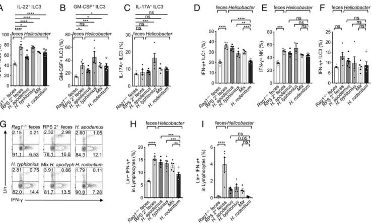
Helicobacter spp. Activate ILC Function and Induce Inflammation.We further determined the effect of Helicobacter spp. on the function of ILCs. We observed increased IL-22+, GM-CSF+, or IL-17A+ ILC3 frequencies (Fig. 6 A–C), and their absolute numbers were decreased or unchanged in RPS feces-gavaged mice and in mice transfaunated with Helicobacter spp. except for H. rodentium (SI Appendix, Fig. S6 E–G). These data suggest that Helicobacter spp. induce activation of ILC3s, consistent with previous reports (14–17); however, H. typhlonius and H. apodemus but not H. rodentium reduce ILC3 numbers. Similar to the RPS 2° feces gavage, transfaunation of mice with either H. typhlonius or H. apodemus induced a strong IFN-γ response, notably expressed by ILC1s and infiltrating NK cells but not by ILC3s (Fig. 6 D–F).
We also observed an increase of a lineage+(CD3, CD19, and Ly6g) IFN-γ+population of cells in mice gavaged with RPS 2°
feces, H. typhlonius, H. apodemus, or mixed treatments that was absent in H. rodentium-gavaged mice (Fig. 6 G–I). In Rag1−/−
mice, this lineage+population is likely infiltrating Ly6g+ neutro-phils. These data indicate that H. apodemus and H. typhlonius, may cooperatively contribute to the loss of T-bet+ILCs and the induced inflammation. Indeed, the mixed 50:50 Helicobacter transfaunation treatment most closely resembled results obtained in mice gavaged with RPS 2° feces (Fig. 5B). However, this does not exclude the possibility that other bacterial species endogenous to the resident mouse colony or enriched in the RPS feces, in-cluding Prevotella and Mucispirillum spp., potentially contribute to the described phenotypes (Fig. 4 B and H). Together, these data demonstrated that Helicobacter spp. activate ILC function by en-hancing their cytokine production.
We speculated that the loss of ILC3s might result in altered immunity of mice transfaunated with H. typhlonius or H. apo-demus. To this end, we determined the action of Helicobacter in 2 models of colitis, dextran sodium sulfate (DSS)-induced colitis and Citrobacter rodentium infection, both of which depend on ILC3s for protective immunity (25, 34, 35). We induced DSS colitis after Helicobacter spp. gavage and found that Helicobacter-gavaged mice had increased weight loss compared to Rag1‒/‒ feces-gavaged control mice (SI Appendix, Fig. S7 A and B). In-triguingly, the H. rodentium-gavaged control mice, which did not show a reduction of ILC3s after gavage (Fig. 5A), had increased weight loss, consistent with more inflammatory cytokine pro-duction (e.g., IFN-γ) by ILC3s (Fig. 6 A–C and SI Appendix,
Fig. S6 E–G) that can influence the DSS colitis phenotype
Fig. 5. H. typhlonius and H. apodemus promote the loss of ILC3s in the large intestine. (A) FACS plots of RORγt and T-bet staining gated on Lineage−LI LPLs.
(B) Quantification of ILC3, (C) T-bet+, and (D) T-bet−ILC3 frequencies in LI LPLs of mice gavaged with feces or Helicobacter spp. (E) FACS plots of EOMES and
T-bet staining gated on Lineage−RORγt−LI LPLs. (F) Quantification of ILC1 (RORγt−T-bet+EOMES−) and (G) NK cells (RORγt−T-bet+EOMES+) in LI LPLs of mice
gavaged with feces or Helicobacter spp. (H) FACS plots of GATA3 and KLRG1 staining gated on Lineage−LI LPLs. (I) Quantification of ILC2 frequencies in LI LPLs
of mice gavaged with feces or Helicobacter spp. Data are pooled from 3 independent experiments (n= 5); one-way ANOVA. Error bars indicate SEM. *P < 0.05,
**P< 0.01, ***P < 0.001, ****P < 0.0001; ns, not significant.
(e.g., wasting disease) (SI Appendix, Fig. S7B). Since DSS treatment caused ILC3 reduction in Rag1−/−mice compared to Rag1−/−mice at the steady state (SI Appendix, Figs. S7C and S1 A and B), the role of Helicobacter spp. in regulating ILC3-mediated protection was unclear in the aforementioned model.
Consistent with DSS-induced wasting disease, during C. rodentium infection, Helicobacter spp. gavage led to more body weight loss (SI Appendix, Fig. S7 D and E). Moreover, Helicobacter-gavaged mice had more dissemination of bacteria to the spleen compared to Rag1‒/‒mouse feces-gavaged control mice (SI Ap-pendix, Fig. S7F). Together, these data suggest that Helicobacter spp. may induce prolonged colonic inflammation and tissue damage that lead to dissemination and wasting disease. Discussion
We started our investigation examining the role of PSGL-1 in ILC biology, but discovered an important interaction between the microbiota and host immune cells. We favored the possibility that RPS mice acquired, by chance, pathobiont Helicobacter species in our mouse facility. These Helicobacter function as key contributors to the reduction of ILC3s in the large intestines of mice that lack adaptive immunity. However, we could not rule out the possibility that PSGL-1 deficiency helped initiate the fortuitous outgrowth of the Helicobacter spp. in RPS mice. Fur-ther study is needed to determine if loss of PSGL-1 is an initiating factor or dispensable for the observed phenotype. Nevertheless, our data show that in Helicobacter-gavaged Rag1−/− mice, in-flammation and pathology, as noted by increased cell infiltration and cytokine expression, is negatively correlated with the fre-quency of ILC3s. It remains to be determined if the loss of ILC3s is responsible for gut inflammation or vice versa.
Individual ILC3 subset (T-bet+vs. T-bet–) responses to RPS feces or Helicobacter spp. gavage differed in our experiments. Our data showed that treatment of RPS feces-transferred Rag1−/−mice with antibiotics results in more significant restoration of the T-bet– ILC3 population. However, Helicobacter spp. treatment suppressed the T-bet+but not T-bet–ILC3s. The 16S rRNA gene sequencing shows in addition to Helicobacter spp., the expansion/enrichment of Prevotella, Mucispirillum, and other colitis-associated bacteria in the RPS feces. The differences in the ILC3 subset responses to RPS feces versus Helicobacter spp. gavage may be influenced by these additional bacteria. In addition, changes in proliferation observed after RPS feces or Helicobacter spp. gavage distinctly affected T-bet−, but not T-bet+, ILC3s.
There are 2 major subsets of ILC3s. T-bet+ILC3s are CCR6− and can up-regulate NKp46. On the other hand, T-bet−ILC3s may or may not express CCR6 or CD4 (18–20). Previous work has shown that T-bet+CCR6−ILC3 numbers are influenced by signals from the microbiota, as germ-free mice had lower num-bers compared to mice raised in specific pathogen-free (SPF) conditions (18–20). Our data showed that the numbers of T-bet+
CCR6−ILC3s were reduced by the introduction of Helicobacter spp. Precise mechanisms remain to be determined and may in-volve either a direct impact on those specific ILC3 subsets or an indirect mechanism of dysbiosis, consistent with the reduced microbial diversity upon RPS fecal treatment. In addition, it has been shown that T-bet–CCR6–ILC3s can up-regulate T-bet, some of which can lose RORγt (18, 23). Thus, plasticity of ILC3s may also account for the reduction of T-bet+ILC3s. Together, these data indicate multiple mechanisms may affect the maintenance of T-bet−versus T-bet+ILC3s in mice gavaged with RPS feces or Helicobacter spp.
Fig. 6. H. typhlonius and H. apodemus enhance ILC function. (A) Quantification of IL-22+, (B) GM-CSF+, and (C) IL-17A+ILC3s frequencies in LI LPLs of mice
gavaged with feces or Helicobacter spp.; one-way ANOVA compared to the mean of Rag1‒/‒control. (D) Quantification of IFN-γ+ILC1, (E) NK, and (F)
ILC3 frequencies after gavage with feces or Helicobacter spp. (G) FACS plots of IFN-γ and Lineage (CD3, CD19, and Ly6g) staining gated on LI LPLs. (H)
Quantification of Lin+and (I) Lin−IFN-γ+LI LPLs from mice gavaged with feces or Helicobacter spp. Data are pooled from 3 independent experiments (n= 5);
one-way ANOVA. Error bars indicate SEM. *P< 0.05, **P < 0.01, ***P < 0.001, ****P < 0.0001; ns, not significant.
IMM
UNOLOGY
AND
INFLAM
Novel nongastric Helicobacter spp. are increasingly identified in cases of diarrhea and bacteremia in humans and animals (10, 36, 37). Helicobacter hepaticus infection in immunocompro-mised mice has been widely used as a model of inflammatory bowel disease, and chronic inflammation induced by H. hepaticus infection in Rag2−/−mice promotes tumor formation in the colon (9, 14, 38). Similarly, we identified increased proinflammatory cytokines after H. apodemus or H. typhlonius gavage in Rag1‒/‒mice. Powell (17) showed that H. typhlonius promoted gastrointestinal pathology in Tbx21−/−Rag2−/− ulcerative colitis (TRUC) mice. Additionally, they found that IL-17A–producing ILCs contributed to the colitis, implicating ILC3s (17). Accordingly, treatment with agents that deplete ILCs (e.g., CD90 and anti–IL-7Ra anti-bodies) reduced the pathology of Helicobacter-induced colitis (14, 17). Consistently, our data showed that transfaunation of Heli-cobacter spp. can increase production of IL-17A, IFN-γ, and GM-CSF by ILCs in Rag1−/−mice. Thus, these data indicate that ILC3s may play a pathogenic role by producing proinflammatory cyto-kines in immune-compromised mice lacking the adaptive immune system (14–16).
In the presence of adaptive immunity, ILC3 induction/activa-tion by Helicobacter spp. plays a protective role by limiting ef-fector T cell responses toward Helicobacter spp. Indeed, a recent study found that ILC3s negatively regulate the adaptive immune response to H. typhlonius (39). Antigen presentation by ILC3s suppresses the effector T cell response to H. typhlonius, suggesting that ILC3s, along with regulatory T cells, may be responsible for limiting effector T cell-mediated pathology in response to Heli-cobacter spp. in the colon. H. typhlonius and H. apodemus are both potent inducers of antigen-specific effector and regulatory T cell responses (12). The addition of IL-10 receptor blocking antibodies in a model of DSS-induced colitis results in the expansion of H. typhlonius and H. apodemus in the population of mucosa-associated bacteria. This is consistent with previous reports indicating that IL-10–deficient mice are particularly susceptible to colonization by enteric Helicobacter spp., and that regulatory T cells suppress Helicobacter-induced pathology, dependent on IL-10 (13, 33). In our study, C57BL/6 wild-type mice gavaged with Helicobacter do not lose ILC3s, unlike Rag1-deficient mice, suggesting that the adaptive immunity could sustain ILC3s in the presence of Helicobacter spp. These data suggest a balance between the in-duction of innate immunity and effector T cells by Helicobacter spp., and the induction of regulatory responses (e.g., regulatory T cells). Factors from H. apodemus or H. typhlonius may work directly or indirectly on immune cells in the colon. Further study is re-quired to identify and test specific factors from these species. Genomic sequencing of H. typhlonius and H. apodemus indicate that these bacteria contain a number of virulence factors whose contribution to the ILC3 phenotype would need to be tested through knockout mutations (40, 41). Nonetheless, in this study we discovered that H. apodemus and H. typhlonius introduction into immunocompromised mice are suppressive of ILC3s, and can serve as a model for future investigations of the host–microbe interactions that maintain ILC3s in the gut.
Materials and Methods
Mice. All of the mice in this study were maintained in SPF facilities at Northwestern University or later at the University of Florida. Mice were of both sexes, littermates, and 6 to 10 wk old unless otherwise indicated. C57BL/6
and Rag1−/−were purchased from The Jackson Laboratory. Rag1−/−mice
were crossed with Selplg−/−mice to generate Rag1−/−Selplg−/−mice. The
institutional animal care and use committees of Northwestern University and the University of Florida approved all studies with mice.
Bacterial Cultures, Fecal Collection, and Administration. H. apodemus (MIT 18-1095S), H. typhlonius (MIT 18-1095F), which were isolated from the feces of RPS mice in the same colony, and H. rodentium (ATCC700285) were grown
under microaerobic conditions in a vented jar containing N2, H2, and CO2
(80:10:10) at 37 °C on 5% sheep blood agar plates for 2 to 3 d. Bacteria were
collected and resuspended in Brucella broth with 20% glycerol, and the
bacteria concentration was adjusted to 1 OD600/mL. Mice were orally gavaged
with 200μL inoculum every other day, a total of 3 times. Freshly collected
feces from Rag1−/−, RPS, or mice previously gavaged with RPS feces were
homogenized in 20% glycerol in sterile PBS and stored as a slurry at −80 °C. Fecal slurries were pooled, centrifuged, and washed with sterile
PBS before resuspension in sterile PBS for gavage (200μL per mouse, once).
Antibiotics Treatments. Mice were treated for 12 d with a broad-spectrum mixture of antibiotics (ampicillin [1 g/L], vancomycin [0.5 g/L], metronida-zole [1 g/L], neomycin [1 g/L], and gentamicin [1 g/L]) or with individual antibiotics at the indicated concentrations in their drinking water. Isolation of Lymphocytes from Intestinal Lamina Propria and Flow Cytometry. Isolation of intestinal lamina propria cells and flow cytometry were done as previously described (28). CD16/32 antibody (eBioscience) was used to block the nonspecific binding to Fc receptors before surface staining. Lymphocytes isolated from intestinal lamina propria were stained with antibodies against
the following markers: GATA3 (APC, PE-Cy7), RORγt (Brilliant Violet 421, PE),
T-bet (PE-Cy7), Eomes (PerCP-eFluor 710), Ahr (APC), CD117 (PE), KLRG1 (PerCP-eFluor 710), CD3 (FITC), CD4 (Alexa Fluor 488, Brilliant Violet 510), NKp46 (PerCP-eFluor 710, Brilliant Violet 510), GM-CSF (Alexa Fluor 488),
IFN-γ (APC), IL-22 (APC), IL-17A (eFluor 710), CD45.2 (APC,
PerCP-Cy5.5), CD11b (PerCP/PerCP-Cy5.5), CD11c (PE-Cy7), and Siglec-F (Brilliant Violet 421). For staining of ILC1 and NK cells, Lineage marker mix (Lin) contained APC-Cy7 or APC-eFluor 780-CD3, CD19, and Ly6G. For staining of ILC2 and ILC3 cells, Lin referred to a combination of lineage markers: Cy7 or APC-eFluor 780-CD3, CD5, CD19, B220, CD11b, CD11c, Ter119, and Ly6G. For nuclear transcription factor staining, cells were fixed and permeabilized with Foxp3 staining buffer kit (eBioscience). For cytokine staining, cells were stimulated with 50 ng/mL phorbol 12-myristate 13-acetate and 500 ng/mL
ionomycin for 4 h and Brefeldin A (2μg/mL) was added 2 h before cells were
harvested. The live and dead cells were discriminated by Live/Dead violet viability kit (Invitrogen) or Zombie Aqua Fixable Viability Kit (Biolegend).
qRT-PCR. RNA from proximal colon tissue (30 mg) from Rag1−/−mice gavaged
with Rag1−/−feces or RPS 2° feces was isolated with TRIzol reagent
(Invi-trogen). cDNA was synthesized using GoScript Reverse Transcription kit (Promega). Fecal DNA was extracted by Quick-DNA Fecal/Soil Microbe kit (Zymogen Research) or E.Z.N.A. Stool DNA kit (Omega Bio-tek). RT-PCR was
performed using SYBR Green (Biorad) with the primers inSI Appendix, Table
S1. Measurements were made in duplicate wells, and results were
normal-ized to those obtained with Actin for mouse genes and universal 16S rRNA gene primers for bacteria.
RNA-Seq and Analysis. Whole proximal colon tissue (30 mg) from Rag1−/−mice
gavaged with Rag1−/−feces or RPS 2° feces (2 biological replicates for each
group) was homogenized in TRIzol reagent (Invitrogen). RNA was sub-sequently extracted and total RNA was treated with a poly-A enrichment kit and RNA-seq libraries were generated. Barcoded samples were pooled and sequenced over 1 lane on an Illumina HiSeq 2500 instrument (University of Chicago Genomics Core) to produce 50-bp single-end reads. Demultiplexed raw data files from the sequencing core were analyzed for quality control using FastQC (Babraham Bioinformatics). Reads were mapped (STAR aligner) to the mm10 assembly of the Mus musculus genome (National Center for Biotechnology Information) and filtered for uniquely mapped reads. Genome visualization tracks (bedgraph files) were uploaded to the University of Cal-ifornia Santa Cruz Genome Browser for visual comparison of expression levels. Quantitated relative mRNA expression levels (normalized read counts) were calculated based on exon regions using STAR and the mm10 reference ge-nome annotations. Significantly changed genes were identified by DESeq2.
Genes found to be significantly changed (q value≤0.05; fold change ≥1.5)
were used for pathway analysis with GSEA software and the Molecular Sig-nature Database (MSigDB).
16S rRNA Gene Sequencing and Analysis. For microbiome analyses, fecal DNA was isolated and amplified with Illumina MiSeq compatible primers, tar-geting the 16S rRNA gene V4–V5 region. Amplicons were purified by QIAquick Gel Extraction kit (Qiagene, Madison, WI) and quantified by Qubit 2.0 Fluorometer (Invitrogen, Grand Island, NY) and Kapa SYBR fast qPCR kit (Kapa Biosystems, Inc., Woburn, MA). Equal amounts of amplicons were pooled with 10% of Phix control to generate the DNA library. Sequencing was performed on the Illumina MiSeq (Illumina, Inc., San Diego, CA). Se-quence analyses were performed using QIIME v.1.9.0. After checking the quality of the sequenced reads, 8-nucleotide (nt) barcodes were extracted
from both forward and reverse reads to generate a barcode library. Forward and reverse reads were then joined, and sequence libraries were split based on their corresponding barcodes. We used an open reference operational taxonomic unit (OTU) picking strategy to select OTUs (with 97% identity threshold). Taxonomy was assigned based on the Greengenes reference database. A taxonomic table for each taxonomic level was generated based on the OTU table and bar charts were generated. Differentially significant
features at each level were identified by Mann–Whitney U (pairwise
com-parisons) or Kruskall–Wallis test (P < 0.05).
Bone Marrow Transfer. Bone marrow (4× 10⁶cells in total) from Rag1−/−or
RPS mice was i.v. injected into Rag1−/−mice irradiated at 550 rads twice with
5-h intervals. Recipient mice were treated with antibiotics (sulfamethoxazole and trimethoprim suspension, Hi-Tech Pharmacal) for 2 wk after injection and analyzed 11 wk after transfer.
C. rodentium Infection and Colony-Forming Units. C. rodentium (DBS100, ATCC51459) was cultured overnight in LB medium and the cell density was
determined by OD600measurement. A total of 1010colony-forming units
(CFUs) of bacteria in 200μL PBS was gavaged orally into each mouse. Body
weight was monitored at indicated time points. Fecal contents were plated on MacConkey plates after serial dilution, and the CFUs of C. rodentium were counted and normalized to fecal weight after incubation at 37 °C for 24 h.
DSS-Induced Colitis. DSS colitis was induced in mice by providing 2.5% (wt/vol)
DSS (molecular mass= 36 to 50 kDa, MP Biomedicals) in drinking water for
6 d. Mice were monitored daily for body weight loss, stool consistency, and blood in the stool.
Statistical Methods. Unless otherwise noted, statistical analysis was performed
with the unpaired 2-tailed Student’s t test on individual biological samples
with GraphPad Prism 8.0. One-way ANOVA analyses are corrected for
mul-tiple comparisons. *P< 0.05, **P < 0.01, ***P < 0.001, ****P < 0.0001; ns,
not significant.
Data Availability. All data discussed in the paper have been made available to readers.
ACKNOWLEDGMENTS. We thank the entire L.Z. laboratory for help and suggestions. We thank Jacob Weiderman for assistance with the preparation and performance of 16S rRNA gene sequencing. We thank the Genomics Facility (The University of Chicago) for sequencing services and assistance. The work was supported by the NIH (AI132391 and DK105562 to L.Z.). L.Z. is a Pew Scholar in Biomedical Sciences, supported by the Pew Charitable Trusts, and an Investigator in the Pathogenesis of Infectious Disease, supported by the Burroughs Wellcome Fund. J.W.B. is partially supported by a supplement to DK105562. This work was made possible in part by NIH Instrumentation Grant 1S10 OD021676-01.
1. J. W. Bostick, L. Zhou, Innate lymphoid cells in intestinal immunity and inflammation. Cell. Mol. Life Sci. 73, 237–252 (2016).
2. M. Ebbo, A. Crinier, F. Vély, E. Vivier, Innate lymphoid cells: Major players in inflam-matory diseases. Nat. Rev. Immunol. 17, 665–678 (2017).
3. G. Gasteiger, X. Fan, S. Dikiy, S. Y. Lee, A. Y. Rudensky, Tissue residency of innate lymphoid cells in lymphoid and nonlymphoid organs. Science 350, 981–985 (2015). 4. Y. Huang et al., IL-25-responsive, lineage-negative KLRG1(hi) cells are multipotential
‘inflammatory’ type 2 innate lymphoid cells. Nat. Immunol. 16, 161–169 (2015). 5. M. B. M. Teunissen et al., Composition of innate lymphoid cell subsets in the human
skin: Enrichment of NCR(+) ILC3 in lesional skin and blood of psoriasis patients. J. Invest. Dermatol. 134, 2351–2360 (2014).
6. F. Villanova et al., Characterization of innate lymphoid cells in human skin and blood demonstrates increase of NKp44+ ILC3 in psoriasis. J. Invest. Dermatol. 134, 984–991 (2014).
7. W. Haddad et al., P-selectin and P-selectin glycoprotein ligand 1 are major determi-nants for Th1 cell recruitment to nonlymphoid effector sites in the intestinal lamina propria. J. Exp. Med. 198, 369–377 (2003).
8. K. M. Veerman et al., Interaction of the selectin ligand PSGL-1 with chemokines CCL21 and CCL19 facilitates efficient homing of T cells to secondary lymphoid organs. Nat. Immunol. 8, 532–539 (2007).
9. J. G. Fox, Z. Ge, M. T. Whary, S. E. Erdman, B. H. Horwitz, Helicobacter hepaticus in-fection in mice: Models for understanding lower bowel inflammation and cancer. Mucosal Immunol. 4, 22–30 (2011).
10. J. G. Fox, The non-H pylori helicobacters: Their expanding role in gastrointestinal and systemic diseases. Gut 50, 273–283 (2002).
11. J. G. Fox, T. C. Wang, Inflammation, atrophy, and gastric cancer. J. Clin. Invest. 117, 60–69 (2007).
12. J. N. Chai et al., Helicobacter species are potent drivers of colonic T cell responses in homeostasis and inflammation. Sci. Immunol. 2, eaal5068 (2017).
13. K. J. Maloy et al., CD4+CD25+ T(R) cells suppress innate immune pathology through cytokine-dependent mechanisms. J. Exp. Med. 197, 111–119 (2003).
14. S. Buonocore et al., Innate lymphoid cells drive interleuk23-dependent innate in-testinal pathology. Nature 464, 1371–1375 (2010).
15. M. Coccia et al., IL-1β mediates chronic intestinal inflammation by promoting the accumulation of IL-17A secreting innate lymphoid cells and CD4(+) Th17 cells. J. Exp. Med. 209, 1595–1609 (2012).
16. C. Pearson et al., ILC3 GM-CSF production and mobilisation orchestrate acute intes-tinal inflammation. eLife 5, e10066 (2016).
17. N. Powell et al., The transcription factor T-bet regulates intestinal inflammation mediated by interleukin-7 receptor+ innate lymphoid cells. Immunity 37, 674–684 (2012).
18. C. S. Klose et al., A T-bet gradient controls the fate and function of CCR6-RORγt+ innate lymphoid cells. Nature 494, 261–265 (2013).
19. L. C. Rankin et al., The transcription factor T-bet is essential for the development of NKp46+ innate lymphocytes via the Notch pathway. Nat. Immunol. 14, 389–395 (2013).
20. G. Sciumé et al., Distinct requirements for T-bet in gut innate lymphoid cells. J. Exp. Med. 209, 2331–2338 (2012).
21. S. Li et al., Aryl hydrocarbon receptor signaling cell intrinsically inhibits intestinal group 2 innate lymphoid cell function. Immunity 49, 915–928.e5 (2018).
22. J. H. Bernink et al., Interleukin-12 and -23 control plasticity of CD127(+) group 1 and group 3 innate lymphoid cells in the intestinal lamina propria. Immunity 43, 146–160 (2015).
23. C. Vonarbourg et al., Regulated expression of nuclear receptor RORγt confers distinct functional fates to NK cell receptor-expressing RORγt(+) innate lymphocytes. Immunity 33, 736–751 (2010).
24. M. L. Robinette et al., IL-15 sustains IL-7R-independent ILC2 and ILC3 development. Nat. Commun. 8, 14601 (2017).
25. S. Sawa et al., RORγt+ innate lymphoid cells regulate intestinal homeostasis by in-tegrating negative signals from the symbiotic microbiota. Nat. Immunol. 12, 320–326 (2011).
26. E. A. Kiss et al., Natural aryl hydrocarbon receptor ligands control organogenesis of intestinal lymphoid follicles. Science 334, 1561–1565 (2011).
27. J. S. Lee et al., AHR drives the development of gut ILC22 cells and postnatal lymphoid tissues via pathways dependent on and independent of Notch. Nat. Immunol. 13, 144–151 (2011).
28. J. Qiu et al., The aryl hydrocarbon receptor regulates gut immunity through modu-lation of innate lymphoid cells. Immunity 36, 92–104 (2012).
29. J. U. Scher et al., Expansion of intestinal Prevotella copri correlates with enhanced susceptibility to arthritis. eLife 2, e01202 (2013).
30. M. G. Rooks et al., Gut microbiome composition and function in experimental colitis during active disease and treatment-induced remission. ISME J. 8, 1403–1417 (2014). 31. R. Caruso et al., A specific gene-microbe interaction drives the development of
Crohn’s disease-like colitis in mice. Sci. Immunol. 4, eaaw4341 (2019).
32. C. L. Franklin et al., Enteric lesions in SCID mice infected with“Helicobacter typhlo-nicus,” a novel urease-negative Helicobacter species. Lab. Anim. Sci. 49, 496–505 (1999).
33. J. G. Fox et al., A novel urease-negative Helicobacter species associated with colitis and typhlitis in IL-10-deficient mice. Infect. Immun. 67, 1757–1762 (1999). 34. J. W. Collins et al., Citrobacter rodentium: Infection, inflammation and the microbiota.
Nat. Rev. Microbiol. 12, 612–623 (2014).
35. K. Sugimoto et al., IL-22 ameliorates intestinal inflammation in a mouse model of ulcerative colitis. J. Clin. Invest. 118, 534–544 (2008).
36. H. Araoka et al., Risk factors for recurrent Helicobacter cinaedi bacteremia and the efficacy of selective digestive decontamination with kanamycin to prevent recurrence. Clin. Infect. Dis. 67, 573–578 (2018).
37. Y. Fujiya et al., Successful treatment of recurrent Helicobacter fennelliae bacteraemia by selective digestive decontamination with kanamycin in a lung cancer patient re-ceiving chemotherapy. JMM Case Rep. 3, e005069 (2016).
38. Z. Ge et al., Helicobacter hepaticus cytolethal distending toxin promotes intestinal carcinogenesis in 129Rag2-deficient mice. Cell. Microbiol. 19, e12728 (2017). 39. F. Melo-Gonzalez et al., Antigen-presenting ILC3 regulate T cell-dependent IgA
re-sponses to colonic mucosal bacteria. J. Exp. Med. 216, 728–742 (2019).
40. J. Kim et al., Complete genome sequencing and comparative genomic analysis of Helicobacter Apodemus isolated from the wild Korean striped field mouse (Apodemus agrarius) for potential pathogenicity. Front. Pharmacol. 9, 838 (2018).
41. J. Frank et al., The complete genome sequence of the murine pathobiont Heli-cobacter typhlonius. Front. Microbiol. 6, 1549 (2016).
IMM
UNOLOGY
AND
INFLAM