First International Conference on Bio-based Building Materials June 22nd - 24th 2015
Clermont-Ferrand, France
CHARACTERIZATION OF LIVE, DEAD, STARVED, AND HEAT-TREATED S.
PASTEURII CELLS: IMPLICATIONS FOR BIOMINERALIZATION IN
CONSTRUCTION MATERIALS
Sarah L. Williams, Mary Jo Kirisits, Raissa Douglas Ferron*Department of Civil, Architectural, and Environmental Engineering, The University of Texas at Austin, 301 East Dean Keeton Street, Austin, TX 78712, USA.
*Corresponding author; email: rferron@mail.utexas.edu
Abstract
Recently, interest has focused on leveraging the ability of certain microorganisms to improve the performance of porous infrastructure materials by inducing biogenic calcium carbonate precipitation. This biomineralization shows promise in the remediation of cracks in concrete. One concern for biomineralization applications in cementitious materials is that harsh conditions, such as elevated temperature and pH and lack of nutrients, might hinder or prevent bacterial carbonate production in situ. In this study, the surface charge and urease activity of Sporosarcina
pasteurii cells that were exposed to moderate heat (55 °C for 4 hours) or suspended in simulated
cement paste pore solution (absence of nutrients coupled with pH 13.6) were compared to those of dead cells (killed by autoclaving) or untreated cells. The zeta potential of S. pasteurii cells, which was used to approximate surface charge, was minimally affected by the treatments employed in this study. However, urease activity was greatly influenced. Heat-treated cells exhibited a considerable reduction in urease activity compared to untreated cells, and autoclaved cells or cells suspended in simulated pore solution for 1 day or longer did not exhibit any urease activity. Calcite content in bacterial cement pastes (i.e., cement mixed with bacterial cultures that hydrolyzed urea to produce ammonia and carbonates) was substantially increased as compared to that in neat paste. The results of this study suggest that urea hydrolysis (and concomitant production of carbonates) might be impaired or halted under environmental conditions that occur in cement pastes. However, bacterial surface charge can persist under these conditions, and the bacteria could be important for nucleation of calcium carbonate.
Keywords: Calcium Carbonate, Biomineralization, MICCP, Zeta Potential, Urease Activity
1 INTRODUCTION
Biomineralization, the process by which organisms produce minerals, has been identified as a possible method to enhance the properties of porous materials [DeJong 2006; Ramachandran 2001; De Muynck 2008]. It has been widely recognized that this process can improve the cohesion and stiffness of soil [Whiffin 2007; DeJong 2010], and it has been shown to improve the durability of portland cement concrete by reducing permeability [Achal 2011; Achal 2013; De Muynck 2008]. Crack remediation [Achal 2013; Van Tittelboom 2010] and corrosion prevention of steel reinforcement [Achal 2012] also have been identified as prospective applications of biomineralization in concrete.
A common means by which biomineralization is achieved in construction materials is by using ureolytic bacteria, which possess the urease enzyme. Urease activity triggers a series of chemical reactions known as microbial-induced calcium
carbonate precipitation (MICCP), which is an example of biomineralization. Urease catalyzes the hydrolysis of urea, which results in the production of ammonia and carbon dioxide (Reaction 1). These products participate in acid-base reactions (Reactions 2-4), which result in an increase in pH that induces calcium carbonate precipitation if dissolved calcium is present (Reaction 5) [Stocks-fischer 1999; Mitchell 2006; Sarda 2009].
CO(NH2)2 + H2O 2NH3 + CO2 (1)
NH4+ NH3 + H+ pKa = 9.3 (2)
H2CO3* HCO3- + H+ pKa = 6.3 (3)
HCO3- CO32- + H+ pKa = 10.3 (4)
CaCO3 CO32- + Ca2+ Ksp = 3.8 x 10⁻⁹ (5)
Perhaps the strongest motivation to incorporate MICCP in cement-based systems is to create a material that is capable of self-healing. Formation of microscopic and macroscopic cracks (due to shrinkage, mechanical loads, etc.) is an inevitable occurrence in concrete systems, and these cracks can contribute to severe reductions in strength and
durability over the service life of a concrete structure. Bio-inspired, self-healing, cement-based materials have received great interest because they could allow for the precipitation of biogenic calcium carbonate to seal these cracks [Patil 2008; Li 2012; Wang 2014; Van Tittelboom 2010; Jonkers 2007; Jonkers 2010].
However, a critical issue that must be resolved for long-term use of MICCP in concrete is ensuring that the bacteria can remain metabolically active (i.e., vegetative) during the service life of the material. In bacterial cement paste, where vegetative cells of
Sporosarcina pasteurii were added to the cement
paste, Basaran [2013] reported that only 0.4% of the inoculated cells remained viable after 28 days. Similarly, Jonkers [2007] found that just 2% of
Bacillus cohnii and Bacillus halodurnas spores and
7% of Bacillus pseudofirmus spores survived 10 days after inoculation to cement stone.
The death of cells incorporated into cement paste is likely attributable to sudden environmental changes that occur in the mixing process, including an increase in temperature (due to heat generated by cement hydration), lack of nutrients, and a rapid increase in pH. Although studies have addressed ways to prolong the viability of microorganisms in cement paste and mortar, such as encapsulating or immobilizing cells in a protective carrier [Wang 2014; Jonkers 2010; Ricca 2003], little attention has been aimed at understanding how vegetative cells are affected by environmental conditions specific to cement-based systems. Furthermore, the role of dead cells and endospores (i.e., dormant structures) in these systems remains uncertain. Specifically, it is unclear how urease activity and surface charge are different in dead cells and endospores as compared to vegetative cells. For instance, it is unclear if dead cells might aid in MICCP by serving as nucleation sites for calcium carbonate precipitation [Bundur 2015]. Most bacterial cells have a negative surface charge in neutral and basic environments, and it has been suggested that this charge can attract oppositely charged calcium ions and induce calcium carbonate precipitation through heterogeneous nucleation on bacterial cell walls [Achal 2009; Hammes 2002; Van Tittelboom 2010].
In this study, the ability of S. pasteurii to precipitate biogenic calcium carbonate when exposed to non-ideal conditions in cement-based materials was investigated by examining the effects of heat, nutrient depletion, and cell death on S. pasteurii surface charge and urease activity. Zeta potential was used to approximate surface charge of bacterial cells [Wilson 2001].
2 MATERIALS
2.1 Microorganism and media
American Type Culture Collection (ATCC) 6453
Sporosarcina pasteurii was grown in Urea-Yeast
Extract (UYE) medium; the medium contained 15.75 g Tris base, 20 g yeast extract, and 10 g urea per liter distilled deionized (DDI) water. The pH was adjusted to 9 with hydrochloric acid. S. pasteurii cultures were grown aerobically at 30°C with shaking. A representative growth curve, displayed in Fig. 1, was obtained by monitoring the optical density at 600 nm (OD600) for triplicate 100-mL bacterial
cultures in 250-mL flasks for up to 24 hours using a
BIO-TEK Synergy HT spectrophotometer (Winooski, VT, United States).
Fig. 1: Growth curve for S. pasteurii (ATCC 6453). S. pasteurii is ureolytic, meaning it can catalyze the
decomposition of urea into NH3 and carbon dioxide
(Reaction 1) as described in Section 1. This is a critical step in the MICCP process. A relationship was established between NH3 concentration and
time in S. pasteurii cultures, and this relationship was used to estimate the concentration of available carbonates at different stages of growth according to Reactions 1-4.
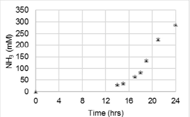
To monitor urea hydrolysis, NH3 concentration was
monitored in triplicate 100-mL cultures in 250-mL flasks using a Thermo ScientificTM OrionTM High-Performance Ammonia Electrode (Waltham, MA, United States) for up to 24 hours. The relationship between NH3 concentration and time is displayed in
Fig. 2. Because the UYE nutrient medium contained 10 g/L urea, or 167 mM urea, the maximum amount of NH3 that can be produced in a culture (when all of
the urea has been hydrolyzed) is 333 mM.
Fig. 2: NH3 concentration vs time for S. pasteurii (ATCC 6453). Error bars represent the standard
deviation for triplicate cultures
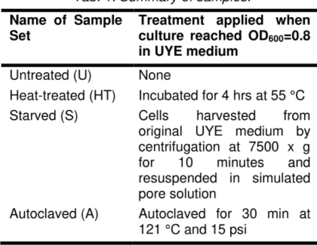
2.2 Summary of samples
A summary of the four sample sets investigated in this study is presented in Tab. 1. First, an untreated sample set was defined to represent live, vegetative cells and serve as the control. Then, three other sample sets were chosen to examine the effects of conditions designed to mimic those present in cement-based materials on surface charge and urease activity of S. pasteurii cells. Heat-treatment was used to investigate the effect of the heat liberated from the cement hydration process. The maximum allowable concrete temperature is commonly specified as 57 °C, and a typical temperature rise for concrete using Type I/II cement can range up to 30°C above the placement temperature [Gajda 2002]. The heat-treated sample set in this study was exposed to 55 °C for four hours.
The starved sample set was used to evaluate the effect of the absence of nutrients and a high pH. It should be noted that starved cells in this study were separated from their nutrients by centrifugation and suspended in simulated pore solution, while cells that are added to cement paste in typical MICCP studies are often inoculated with their nutrients. As a result, the degree of nutrient depletion and pH shock endured by these cells might be more severe than what is actually present in concrete. The autoclaved sample set was employed to investigate the impact of dead cells. The cells were classified as dead based on their inability to grow in liquid or solid UYE medium.
Tab. 1: Summary of samples.
Name of Sample Set
Treatment applied when culture reached OD600=0.8
in UYE medium Untreated (U) None
Heat-treated (HT) Incubated for 4 hrs at 55 °C Starved (S) Cells harvested from
original UYE medium by centrifugation at 7500 x g for 10 minutes and resuspended in simulated pore solution
Autoclaved (A) Autoclaved for 30 min at 121 °C and 15 psi
2.3 Simulated pore solution
A solution containing 17.94 g KOH and 5.24 g NaOH per liter DDI water [Moser 2011] was used to simulate the high alkalinity environment that is intrinsic to the pore solution of cement paste. This simulated pore solution was used for starved samples, with a pH of approximately 13.6.
2.4 Cement
Texas Lehigh Type I/II (Buda, TX) portland cement was used for all cement paste mixtures, and the mass composition of oxides is presented in Tab. 2.
Tab. 2: Type I/II portland cement oxide composition. LOI: Loss on ignition.
Oxides % (w/w) composition CaO 63.63 SiO2 20.14 Al2O3 5.42 Fe2O3 2.47 MgO 1.32 SO3 3.09 Na2O 0.17 K2O 0.95 LOI 2.82 3 METHODOLOGY
3.1 Electrophoretic mobility and zeta potential
S. pasteurii cells in each sample set were grown in
UYE medium (section 2.1) to OD600=0.8, and treated
according to Tab. 1. Then, cells were harvested by
centrifugation at 4000 x g for 6 minutes, washed, and resuspended in sterile 20 mM Tris buffer at pH 9 for testing. A Malvern Zetasizer Nano ZS (Malvern, Worcestershire, United Kingdom) was used to measure electrophoretic mobility, and the Henry equation (Equation 7) was used to calculate zeta potential [Olson 2012].
(7) where UE is the electrophoretic mobility, ε is the
dielectric constant, z is the zeta potential, f(Ka) is Henry’s function, and η is viscosity.
For Henry’s function, f(Ka) = 1.5, or the Smoluchowski approximation, was used. Electrophoretic mobility was measured immediately after treatment, and samples were then stored at room temperature (23 °C) until the next analysis. Electrophoretic mobility was measured again at 3, 7, and 28 days after treatment to assess whether untreated, heat treated, starved, and autoclaved cells would maintain their surface charge over time. Triplicate specimens were prepared for each sample set, and three runs were performed on each specimen.
3.2 Urea hydrolysis
Urease activity was examined for S. pasteurii sample sets by monitoring ammonia production from urea hydrolysis. After growth in UYE medium (section 2.1) to OD600=0.8, cells in each sample set were treated
according to Tab. 1. Then, cells were harvested by centrifugation at 7500 x g for 10 minutes, resuspended in fresh UYE medium, and incubated at 30 °C with shaking. Following resuspension, NH3
concentration was monitored using a Thermo ScientificTM OrionTM High-Performance Ammonia
Electrode at discrete intervals for 10 hours. Because a small amount of NH3 was present in the fresh
medium due to heat-induced decomposition of urea during the autoclaving process, the NH3
concentration was measured immediately after resuspension of the cells (t=0), and this value was subtracted from all subsequent measurements. The cumulative percentage of urea consumed at each time point was then calculated by the stoichiometry of Reaction 1, where 1 mole of urea is hydrolyzed to form two moles of NH3 and one mole
of carbon dioxide. Each medium contains 167 mM urea, so the maximum amount of NH3 that can be
produced in each culture is 333 mM. The amount of NH3 present in the medium at t=0 was subtracted
from both the ammonia concentration at the time of interest and the maximum possible ammonia concentration in the culture. The percent urea consumed was calculated as shown in Equation 6.
(6) 3.3 X-Ray diffraction (XRD)
Sample preparation and procedure
XRD was conducted using a Siemens Bruker X-ray Diffractometer (Madison, WI, United States) to quantify CaCO3 in each sample. In general, three
types of pastes were prepared:
• Neat paste: prepared with distilled water and cement;
• Nutrient paste: prepared with UYE medium and cement;
• Bacterial paste: prepared for untreated, heat-treated, and autoclaved sample sets with S.
pasteurii cells grown in UYE medium to
OD600=0.8 and treated according to Tab. 1 (in
their original UYE nutrient medium) and cement; For all pastes, the solution to cement ratio (s/c) was 0.50. Pastes were prepared by mixing 40 g of cement with 20 g of either distilled water (for neat pastes), UYE medium (for nutrient pastes), or bacterial cultures (for bacterial pastes) by hand stirring for 2 minutes. Then, the samples were cast into 3 x 3 cm cylindrical molds and cured at 100% relative humidity at 25°C for 24 hours.
After curing, the molds were removed, and the samples were prepared for testing. The cement paste sample was crushed with a pestle, and a representative aliquot was obtained from the core. Representative aliquots were pulverized with a mortar and pestle, such that the resulting powder was finer than 53 µm. Then, the sample was ground with ethanol to stop hydration [Zhang 2011], and 10% (w/w) zincite was added as an internal standard. The sample was mixed well to ensure homogeneity.
Samples were stored in a vacuum desiccator until the time of testing. Analysis was conducted from 25-40° 2θ with 6 seconds dwell time. The diffractometer was operated at 40 keV and 30 mA, at a step size of 0.02° 2θ. Triplicate specimens were prepared for each sample set.
Reference Intensity Ratio (RIR) procedure
Calcite was quantified in each sample using the RIR method [Hubbard 1988]. A ratio was established between the intensities of the primary zincite (internal standard) and calcite peaks by creating samples with known mass proportions and plotting the results on a standard curve. Calcite content was varied from 2% to 18% while zincite was kept at 10%. NaCl was selected as a filler material because it is crystalline and its peaks do not overlap with calcite or zincite. The proportions for each sample representing a point on the standard curve are presented in Tab. 3, and the RIR standard curve is displayed in Fig. 3.
Calcite was quantified in each sample using the equation Y = 0.0476X + 0.0427, where Y is the intensity of the primary calcite peak divided by the intensity of the primary zincite peak and X is wt. % calcite.
Tab. 3: Mass composition of samples representing points on RIR standard curve.
Fig. 3: RIR standard curve.
4 RESULTS AND DISCUSSION
4.1 Electrophoretic mobility and zeta potential Results of electrophoretic mobility measurements are presented in Tab. 4, and corresponding zeta potential values are presented in Fig. 4.
Tab. 4: Electrophoretic mobility at 0, 3, 7, and 28 days after treatment. Error represents the standard
deviation for triplicate samples.
Sample Set Electrophoretic Mobility (µm-cm/V-s) 0 days 3 days 7 days 28 days Untreated -2.79 ± 0.22 -2.77 ± 0.07 -2.60 ± 0.13 -2.81 ± 0.09 Heat-treated -2.43 ± 0.22 -2.18 ± 0.15 -2.30 ± 0.13 -2.55 ± 0.17 Starved -2.65 ± 0.06 -2.65 ± 0.22 -2.61 ± 0.07 -2.51 ± 0.07 Autoclaved -2.64 ± 0.05 -2.61 ± 0.12 -2.48 ± 0.19 -2.54 ± 0.15
Fig. 4: Zeta potential at 0, 3, 7, and 28 days after treatment of S. pasteurii cells. Error bars represents
the standard deviation for triplicate samples. S. pasteurii cells harvested from each sample set
exhibited similar zeta potential values, indicating that cells exposed to heat, lack of nutrients combined with high pH, and autoclaving maintained a robust negative surface charge similar to that of the untreated cells. The zeta potential of S. pasteurii cells was reasonably constant for up to 28 days in each sample set, indicating that even stressed or dead cells could sustain a negative surface charge over time. Surface charge is considered to be an Point Calcite (%) Zincite (%) NaCl filler (%)
1 2 10 88 2 4 10 86 3 6 10 84 4 8 10 82 5 10 10 80 6 12 10 78 7 14 10 76 8 16 10 74 9 18 10 72
influential parameter in the nucleation process of minerals, and it has been proposed that bacterial cells in cement paste could promote calcium carbonate precipitation by serving as nucleation sites [Achal 2009; Hammes 2002; Van Tittelboom 2010] via the electrostatic attraction between the negatively charged bacteria and Ca2+. If this mechanism can occur, it seems that all cells, even stressed or dead cells, could contribute to calcium carbonate formation by providing sites for heterogeneous nucleation to occur.
4.2 Urea hydrolysis
NH3 profiles for the untreated and heat-treated
sample sets are presented in Fig. 5. No discernible increase in NH3 concentration was observed for
autoclaved samples or for samples starved in simulated pore solution for 1 day or longer. Percentages of urea consumed for untreated and heat-treated samples, which were calculated from NH3 measurements as described in Section 3.3, are
presented in Fig. 6.
Fig. 5: NHs profiles for untreated and heat-treated S.
pasteurii cells. Error bars represent the standard
deviation for triplicate samples.
Fig. 6: Urea consumed by S. pasteurii for untreated and heat-treated samples (obtained by calculation).
Error bars represent the standard deviation for triplicate samples.
From Figs. 5 and 6, it is evident that heat-treatment substantially impeded urea hydrolysis by S. pasteurii cells as compared to untreated samples. After 10 hours of incubation in fresh UYE medium at 30 °C with shaking, heat-treated cells were able to decompose approximately 50% of the urea available in the fresh medium, while untreated cells decomposed nearly 100% of the urea available. There was no discernible increase in NH3 detected in
autoclaved samples or samples starved in simulated pore solution for 1 day or longer, indicating lack of urease activity due to those treatments.
4.3 XRD
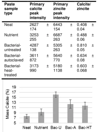
Using the calcite and zincite data in Tab. 5, the mass percentages of calcite were calculated (Fig. 7).
Tab. 5: Intensities of primary calcite and zincite peaks, and calcite intensity divided by zincite intensity at 1 day. Error represents the standard
deviation of triplicate samples.
Paste sample type Primary calcite peak intensity Primary zincite peak intensity Calcite/ zincite Neat 2627 ± 174 6443 154 ± 0.408 ± 0.04 Nutrient 3253 ± 255 6687 263 ± 0.488 ± 0.06 Bacterial- untreated 4287 138 ± 5305 263 ± 0.810 ± 0.05 Bacterial- autoclaved 3611 872 ± 5640 770 ± 0.634 ± 0.08 Bacterial- heat-treated 3173 ± 990 5180 1138 ± 0.603 ± 0.068
Fig. 7: Mass percentages of calcite at 1 day. Error bars represent the standard deviation of triplicate
samples. Bac-U: Bacterial-untreated. Bac-A: Bacterial-autoclaved. Bac-HT: Bacterial-heat-treated.
From Fig. 7, calcite content increased substantially in all bacterial pastes as compared to neat and nutrient pastes at 1 day, and this increase could be attributed to MICCP by S. pasteurii cells. As compared to neat paste, bacterial paste with untreated cells contained 110% more calcite and bacterial pastes with autoclaved and heat-treated cells contained 62% and 53% more calcite, respectively. Calcite content was higher in the autoclaved bacterial paste as compared to neat paste despite the fact that the autoclaved cells showed no urease activity when resuspended in fresh UYE medium (section 3.2). Because the autoclaved cells could not continue urea hydrolysis after the autoclaving process, this increase in calcite is likely attributable to urease activity that occurred prior to autoclaving. Additionally, because zeta potential measurements showed that cells in the autoclaved sample set maintained a negative surface charge, dead cells in autoclaved cement paste might have promoted the formation of calcite by serving as nucleation sites.
Calcite content also increased substantially in heat-treated bacterial paste as compared to neat paste even though urease activity was impeded when heat-treated cells were resuspended in fresh UYE medium (section 3.2). Similar to autoclaved cement paste, it is likely that this increase in calcite is
primarily attributable to urease activity in the cultures prior to heat-treatment, and calcium carbonate formation also might have been induced by heat-treated cells acting as nucleation sites. It should also be noted that the lower amount of calcite precipitation observed in the autoclaved and heat-treated pastes (compared to unheat-treated paste) might also have been due to loss of carbon dioxide (and therefore a reduction in the total carbonates) during the treatments at elevated temperatures.
5 CONCLUSIONS
The role of dead cells in MICCP in construction materials was explored for the first time in this study, and the role of stressed cells also was evaluated. To gain insight to the physical and metabolic state of microorganisms in cement-based materials, the effect of environmental stressors designed to mimic conditions inside cement paste on the surface charge and urease activity of S. pasteurii cells was investigated. Zeta potential was used to approximate surface charge, and results showed that heat-treatment, lack of nutrients coupled with high pH, and death induced by autoclaving had no discernible effect on the zeta potential of S. pasteurii cells as compared to untreated cells. It also was found that the zeta potential of untreated, heat-treated, starved, and autoclaved cells remained reasonably constant for up to 28 days after treatment (in a liquid medium). This indicated that cells from each sample set, even dead cells, could maintain a negative surface charge over time. S. pasteurii cells could be good candidates for nucleation sites of calcium carbonate because of their small size and ample surface charge. Results suggested that although dead cells are no longer able to promote calcium carbonate precipitation by carrying out urea hydrolysis in cement paste, their negative surface charge might allow them to aid in precipitation by serving as nucleation sites.
Although surface charge was minimally affected by the treatments employed in this study, urea hydrolysis by S. pasteurii was greatly impacted. Less ammonia production (likely a direct result of less urea hydrolysis) was observed in the heat-treated S.
pasteurii cells as compared to the untreated cells,
and ammonia production ceased altogether for cells starved in simulated cement paste pore solution for longer than 1 day and for autoclaved cells. By extension, the production of carbonates by S.
pasteurii cells would be substantially hindered by
exposure to heat (55 °C for 4 hours) and was stopped by submergence in simulated cement paste pore solution (absence of nutrients coupled with pH 13.6). Cells killed by autoclaving did not produce ammonia from urea hydrolysis nor, by extension, produce carbonates for MICCP. Since urease activity is substantially impacted by conditions which might occur in cement paste (e.g. increased temperature, lack of nutrients, high pH), it seems unlikely that S.
pasteurii could produce new carbonates over time in situ (in the cement paste) unless measures are taken
to alleviate these conditions. For example, the use of low heat cements and/ or supplementary cementing materials such as fly ash could mitigate the increase in heat generated by cement hydration [Ballim 2009], and the high pH of fresh cement paste could be
reduced through use of cements with lesser contents of alkalis and gypsum [Mansur 2008].
Quantitative XRD revealed that bacterial cement paste mixed with untreated, heat-treated, and autoclaved bacterial cultures contained 110%, 53%, and 62% more calcite than did neat cement paste. These increases were attributed to MICCP by S.
pasteurii cells. Calcite content increased most
substantially in untreated bacterial paste, which could be attributed to urea hydrolysis by S. pasteurii cells and possibly heterogeneous nucleation of calcite on the bacterial cell walls promoted by their negative surface charge. Finally, it was proposed that greater calcite contents observed in autoclaved and heat-treated cement pastes as compared to neat cement paste could be attributed to urea hydrolysis carried out before the cells were autoclaved or heat-treated, and, like untreated cells, autoclaved and heat-treated cells might have been able to further promote calcium carbonate precipitation by acting as nucleation sites.
6 REFERENCES
[Achal 2013] Achal, V.; Mukerjee, A.; Reddy, M.; Biogenic treatment improves the durability and remediates the cracks of concrete structures. Construction and Building Materials, 2013, 48, 1-5. [Achal 2012] Achal, V.; Mukherjee, A.; Goyal, S.; Reddy, M.; Corrossion prevention of reinforced concrete with microbial calcite precipitation. ACI Materials Journal, 2009, 109, 2, 157-163.
[Achal 2011] Achal, V.; Mukherjee, A.; Reddy, M.; Effect of calcifying bacteria on permeation properties of concrete. Journal of Industrial Microbiology and Biotechnology, 2011, 38, 1229-1234.
[Achal 2009] Achal, V.; Mukherjee, A.; Basu, P.; Reddy, M.; Lactose mother liquor as an alternative nutrient source for microbial concrete production by
Sporosarcina Pasteurii. Journal of Industrial
Microbiology and Biotechnology, 2009, 36, 433-438. [Ballim 2009] Ballim, Y.; Graham, P.; The effects of supplementary cementing materials in modifying the heat of hydration of concrete. Materials and Structures, 2009, 42, 6, 803-811.
[Basaran 2013] Basaran, Z. Biomineralization of cement based materials: inoculation of vegetative cells. Austin: The University of Texas at Austin, 2013. [Bundur 2015] Bundur, Z.; Kirisits, M.; Ferron, R. Biomineralized cement-based materials: Impact of inoculating vegetative bacterial cells on hydration and strength. Cement and Concrete Research, 2015, 67, 237 - 245.
[DeJong 2010] DeJong, J.; Mortensen, B.; Martinez, B.; Nelson, D.; Bio-mediated soil improvement. Ecological Engineering, 2010, 36, 197-210.
[DeJong 2006] DeJong, J.; Fritzges, M.; Nusslein, K.; Microbially induced cementation to control sand response to undrained shear. Journal of Geotechnical and Geoenvironmental Engineering, 2006, 132, 11, 1381-1392.
[De Muynck 2008] De Muynck, W.; Debrouwer, D.; De Belie, N.; Verstraete, W.; Bacterial carbonate precipitation improves the durability of cementitious materials. Cement and Concrete Research, 2008, 38, 1005-1014.
[Gajda 2002] Gajda, J.; Vangeem, M.; Controlling temperatures in mass concrete. Concrete International, January 2002, 59-62.
[Hammes 2002] Hammes, F.; Verstraete, W.; Key roles of pH and calcium metabolism in microbial carbonate precipitation. Reviews in environmental science and biotechnology, 2002, 1, 1, 3-7.
[Hubbard 1988] Hubbard, C. R.; Snyder, R. L.; (1988). RIR - Measurement and use in quantitative XRD. Powder Diffraction, 1988, 3, 02, 74-77.
[Jonkers 2010] Jonkers, H. M.; Thijssen, A.; Muyzer, G.; Copuroglu, O.; Schlangen, E.; Application of bacteria as self-healing agent for the development of sustainable concrete. Ecological Engineering, 2010, 36, 230-235.
[Jonkers 2007] Jonkers, H.M.; Schlangen, E.; Self-healing of cracked concrete : A bacterial approach. Proceedings of FRACOS6: fracture mechanics of concrete and concrete structures. Catania, Italy. [Li 2012] Li, V.; Herbert, E.; Robust self-healing concrete for sustainable infrastructure. Journal of Advanced Concrete Technology, 2012, 10, 207-218. [Mansur 2008] Mansur, A.; Do Nascimento, O.; Mansur, H.; Controlling alkalinity of cement matrix: A key approach to improve fiber-cement durability. Proceedings of IIBCC: 11th international inorganic-bonded fiber composites conference. 2008, Madrid, Spain: 236-243.
[Mitchell 2006] Mitchell, A. C.; Ferris, F. G.; The Influence of Bacillus pasteurii on the nucleation and growth of calcium carbonate. Geomicrobiology Journal, 2006, 23, 213-226.
[Olson 2012] Olson, E.; Zeta potential and colloid chemistry. Journal of GXP Compliance, 2012, 1, 16, 81.
[Patil 2008] Patil, H.; Prashant, H.; Raijiwala, D.; Vijay, B.; Bacterial concrete--a self healing concrete. International Journal of Applied Engineering Research, 2008, 3, 12, 1719-1725.
[Ramachandran 2001] Ramachandran, S.; Ramakrishnan, V.; Bang, S.; Remediation of concrete using micro-organisms. ACI Materials Journal, 2001, 98, 3-9.
[Ricca 2003] Ricca, E.; Cutting, S.; Emerging applications of bacterial spores in nanobiotechnology. Journal of Nanobiotechnology, 2013, 1, 6.
[Sarda 2009] Sarda, D.; Choonia, H. S.; Sarode, D. D.; Lele, S.S.; Biocalcification by Bacillus pasteurii urease : a novel application. Journal Of Industrial Microbiology, 2009, 36, 1111-1115.
[Stocks-fischer 1999] Stocks-fischer, S.; Galinat, J. K.; Bang, S. S.; Microbiological precipitation of CaCO3. Soil Biology and Biochemistry, 1999, 31,
1563-1571.
[Van Tittelboom 2010] Van Tittelboom, K.; De Belie, N.; De Muynck, W.; Verstraete, W.; Use of bacteria to repair cracks in concrete. Cement and Concrete Research, 2010, 40, 157-166.
[Wang 2014] Wang, J.; Soens, H.; Verstraete, W.; De Belie, N.; Self-healing concrete by use of microencapsulated bacterial spores. Cement and Concrete Research, 2014, 56, 139-152.
[Whiffin 2007] Whiffin, V. S.; van Paassen, L. A.; Harkes, M. P.; Microbial carbonate precipitation as a soil improvement technique. Geomicrobiology Journal, 2007, 24 5, 417 - 423.
[Wilson, 2001] Wilson, W.; Wade, M.; Holman, S.; Champlin, F.; Status of methods for assessing bacterial cell surface charge properties based on zeta potential measurements. Journal of Microbiological Methods, 2001, 43, 153-164.
[Zhang 2011] Zhang, J.; Scherer, G. W.; Comparison of methods for arresting hydration of cement. Cement and Concrete Research, 2011, 41, 1024– 1036.