RESEARCH OUTPUTS / RÉSULTATS DE RECHERCHE
Author(s) - Auteur(s) :
Publication date - Date de publication :
Permanent link - Permalien :
Rights / License - Licence de droit d’auteur :
Bibliothèque Universitaire Moretus Plantin
Institutional Repository - Research Portal
Dépôt Institutionnel - Portail de la Recherche
researchportal.unamur.be
University of Namur
Morphological analysis of the sheathed flagellum of Brucella melitensis
Ferooz, J.; Letesson, Jean-Jacques
Published in: BMC Res Notes DOI: 10.1186/1756-0500-3-333 Publication date: 2010 Document VersionPublisher's PDF, also known as Version of record Link to publication
Citation for pulished version (HARVARD):
Ferooz, J & Letesson, J-J 2010, 'Morphological analysis of the sheathed flagellum of Brucella melitensis', BMC
Res Notes, vol. 3. https://doi.org/10.1186/1756-0500-3-333
General rights
Copyright and moral rights for the publications made accessible in the public portal are retained by the authors and/or other copyright owners and it is a condition of accessing publications that users recognise and abide by the legal requirements associated with these rights. • Users may download and print one copy of any publication from the public portal for the purpose of private study or research. • You may not further distribute the material or use it for any profit-making activity or commercial gain
• You may freely distribute the URL identifying the publication in the public portal ?
Take down policy
If you believe that this document breaches copyright please contact us providing details, and we will remove access to the work immediately and investigate your claim.
S H O R T R E P O R T
Open Access
Morphological analysis of the sheathed flagellum
of Brucella melitensis
Jonathan Ferooz
1,2, Jean-Jacques Letesson
1*Abstract
Background: It was recently shown that B. melitensis is flagellated. However, the flagellar structure remains poorly described.
Findings: We analyzed the structure of the polar sheathed flagellum of B. melitensis by TEM analysis and
demonstrated that the Ryu staining is a good method to quickly visualize the flagellum by optical microscopy. The TEM analysis demonstrated that an extension of the outer membrane surrounds a filament ending by a club-like structure. TheΔftcR, ΔfliF, ΔflgE and ΔfliC flagellar mutants still produce an empty sheath.
Conclusions: Our results demonstrate that the flagellum of B. melitensis has the characteristics of the sheathed flagella. Our results also suggest that the flagellar sheath production is not directly linked to the flagellar structure assembly and is not regulated by the FtcR master regulator.
Background
Brucellae are gram-negative, intracellular pathogenic bacteria that cause brucellosis in a variety of mammals, including humans. During a long time, they were con-sidered as unflagellated. However, the presence of a sheathed flagellum has recently been discovered in Bru-cella melitensis[1,2].
The fact that the flagellum was never previously observed in Brucella is probably due to the very short period (the early exponential growth phase in rich liquid medium) during which the flagellum is produced. The availability of the genome sequences of Brucella meliten-sisand Brucella suis revealed the presence of all the fla-gellar genes needed for the construction of a functional flagellum, except the genes encoding the chemotactic system [3,4]. Today, the sequence of the genome of B. abortus, B. ovis, B. canis and B. microti are available, showing that flagellar genes are also present in these species [5-7]. Interestingly, the presence of mutations in several flagellar genes in the different Brucella species suggest that the flagellar regulon contains pseudogenes and that the filamentous appendage observed is a flagel-lum-derived structure [5].
The flagellum is a complex machine divided in three main structures [8]. The basal body structure is embedded in the membranes and functions like a tiny engine containing a motor, a stator and a secretion sys-tem. The hook structure is a universal joint transmitting the energy generated by the basal body to the filament. Finally, the filament is the most visible part of the flagel-lum and is made of about 20,000 monomers of flagellin with a length of severalμm. The control of the flagellar assembly is a fine-tuned mechanism well described in Salmonella entericaserovar Typhimurium, Escherichia coli and Caulobacter crescentus (for reviews, see the references [9-12]).
It was recently described that flagellar regulation of B. melitensiswas controlled by the FtcR flagellar master regulator which activates flagellar expression by binding directly on fliF promoter [13]. The mutation of ftcR decreases fliF expression and induces the extinction of the flagellin and hook proteins. The two LuxR-type reg-ulators VjbR and BlxR are also required for flagellar activation [14,15]. VjbR is involved in the quorum sen-sing of B. melitensis and mediates the inhibitory effect of the N-dodecanoyl-DL-homoserine lactone (C12-HSL)
[16]. In the early steps of the hierarchical flagellar regu-lation of B. melitensis, it was proposed that VjbR con-trols the expression of ftcR [13]. More recently, it was proposed that flagellar hierarchy is divided in three
* Correspondence: jean-jacques.letesson@fundp.ac.be 1
Unité de Recherche en Biologie Moléculaire (URBM), Facultés Universitaires Notre-Dame de la Paix (FUNDP), 61 rue de Bruxelles, B-5000 Namur, Belgium Full list of author information is available at the end of the article
© 2010 Letesson et al; licensee BioMed Central Ltd. This is an Open Access article distributed under the terms of the Creative Commons Attribution License (http://creativecommons.org/licenses/by/2.0), which permits unrestricted use, distribution, and reproduction in any medium, providedthe original work is properly cited.
classes in B. melitensis (J. Ferooz et al., unpublished). In this later work, FlbT was described as the checkpoint regulator between the class II and III genes, and acti-vates the flagellin production.
Here, we demonstrate that the extracellular appendage produced by B. melitensis is a flagellum with all charac-teristics of the sheathed flagella. Moreover,ΔftcR, ΔfliF, ΔflgE, and ΔfliC mutants still produce a filament, prob-ably an empty sheath regulated independently of the ftcRand vjbR flagellar pathway.
Materials and methods
Bacterial strains and culture conditions
All strains used in this study are listed in Table 1. All Bru-cellastrains used in this study derive from B. melitensis 16 M Nalr (spontaneous nalidixic acid resistant mutant selected from B. melitensis 16 M, received from A. Mac-millan, Central Veterinary Laboratory, Weybridge, UK). The growth was measured by reading the optical density of the cultures at 600 nm. B. melitensis 16 M growth curves in rich medium (1% yeast extract, 1.6% peptone, 0.5% NaCl) were performed from a late-exponential over-night culture obtained in liquid 2YT medium. B. melitensis 16 M strains grew with shaking at 37°C in rich medium containing appropriate antibiotics from an initial OD600of
0.05. C. crescentus CB15N grew in peptone-yeast extract (PYE complex media) at 30°C. Antibiotics were used at the following final concentrations: kanamycin, 50μg ml-1; nalidixic acid, 25μg ml-1.
Flagellum staining and visualization by phase-contrast microscopy
Bacterial flagella were stained using the“Ryu staining” method as described previously [17-19]. The Ryu stain has two components. Solution I (the mordant) contains 10 ml of 5% aqueous solution of phenol, 2 g of tannic acid, and 10 ml of saturated aqueous solution of alumi-num potassium sulfate-12 hydrate. Solution II (the stain) is a saturated ethanolic solution of crystal violet
(3 g in 25 ml of 95% ethanol). The final stain was pre-pared just before use by mixing 1 part of solution II with 10 parts of solution I and then by filtering the mix-ture through filter paper to remove coarse precipitate. A drop of cell culture was transferred onto the clean slide and covered with a cover slip. After 5 to 10 min, two drops of Ryu stain were applied to the edge of the cover slip and flowed under the cover slip by capillarity and mixed with the cell suspension. The cells were examined for flagella after 5 to 15 min at room temperature under a phase-contrast microscope. The edges of the cover slip on the slide were sealed with nail varnish.
Visualization of the flagella by transmission electron microscopy (TEM)
The bacteria were grown in rich medium at 37°C to an OD600of 0.25. Bacteria were centrifuged at 1000 r.p.m.
for 20 min (Jouan), washed in PBS and fixed for 20 min in 50 μl of 4% paraformaldehyde pH 7.3. Samples were stored at 4°C. A drop (15 to 35 μl) of a solution of 1% Alcian blue was placed on a sheet of Parafilm. A carbon Formvar-coated grid was placed on a drop of solution for 5 min, carbon side down, washed five times in dis-tilled water and then placed on a drop of bacterial sus-pension for 10 min on the same parafilm sheet. Grids with adherent cells were either (i) negatively stained for direct visualization on transmission electron microscope (TEM) or (ii) labeled with the anti-Brucella LPS O-chain, M epitope, mAb A156b3b2 [20,21] before staining as described previsously by Fretin et al.[1]. For immunolabeling, after 10 min on the drop of bacterial suspension, the carbon Formvar-coated grid was placed onto drops (15 to 35 μl) of the following reagents on the same parafilm sheet: 5 washes in phosphate-buffered saline (PBS)-glycine 5% (3 sec each), PBS-bovine serum albumin (BSA)(1 min), monoclonal antibody A156b3b2 diluted 1/20 in PBS-BSA 5% (1 h), five washes in PBS (10 sec each), rabbit anti-mouse immunoglobulin conju-gated to ± 15 nm colloidal gold diluted 1/20 in
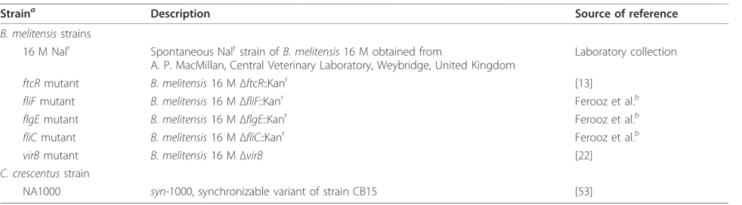
PBS-Table 1 Bacterial strains
Straina Description Source of reference
B. melitensis strains
16 M Nalr Spontaneous Nalrstrain of B. melitensis 16 M obtained from
A. P. MacMillan, Central Veterinary Laboratory, Weybridge, United Kingdom
Laboratory collection
ftcR mutant B. melitensis 16 MΔftcR::Kanr [13] fliF mutant B. melitensis 16 MΔfliF::Kanr Ferooz et al.b flgE mutant B. melitensis 16 MΔflgE::Kanr Ferooz et al.b fliC mutant B. melitensis 16 MΔfliC::Kanr Ferooz et al.b
virB mutant B. melitensis 16 MΔvirB [22]
C. crescentus strain
NA1000 syn-1000, synchronizable variant of strain CB15 [53]
a
Nalr
, nalidixic acid resistant; Kanr
, kanamycin resistant.b
Submitted for publication.
Ferooz and Letesson BMC Research Notes 2010, 3:333 http://www.biomedcentral.com/1756-0500/3/333
BSA (1 h), three washes in PBS (10 sec each), 2 washes in distilled water (10 sec each). After a completed treat-ment protocol, the grid was negatively stained with 2% aqueous solution of uranyl acetate for 10 sec, the excess fluid was removed with a filter paper, and the grid was air dried. Specimens were examined using a Philips Technai 10 TEM. Note that protein A-colloidal gold were also used instead of rabbit anti-mouse immunoglo-bulin with the same results.
Results
The Ryu staining is a simple technique for the detection of the flagellum ofBrucella melitensis
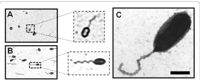
Due to the short period of flagellar production (only at early exponential growth) and the low percentage of fla-gellated bacteria in B. melitensis, the visualization of the flagellum by TEM is difficult [1]. To easily detect the flagellated Brucella, we used the Ryu staining. An advantage of that technique is that the flagellum of B. melitensiscan be visualized directly without any prior centrifugation (required for the TEM technique), mini-mizing the manipulation of the culture containing the pathogen. As positive control, we used Caulobacter cres-centus because its flagellar expression is cell-cycle dependent and the flagella were previously visualized by Ryu staining [19]. We were able to visualize the flagel-lum of B. melitensis using the Ryu staining by phase-contrast microscopy (Figure 1A). Although the number of flagellated B. melitensis in the population is fewer than the flagellated C. crescentus (Figure 1B), the stained flagellum produced by B. melitensis is clearly detected by optical microscope. This result demonstrates that the Ryu staining is a simple technique allowing a rapid visualization of the flagellum of B. melitensis and with minimal sample manipulation.
Ultrastructure analysis of the sheathed flagellum of B. melitensis by TEM
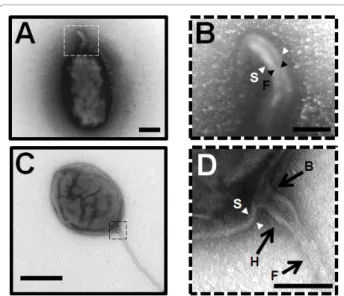
We used the transmission electron miscroscopy (TEM) to analyze precisely the ultrastructure of the flagellum in B. melitensis. A sample of a B. melitensis culture at the early exponential growth phase in rich medium was stained with uranyl acetate 2% and visualized by TEM as described in the Materials and Methods section. We also labeled the bacteria with an anti-Brucella lipopoly-saccharide (LPS) antibody and confirmed the presence of LPS on both the cell surface and the sheathed flagel-lum of B. melitensis (Figure 1C). B. melitensis produces a polar sheathed flagellum of about 50 nm diameter, showing an inner filament of about 11 nm diameter clearly surrounded by a sheath, which seems to be most likely a continuous extension of the bacterial outer membrane (Figure 1C and 2). As usually seen in other bacterial species producing a sheathed flagellum, the
flagellum of B. melitensis is ending by a club-like struc-ture (Figure 3A). A polar bended strucstruc-ture is also usually observed, like a nascent flagellum during the first steps of flagellar assembly (Figure 3B). This bended structure, and sometimes a flagellum, is visualized at the septum of division between two daughter cells, suggest-ing that flagellar assembly occurs at this site (Figure 3C). Taken together, these data clearly demonstrate that the appendage produces by B. melitensis has the typical features of the sheathed flagella and not a pilus-like structure or a remaining part of the flagellum.
The sheath production is not dependent of theftcR pathway and the flagellar structure
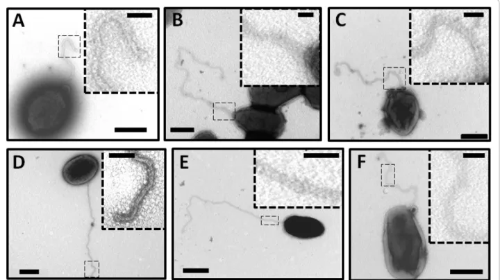
To determine whether the mutation of flagellar genes affect flagellar assembly,ΔfliF, ΔflgE and ΔfliC mutants were analyzed by TEM. These mutants were created by gene replacement with the aphA4 cassette (J. Ferooz et al., unpublished). Surprisingly, these three mutants still produce a filamentous structure (Figure 4A, B and 4C). We previously demonstrated that FlgE and FliC protein cannot be detected by Western blot analysis in ΔflgE and ΔfliC respectively. Therefore, we propose that the appendage observed inΔfliF, ΔflgE and ΔfliC is an empty sheath. The genome of B. melitensis contains the clusters of genes coding for only two extracellular struc-tures: a type IV secretion system (T4SS) expressed at the stationary growth phase and a flagellum. In order to know whether the structure seen is a T4SS or not, a ΔvirB mutant was visualized by TEM. This ΔvirB mutant was made by removing the 12 open reading frames (ORFs) encoding the T4SS of B. melitensis 16 M [22]. As seen in the wild-type strain (Figure 4D),ΔvirB also produces a similar flagellar structure at the early exponential growth phase (Figure 4E). This result demonstrates that the extracellular appendage observed in flagellar mutants is not a T4SS.
Figure 1 Visualization of flagella. Ryu staining of B. melitensis (A) C. crescentus (B) observed by phase-contrast microscopy. The samples were treated by the Ryu staining method as described in Materials and Methods. A flagellated bacterium is enlarged in a dotted square. (C) Negative-staining EM images of the sheathed polar flagellum of B. melitensis stained with uranyl acetate 2% and labeled with anti-LPS antibody conjugated to ± 15 nm gold particles. Bar, 500 nm.
The flagellar expression is not affected by mutation of the structural genes, indeedΔfliF and ΔflgE still produce the flagellin FliC (J. Ferooz et al., unpublished). How-ever, the flagellar master regulator FtcR is needed for fliF expression and the FlgE and FliC synthesis [13]. By intensively observing ΔftcR by TEM, we also found some bacteria producing a filament-like structure, sug-gesting that sheath synthesis and assembly is likely inde-pendent of the FtcR pathway (Figure 4F).
Discussion and Conclusions
Although it was demonstrated that B. melitensis pro-duces a polar sheathed flagellum under specific condi-tions [1], there were still some doubts concerning the production of a classical flagellum by Brucella [5,23]. The conditions in which the flagellum is observed are not optimal because Brucella produces a flagellum only at the early exponential growth phase in rich medium [1]. Moreover, Brucella must be handled according to level 3 biosafety precautions [24], making difficult its manipulation for the preparation of sample for TEM analysis. This is the reason why we tested the Ryu stain-ing technique, that allows an easier detection of flagel-lum than TEM as described in several flagellated bacteria such as C. crescentus, L. monocytogenes, Bacillus subtilisand Salmonella Typhi [19,25-27]. Interestingly, even with a low number of flagellated bacteria, this technique allows a quick and direct manipulation of the culture before the extinction of flagellar expression. In this work, we showed that the flagellum of B. melitensis can be easily visualized using the Ryu staining.
The structure of the flagellum produces by B. meliten-siswas analyzed by the TEM technique and several fea-tures of the sheathed flagella of other species were highlighted. At the present time, little is known about the formation, composition or function of flagellar sheaths in bacteria, and interestingly, Brucella is the only one rhizobiale to produce a sheathed flagellum [28,29]. Due to the sheath around the filament, the visi-ble flagellum of B. melitensis has a diameter of 50 nm, which is larger than an unsheathed flagellum. However, the diameter of the bacterial filament is usually about 20 nm but we showed that the diameter of the inner filament in the sheath of B. melitensis is only 11 nm. This smaller diameter could be linked to the shorter amino acid sequence of the unique flagellin composed of only 282 amino acids in B. melitensis. In comparison, the flagellin sequence of E. coli, S. enterica serovar Typhimurium and Pseudomonas aeruginosa are between 488 and 498 amino acids.
Among the features of sheathed flagellum, the shape at the end of the flagella of B. melitensis is similar to a club-like structure, also described in other bacteria like B. bacteriovorus, H. pylori and V. fischeri [30-32]. This extension can be viewed as the continuity of the sheath at the end of the filament. The genome of Brucella lacks the FliD (or HAP2) homologue, a protein involved in the flagellin assembly at the top of the filament. One hypothetical function of this club-like structure could be the formation of a confined space facilitating the self-assembly of flagellin monomers into a filament, despite the lack of a FliD homologue. Similarly in Vibrio algino-lyticus, it was assumed that the sheath could trap
Figure 2 Visualization of the flagellar sheath. (A and B) Negative-staining EM images of the sheathed polar flagellum of B. melitensis stained with uranyl acetate 2%. (B)The central filament is indicated by black arrowheads with F and the sheath by white arrowheads with S. Bar, 200 nm. (C and D) Capture of the basal region of the flagellum. The image shows the basal body region (B with black arrow), the hook (H with black arrow) and is finished by the filament (F with black arrow). The structure is surrounded by a sheath extended from the outer membrane (S, white arrowheads). Bar, 100 nm. The images B and D are enlarged from the dotted square in image A and C respectively.
Figure 3 Negative-staining EM images of the sheathed polar flagellum fromB. melitensis stained with uranyl acetate 2%. (A) The flagellum is ended by a club-like structure (black arrowhead). (B) Image of a curve structure at the pole of the cell. (C) Predivisional cells exhibited a flagellum at the septum division. A higher magnification of a part of flagellum (black square) is showing (dotted square). Bars, 500 nm in B and C; Bars, 100 nm (A, dotted squares in B and C).
Ferooz and Letesson BMC Research Notes 2010, 3:333 http://www.biomedcentral.com/1756-0500/3/333
excreted flagellin to allow polymerization independently of FliD [33]. Secondly, the sheath of the flagellum of B. melitensis is likely an extension of the outer mem-brane and contains LPS, which is also observed in H. pylori, B. bacteriovorus and some Vibrio spp., [1,32,34-38]. Even if the sheath contains LPS, the sheath composition of Vibrio and Helicobacter is different from the outer membrane [30,39,40]. To the best of our knowledge, all these features have never been observed in a pilus-like structure, confirming that this appendage is a flagellum and not a pilus or flagellum-like structure.
Similarly to other a-proteobacteria, Brucella and C. crescentusare morphologically asymmetric [41]. C. cres-centuscouples flagellar biogenesis with cell cycle allowing the production of a flagellum at the swarmer pole of predi-visional cell and a stalk at the other pole [11]. Surprisingly, we occasionally observed a bended structure or a flagellum at the septum division of B. melitensis rather than at the poles of predivisional cells. The localization of the flagel-lum at the septum of division is rarely observed in bacteria but is not unusual and is described in B. bacteriovorus and Treponema phagedenis[42,43].
In the last part of this work, we demonstrated that pro-duction of the sheath is probably unlinked to flagellar assembly in B. melitensis. Indeed,ΔfliF, ΔflgE and ΔfliC structural flagellar mutants still produce a filamentous
appendage despite the absence of FlgE or FiC proteins. However, persistence of an empty sheath in flagellar mutants was often described in bacteria producing a sheathed flagellum as Vibrio species and H. pylori [44-48]. In B. bacteriovorus, deletion of flagellin fliC3 caused the synthesis of copious, disordered, tubular material resembling outer membrane [49,50]. The authors suggest that this structure is a disordered sheath without a normal flagellum inside. It is important to note that counting the number of flagellated bacteria in the population of the mutants in order to estimate a percen-tage of flagellation compared to the wild-type strain is not relevant due to the variability and the low number of flagellated bacteria detected between samples. Thus, it is possible that the population of flagellar mutants is less flagellated than the wild type population, but better con-ditions to enhance flagellation of B. melitensis will be needed.
In spite the fact that FtcR is the master regulator of flagellar assembly in Brucella, persistence of sheath pro-duction inΔftcR mutant also demonstrate that sheath expression is not dependent of FtcR and suggests that another unknown regulator is involved. Since the sheath surrounds the filament and is produced at one pole of the cell, even in flagellar mutants, it is possible that a flagellar pole marker coordinates both flagellum and
Figure 4 Detection of a flagellum inBrucella’s flagellar mutants. Negative-staining EM images of the wild-type (D) compared to ΔfliF (A), ΔflgE (B), ΔfliC (C), ΔvirB (E) and ΔftcR (F) stained with uranyl acetate 2%. In dotted squares: higher magnification of a flagellum section. Bars, 500 nm and 100 nm in dotted squares.
sheath biogenesis at the cell pole before the FtcR activa-tion. In Vibrio alginolyticus and C. crescentus, the locali-zation of the flagellum at the pole is dictated by FlhF and PflI regulators respectively [51,52]. Polar localization of the pole marker PflI is independent of FliF, whose oligomerization into the MS-ring probably allows the definition of the site of flagellar synthesis, suggesting that PflI acts before or independently of this event [52]. Similarly, a regulator could couple flagellar assembly and sheath production independently of FtcR in B. melitensis.
Altogether, the data presented in this study proved that B. melitensis produces a flagellum with the charac-teristics of sheathed flagella described in other organ-isms. Flagellar assembly and sheath production are a complex regulatory mechanism that remains to be further investigated to gain a better understanding of the flagellum’s function during Brucella’s infection.
Acknowledgements
We thank Dr Valérie Haine for her critical and careful reading of this manuscript and helpful discussion. We are grateful to Bettina Battisti for the helpful manuscript revision. We thank the anonymous reviewers for their insightful comments and suggestions. We acknowledge the‘Service Interfacultaire de Microscopie Electronique’ of the University of Namur and for its expertise in TEM. This work was supported by grants to Jonathan Ferooz from the Fonds Adrien Bauchau and the FNRS (Fonds National de la Recherche Scientifique). This work was supported by the Commission of the European Communities (contract no. QLK2-CT-1999-00014), the FRFC (Fonds de la Recherche Fondamentale Collective, conventions 2.4521.04 and 2.4521.08) and the ARC fellowship (Actions de Recherches Concertées, conventions 04/09-325 and 08/13-015). Jonathan Ferooz held a Belgian specialization grant from the FRIA (Fonds pour la Formation à la Recherche dans l’Industrie et dans l’Agriculture).
Author details 1
Unité de Recherche en Biologie Moléculaire (URBM), Facultés Universitaires Notre-Dame de la Paix (FUNDP), 61 rue de Bruxelles, B-5000 Namur, Belgium. 2GlaxoSmithKline Biologicals, 20 Avenue Fleming, B-1300 Wavre, Belgium.
Authors’ contributions
JF and JJL designed the experiments and interpreted the data. JF performed the experiments. JF wrote the manuscript. All authors read and approved the final manuscript.
Competing interests
The authors declare that they have no competing interests.
Received: 12 September 2010 Accepted: 9 December 2010 Published: 9 December 2010
References
1. Fretin D, Fauconnier A, Kohler S, Halling S, Leonard S, Nijskens C, Ferooz J, Lestrate P, Delrue RM, Danese I, et al: The sheathed flagellum of Brucella melitensis is involved in persistence in a murine model of infection. Cell Microbiol 2005, 7:687-98.
2. Ferooz J: The flagellum of Brucella melitensis: caracterization of the flagellar structure and regulation (in french). Namur: Facultés universitaires Notre-Dame de la Paix (FUNDP) 2009, PhD thesis.
3. Paulsen IT, Seshadri R, Nelson KE, Eisen JA, Heidelberg JF, Read TD, Dodson RJ, Umayam L, Brinkac LM, Beanan MJ, et al: The Brucella suis genome reveals fundamental similarities between animal and plant pathogens and symbionts. Proc Natl Acad Sci USA 2002, 99:13148-53.
4. DelVecchio VG, Kapatral V, Redkar RJ, Patra G, Mujer C, Los T, Ivanova N, Anderson I, Bhattacharyya A, Lykidis A, et al: The genome sequence of the facultative intracellular pathogen Brucella melitensis. Proc Natl Acad Sci USA 2002, 99:443-8.
5. Chain PS, Comerci DJ, Tolmasky ME, Larimer FW, Malfatti SA, Vergez LM, Aguero F, Land ML, Ugalde RA, Garcia E: Whole-genome analyses of speciation events in pathogenic Brucellae. Infect Immun 2005, 73:8353-61. 6. Tsolis RM, Seshadri R, Santos RL, Sangari FJ, Lobo JM, de Jong MF, Ren Q,
Myers G, Brinkac LM, Nelson WC, et al: Genome degradation in Brucella ovis corresponds with narrowing of its host range and tissue tropism. PLoS One 2009, 4:e5519.
7. Audic S, Lescot M, Claverie JM, Scholz HC: Brucella microti: the genome sequence of an emerging pathogen. BMC Genomics 2009, 10:352. 8. Chevance FF, Hughes KT: Coordinating assembly of a bacterial
macromolecular machine. Nat Rev Microbiol 2008, 6:455-65. 9. McCarter LL: Regulation of flagella. Curr Opin Microbiol 2006, 9:180-6. 10. Aldridge P, Hughes KT: Regulation of flagellar assembly. Curr Opin
Microbiol 2002, 5:160-5.
11. England JC, Gober JW: Cell cycle control of cell morphogenesis in Caulobacter. Curr Opin Microbiol 2001, 4:674-80.
12. Soutourina OA, Bertin PN: Regulation cascade of flagellar expression in Gram-negative bacteria. FEMS Microbiol Rev 2003, 27:505-23.
13. Leonard S, Ferooz J, Haine V, Danese I, Fretin D, Tibor A, de Walque S, De Bolle X, Letesson JJ: FtcR is a new master regulator of the flagellar system of Brucella melitensis 16 M with homologs in Rhizobiaceae. J Bacteriol 2007, 189:131-41.
14. Rambow-Larsen AA, Rajashekara G, Petersen E, Splitter G: Putative quorum-sensing regulator BlxR of Brucella melitensis regulates virulence factors including the type IV secretion system and flagella. J Bacteriol 2008, 190:3274-82.
15. Delrue RM, Deschamps C, Leonard S, Nijskens C, Danese I, Schaus JM, Bonnot S, Ferooz J, Tibor A, De Bolle X, et al: A quorum-sensing regulator controls expression of both the type IV secretion system and the flagellar apparatus of Brucella melitensis. Cell Microbiol 2005, 7:1151-61. 16. Uzureau S, Godefroid M, Deschamps C, Lemaire J, De Bolle X, Letesson JJ:
Mutations of the quorum sensing-dependent regulator VjbR lead to drastic surface modifications in Brucella melitensis. J Bacteriol 2007, 189:6035-47.
17. Ryu E: A simple method of staining bacterial flagella. Kitasato Arch Exp Med 1937, 14:218-219.
18. Kodaka H, Armfield AY, Lombard GL, Dowell VR Jr: Practical procedure for demonstrating bacterial flagella. J Clin Microbiol 1982, 16:948-52. 19. Heimbrook ME, Wang WL, Campbell G: Staining bacterial flagella easily. J
Clin Microbiol 1989, 27:2612-5.
20. Weynants V, Gilson D, Cloeckaert A, Tibor A, Denoel PA, Godfroid F, Limet JN, Letesson JJ: Characterization of smooth lipopolysaccharides and O polysaccharides of Brucella species by competition binding assays with monoclonal antibodies. Infect Immun 1997, 65:1939-43.
21. Cloeckaert A, Zygmunt MS, Dubray G, Limet JN: Characterization of O-polysaccharide specific monoclonal antibodies derived from mice infected with the rough Brucella melitensis strain B115. J Gen Microbiol 1993, 139:1551-6.
22. Nijskens C, Copin R, De Bolle X, Letesson JJ: Intracellular rescuing of a B. melitensis 16 M virB mutant by co-infection with a wild type strain. Microb Pathog 2008, 45:134-41.
23. Pallen MJ, Matzke NJ: From The Origin of Species to the origin of bacterial flagella. Nat Rev Microbiol 2006, 4:784-90.
24. Robichaud S, Libman M, Behr M, Rubin E: Prevention of laboratory-acquired brucellosis. Clin Infect Dis 2004, 38:e119-22.
25. Todhanakasem T, Young GM: Loss of flagellum-based motility by Listeria monocytogenes results in formation of hyperbiofilms. J Bacteriol 2008, 190:6030-4.
26. Kinsinger RF, Shirk MC, Fall R: Rapid surface motility in Bacillus subtilis is dependent on extracellular surfactin and potassium ion. J Bacteriol 2003, 185:5627-31.
27. Ramirez K, Capozzo AV, Lloyd SA, Sztein MB, Nataro JP, Pasetti MF: Mucosally delivered Salmonella typhi expressing the Yersinia pestis F1 antigen elicits mucosal and systemic immunity early in life and primes the neonatal immune system for a vigorous anamnestic response to parenteral F1 boost. J Immunol 2009, 182:1211-22.
Ferooz and Letesson BMC Research Notes 2010, 3:333 http://www.biomedcentral.com/1756-0500/3/333
28. McCarter LL: Polar flagellar motility of the Vibrionaceae. Microbiol Mol Biol Rev 2001, 65:445-62, table of contents.
29. Sjoblad RD, Emala CW, Doetsch RN: Invited review: bacterial flagellar sheaths: structures in search of a function. Cell Motil 1983, 3:93-103. 30. Thomashow LS, Rittenberg SC: Isolation and composition of sheathed
flagella from Bdellovibrio bacteriovorus 109J. J Bacteriol 1985, 163:1047-54. 31. Geis G, Leying H, Suerbaum S, Mai U, Opferkuch W: Ultrastructure and
chemical analysis of Campylobacter pylori flagella. J Clin Microbiol 1989, 27:436-41.
32. Allen RD, Baumann P: Structure and arrangement of flagella in species of the genus Beneckea and Photobacterium fischeri. J Bacteriol 1971, 107:295-302.
33. Nishioka N, Furuno M, Kawagishi I, Homma M: Flagellin-containing membrane vesicles excreted from Vibrio alginolyticus mutants lacking a polar-flagellar filament. J Biochem 1998, 123:1169-73.
34. Geis G, Suerbaum S, Forsthoff B, Leying H, Opferkuch W: Ultrastructure and biochemical studies of the flagellar sheath of Helicobacter pylori. J Med Microbiol 1993, 38:371-7.
35. Fuerst JA, Perry JW: Demonstration of lipopolysaccharide on sheathed flagella of Vibrio cholerae O:1 by protein A-gold immunoelectron microscopy. J Bacteriol 1988, 170:1488-94.
36. Millikan DS, Ruby EG: Vibrio fischeri flagellin A is essential for normal motility and for symbiotic competence during initial squid light organ colonization. J Bacteriol 2004, 186:4315-25.
37. Thomashow LS, Rittenberg SC: Waveform analysis and structure of flagella and basal complexes from Bdellovibrio bacteriovorus 109J. J Bacteriol 1985, 163:1038-46.
38. Glauert AM, Kerridge D, Horne RW: The Fine Structure and Mode of Attachment of the Sheathed Flagellum of Vibrio Metchnikovii. J Cell Biol 1963, 18:327-36.
39. Doig P, Trust TJ: Identification of surface-exposed outer membrane antigens of Helicobacter pylori. Infect Immun 1994, 62:4526-33.
40. Jones AC, Logan RP, Foynes S, Cockayne A, Wren BW, Penn CW: A flagellar sheath protein of Helicobacter pylori is identical to HpaA, a putative N-acetylneuraminyllactose-binding hemagglutinin, but is not an adhesin for AGS cells. J Bacteriol 1997, 179:5643-7.
41. Hallez R, Bellefontaine AF, Letesson JJ, De Bolle X: Morphological and functional asymmetry in alpha-proteobacteria. Trends Microbiol 2004, 12:361-5.
42. Burnham JC, Hashimoto T, Conti SF: Ultrastructure and cell division of a facultatively parasitic strain of Bdellovibrio bacteriovorus. J Bacteriol 1970, 101:997-1004.
43. Izard J, Samsonoff WA, Kinoshita MB, Limberger RJ: Genetic and structural analyses of cytoplasmic filaments of wild-type Treponema phagedenis and a flagellar filament-deficient mutant. J Bacteriol 1999, 181:6739-46. 44. Richardson K: Roles of motility and flagellar structure in pathogenicity of
Vibrio cholerae: analysis of motility mutants in three animal models. Infect Immun 1991, 59:2727-36.
45. Richardson K, Nixon L, Mostow P, Kaper JB, Michalski J: Transposon-induced non-motile mutants of Vibrio cholerae. J Gen Microbiol 1990, 136:717-25.
46. Josenhans C, Labigne A, Suerbaum S: Comparative ultrastructural and functional studies of Helicobacter pylori and Helicobacter mustelae flagellin mutants: both flagellin subunits, FlaA and FlaB, are necessary for full motility in Helicobacter species. J Bacteriol 1995, 177:3010-20. 47. Ryan KA, Karim N, Worku M, Penn CW, O’Toole PW: Helicobacter pylori
flagellar hook-filament transition is controlled by a FliK functional homolog encoded by the gene HP0906. J Bacteriol 2005, 187:5742-50. 48. Schirm M, Soo EC, Aubry AJ, Austin J, Thibault P, Logan SM: Structural,
genetic and functional characterization of the flagellin glycosylation process in Helicobacter pylori. Mol Microbiol 2003, 48:1579-92.
49. Lambert C, Evans KJ, Till R, Hobley L, Capeness M, Rendulic S, Schuster SC, Aizawa S, Sockett RE: Characterizing the flagellar filament and the role of motility in bacterial prey-penetration by Bdellovibrio bacteriovorus. Mol Microbiol 2006, 60:274-86.
50. Iida Y, Hobley L, Lambert C, Fenton AK, Sockett RE, Aizawa S: Roles of multiple flagellins in flagellar formation and flagellar growth post bdelloplast lysis in Bdellovibrio bacteriovorus. J Mol Biol 2009, 394:1011-21. 51. Kusumoto A, Shinohara A, Terashima H, Kojima S, Yakushi T, Homma M:
Collaboration of FlhF and FlhG to regulate polar-flagella number and localization in Vibrio alginolyticus. Microbiology 2008, 154:1390-9.
52. Obuchowski PL, Jacobs-Wagner C: PflI, a protein involved in flagellar positioning in Caulobacter crescentus. J Bacteriol 2008, 190:1718-29. 53. Evinger M, Agabian N: Envelope-associated nucleoid from Caulobacter
crescentus stalked and swarmer cells. J Bacteriol 1977, 132:294-301.
doi:10.1186/1756-0500-3-333
Cite this article as: Ferooz and Letesson: Morphological analysis of the sheathed flagellum of Brucella melitensis. BMC Research Notes 2010 3:333.
Submit your next manuscript to BioMed Central and take full advantage of:
• Convenient online submission
• Thorough peer review
• No space constraints or color figure charges
• Immediate publication on acceptance
• Inclusion in PubMed, CAS, Scopus and Google Scholar
• Research which is freely available for redistribution
Submit your manuscript at www.biomedcentral.com/submit