RESEARCH OUTPUTS / RÉSULTATS DE RECHERCHE
Author(s) - Auteur(s) :
Publication date - Date de publication :
Permanent link - Permalien :
Rights / License - Licence de droit d’auteur :
Bibliothèque Universitaire Moretus Plantin
Institutional Repository - Research Portal
Dépôt Institutionnel - Portail de la Recherche
researchportal.unamur.be
University of Namur
Transmission electron microscopy of unstained hybrid Au nanoparticles capped with
PPAA (plasma-poly-allylamine): Structure and electron irradiation effects
Gontarda,, Lionel C.; Fernández, Asunción ; Dunin-Borkowski, Rafal E. ; Lucas, Stéphane
Published in:
Micron
Publication date:
2014
Document Version
Première version, également connu sous le nom de pré-print
Link to publication
Citation for pulished version (HARVARD):
Gontarda, LC, Fernández, A, Dunin-Borkowski, RE & Lucas, S 2014, 'Transmission electron microscopy of
unstained hybrid Au nanoparticles capped with PPAA (plasma-poly-allylamine): Structure and electron irradiation
effects', Micron, VOL. 67, p. 1-9. <http://www.sciencedirect.com/science/article/pii/S0968432814001218#>
General rights
Copyright and moral rights for the publications made accessible in the public portal are retained by the authors and/or other copyright owners and it is a condition of accessing publications that users recognise and abide by the legal requirements associated with these rights. • Users may download and print one copy of any publication from the public portal for the purpose of private study or research. • You may not further distribute the material or use it for any profit-making activity or commercial gain
• You may freely distribute the URL identifying the publication in the public portal ? Take down policy
If you believe that this document breaches copyright please contact us providing details, and we will remove access to the work immediately and investigate your claim.
ContentslistsavailableatScienceDirect
Micron
jo u r n al h om ep a g e :w w w . e l s e v i e r . c o m / l o c a t e / m i c r o n
Transmission
electron
microscopy
of
unstained
hybrid
Au
nanoparticles
capped
with
PPAA
(plasma-poly-allylamine):
Structure
and
electron
irradiation
effects
Lionel
C.
Gontard
a,∗,
Asunción
Fernández
a,
Rafal
E.
Dunin-Borkowski
b,
Takeshi
Kasama
c,
Sergio
Lozano-Pérez
d,
Stéphane
Lucas
eaInstitutodeCienciadeMaterialesdeSevilla(CSIC),41092Sevilla,Spain
bErnstRuska-CentreforMicroscopyandSpectroscopywithElectronsandPeterGrünbergInstitute,ForschungszentrumJülich,D-52425Jülich,Germany cCenterforElectronNanoscopy,TechnicalUniversityofDenmark,DK-2800KongensLyngby,Denmark
dUniversityofOxford,DepartmentofMaterials,ParksRoad,OxfordOX13PH,UK
eNARILIS–NAmurResearchInstituteforLIfeSciences,ResearchCenterinPhysicsofMatterandRadiation(PMR),Laboratoired’AnalysesparRéactions Nucléaires(LARN),FUNDPUniversityofNamur,Belgium
a
r
t
i
c
l
e
i
n
f
o
Articlehistory: Received12May2014
Receivedinrevisedform11June2014 Accepted12June2014
Availableonline20June2014 Keywords:
Poly-allylamine Hybridnanoparticles
Organic–inorganicnanoparticles Transmissionelectronmicroscopy EFTEM
Irradiationeffects
a
b
s
t
r
a
c
t
Hybrid(organic shell–inorganic core) nanoparticleshave important applicationsin nanomedicine. Althoughtheinorganiccomponentsofhybridnanoparticlescanbecharacterizedreadilyusing con-ventionaltransmissionelectronmicroscopy(TEM)techniques,thestructuralandchemicalarrangement oftheorganicmolecularcomponentsremainslargelyunknown.Here,weapplyTEMtothe physico-chemicalcharacterizationofAunanoparticlesthatarecoatedwithplasma-polymerized-allylamine,an organiccompoundwiththeformulaC3H5NH2.Wediscusstheuseofenergy-filteredTEMinthe low-energy-lossrangeasacontrastenhancementmechanismforimagingtheorganicshellsofsuchparticles. Wealsostudyelectron-beam-inducedcrystallizationandamorphizationoftheshellsandtheformation ofgraphitic-likelayersthatcontainbothCandN.Theresistanceofthesamplestoirradiationby high-energyelectrons,whichisrelevantforopticaltuningandforunderstandingthedegreetowhichsuch hybridnanostructuresarestableinthepresenceofbiomedicalradiation,isalsodiscussed.
©2014ElsevierLtd.Allrightsreserved.
1. Introduction
In the past decade, the study of hybrid nanoparticles(NPs) hasbecomeamajorfieldofinvestigationincolloidand materi-alsscienceandhasledtoavarietyofapplications,especiallyin nanomedicine(Bawarski et al.,2008).Hybridorganic–inorganic NPs that are capped with polymers and naturally occurring biomolecules areof great interest for targeting vascular, extra-cellular and cell surface receptors. Although the inorganic components of hybrid NPs can be characterized readily using conventionaltransmissionelectronmicroscopy(TEM)techniques, a knowledge of the structural arrangement of the surrounding organiccomponents,whichisrequiredtoestablishabetter under-standingand controlover NPsynthesisand properties,remains largelyunknown.Thissituationresults,inpart,fromthefactthat
∗ Correspondingauthor.
E-mailaddress:[email protected](L.C.Gontard).
softmaterialshavepoorelectron-opticalimagecontrastandare sensitiveto the ionizing radiation that is used in conventional TEMs(Egertonetal.,2004).Theuseofheavy-atomstainingisan alternativeapproach (Chenetal., 2006), but itdegradesspatial resolutionandisdifficulttocontrol,whileelectron-beam-induced effects,suchasheating,hydrocarboncontaminationandcharging, canaffectthestructuresandstabilities ofsuchsamples(Martin et al., 2005;Libera and Egerton, 2010). Toan extent, electron-beam-inducedheatingandcontaminationcanbereducedbyusing cryogenic stages, while chargingcan bereduced by using con-ductive coatings.Insomecases,core–polymeric–shellNPshave beencharacterizedsuccessfullyusingTEM(KangandTaton,2005; Liet al.,2009)and high-angleannulardark-field scanningTEM (HAADFSTEM)(Krivaneketal.,2010;vanSchooneveldetal.,2010), butingeneralonemustattempttodelaytheonsetofdamageby usinglow-dosetechniques(Malacetal.,2007),lowaccelerating voltages(Drummyetal.,2004),lowspecimentemperatures(Libera andEgerton,2010)and,morerecently,directelectrondetection (Gontardetal.,2014a).AlthoughphaseplatesforTEM,including moreunusualsuggestionssuchaslaser-basedphaseplatesbased
http://dx.doi.org/10.1016/j.micron.2014.06.004
2 L.C.Gontardetal./Micron67(2014)1–9
ontheKapitza–Diraceffect(Mülleretal.,2010), mayprovide a solutiontosomeoftheseproblemsinthefuture,these technolo-giesare currentlyeitherunderdevelopmentor stillimpractical forcontinuoususe(NagayamaandDanev,2008).Nevertheless,in conventionalelectronmicroscopes,damageofelectrically insulat-ingpolymersisunavoidable,inpartbecausefreeelectronsinthe specimencannotcompensateforradiation-inducedfreeradicals onatimescalethatisshorterthanthatrequiredforotherchemical processestooccur.
Here,we assess theapplication of differentTEMtechniques forthecharacterizationof AuNPsthat haveamino functionali-ties.TheNPsaresynthesizedusingplasmavapordepositionofAu (cores)andallylamine(caps),resultingintheformationof plasma-polymerized-allylamineshells.Suchcappednanoconjugateshave beensuccessfullycovalentlyimmobilizedviaanamidelinkageto Cetuximabmonoclonalantibodies(Maregaet al.,2012).Invivo studiesofthenanoconjugatesdemonstratedactivetumor target-ing,openingnewpossibilitiesforcancertreatment(Karmanietal., 2013).
Polymers (including plasma-poly-allylamine (PPAA)) consist largelyoflightelements, whoseelasticinteractionswithhighly energeticelectronsarerelatively weak,while inelastic (energy-loss)processes arerelativelystrong(Liberaand Egerton,2010). Therelativelyhighfractionofinelasticallyscatteredelectronsfor lighter elementsin polymers providesan alternative source of contrast,by using electron energy-loss spectroscopy (EELS) for mappinglocal changes in composition and the distributions of phaseswithouttheneedtouseheavy-elementstains.The versa-tilityofthistechnique,whichhasbeenusedtostudymultiphase polymermorphologyinblends,compositesandblockcopolymers, resultsfromthefactthatincidenthigh-energyelectronscanexcite valence electrons, inter- or intra-band transitions or plasmons (collectivelongitudinaloscillationsofvalenceorconductionband electrons).Inaromaticpolymers,acharacteristicenergy-losspeak at∼7eVhasbeenassignedtoanelectronic–*transition(Liand Egerton,2004).However,itcanalsoresultfromelectronirradiation duetohydrogenabstractionfollowedbyareactionbetween adja-centprimary-chaincarbonatoms.SurfaceplasmonsinNPs,which featureinthesub-10-eV(optical)regionsofEELspectra,havebeen studiedintensivelyasaresultoftheirimportanceinplasmonics andbiomedicine(Jainetal.,2007;Haridasetal.,2008).In con-trast,therehasnotbeenanequivalenttheoreticaleffortaimedat understandingbulkplasmons,whichtypicallyhaveenergiesabove 10eV.
Theselectionofinelasticallyscatteredelectronsthatliewithin anarrowenergyrangeformsthebasisofcompositionalmapping usingeitherspectrum imagingor energy-filteredTEM(EFTEM). EFTEM core-loss imaging of C, N and O has been invaluable forprovidingmicrochemicalandtopochemicalinformationabout polymerfilmsandcomposites(DuChesne,1999).However,forNPs, damageandcontaminationofthespecimenbytheelectronbeam resultsinuncertaintyintheanalysisofthesignalsandinaloss ofresolution.Recentdevelopmentsincludetheuseofdedicated insituplasmacleaningintheTEMforEFTEMCK-edgestudiesof hybridAu@polymerspecimens(Horiuchietal.,2009).Although low-lossEFTEMinthesurfaceplasmonrange(<5eV)isnowused intensively(e.g.,Nelayahetal.,2009)thebulkplasmonrangeis muchlessexploredbecauseofthedifficultyofdatainterpretation (Howie,2003).Howeverbulkplasmonscanbebeenusedtocreate high-contrastimageswithhighsignal-to-noiseratiostodistinguish betweendifferentcarbonaceousmaterials(Huntetal.,1995;Du Chesne,1999;Danielsetal.,2003;Linaresetal.,2009).
Here,weuseaberration-correctedTEMandthinsample sup-portsatroomtemperaturetorecordbothlow-lossandcore-loss EFTEMimagesofunstainedAuNPsthat arecoated withPPAA, anorganiccompoundwiththeformulaC3H5NH2(Moreauetal.,
2009).ThesizesoftheAuparticlecoresrangebetween1and10nm, while the thicknessesof the organicshells are typically below 2nm.Weshowthatitispossibletoobtainhighspatialresolution physico-chemicalinformationabouttheorganicshellsanddiscuss theapplicabilityofbulk-plasmonEFTEMforimagingtheshellswith highcontrast.Wealsodiscusstheeffectsofelectron-beam-induced degradationinthecontextoftheeffectsofbiomedicalradiation techniquesonsuchNPs.
2. Methods
2.1. Specimenpreparation
Several methods of capping Au NPs with PPAA have been devised,includingaqueous-phasetechniques(Sardaretal.,2007) and layer-by-layer deposition of polyelectrolytes (Gittins and Caruso,2001;Masereeletal.,2011).Here,weusedagas-phase physicalprocessthatprovidesamethodfor NPproductionthat isindependentofthesubstrate.Inordertoproducelarge quan-titiesofmaterial,werepeatedthedepositionofseveralstacksof NaCl/Au/PPAAonastainlesssteelsubstrate(3in.).After deposit-ing more than 30 of these stacks sequentially, the substrate wasimmersedinwaterin ordertotransferthecapped AuNPs (Au@PPAA)intosolutionthankstotheNaCldissolution(Moreau etal.,2009).PPAAcappingpreventsaggregationandprecipitation andissuitableformABlabeling.
Theprocedurefortheproductionofeachstackwasasfollows. WeusedaPVDvacuumsystemfromAJAInternationalequipped withtwomagnetronsputteringsources(bottomupconfiguration). Thechamberwaspumpeddownto10-4Paprior totheprocess
(turbomolecularpump).Thespecimenholderwaselectrically iso-latedfromthechamber,andconnectedtoaradiofrequency(RF) powersupply(13.56MHz).Goldnanoparticleswereproducedby usingonemagnetrongunequippedwitha99.99%goldtarget(2in. diameter),poweredinDCmodeat75Wandat24Pafor2s.This highpressureensuresgoldNPformation.Thefinalmonolayer(ML) equivalentthicknesswas0.5nm.Once thesample wascovered withAuNPs,PPAAfunctionalizationtookplacethankstoanRF ally-lamineplasmaimpingingonthesubstrateholderat10WRF,8Pa for20s.ThePPAAthicknesswasabout1nm.Thesamplewasthen movedabovethesecondmagnetrongunequippedwithastainless steelcrucible(2in.diameter)filledwithNaCl(Merck).NaCl depo-sitionwascarriedoutat30WRF,13Pafor60sforatotalthickness of30nm.Theprocedurewasthenrepeatedabout30times:the AJAsystemisfullyautomatedanda recipewassetuptoensure repeatabilityfromproductiontoproduction.
2.2. Characterization
ForTEMobservation,adropofthesolutionwasdepositedonto athinCsupportfilmonaCuTEMgridanddriedinair.The sup-portingC filmsusedin thepresentexperiments wereobtained fromTedPellaandhadthicknessesofbelow10nm.Bright-field andhigh-resolutionTEMimageswereacquiredat300kVwithan aberration-correctedFEITitanfieldemissiongun(FEG)TEMusing a2048×2048pixelcharge-coupled-device(CCD)camerawithout binningandatypicalacquisitiontimeof2s.EFTEMimageswere acquiredusingaGatanimagingfilter(GIF)thathadameasured energyresolution (fullwidth athalfmaximum ofthezero-loss peak) of ∼1.2eV.1024×1024 pixel EFTEMimages in the bulk plasmonrangewereacquiredusing2×hardwarebinning,a4eV energy-selectingslit,anacquisitiontimeof7sandanedgeonset energyof 27eV.Core-loss EFTEMwasusedtoidentify elemen-taldistributionsofNusingthethree-windowsmethod,whereby images are recorded using two pre-edge and one post-edge
window.Thepre-edgeimagesareusedtosubtractthebackground inthepost-edgewindow,assumingapowerlawbackgroundofthe formAE−r,whereAandrarefittingparametersandEistheenergy loss.512×512pixelN core-losselementalmapswereacquired using4×hardwarebinning,a30eVenergy-selectingslit, acqui-sitiontimesof10s,edgeonset energiesof353,383and416eV andthree-windowbackgroundsubtractioninDigitalMicrograph softwarefromGatan.EELspectrawereacquiredbyoperatingthe TEMinspectroscopymode.Adispersionof0.5eV/channelwasused andthespectrawereprocessedusingDigitalMicrographsoftware fromGatan.Energy-dispersiveX-rayspectroscopy(EDXS)was per-formedat300kV usingaTecnai F30FEGTEMequippedwitha windowlessX-MAXSidriftdetectorfromOxfordInstrumentswith adispersionof5eV/channelanda6spulseprocessingtime.The samplewastiltedby30◦ andspectrawereprocessedusingTIA softwarefromFEICompany.
Atomisticmodelsofcore–shellNPsweregeneratedusinga ded-icatedsoftwareprogramwritteninMatlab.Theshellwasmodeled bystartingfromahypotheticalC3Ncrystal(withthecorrectC/N
ratioforPPAA)comprisingafacecenteredcubicstructurewithone Catomateachcorneroftheunitcellanda Natomatthe cen-terofeachface.Theunitcelldimensionwaschosentobe0.2nm, sothat theaveragedistancebetweentheC andNatoms corre-spondstothetypicalbondlengthexpectedinamines(∼0.148nm). Inordertomodelanamorphousshell,thecrystallinepositionof eachatomwaschangedfromitsstartingpositionbyarandom num-bermultipliedby20%oftheunitcelldimension.Thismodelwas refinedbydeletingouteratomsselectivelyuntiltheshapeofthe shellresembledthatobservedinexperimentalimages.Aholewas thencreatedinthecenteroftheshelltoaccommodateaAuNP. High-resolutionTEMimagesweresimulatedusingthemultislice algorithminJEMSsoftware,withthetransferfunctionofthe micro-scope(defocus+90nm;defocusspread20nm;beamconvergence semi-angle2mrad)chosentoprovideamatchtothe experimen-tallyobservedcontrastand 1%randomnoiseaddedtothefinal simulations.Thelargedefocususedintheexperimentalimagesand
simulationsisareflectionofthefactthatcontrastenhancementof theshellsaroundtheNPsisneeded.
Casinov3.2softwarewasusedtoperformMonte Carlo sim-ulations of inelastic electron scattering events and trajectories (Demersetal.,2011).Inthesimulations,whichwereperformed with100electronsandparallelillumination,spherical5nmAuNPs weresemi-buriedin10nm3ofamorphousC.
3. Results
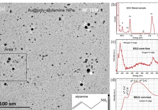
Fig.1ashowsa conventionalBFTEMimageoftheAu@PPAA NPssupportedonultrathinC.Thefieldofviewis300nmandthe darkparticlesofdifferentsizecorrespondtotheAucores.Shells arevisiblearoundthelargerparticles.Thetwo mainsourcesof contrastinBFTEMimagesaremass-thicknessanddiffraction con-trast.Theshellshaveuniformintensityirrespectiveoftheirsize, suggestingthattheyareamorphous.Fig.1b–dshows representa-tiveresultsofchemicalanalysisperformedbyaveragingEDXSand EELSsignalsoveranareaofthespecimensimilartothatshown inFig.1a.TheEDXspectruminFig.1aconfirmsthepresenceof NbutalsoindicatesthepresenceofSi.Thelatterpeakmaybean artifactoriginatingfromthewindowlessSidetector.Fig.1cshows anEELspectrumofthecore-lossNK-edge(401eV)andOK-edge (532eV),whileFig.1dshowsacorrespondingspectrumoftheC K-edge(284eV).TheNpeakistheprimarysignatureofthepresence ofallylamine(C3H5NH2)inthespecimen.Althoughpristine
ally-laminecontainssingleC Nbonds,sharpNandOedgeonsetsmay indicatethepresenceofC OandC Ngroups(GarvieandBuseck, 2004).Suchgroupsmayformduringtheplasmadischargewhen theprecursor(allylamine)isdissociatedintofragmentsofdifferent masscontainingC C,C N,C HandN Hbonds.Thebreakingof N HbondscanleadtotheformationofC Ngroupsduring recom-bination.PPAAcoatingsmayalsooxidizeinambientair,resulting intheformationofC OandC Obondsduetopolymeroxidation (Masseyetal.,2010).
Fig.1. (a)Bright-fieldTEMimageofhybridAunanoparticlescappedwithallylaminesupportedonultrathin(approx.10nm)amorphousCacquiredafteranelectrondose exposureof250C/cm2.TheAucores(darker)aresurroundedbyshellsoflowerdensity(lighter).Theshellsdonotencloselargerparticlescompletely.(b)EDXspectrum showingpeaksofN,AuandasmallamountofSi.(c)Core-lossEELspectrumshowingNandOK-edges.(d)Core-lossEELspectrumshowingtheCK-edge.ThestrongCu peaksoriginatefromthegridbarsoftheTEMCugrid.
4 L.C.Gontardetal./Micron67(2014)1–9
TheC K-edgeinFig.1dissimilartothatexpectedfor amor-phousC,withthelossofstructuralorderresultinginarelaxation oftheselectionrulesforthe1sto*transitionandproducinga broadfeaturelesspeakat290–310eV.Thespectrumcontainstwo strongpeaksat288.5and300eV,correspondingtoC C*and C C*bonds(GarvieandBuseck,2004).The*to*ratiocanbe calculatedbyassigningenergylossesof282–291eVto*states andenergylossesof294–301eVto*statesinthe
background-subtractedCK-edgespectrumshowninFig.1d.Weobtainavalue forthe*/*ratioof0.4,suggesting alackoflong-rangeorder (Katrinaketal.,1992).Thecontributiontothesignalfromthe amor-phousCsupportfilmmayaccountforthedominantsignatureof *typebonds.Ontheotherhand,C C*peakscanindicatethe
presenceof aromaticringsandunsaturated bondsof hydrocar-bons(contamination)typicallypresentinthecarbonfilmsusedto supportTEMsamples.Also,C C*peakscanoriginatefrom
unsat-uratedbondsthatremaininthePPAAdeposition,asC Cbonds arepresentinallylamine(CH2 CH CH2 NH2);correspondingto
incompletepolymerizationoftheallylamine.Therefore,itis nec-essarytouseanalternativeapproachtodeterminetheoriginofthe C C*signal,forexampleusinglow-lossEFTEMinordertomap thecharacteristicenergy-losspeakof∼7eVthathasbeenassigned toanelectronic–*transitionofaromaticCrings(LiandEgerton, 2004).
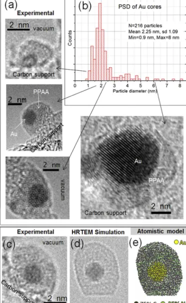
Fig.2ashowsrepresentativeexamplesofhigh-resolutionTEM [email protected] elec-tronirradiationresultedintearingoftheCsupportfilm,permitting clearvisualizationofNPshellsforcoresizesdownto1nm.The shellstypicallyhavethicknessesof1–2nmanddonotenclosethe NPsuniformly.Fig.2bshowstheparticlesizedistribution(PSD) measuredfrom216NPsbyapplyingasemi-automated segmenta-tionalgorithm(Gontardetal.,2011)totheimageshowninFig.1. ThePSDindicatesthattheAucoreshavesizesofbetween0.9and 8nm,withanaveragevalueof2.25nmandastandarddeviationof 1.09nm.
Fig.2cshowsahigh-resolutionTEMimageofahybridNPwith acoresizeof1.5nmattachedtotheedgeoftheCsupport.Fig.2d showsasimulatedhigh-resolutionTEMimageobtainedfromthe atomisticmodelshowninFig.2e,whichhasa1.5nmAucoreand anamorphousshellmadeof75%Cand25%N.Thecontrastinthe simulatedimagematchesthatintheexperimentalimage qualita-tively.ThereplacementofNatomsinthemodelbyCatomsdidnot resultinasubstantialchangeinthesimulatedimage,confirming thelackofsensitivityofhigh-resolutionTEMtochemicalchanges forsmalldifferencesinatomicnumber(Z=6forCandZ=7forN). ThebrightrimaroundtheAucoreinthesimulationinFig.2dis associatedwiththefactthatinthemodelthereisagapbetween thecoreandtheshell.Theabsenceofsuchabrightriminthe exper-imentalimagesuggeststhatthecoreiscappedcoherentlybythe PPAA.Thebrightrimonthesurfaceoftheshellisconsistentwith thedefocusvalueof+90nmusedinthesimulation.
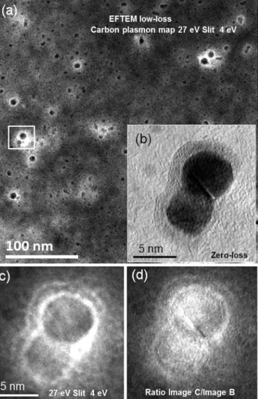
Fig.3ashowsalow-lossEFTEMimageacquiredfromthe spec-imenshown inFig.1atanenergy lossof27eV,which isclose tothebulk plasmonenergyexpectedfor Cthatcontains short-range(graphitic)order.Fig.3bandcshowszero-lossandlow-loss images,respectively, oftwo interactingparticlesindicated by a whitesquareinFig.3a.Fig.3dshowsaratioofthetwoimages. Thegraylevelsprovideameasureoftheplasmonexcitation prob-abilityatagivenpositionintheimageintegratedoverthewidthof theenergywindow(Stöcklietal.,2000).Theratioimageindicates thattheplasmonresonancesareparticularlyintenseattheAucore, theAu–shellinterfaceandtheoutersurfaceoftheshell.
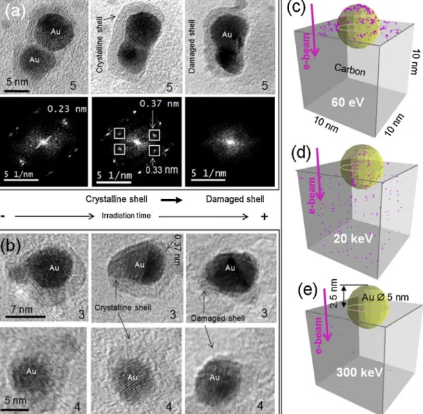
Fig.4illustratestheeffectoftheelectronbeamonthespecimen. During TEM observation, the C support film thinned progres-sivelyuntilholesformed.SomeNPsthatwereclosetoeachother coalescedorchangedinshape,whiletheshellsaroundtheNPs
Fig.2.(a)Examplesofhigh-resolutionTEMimagesofAu@PPAAnanoparticleswith coresofdifferentsizesupportedonthinC.(b)Measuredparticlesizedistribution oftheAucores(mean=2.25nmandSD=1.09nm).(c)High-resolutionTEMimage ofahybridnanoparticlewitha1.5nmcoreattachedtotheedgeoftheCsupport. (d)Simulatedhigh-resolutionTEMimageintensitycalculatedusingtheatomistic modelshownin(e).Themodelhasa1.5nmAucoreandanamorphousshellthat contains75%Cand25%N.
changeddramatically.Fig.4aandthecorrespondingdiffractograms below it show theevolution of two large NPs(numbered 5 in
Fig.5),whichfusedintooneparticleandbecameenclosedbya commonshell.ThefirstimageinthesequenceshowstwoAuNPs thatareincontactandsurroundedbyanamorphousPPAAlayer. Thesecondimageshowsthattheparticlesstarttocoalesceand thata commonshellis createdaround bothparticles.Theshell isnowcrystallinewith∼6independentgraphene-likelayerswith twodifferentinterplanarspacingsvisiblein twodirections.The firstspacingof0.33nmissimilartothec-axisspacing between graphene sheets in graphite of 0.34nm. The second spacing of 0.37nmissimilartothatreportedforgraphene/Cshells surround-ingAuNPssynthesizedusingchemicalvapordeposition(Chopra etal.,2009).Inthethirdimage,thetwoNPshavefusedtogether andtheshellisnowdamaged.ItisremarkablethattheNPswere able tocoalesceunder intense electron beamirradiation while retainingtheshellaroundthem.Inarelatedexperiment,Auhas beenobservedtoflowbetweendefectivegrapheneparticles(Sun etal.,2008).TheimagesequencesinFig.4billustratethesame
Fig.3.(a)EFTEMimageacquiredinthelow-energy-lossrangeforbulkplasmons usinga4eVenergy-selectingslitcenteredonanenergylossof27eV,whichisclose tothebulkplasmonenergyforCwithshort-range(graphitic)orderandalsoof Au(∼25eV).(b)Zero-lossimageofadimerofinteractingAuNPsindicatedwitha whitesquarein(a).(c)Low-lossimageoftheparticlesshownin(b).(d)Ratioof image(c)dividedbyimage(b).Thegraylevelsin(d)provideadirectmeasureofthe plasmonexcitationprobabilityatagivenpositionintheimageintegratedoverthe energywindow.Theplasmonresonancesareparticularlyintenseintheparticles, attheAu–shellinterface,inthecontactregionbetweentheAuparticlesandatthe outermostsurfaceoftheshell.
irradiation-inducedtransitioninotherparticles (numbered3in
Fig.5).Fig.4c–eshows simulationsof theexpectednumber of inelasticevents(showninpink)generatedbytheimpactof100 electronsacceleratedby60V,20kVand300kV,respectively.For 60V,theyieldis1andtheinelasticeventsremainatthesurface. For20kV,theyielddecreasesbutthe“damage”affectstheentire volumeofthespecimen.For300kV,thenumberofinelasticevents issmallerthanfor20kV.
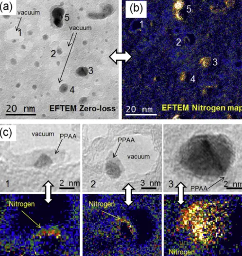
InordertoconfirmthattheorganicshellisPPAA,weusedEFTEM tomeasurethepresenceandlocaldistributionofN,whichisa con-stituentofallylamine,withhighspatialresolution.Imageswere acquiredatatimewhenthePPAAcoatinghadalreadystartedtobe damagedbytheelectronbeam.Fig.5ashowsazero-lossEFTEM imagerecorded from“area1”indicated in Fig.1.Fig. 5band c showscorelossEFTEMimagesacquiredattheNK-edge(401eV). Althoughspecimendriftand/ordamagewillhaveaffectedpartsof thebackground-subtractedelementalmapsshowninFig.5bandc, theresultsarestronglysuggestiveofthepresenceofNaroundthe AuNPs.
4. Discussion
We firstdiscussthepossibleoriginof thehighcontrast fea-turesinthelow-lossEFTEMimagesshowninFig.3.Second,we analyzetheeffectsofelectronirradiationontheintegrityofthe specimen.Third,weassessthephasetransformationsthatoccur afterprolongedelectronirradiation,includingcrystallizationand amorphization.
4.1. EFTEMcontrast:bulkorsurfaceplasmons?
InFig.3,thehighrecordedintensityattheAu–PPAAinterface andattheoutermostsurfaceofthecappinglayerislikelytobe associatedwiththeexcitationofplasmonsintheshelland/orthe Aucore.Theimageswererecordedatanenergylossof27eV.For aAuNP,thebulkplasmonenergyistypically∼25eV,althoughthe precisevaluecandependonthenatureofthesurfaceandthe oxi-dationstate(Tsivadzeetal.,2013).Surfaceplasmonscanalsobe excitedattheAucore,buttheyaretypicallyobservedintheoptical rangenear530nmor2.3eV.Boththeoreticallyand experimen-tally,shiftsofsurfaceplasmonenergiesinNPshavebeenshownto dependontheelectronicpropertiesoftheparticles.However,such energyshiftsaremostlyblue-shiftsandtheirmagnitudesrarely exceed10eV(Jainetal.,2007;Haridasetal.,2008).Hence,ashift ofthesurfaceplasmonenergyoftheAucoreisnotlikelytobethe sourceofthestrongintensityintheshellsobservedinFig.3.
ThebulkplasmonenergyforCisbetween22and27eV, depend-ing on the degree of short-range order present. For graphitic structures,theelectronicpropertiesandhencetheplasmon ener-giesindirectionsparallelandperpendiculartothegraphiticshells aredifferent(Stöcklietal.,2000).Insomecases,surfaceplasmon excitationprobabilitiesfornanotubeshaveresultedin the exci-tationof higher ordersurface plasmonresonance modesabove 10eVduetothefinitelengthsofthenanostructures,whichimpose alowerboundonthewavevectortransferoftheelectronsthat excitestheplasmons(Stöcklietal.,2000).
Giventhedifferentpossiblecontributionstothestrongcontrast observedinFig.3,weareforcedtoconcludethatwedonotyet haveaclearinterpretationofitsorigin,whichislikelytoinclude theroleofinterfacesandelectriccharge.TheenergylossesinFig.3
areintheexpectedrangeforbulkplasmonexcitationsinAuand C,buttheirspatialdistributionisnotdistributeduniformly, sug-gesting thatthedetailedmorphologyofthecore-shelldimerin
Fig.3bandthestructure androleofthesupportmustbetaken intoaccountfor afullinterpretationofthecontrast(Wangand Cowley,1987).Oscillationsofsurfacechargesdependsensitively onthedielectricpropertiesofthematerialand,moreimportantly, onthegeometricalconfigurationoftheinterfaces.Forexample, energy lossesof 20eVhave beenmeasuredusingX-ray photo-electronspectroscopyatthesurfacesofamorphousCfilmswith superficial“polymeric-like”monolayergrafting(Godetetal.,2009). Thissignalwasunderstoodtooriginatefromsurfaceplasmonsin theClayer,ratherthanfrombulklossesinthe“polymeric”grafting. Similarly,thehigherprobabilityofenergylossesfoundatthe inter-facesoftheparticlesinFig.3maybeassociatedwithbulkplasmons originatingfromthecappinglayer.
4.2. Electronirradiationdamage
ThesupportingCfilmthinneddownandthePPAAshell amor-phizedanddamagedafterlongexposuretotheelectronbeam.At theacceleratingvoltageusedinthepresentexperiments,knock-on damage,i.e.,thedisplacementofanatomfromitsoriginalsitebythe impactoftheenergeticelectronbeam,isinevitable.Ofparticular relevanceisthelossofatomsthatareexpelledintovacuum (sput-tered)fromthespecimenexitsurface.Theprobabilityofknock-on
6 L.C.Gontardetal./Micron67(2014)1–9
Fig.4.Illustrationoftheeffectoftheelectronbeamonthespecimen.AfterTEMobservation,theCsupportfilmthinneddownandsubsequentlyholesopenedacrossit. Theprocessstartedslowlyandthenaccelerated.Someparticlesthatwereclosetoeachothercoalescedorchangedshape,whiletheshellssurroundingtheNPsunderwent dramatictransformations.(a)High-resolutionTEMimagesandcorrespondingdiffractogramsshowingtheevolutionoftwolargeparticlesthatfusedintooneparticleand becameenclosedbyacommongraphiticshell.(b)Exampleofthetransformationofashellintoacrystallineform.(c–e)MonteCarlosimulationsofinelasticinteractions (pinkspots)generatedby60eV,20keVand300keVelectronsirradiatinga5nmAunanoparticlesemi-buriedinamorphousC.(Forinterpretationofthereferencestocolor inthisfigurelegend,thereaderisreferredtothewebversionofthearticle.)
damagedecreaseswithincreasingatomicnumberandistherefore lesslikelyforAuthanforH,C,OorN.Despitethenoticeable trans-formationofthehybridNPs(Fig.4),wemeasuredthepresence ofNintheirshellsafter2minofelectronirradiation(Fig.5)even aftertheshellbecameamorphous.Justascoatingoneorboth sur-facesofaspecimenwithCcanhelptopreserveitscrystallinityand toreducemassloss,thepresenceofaC supportfilmmayhave preventedsputteringoflightatomsorothermoleculesfromthe organicshellsintovacuum(Egertonetal.,2004).Oneproposed explanationisthatareturntotheoriginalmolecularstate (heal-ingofthebrokenbond)ofanorganicmoleculeismorelikelyifthe escapeofvolatileelementsisprevented(FryerandHolland,1983). Anotherimportantsourceofdamageinorganiccompoundsis inelasticdamagecausedbyvalenceelectron(ratherthancore-loss) excitation. Even relatively radiation-resistant organic materials mayundergosomeformofdamagewhenexaminedinanelectron microscopeatenergiesbelowtheknock-onthresholdenergy.It isinterestingtonotethatholesfrequentlyformedclosetoNPs. ThesimulationinFig.4eindicatesthatat300keVinelasticevents aremore likely tohappenin theAuNPsthan inC. Hence,the
breakingofC CbondsbyradiolysismaysoftentheCfilmnearthe particles,enhancingthesputteringrateofCatoms.Anotherform ofradiation-induceddamageisthroughheatdissipationfollowing energy transferin inelasticevents. Also, theelectronbeamcan breakhydrocarbonresiduesandtypicallyresultinapolymerization processthatcanfrequentlycoverareasofthesampleunderstudy, degrading thequality of data acquired.Although the electron-beam-inducedriseinspecimentemperatureisexpectedtobelow fortheexperimentalconditionsusedhere(LiandEgerton,2004), moleculardynamicssimulationshaveshownthatholeformation inthin C filmscanoccurwhen theelectron beamcausessmall clustersofamorphousC(e.g.,fromhydrocarboncontamination)to undergothermalexplosions(Börrnertetal.,2012).
A more profoundconsequence of inelasticscattering is that it leads to chemical changes in the specimen induced by the generation of low-energy (secondary) electrons. The specimen wasirradiated witha highcurrent flux(25A/cm2), wellabove
the critical electron dose for damage of PPAA, approximately 0.9×10⁻²C/cm2, based on its melting temperature, (216◦C)
Fig.5. (a)Zero-lossEFTEMimageof“Area1”indicatedinFig.1.After2minofirradiationwith200keVelectrons(totaldose=3×10³C/cm2),theCfilmbecamenoticeably thinnerandholesformed.(b)Background-subtractedNEFTEMelementalmap.(c)Detailsofzero-lossimagesandNmapsofthreeparticlesin(a)and(b).Althoughspecimen driftand/orspecimendamagemayhaveaffectedpartsoftheelementalmapsshownin(b)and(c),theresultsaresuggestiveofthepresenceofNassociatedwiththepositions oftheAuparticles.
Figs. 4and 5 wereacquired afterexposures of 2min,resulting in a total dose of3×103C/cm2. Low-energy electronsthat are
produced by irradiation with high-energy particles may cause primary chain breakage and other molecularfragmentation, as wellastheformationofdoublebondsandcross-linking(Sanche, 2002;Waskeetal.,2012).Secondaryelectronshavelowenergies (usuallybelow70eV)andthermalizationdistancesontheorder of nm, which define theinitial volumesfor energy deposition. ConsideringthestrongergenerationofsecondaryelectronsinAu thaninC,apolymer-metalinterfaceofahybridNPislikelytobe veryvulnerabletoradiationdamage.ExperimentsonPPAAhave demonstratedthataminegroups(CN)remainstableduring irra-diationwithlow-energy(1–60eV)electronsatlowdosesandthat theprimarychemicalmodificationsarealossofHandO(surface deoxidation)(Masseyetal.,2010).Emissionthresholdsforcations andanionsinirradiatedPPAA werefoundtobebetween7and 25eV,whichiswithintherangeofenergiesofsecondaryelectrons and collective-excitations (inter band, intra bandand plasmon resonances)generatedbyhigh-energyelectronirradiation(Liand Egerton,2004;Inadaetal.,2011).Theelectron-induced decompo-sitionofadsorbedorganiclayerswithamine(NH2)groupsonAu
substrateshasbeenobservedtobeinitiatedbysecondaryelectrons, transformingthelayerintoanamorphouscarbonnitridethinfilm (Wnuketal.,2009).MonteCarlosimulations(seeFig.4)showthat for60eVincidentelectronstheyieldofinelasticeventsis1andthat theyoccurmainlyatthesurfaceofthesample.Inourexperiments,
irradiationwasperformedusing300keVelectrons,whichhavea much higherpenetrationdepth(see Fig.4),generatinginelastic eventsinside thevolume of the specimen and not only onits surface. For our experimental specimenthickness and incident electron energy,we calculated a yieldfor inelasticcollisionsof 5% of the number of incoming electrons. Even after applying a correctionfor the yield, the high current fluxes used in our experimentswereseveralordersofmagnitudehigherthanthose usedbyMasseyetal.(2010),indicatingthatthegenerationofa largenumberofsecondaryelectronsislikelytohavecontributed todamageofthePPAAshellsofourNPs.
4.3. Electron-irradiation-inducedcrystallization
Figs.4and5showaphasetransitionsufferedbytheorganic cappingoftheAu@PPAANPs,firstbyare-orderingofthelayers andthenbyamorphizationandsubsequentdamage.Inparticular, theformationofgraphite-likelayerswasobserved.AlthoughAuis anoblemetalthatistypicallynotabletocatalyzetheformationof C-basedmaterials,thegrowthofCshellsinthevicinityofAuNPs hasbeenobservedinsituintheTEM.Graphenefragmentsand lay-ersformed,suggestingthatacrystallineAusurfacecancatalyzeand provideatemplatefortheorderingandcrystallizationofC. More-over,underintenseelectronirradiation(50A/cm2),thegraphene
fragmentscancloseandformregularsymmetriclayersaroundthe NPsuntilcompleteshellclosureisachieved(Sutteretal.,2005).
8 L.C.Gontardetal./Micron67(2014)1–9
However,thelatterexperimentswerecarriedoutataspecimen temperatureof 550◦C, whiletheexperimentsshown herewere carriedoutatroomtemperature.
Electron-induced graphitization of C is a well-known phe-nomenon, arising from cross-linking between neighboring moleculesatlowtemperature.Asaresultofdifferent hybridiza-tion,Cisabletoformdifferentallotropes,ofwhichgraphiteand diamondarethemostwellknown(Banhart,1999;Shakerzadeh et al.,2012).In particular, high-energy electronirradiation can graphitizefree-standingamorphousCandformConions (spher-icalstructureswithnodanglingbondsthathaveauniformstrain distribution)(Börrnertetal.,2012).Thegraphitization(formation ofC Cbonds)inpolystereneandPPAAasaresultofirradiation withlow-energyelectronshasbeenobserved(Masseyetal.,2010; Leeetal.,2008),inadditiontothelossofOandH.Afterprolonged irradiation,weobservedthatthePPAAshellsweredamaged,but westillmeasuredthepresenceofNusingEFTEM.Therefore,we canconcludethataftercrystallizationC and Nremainedinthe shellsoftheNPsinourspecimen.ThesubstitutionofCbyNin graphiteinaregularfashionmayhaveresultedintheformation of a “carbonnitride” (e.g., C3N4, C3N2, C3N, C5N or C10N3), or
evengraphiticg-C3N4(Thomasetal.,2008).Subsequent
electron-beam-induced decomposition may therefore have transformed thelayerintoanamorphouscarbonnitride,aspreviouslyreported for 1,2-diaminopropane films irradiated with electrons (Wnuk etal.,2009).
5. Conclusions
We have undertaken a TEM investigation of hybrid (metal/ polymer)AuNPscappedwithPPAA,whichareofinterestfor appli-cationsinnanomedecine,suchasselectivetargetingoftumorsites. OurresultsshowthatthePPPAcoatingsofunstainedhybridAuNPs canbeimagedintheTEMwithsufficientcontrastatan accelerat-ingvoltageof300kVatambienttemperaturewithouttheneedfor staining.ThethicknessofthePPAAcappingwastypicallybelow 2nmandoftennon-uniform.
FeaturesinEELspectraarelikelytobedominatedbythe sig-nalfromtheC supportfilm.Thisissuemightbealleviatedin a futurestudybyusinganimpregnationtechniqueforsupporting thecolloidofhybridnanoparticles(Gontardetal.,2014b).EFTEM imagesrecordedinthebulkplasmonenergyregimeshowedstrong intensityattheAu–polymerinterfaceandattheexternalsurface ofthepolymershell.Qualitatively,theresultspresentedhereshow thatlow-lossEFTEMcanbeanefficientsourceofhighcontrastin imagesofhybridnanostructures.However,thefullinterpretation ofsuchimagesrequiresfurtherworkbecausetheplasmonmodes ofhybridcore–shellNPscannotbetreatedasadditiveindividual contributionsofthecoreandshellmodes(Chuntonovetal.,2012) andareverysensitivetosmallchangesinlocalmorphologyand electricalstate(Suetal.,2012;Jiangetal.,2014).
WhereasPPAAispredictedtobeelectron-beam-resistantatlow doses,thePPAAlayersofhybridNPsdamagedandbecame amor-phousunderelectronbeamirradiationat300keVwhenhighdoses wereused,althoughwecouldstillmeasurethepresenceofNin theshells.Knock-ondamageishighattheacceleratingvoltages usedinourwork.However,theuseofaCsupportfilmmayreduce sputteringfromtheshellsandexplaintheirstability.Ontheother hand,MonteCarlosimulationsshowthatthenumberofinelastic eventsclosetotheparticlesincreasesatlowaccelerationvoltages (≤20kV).Inafuturestudy,itwillbeofgreatinteresttocompare thepresentresultswithimagesacquiredatanintermediate accel-eratingvoltage(60–80kV).
Before the shells were destroyed, we observed the forma-tionofcrystallineorder aroundlargerNPs,aswellas graphitic planes.Polymericshellscontainingnitrogenwereobservedafter
irradiationwithveryhighelectrondoses,indicatingthatthe parti-clesarelikelytobemorestableattherelativelylowerdosesused inbiomedicalapplications.Also,suchexperimentsinvolvinginsitu TEMirradiationofpolymersmaybeimportantforunderstanding electron-beam-induced structural changes in polymers and for refiningapproaches fortuningtheopticalpropertiesofNPs.For example, polysterene homopolymer films have been shown to becomeluminescentinvisiblelightunder50keVelectron irradi-ation,whichinducedthere-formationofaromaticrings(Leeetal., 2008).
Acknowledgment
We are grateful to the European Union for support under project reference REGPOT-CT-2011-285895-Al-NANOFUNC and underGrantAgreement312483–ESTEEM2(Integrated Infrastruc-tureInitiative–I3).
References
Banhart,F.,1999.Irradiationeffectsincarbonnanostructures.Rep.Prog.Phys.62, 1181–1221.
Bawarski,W.E.,Chidlowsky,E.,Bharali,D.J.,Mousa,S.A.,2008.Emerging nanophar-maceuticals.Nanotechnology4,273–282.
Börrnert,F.,Avdoshenko,S.M.,Bachmatiuk,A.,Ibrahim,A.,Büchner,B.,Cuniberti, G.,Rümmeli,M.H.,2012.Amorphouscarbonunder80kVelectronIrradiation:a meanstomakeorbreakgraphene.Adv.Mater.24(41),5630–5635.
Chen,C.,Daniel,M.C.,Quinkert,Z.T.,De,M.,Stein,B.,Bowman,V.D.,Chipman,P.R., Rotello,V.M.,Kao,C.C.,Dragnea,B.,2006.Nanoparticle-templatedassemblyof viralproteincages.NanoLett.6(4),611–615.
Chopra,N.,Bachas,L.G.,Knecht,M.R.,2009.Fabricationandbiofunctionalizationof carbon-encapsulatedAunanoparticles.Chem.Mater.21,1176–1178.
Chuntonov, L., Bar-Sadan, M., Houben, L., Haran, G., 2012. Correlating elec-trontomographyandplasmonspectroscopyofsinglenoblemetalcore–shell nanoparticles.NanoLett.12,145–150.
Daniels,H.R.,Brydson,R.,Brown,A.,Rand,B.,2003.Quantitativevalenceplasmon mappingintheTEM:viewingphysicalpropertiesatthenanoscale. Ultrami-croscopy96,547–558.
Demers,H.,Poirier-Demers,N.,Couture,A.R.,Joly,D.,Guilmain,L.,DeJonge,N., Drouin,D.,2011.Three-dimensionalelectronmicroscopysimulationwiththe CASINOMonteCarlosoftware.Scanning133,135–146.
Drummy,L.F.,Yang,J.,Martin,D.C.,2004.Low-voltageelectronmicroscopyof poly-merandorganicmolecularthinfilms.Ultramicroscopy99,247–256.
DuChesne,A.,1999.Energyfilteringtransmissionelectronmicroscopyofpolymers –benefitandlimitationsofthemethod.Macromol.Chem.Phys.200,1813–1830.
Egerton,R.F.,Li,P.,Malac,M.,2004.RadiationdamageintheTEMandSEM.Micron 35,399–409.
Fryer,J.R.,Holland,F.,1983.Thereductionofradiationdamageintheelectron microscope.Ultramicroscopy11,67–70.
Garvie,L.A.J.,Buseck,P.R.,2004.Nanosizedcarbon-richgrainsincarbonaceous chon-dritemeteorites.EarthPlanet.Sci.Lett.224,431–439.
Gittins,D.I.,Caruso,F.,2001.Tailoringthepolyelectrolytecoatingofmetal nanopar-ticles.J.Phys.Chem.B105,6846–6852.
Godet,C.,David,D.,Sabbah,H.,Ababou-Girard,S.,Solal,F.,2009.Bulkandsurface plasmonexcitationsinamorphouscarbonmeasuredbycore-level photoelec-tronspectroscopy.Appl.Surf.Sci.255,6598–6606.
Gontard,L.C.,Knappett,B.R.,Wheatley,A.E.H.,Chang,S.L.-Y.,Fernández,A.,2014a.
ImpregnationofcarbonblackfortheexaminationofcolloidsusingTEM.Carbon 76,464–468.
Gontard,L.C.,Moldovan,G.,Carmona-Galán,R.,Lin,C.,Kirkland,A.I.,2014b. Detec-tingsingle-electroneventsinTEMusinglow-costelectronicsandasiliconstrip sensor.Microscopy63(2),119–130.
Gontard,L.C.,Ozkaya,D.,Dunin-Borkowski,R.E.,2011.Asimplealgorithmfor mea-suringparticlesizedistributionsonanunevenbackgroundfromTEMimages. Ultramicroscopy111,101–106.
Haridas,M.,Srivastava,S.,Basu,J.K.,2008.Tunablevariationofopticalpropertiesof polymercappedgoldnanoparticles.Eur.Phys.J.D49,93–100.
Horiuchi,S.,Hanada,T.,Ebisawa,M.,Matsuda,Y.,Kobayashi,M.,Takahara,A.,2009.
Contamination-freetransmissionelectronmicroscopyforhigh-resolution car-bonelementalmappingofpolymers.ACSNano3(5),1297–1304.
Howie,A.,2003.Valenceexcitationsinelectronmicroscopy:resolvedand unre-solvedissues.Micron34,121–125.
Hunt,J.A.,Disko,M.M.,Behal,S.K.,Leapman,R.D.,1995.Electronenergy-loss chem-icalimagingofpolymerphases.Ultramicroscopy58,55–64.
Jain,P.K.,Huang,X.,El-Sayed,I.H.,El-Sayed,M.A.,2007.Reviewofsomeinteresting surfaceplasmonresonance-enhancedpropertiesofnoblemetalnanoparticles andtheirapplicationstobiosystems.Plasmonics2,107–118.
Jiang,N.,Shao,L.,Wang,J.,2014.(Goldnanorodcore)/(polyanilineshell) plas-monicswitcheswithlargeplasmonshiftsandmodulationdepths.Adv.Mater.,
Kang,Y.,Taton,T.A.,2005.Controllingshellthicknessincore–shellgold nanopar-ticles via surface-templated adsorption of block copolymer surfactants. Macromolecules38,6115–6121.
Karmani,L.,Labar,D.,Valembois,V.,Bouchat,V.,Nagaswaran,P.G.,Bol,A.,Gillart,J., Levêque,P.,Bouzin,C.,Bonifazi,D.,Michiels,C.,Feron,O.,Grégoire,V.,Lucas,S., Borght,T.V.,Gallez,B.,2013.Antibody-functionalizednanoparticlesforimaging cancer:influenceofconjugationtogoldnanoparticlesonthebiodistributionof
89Zr-labeledCetuximabinmice.ContrastMediaMol.Imag.8,402–408.
Katrinak,K.A.,Rez,P.,Buseck,P.R.,1992.Structuralvariationsinindividual carbona-ceousparticlesfromanurbanaerosol.Environ.Sci.Technol.26,1967–1976.
Krivanek,O.J.,Dellby,N.,Murfitt,M.F.,Chisholm,M.F.,Pennycook,T.J.,Suenaga,K., Nicolosi,V.,2010.GentleSTEM:ADFimagingandEELSatlowprimaryenergies. Ultramicroscopy110,935–945.
Kumar,S.,Adams,W.W.,1990.Electronbeamdamageinhightemperature poly-mers.Polymer31,15–19.
Inada,H.,Su,D.,Egerton,R.F.,Konno,M.,Wu,L.,Ciston,J.,Wall,J.,Zhu,Y.,2011.
Atomicimagingusingsecondaryelectronsinascanningtransmissionelectron microscope:experimentalobservations andpossiblemechanisms. Ultrami-croscopy111,865–876.
Lee,H.M.,Kim,Y.N.,Kim,B.H.,Kim,S.O.,Cho,S.O.,2008.Fabricationof lumines-centnanoarchitecturesbyelectronirradiationofpolystyrene.Adv.Mater.20, 2094–2098.
Li,D.,He,Q.,Li,J.,2009.Smartcore/shellnanocomposites:intelligentpolymers modifiedgoldnanoparticles.Adv.ColloidInterfaceSci.149,28–38.
Li,P.,Egerton,R.F.,2004.Radiationdamageincoronene,rubreneandp-terphenyl, measuredforincidentelectronsofkineticenergybetween100and200keV. Ultramicroscopy101,161–172.
Libera,M.R.,Egerton,R.F.,2010.Advancesinthetransmissionelectronmicroscopy ofpolymers.Polym.Rev.50(3),321–339.
Linares,E.M.,Leite,C.A.P.,Valadares,L.F.,Silva,C.A.,Rezende,C.A.,Galembeck,F., 2009.Molecularmappingbylow-energy-lossenergy-filteredtransmission elec-tronmicroscopyimaging.Anal.Chem.81,2317–2324.
Malac,M.,Beleggia,M.,Egerton,R.,Zhu,Y.,2007.Bright-fieldTEMimagingofsingle molecules:dreamornearfuture?Ultramicroscopy107,40–49.
Marega,R.,Karmani,L.,Flamant,L.,Nageswaran,P.G.,Valembois,V.,Masereel, B.,Feron,O.,Borght,T.V.,Lucas,S.,Michiels,C.,Gallez,B.,Bonifazi,D.,2012.
Antibody-functionalizedpolymer-coatedgoldnanoparticlestargetingcancer cells:aninvitroandinvivostudy.J.Mater.Chem.22,21305–21312.
Martin,D.C.,Chen,J.,Yang,L.F.,Drummy,L.F.,Kübel,C.,2005.Highresolution electronmicroscopy ofordered polymers andorganic molecular crystals: recentdevelopmentsandfuturepossibilities.J.Polym.Sci.B:Polym.Phys.43, 1749–1778.
Masereel,B.,Dinguizli,M.,Bouzin,C.,Moniotte,N.,Feron,O.,Gallez,B.,Borght, T.V.,Michiels,C.,Lucas,S.,2011.Antibodyimmobilizationongold nanopar-ticles coated layer-by-layer with polyelectrolytes. J. Nanopart. Res. 13, 1573–1580.
Massey,S.,Gallino,E.,Cloutier,P.,Tatoulian,M.,Sanche,L.,Mantovani,D.,Roy, D., 2010. Low-energy electrons and X-ray irradiation effects on plasma-polymerizedallylaminebioactivecoatingsforstents.Polym.Degrad.Stab.95, 153–163.
Moreau,N.,Michiels,C.,Masereel,B.,Feron,O.,Gallez,B.,Borght,T.V.,Lucas,S., 2009.PVDsynthesisandtransferintowater-basedsolutionsoffunctionalized goldnanoparticles.PlasmaProcess.Polym.6(S1),888–892.
Müller,H.,Jin,J.,Danev,R.,Spence,J.,Padmore,H.,Glaeser,R.M.,2010.Designofan electronmicroscopephaseplateusingafocusedcontinuous-wavelaser.NewJ. Phys.12,073011.
Nagayama,K.,Danev,R.,2008.Phasecontrastelectronmicroscopy:development ofthin-filmphaseplatesandbiologicalapplications.Phil.Trans.R.Soc.B363, 2153–2162.
Nelayah,J.,Gu,L.,Sigle,W.,Koch,C.T.,Pastoriza-Santos,I.,Liz-Marzán,L.M.,van Aken,P.A.,2009.Directimagingofsurfaceplasmonresonancesonsingle trian-gularsilvernanoprismsatopticalwavelengthusinglow-lossEFTEMimaging. Opt.Lett.34(7),1003–1005.
Sardar,R.,Park,J.-W.,Shumaker-Parry,J.S.,2007.Polymer-inducedsynthesisof sta-blegoldandsilvernanoparticlesandsubsequentligandexchangeinwater. Langmuir23,11883–11889.
Sanche,L.,2002.Nanoscopicaspectsofradiobiologicaldamage:fragmentation inducedbysecondarylow-energyelectrons.MassSpectrom.Rev.21,349–369.
Shakerzadeh,M.,Loh,G.C.,Xu,N.,Chow,W.L.,Tan,C.W.,Lu,C.,Yap,R.C.C.,Tan,D., Tsang,S.H.,Teo,E.H.T.,Tay,B.K.,2012.Re-orderingchaoticcarbon:originsand applicationoftexturedcarbon.Adv.Mater.24,4112–4123.
Stöckli,T.,Bonard,J.-M.,Châtelain,A.,Wang,Z.L.,Stadelmann,P.,2000.Plasmon excitationsingraphiticcarbonspheresmeasuredbyEELS.Phys.Rev.B61(8), 5751–5759.
Su,H.,Zhong,Y.,Ming,T.,Wang,J.,Wong,K.M.,2012.Extraordinarysurface plas-moncoupledemissionusingcore/shellgoldnanorods.J.Phys.Chem.C116, 9259–9264.
Sun,L.,Krasheninnikov,A.V.,Ahlgren,T.,Nordlund,K.,Banhart,F.,2008.Plastic deformationofsinglenanometer-sizedcrystals.Phys.Rev.Lett.101,156101.
Sutter,E.,Sutter,P.,Zhu,Y.,2005.AssemblyandinteractionofAu/Ccore–shell nano-structures:insituobservationinthetransmissionelectronmicroscope.Nano Lett.5(19),2092–2096.
Thomas,A.,Fischer,A.,Goettmann,F.,Antonietti,M.,Müller,J.-O.,Schlögl,R., Carls-son,J.M.,2008.Graphiticcarbonnitridematerials:variationofstructureand morphologyandtheiruseasmetal-freecatalysts.J.Mater.Chem.18,4893–4908.
Tsivadze,A.Y.,Ionova,G.V.,Mikhalko,V.K.,Ionova,I.S.,Gerasimov,G.A.,2013.The finestructureofplasmonbandsingoldnanoparticles.Prot.Met.Phys.Chem. Surf.49(2),166–168.
vanSchooneveld,M.M.,Gloter,A.,Stephan,O.,Zagonel,L.F.,Koole,R.,Meijerink, A.,Mulder,W.J.M.,deGroot,F.M.F.,2010.Imagingandquantifyingthe mor-phologyofanorganic–inorganicnanoparticleatthesub-nanometrelevel.Nat. Nanotechnol.5,538–544.
Wang,Z.L.,Cowley,J.M.,1987.Surfaceplasmonexcitationforsupportedmetal par-ticles.Ultramicroscopy21,77–94.
Waske,P.A.,Meyerbröker,N.,Eck,W.,Zharnikov,M.,2012.Self-assembled mono-layersofcyclicaliphaticthiolsandtheirreactiontowardelectronirradiation.J. Phys.Chem.C116,13559–13568.
Wnuk,J.D.,Gorham,J.M.,Fairbrother,D.H.,2009.Growthandmicrostructureof nanoscaleamorphouscarbonnitridefilmsdepositedbyelectronbeam irradia-tionof1,2-diaminopropane.J.Phys.Chem.C113,12345–12354.