HAL Id: tel-02882132
https://tel.archives-ouvertes.fr/tel-02882132
Submitted on 26 Jun 2020HAL is a multi-disciplinary open access
archive for the deposit and dissemination of sci-entific research documents, whether they are pub-lished or not. The documents may come from teaching and research institutions in France or abroad, or from public or private research centers.
L’archive ouverte pluridisciplinaire HAL, est destinée au dépôt et à la diffusion de documents scientifiques de niveau recherche, publiés ou non, émanant des établissements d’enseignement et de recherche français ou étrangers, des laboratoires publics ou privés.
Inhibition of UHRF1 protein by epigenetic partners and
epi-drugs
Tanveer Ahmad
To cite this version:
Tanveer Ahmad. Inhibition of UHRF1 protein by epigenetic partners and epi-drugs. Biochemistry, Molecular Biology. Université de Strasbourg, 2019. English. �NNT : 2019STRAJ089�. �tel-02882132�
UNIVERSITÉ DE STRASBOURG
ÉCOLE DOCTORALE DES SCIENCES DE LA VIE ET DE DA SANTE
Laboratoire de Bioimagerie et Pathologies – LBP UMR 7021
THÈSE
présentée par :Tanveer AHMAD
Soutenue le : 20 Novembre 2019Pour obtenir le grade de :
Docteur de l’Université de Strasbourg
Discipline/ Spécialité: DOCTORAT Sciences
Pharmacologie-Pharmacocinétique
Inhibition de la protéine UHRF1 par les
partenaires épigénétiques et les
épi-drogues
THÈSE dirigée par :
M. MOUSLI Marc Chargé de Recherche, INSERM, UMR 7021
RAPPORTEURS :
Mme. EYMIN Béatrice Directeur de Recherche, Institut pour l'Avancée des Biosciences, Université Grenoble Alpes
M. CARTRON Pierre-François Chargé de Recherche, Centre de Recherche en
Cancérologie et Immunologie, Nantes-Angers
AUTRE MEMBRE DU JURY (EXAMINATEUR) :
Mme. DANTZER Françoise Directeur de Recherche, ESBS, UMR7242
MEMBRES INVITÉS :
Mme. DUBREZ Laurence Chargé de Recherche, Université de Bourgogne
THIS DISSERTATION IS DEDICATED TO MY BELOVED
PARENTS
ACKNOWLEDGEMENTS
I acknowledge all the people who helped me to complete this dissertation:
In the first place, to Dr. Marc MOUSLI, my mentor, for his remarkable guidance, extraordinary mentoring, unconditional support, extensive motivation and impressive scientific discussions during last four years. In particular, I am grateful for his time and efforts dedicated to different aspects of my scientific growth including, but not limited to, augmenting productivity, learning of experimental & technical skills, improving scientific thinking and communication skills. I would also like to express my gratitude to my co-supervisor Dr. Christian Bronner for his precious time, worthwhile suggestions, essential support and critical comments regarding project.
On a special note, I am indebted to Professor Yves MELY for his outstanding support, valuable time, extraordinary knowledge, fruitful advices & discussions and his prime example of scientific leadership which inspired me a lot.
I really appreciate my jury members Dr. Béatrice EYMIN, Dr. Laurence DUBREZ, Dr. Françoise DANTZER and Dr. Pierre-François CARTRON for accepting our request to evaluate this thesis.
I am thankful to members of my thesis advisory committee Dr. Ali HAMICHE and Dr. Christian D. MULLER for their time, evaluation, critical discussion, suggestions and providing me lab facilities to complete this project.
Many thanks to Dr. Ludovic RICHERT and Dr. Pascal DIDIER for helping me in acquisition and analysis of FLIM experiments. Thanks a lot to members of Plateforme d’Imagerie Quantitative Romain VAUCHELLES, Dr. Philippe CARL and Philippe RONDE for their help in confocal microscopy and live cell imaging experiments. I would also like to thank Dr. Abdulkhaleg IBRAHIM for his help in protein purification and cell line preparations.
I truly appreciate the permanent staff of Laboratory of Bioimaging and Pathologies for their scientific discussions and help, Dr. Christian BOUDIER, Dr. Emmanuel BOUTANT, Dr.
Julien GODET, Dr. Eleonore REAL, Dr. Nicolas HUMBERT, Dr. Patrice RASSAM, Tania STEFFAN and Thiebault LEQUEU.
Special thanks to Marlyse WERNERT and Ingrid BARTHEL for always helping me in administrative procedures.
Sincere thanks to my small UHRF1 team: Waseem ASHRAF and Liliyana Zaayter. Their amazing personalities, faithful company and endless laughs during good and bad times made this journey easier for me. I really appreciate your care, support and guidance.
Thanks to the current and past lab mates Rajhans, Lesia, Manu, Redouane, Vanille, Krishna, Maaz, Jaganaath, Sarwat, Marianna, Nina, Oleksi, Raphael, Stefano, Tannoo, Hasan, Nario, Tina, Soraya, Sara, Imtiaz, Sani, Antoine, Mario and Marléne for their help, motivation, discussion and enjoyable company. Special and sincere thanks to Malak Abbas, M. Faisal Nadeem and Waseem Ashraf for their kind support, precious time, positive discussions and motivation at each and every step of this journey.
I am grateful to Strasbourg Family especially Wahid, Bilal, Raman, Tajith, Yasmin, Imtiaz, Asad, Salman, Akmal, Sara, Hira Hasan, proteomic friend Arslan, Hira, Aabid, Hanna Gsell, Fareeha, Kareem, Ameer, Irshad, Camille, Jean Martin and Astrid. I cannot forget their unlimited support, time, encouragement, and unforgettable memories.
I would also like to acknowledge Higher Education Commission of Pakistan and Campus France for providing me the financial support to continue my Ph.D in France.
Lastly, it was not possible without pure love, care, sincerity and motivation of my father, mother, sister and brothers who supported me at each phase of my life. They made me what I am today. This thesis belongs to them.
Thank you very much.
i
TABLE OF CONTENTS
LIST OF ABBREVIATIONS ... iv I- INTRODUCTION ... 1 1. Epigenetics... 1 2. Chromatin organization ... 3 3. Epigenetic modifications ... 53.1.A. DNA methylation ... 5
a. DNA methylation chemistry... 5
b. Enzymes involved in DNA methylation ... 6
c. Methylation sites ... 7
d. Maintenance of DNA methylation patterns ... 9
3.1.B. DNMT1 ... 10
a. Structure and domain architecture of DNMT1 ... 10
b. DNMT1 expression ... 10
c. Target selectivity of DNMT1 ... 11
d. Interaction of DNMT1 with other partners... 12
3.1.C. DNMT2 ... 13
3.1.D. DNMT3 family ... 14
a. De novo methylation ... 14
3.1.E. DNA demethylation ... 15
3.2. Histone modification ... 16
3.3. Regulatory ncRNAs (non-coding RNAs) ... 17
4. Ubiquitin PHD RING Finger (UHRF) family ... 19
4.1.A UHRF1 (Ubiquitin-like, containing PHD and RING Finger domains 1) ... 19
4.1.B. Structure and function of UHRF1 domains ... 20
a. Ubiquitin-like domain (UBL) ... 20
b. Tandem Tudor domain (TTD) ... 21
c. Plant homeodomain (PHD) ... 23
d. Set and ring associated (SRA) domain ... 24
e. Really interesting new gene (RING) domain ... 25
4.1.C. Roles of UHRF1 ... 26
ii
b. UHRF1 and its conformational dynamics ... 30
4.1.D. UHRF1 regulation through post-translational modifications... 31
a. Ubiquitination of UHRF1 ... 31
b. Phosphorylation of UHRF1... 33
c. Acetylation of UHRF1 ... 34
4.2. UHRF1 regulation and cell cycle ... 36
4.3. Screening and targeting of UHRF1 ... 37
4.3.A. Targeting of UHRF1 ... 37
a. Chemical compounds targeting UHRF1 ... 38
b. Natural compounds targeting UHRF1 ... 40
4.3.B. Targeting of SRA domain ... 42
4.4. UHRF1 partners ... 45
5. TIP60 (Tat Interactive Protein 60kDa) ... 46
5.1.A. Structure of TIP60 ... 46
5.1.B. Acetyltransferase activity of TIP60 ... 48
5.1.C. Role of TIP60 in transcription regulation ... 50
a. TIP60-mediated regulation of transcription in tumor suppression ... 50
b. TIP60-mediated regulation of transcription in tumorogenesis ... 51
5.1.D. Role of TIP60 in cell cycle regulation ... 54
5.1.E. Regulation of TIP60 in cells ... 55
5.1.F. TIP60 regulation through post-translational modifications ... 56
a. Acetylation of TIP60... 56
b. Phosphorylation of TIP60 ... 57
c. Sumoylation of TIP60 ... 58
5.1.G. Deregulation of TIP60 in cancers ... 59
5.1.H. TIP60 and apoptosis ... 61
II- OBJECTIVES ... 64
III- MATERIALS AND METHODS ... 67
1. Materials ... 67
1.A. Antibodies ... 67
1.B. Plasmid Constructs ... 69
1.C. Bacterial plasmids for expression of TIP60 and its mutants proteins ... 81
1.D. Cell lines ... 83
iii
2.A. Cell Culture ... 83
2.B. Transient transfection ... 83
2.C. Protein isolation ... 84
2.D. Western blot ... 84
2.E. Immunoprecipitation ... 85
2.F. Cell-based ubiquitination assay ... 85
2.G. UHRF1 auto-ubiquitination assay ... 86
2.H. Confocal microscopy ... 86
2.I. FÖster Resonance Energy Transfer-Fluorescence Lifetime Imaging Microscopy (FRET-FLIM) 88 2.J. Apoptosis analysis ... 89
IV- RESULTS ... 91
Manuscript I: Interaction of the epigenetic integrator UHRF1 with the MYST domain of TIP60 inside the cell ... 91
Supplementary Results ... 106
Manuscript II: Intracellular regulation of UHRF1 by TIP60 in human cancer cells ... 110
Manuscript III: A molecular tool targeting the base-flipping activity of human UHRF1 ... 142
Supplementary Results ... 157
Manuscript IV: 2-amino-3-hydroxyanthraquinone (AHAQ) induces cell cycle arrest and apoptosis in cervical cancer cells with a re-expression of p53 and down-regulation of DNMT1/UHRF1 .... 171
V- DISCUSSION AND PERSPECTIVES ... 190
VI-References ... 198
VII-ANNEXURE... 218
VII.A. UHRF1 as a diagnostic tool for cancer ... 218
Review Article: The epigenetic integrator UHRF1: on the road to become a universal biomarker for cancer ... 219
Manuscript V: Maritime pine tannin extract from bark exhibits anticancer properties by inducing expression of p73 and targeting UHRF1 and DNMT1 of epigenetic machinery in cancer cells. .. 237
iv
LIST OF ABBREVIATIONS
5-mC
17-AAG
5-methylcytosine
17-Allylamino-17-demethoxygeldanamycin
Acetyl CoA
AdoMet
AHAQ
AR
ATM
BAH
BAX
BCL2
BPC
BRCA1
Caspase
CCL
CDK
CFP1
CpG
CRD
DAPI
DDR
DHA
DHAQ
DHNQ
DMAPD
DMEM
DNA
DNMT1
E2F1
EDTA
EdU
EGCG
EHMT2 (G9a)
EMT
ER
ERCC1
Acetyl-Coenzyme A
S-Adenosylhomocysteine (SAH)
2-Amino-3-Hydroxyanthraquinone
Androgen Receptor
Ataxia Telangiectasia Mutated
Bromo-adjacent Homology
Bcl-2-Associated X Protein
B Cell Lymphoma
4-Benzylpiperdine-1-Carboximidamide
Breast Cancer 1
Cysteine Aspartate Protease
Chronic Lymphocytic Leukemia
Cyclin kinase dependent
CXXC Finger Protein 1
Cytosine-phosphate Guanine
Chromodomain
4',6-diamidino-2-phenylindole
DNA Damage Response
Dihydroartemisnin
1,3-Dihydroxy-9,10-Anthraquinone-2-carboxylic
acid
5,8-Dihydroxy-1,4-Naphthoquinone
DNA methyltransferase-associated protein 1
domain
Dulbecco’s modified Eagle’s Medium
Deoxyribonucleic Acid
DNA Methyltransferase 1
E2F Transcription Factor 1
Ethylenediaminetetraacetic acid
5-Ethynyl-2´-deoxyuridine
Epigallocatechin-3-Gallate
Euchromatic Histone-Lysine N-Methyltransferase
2
Epithelial-Mesenchymal Transition
Estrogen Receptor
v
ESCs
Esa1
EZH2
eGFP
FACS
FLIM
FRET
FRET
FQI1
GSK-3
H3K9me2
H3K9me3
H3R2
H4K16Ac
HAT
HCC
HDAC1
HDM
HIV
HMT
HNSCC
HPV
HSP90
ICBP90
ING
LIG1
LOPAC
LSF
MBD
MBPs
Mdm2
MPTE
MOF
miRNA
mTOR
MYST
ncRNA
NFκB
NPAT
NR
Embryonic Stem Cells
Essential Sas family acetyltransferase
Enhancer of Zeste 2 Polycomb Repressive
Complex 2 subunit
enhanced Green Fluorescent Protein
Fluorescence-Activated Cell Sorting
Fluorescence Lifetime Imaging Microscopy
Fluorescence Resonance Energy Transfer
Förster Resonance Energy Transfer
Factor Quinolinone Inhibitor
Glycogen Synthase Kinase-3
Histone 3 Lysine 9 dimethylation
Histone 3 Lysine 9 Trimethylation
Histone 3 Arginine 2
Histone 4 Lysine 16 Acetylation
Histone Acetyltransferase
Hepatocellular Carcinoma
Histone Deacetylase 1
Histone Demethylase
Human Immunodeficiency Virus
Histone Methyltransferase
Head and Neck Squamous Cell Carcinoma
Human Papilloma Virus
Heat Shock Protein 90
Inverted CCAAT box Binding Protein of 90 kDa
Inhibitor of Growth protein
DNA Ligase 1
Library of Pharmacologically Active Compounds
Late SV40 Factor
methyl-CpG binding domain proteins
Methyl-binding domain proteins
Mouse double minute 2
Maritime Pine Tannin Extract
Males-absent on the first protein
microRNA
Mammalian Target of Rapamycin
MOZ, Ybf2/Sas3, Sas2 and TIP60
non-coding RNA
Nuclear Factor Kappa B
Nuclear Protein coactivator of histone
Transcription
vi
PBD
PBR
PCNA
PDE1
PHC
PHD
PI
PI5P
PKB
PML
PMSF
PRAK
PRMT5
PUMA
RFTD
RFTS
RING
RNA
ROS
Rb
SAM
SIRT1
SRA
SRSF2
STAT3
SUMO
SUV39H1
TCL
TET
TIP60
TSGs
TRD
TR-FRET
TTD
TQ
UBB
UBC
Ubc9
UBL
UIM
UHRF1
PCNA-binding domain
Polybasic Region
Proliferation Cell Nuclear Antigen
Phosphodiestrase 1
Pericentromeric Heterochromatin
Plant Homeo Domain
Propidium iodide
Phosphatidylinositol 5-Phosphate
Protein Kinase B also known as Akt
Promyelocytic Leukemia
Phenylmethylsulfonyl fluoride
p38-Regulated/Activated protein Kinase
Protein Arginine Methyltransferase 5
p53 Upregulated Modulator of Apoptosis
Replication foci-targeting domain
Replication foci- targeting sequence
Really Interesting New Gene
Ribonucleic acid
Reactive Oxygen Species
Retinoblastoma
S-adenosyl-L-methionine
Sirtuin 1
Set and Ring Associated
Serine and Arginine rich Splicing Factor 2
Signal Transducer and Activator of Transcription
3
Small Ubiquitin-like Modifier
Suppressor of Variegation 39-Homolog 1
T Cell Receptor
Ten Eleven Translocation
Tat Interactive Protein 60 kDa
Tumor Suppressor Genes
Target Recognition Domain
Time Resolved-FRET
Tandem Tudor Domain
Thymoquinone
Ubiquitin-B or Polyubiquitin-B
Ubiquitin-C or Polyubiquitin-C
Ubiquitin Carrier protein 9
Ubiquitin-Like Domain
Ubiquitin Interacting Motif
Ubiquitin like with PHD and Ring Finger domains
1
vii
USP7 also known as
HAUSP
Ubiquitin-specific-processing protease 7
1
I- INTRODUCTION
1. Epigenetics
To answer the diversity of phenotypes within a population arising from the same genome has always attracted the attention of scientists. How stem cells having same genomic sequence can have different phenotypes and they can differentiate into different type of cells and tissues? How homozygote twins or cloned animals having identical DNA sequence can have different phenotypes?
Figure 1: Same genome but different phenotypes. Stem cells of same origin having identical DNA sequence
have developed different phenotypes with specialized functions. Images used in figure are taken from Servier Medical Arts http://servier.com/Powerpoint-image-bank.
Life cycle of a butterfly is a classic example of metamorphosis in insects which explains the essential role of DNA at different stages of life cycle (Figure 2). During different stages (egg, caterpillar, pupa and adult butterfly), butterfly has the same DNA so how it is possible to have four different phenotypes? To answer this question according to genetics is an enigma. A same DNA sequence (genotype) producing different form (phenotypes) indicates that DNA sequence alone is not controlling life patterns.
2
Figure 2: Metamorphosis of a butterfly (Rhopalocera).
Classic genetics alone may not be able to answer these questions. However, epigenetics can answer these questions. Term epigenetics was introduced by C.H.Waddington in 1939 and it is defined as “the casual interactions between genes and their products, which bring phenotype into being.” Later it was defined as “heritable changes in gene function that are not due to any alterations in the DNA sequence” (Esteller, 2008). Arthur Riggs and colleagues defined epigenetics as “the study of mitotically and/or meiotically heritable changes in gene function that cannot be explained by changes in DNA sequence (Russo et al.,1996). Then Adrian Bird summarized the definition as “the structural adaptation of chromosomal regions so as to register, signal or perpetuate altered activity states” (Bird, 2007).
Cells and tissues with different phenotypes are controlled by the gene state which is regulated through epigenetic mechanisms. Epigenetic mechanisms involve chemical modifications on DNA, RNA and proteins. Epigenetic modifications include DNA methylation, histone modifications and non-coding RNAs. Through these modifications epigenetic mechanisms can alter the chromatin accessibility to transcriptional regulation both at local and global level. These mechanisms are very important in cellular functions. Dysregulation of epigenetic mechanisms can persuade to transformed gene activity and pathological state like cancer. Global alterations in genome are hallmark of cancer (Lund & van Lohuizen, 2004; Esteller, 2008; Sharma et al., 2010; Bennett & Licht, 2018).
3 Queen honeybee and her coworker bees have the identical DNA sequence, but which factor makes one as a queen and others as workers? Scientists are now able to answer this with the help of epigenetics. This is because of DNA methylation which decides the rank in honeybee. Through analysis of brain tissues of reproductive and sterile honeybee workers, scientists have discovered more than 550 genes in which DNA methylation patterns are significantly different. It was found that methylation sites were clustered in in areas of genes where splicing of RNA occurs. Methylation can affect splicing which can generate different phenotypes (Lyko et al., 2010).
Figure 3: Caste of honeybee. Queen produces scent (pheromones) and lays eggs. Worker bees represent majority
of population and perform all the necessary work (defense, nectar collection, feeding queen and drones). Drone bees are a small percentage of population and they produce offspring with queen. Figure adapted from http://www.madegood.org/beekeeping/honey-bee-caste/.
2. Chromatin organization
In eukaryotes DNA is assembled into regularly arranged nucleosomes. This organized packing of DNA is very important for gene regulation. Crystal structure has shown that two replicas of each core histone protein (H2A, H2B, H3 and H4) are packed in an octamer form. 145-147 base pair DNA is coiled around this octamer to generate nucleosome. Repeating nucleosomes are organized in arrays to build higher-order structure which is stabilized by H1 linker histone (Figure 4) (Luger et al., 1997; Cole et al., 2016).
Nucleosome being a principal packaging unit of DNA in nucleus determines the accessibility to DNA for gene regulation. Based on its compactness, chromatin is divided into euchromatin and heterochromatin. In euchromatin, DNA is roughly coiled over histones octamer permitting transcription factors to bind and activate transcription. While in heterochromatin, DNA is tightly packed around histones and there is no access for the transcription factors to bind (Figure 5). Chromatin conformation is affected by DNA methylation, histone modifications (phosphorylation, methylation acetylation and ubiquitination), exchange of histone variants (H2A to H2A.Z) and chromatin remodeling complexes which can change
4 DNA and histone interaction. Changing in chromatin dynamics can influence transcription, recombination, replication and DNA repair mechanisms (Allis & Jenuwein, 2016).
Figure 4: Chromatin organization. DNA is tightly wrapped around histones to form chromosome. Positively
charged proteins strongly adhered to negatively charged DNA to build nucleosome. Adapted from (Genetics: A Conceptual Approach, 2nd edition. Pierce 2012).
5
Figure 5: Transcription is regulated by chromatin state. Closed chromatin state represses transcription while
open chromatin state favors transcription. Adapted from (Johnstone 2002).
3. Epigenetic modifications
3.1.A. DNA methylation
DNA methylation is remarkably studied epigenetic modification. First time DNA methylation was identified in calf thymus DNA by Hotchkiss in 1948. Now it is a well-established epigenetic mechanism which is involved in imprinting, control of gene expression, embryonic development, X chromosome inactivation, immune system development, genome integrity, brain function and cellular reprogramming. Abnormal methylation levels are linked with psychological, neurological, atherosclerosis, osteoporosis and immunological diseases as well as associated with initiation and progression of cancer (Robertson, 2005; Jurkowska et al., 2011; Li et al., 2018; Ehrlich, 2019). There are many questions linked with DNA methylation like how methylation marks are settled initially and how they are preserved throughout life cycle? Which are the environmental factors that can affect DNA methylation? Scientists are trying to address these questions but still there are some challenges. DNA methylation impressions are established by de novo methylation process while maintenance methylation process is liable for the faithful transmission and preservation of methylation marks from parent to daughter cells.
a. DNA methylation chemistry
DNA methylation involves methylation of cytosine at 5th position in CpG dinucleotide (Figure 6). DNMTs (DNA methyltransferases) enzymes carry out the methylation of cytosine.
6 DNMTs transfer methyl group (-CH3) from -CH3 donating cofactor S-adenosyl-L-methionine (SAM) to intended cytosine (Robertson, 2005).
Figure 6: Transfer of methyl group from methyl donating cofactor (SAM) to targeted cytosine with help of the
DNA methyltransferase enzyme (DNMT).
b. Enzymes involved in DNA methylation
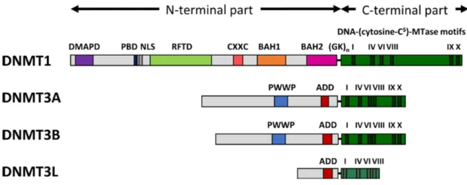
There are four representatives of DNMT (DNA methyltransferases) family: DNMT1, DNMT3A, DNMT3B and DNMT3L. Above mentioned proteins are very essential and their knockout leads to developmental deformities and fatality of embryo (Takebayashi et al., 2007; Liao et al., 2015). DNMT3L lacks enzymatic activity. DNMT1 is the maintenance methyltransferase while DNMT3A and DNMT3B are de novo methyltransferases (Xu et al., 2010; Jurkowska et al., 2011; Jin & Robertson, 2013). DNMT1 maintains the transfer of methylation marks from parent strand to daughter strand by methylating the hemi-methylated (HM) DNA with the help of its interacting partners (UHRF1, PCNA). UHRF1 through its SRA domain perceives HM DNA and engages DNMT1 for preservation of DNA methylation impressions (Bostick et al., 2007; Sharif et al., 2007; Arita et al., 2008; Avvakumov et al., 2008). DNMT3A and DNMT3B hold comparable affinity for HM or non-methylated DNA. While, DNMT3L which lacks catalytic activity, promotes DNMT3A and DNMT3B to perform their role (Xu et al., 2010). Generally, DNMTs have similar structure (Figure 7). DNMTs have two functional parts (C- and N-terminus parts) which are separated by KG repeat. Catalytic center is situated inside the C-terminus domain and this domain has the entire amino acids sequence motifs characteristic for cytosine-C5 methyltransferase fold (Jeltsch & Jurkowska, 2016). Motif I and IX are essential for binding of flipped cytosine and AdoMet (S-adenosyl-L-methionine) (Song et al., 2012) Motif IV, VI and VIII are involved in
7 catalysis (transfer of methyl group). TRD (Target Recognition Domain) which is a non-conserve region located between motif VIII and IX implicates DNA identification and distinction (Jeltsch, 2002; Jurkowska & Jeltsch, 2016). The N-terminus part consists of different domains and this part collaborates with multiple proteins and chromatin and is responsible for focusing and governance of DNMTs (Jeltsch, 2006; Jurkowska & Jeltsch, 2016). N-terminus part guides DNMTs for nuclear localization and helps their communication with proteins, regulative nucleic acids and chromatin (Jurkowska & Jeltsch, 2016).
Figure 7: Architecture of DNMTs and their functional domains. Abbreviations used: DMAPD (DNA
methyltransferase-associated protein1), PBD (PCNA-binding domain), NLS (nuclear localization signal), RFTD (replication foci-targeting domain), CXXC (CXXC domain), BAH1 and BAH2 (bromo-adjacent homology domains 1 and 2), GKn glycine-lysine repeats, PWWP (PWWP domain), ADD (ATRX-DNMT3-DNMT3L) domain. Adapted from (Jeltsch & Jurkowska, 2016).
c. Methylation sites
In humans 70-80% of genomic CpG can undergo methylation (Bird, 2002). These methylated CpG dinucleotides are localized to repetitive elements, centromeres and coded regions of operative genes. Methylation of these distinctive sites is very essential for genomic stability and transcription of gene body (Portela & Esteller, 2010; Biswas & Rao, 2017). Un-methylated CpG dinucleotides are present in promoter region of 70% of human genome. Here CpG nucleotides are clustered in a region called as “CpG island”. CpG island is defined as region having a length of 550 bps and a ratio of observed to statistically expected CpG frequencies of at least 0.6 (Portela & Esteller, 2010; Jurkowska et al., 2011). Approximately 60% of gene promoters are related with CpG and they are normally un-methylated in normal cells. However, a small fraction of these CpG (6%) become methylated during early
8 development or during differentiation of tissues (Portela & Esteller, 2010). Generally, CpG methylation is linked with suppression of genes and transcription repression but not in all contexts. As distribution of CpG is not uniform, their methylation patterns are also not uniform. Methylation patterns of CpG dinucleotides can vary dramatically regarding disease state, cell type and functional state (Ehrlich, 2019; Singer, 2019) (Figure 8). Silencing of gene activity and transcription could be enhanced through two mechanisms. First mechanism involves recognition of CpG islands methylation which can bind with MBPs (methyl-binding domain proteins). MBPs can engage HDACs (histone deacetylases) and other complexes that can mark the chromatin into a repressive state (Wade, 2001; Jurkowska et al., 2011). Second mechanism suggests that methyl group interferes with binding of transcription factors and so supporting repressive state of transcription (Watt & Molloy, 1988; Santoro & Grummt, 2001; Bogdanović & Lister, 2017). Aberrations in methylation of promoter region can cause serious problems like malignant transformation of normal cells. Many studies have reported that hypermethylation in promoter region of various TSGs (like p16, p73, BRCA1, RASSF1,
TIMP3, CDH1, CDH13, ESR1, MLH1, CASP8 and ALKBH3) has been observed in different
cancers. In result, these genes are silenced which allows malignant cells to proliferate in an uncontrolled manner (Merlo et al., 1995; Van Den Broeck et al., 2010; Pei et al., 2011; Stefansson et al., 2011; Stefansson et al., 2017; García-Martínez et al., 2018; Ma et al., 2018; Botezatu et al., 2019; Liyanage et al., 2019). DNA hypermethylation-mediated silencing of pro-apoptotic genes in cancer cells confers resistance against apoptosis which is a hallmark of tumorogenesis (Hervouet et al., 2013).
Global hypomethylation is a hallmark of cancer which favors expression of oncogenes and provokes tumor malignancy and invasion (Ehrlich, 2009; Dawson & Kouzarides, 2012; Hervouet et al., 2013; Klein Hesselink et al., 2017; Veland et al., 2017). Study has suggested that promoter hypermethylation and global hypomethylation can serve as a prognostic marker of cancer (Li et al., 2014; Ehrlich, 2019). Recently, a link between aging and cancer has been suggested as both shared similar genomic distribution of hyper- and hypomethylation patterns. But still there is a need to investigate the methylation pattern relation between aging and cancer in detail (Pérez et al., 2018).
9
Figure 8: DNA hypermethylation regulates gene expression in different fashions. (a) and (b) silencing of gene
expression through suppression of CpG rich promoters and enhancers. (c) and (d) specific cooperation of DNA hypermethylation with gene expression. (e) and (g) positive or negative connection with gene function. (f) effects on nature of transcript which can be established without changing the regulatory state of the transcript. Adapted from (Ehrlich, 2019).
d. Maintenance of DNA methylation patterns
DNA methylation has a critical role in selection and preservation of cell integrity (Bogdanović & Lister, 2017). Once DNA methylation patterns are set then it is very important to maintain and faithfully transfer these methylation patterns. DNMT1 is the main player in maintenance and transmission of methylation patterns during replication (Leonhardt et al., 1992; Singer, 2019). Preference of DNMT1 for binding with hemi-methylated DNA (Jeltsch & Jurkowska, 2014) and its localization at replication fork in S phase favor DNMT1 as a maintenance enzyme.
10
3.1.B. DNMT1
a. Structure and domain architecture of DNMT1
DNMT1 is a substantial enzyme composed of 1616 amino acids. Due to alternative splicing or use of a substitute promoter, this enzyme exists in different isoforms.
Figure 9: Domain composition of DNMT1. Adapted from (Jurkowska & Jeltsch, 2016)
DNMT1 has C- and N-terminus parts which are joined through a KG repeats. N-terminal chunk works as a podium concerning attachment of different proteins which are participating in chromatin remolding and gene activity. DMAPD (DNA methyltransferase-associated protein 1 domain) helps DNMT1 to interact with a transcription repressor (DMAP1) and is also involved in DNMT1 stability. PBD (PCNA-binding domain) recruits DNMT1 to replication fork in S phase. RFTD (replication foci-targeting domain) targets DNMT1 to replication foci and centromere chromatin. It also helps DNMT1 to interact with UHRF1 and ubiquitinated H3. Eight cysteine residues linked with zinc atoms construct CXXC domain which binds with un-methylated DNA. BAH1 and BAH2 (bromo-adjacent homology 1 and 2) domains act as protein-protein modules. C-terminus part harbors catalytic center of DNMT1. For catalytic activity it needs all motifs to be present. Catalytic domain is under the allosteric control of N-terminus domain (Jurkowska & Jeltsch, 2016; Edwards et al., 2017; Ren et al., 2018).
b. DNMT1 expression
DNMT1 expression is low in non-dividing cells while its levels are high in proliferating cells (Robertson et al., 1999). Significant increase in expression levels of DNMT1 mRNA was observed in S phase while before completion of DNA synthesis mRNA expression returned back to basal level (Szyf et al., 1991). Transcriptional regulation of DNMT1 is persuaded by Ras-AP-1 signaling network (Bigey et al., 2000) and pRb-E2F1 pathway (Jung et al., 2007).
11
c. Target selectivity of DNMT1
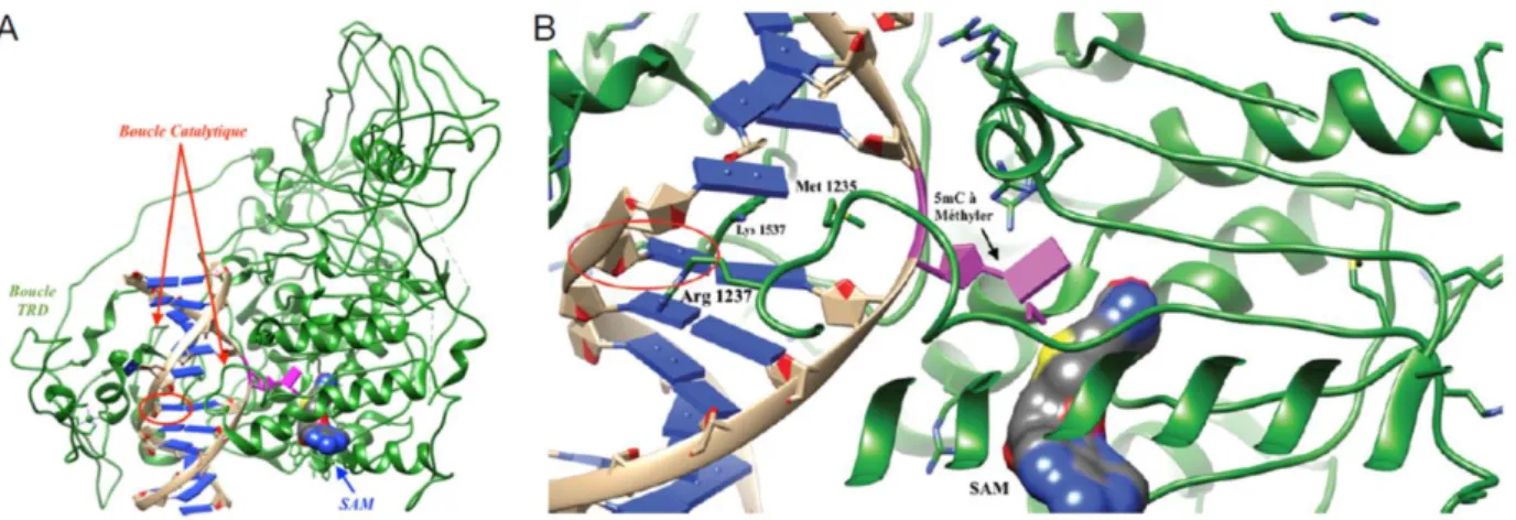
DNMT1 has a high processivity because of its ability to mark methylation tags to long stretches of hemi-methylated DNA. During this process, DNMT1 is not dissociated from substrate. In fact, DNMT1 slides along the DNA (Hermann et al., 2004; Jurkowska & Jeltsch, 2016). DNMT1 also exhibits specificity for hemi-methylated DNA as compared to un-methylated one (Fatemi et al., 2001; Bashtrykov et al., 2012; Song et al., 2012). Inherent tendency of DNMT1 for HM DNA was evaluated approximately 30-40 fold as compared to un-methylated DNA (Jeltsch, 2006; Song et al., 2012; Jeltsch & Jurkowska, 2014). Crystal framework of murine DNMT1 complex with HM DNA has been reported to reveal the preference of DNMT1. Methyl group of methyl cytosine is located inside the hydrophobic pocket within TRD domain. TRD domain is composed of Cysteine 1501, Leucine 1502, Tryptophan 1512, Leucine 1515 and Methionine 1535. This complex formation involves both major (2 TRD loop) and minor (catalytic loop) grooves. Cytosine targeted for methylation is flipped out from DNA duplex and it is fitted inside the catalytic domain which is very close to SAM (Song et al., 2012).
Figure 10: A, Crystal structure of murine DNMT1 with hemi-methylated DNA. DNMT1 (indicated in green
color), bases (indicated in blue color), phosphate bridges (indicated in beige color), targeted cytosine for methylation (indicated in magenta color) and 5mC corresponding to parent strand (indicated as red circle). B, Zoomed on stabilization site of opened DNA duplex. Adapted from (Barthes et al., 2016).
Besides processivity and intrinsic preference of DNMT1, an autoinhibitory regulatory mechanism of DNMT1 at un-methylated CpG sites, has also been proposed. Non-methylated DNA is removed from operating spot of DNMT1 through interaction of CXXC domain. On the other hand, autoinhibitory CXCC-BAH1 linker blocks access of DNA within catalytic pocket of DNMT1 by positioning itself between the substrate DNA and catalytic pocket of enzyme. Next, BAH2-TRD loop gradually pull TRD loop in such a position that TRD is no
12 more able to bind with major groove of DNA. This mechanism suggests that un-methylated CpG sites are preserved from de novo methylation through binding of CXXC domain. In this way efficiency of maintenance methylation increases through inhibition of de novo methylation (Song et al., 2011).
Another study has indicated that several structural changes are necessary for faithful conservation of DNA methylation. First, RFTS domain of DNMT1 resides inside catalytic pocket so DNA cannot access that place. RFTS position is very unique in N-terminus. And its release is very crucial for initiation of DNA methylation activity (Takeshita et al., 2011). Recent study has shown that UHRF1 can release RFTS from catalytic center to help DNMT1 to attain its active conformation (Bashtrykov et al., 2014). Second, CXXC domain occupies the place where DNA has to bind as reported by Song et al. Further, complex with AdoMet flips out C1229 towards the targeted cytosine. C1229 is expected to bind covalently with sixth position of cytosine (Takeshita et al., 2011).
d. Interaction of DNMT1 with other partners
An important partner of DNMT1 is PCNA (proliferating cell nuclear antigen) which is associated not only in DNA replication but also plays an essential role in DNA repair and cell cycle. PCNA serves as a hub which interacts with approximately one hundred proteins including ligases, polymerases, DNA modification enzymes and epigenetic factors. DNMT1 is recruited at replication site which is characterized by PCNA dense regions. Particularly, at its N-terminus DNMT1 has a special PIP box (PCNA interacting protein motif) with a sequence 164QTTITSHF171. DNMT1 is accumulated at replication site in early and mid-S phase. PIP box interacts with PCNA with a comparable binding affinity (Jimenji et al., 2019). Another important partner of DNMT1 is epigenetic integrator UHRF1. UHRF1 by means of its SRA and PHD domain recruits DNMT1 to the replication site for DNA methylation maintenance and transmission. DNMT1/UHRF1/PCNA complex is committed in maintenance of DNA methylation. DNA hypomethylation induced by interruption of this complex can lead to tumorogenesis and tumor transformation (Pacaud, et al., 2014). UHRF1 is also recruited to replication site but in a different way as compared to DNMT1. UHRF1 binds with DNA ligase (LIG1) which has methylated K126 and leads to UHRF1 localization at DNA replication site (Ferry et al., 2017; Kori et al., 2019). UHRF1-DNMT1 interaction pathway will be discussed in detail in UHRF1 section. CFP1 (CXXC finger protein 1) interacts with DNA and cells lacking CFP1 had shown 60% decrease in DNMT1 activity
13 (Butler et al., 2008). Peptide-induced specific disruption of DNMT1/CFP1 and DNMT1/DMAP1 interaction can improve response to chemotherapy (Cheray et al., 2013; Cheray et al., 2014). PARP1 also plays a role in stability of DNMT1. It negatively regulates the UHRF1-mediated ubiquitination of DNMT1 (De Vos et al., 2014). Studies in last two decades have reported variety of proteins interacting with DNMT1 (Figure 11). These studies suggested that there are some additional mechanisms which can increase activity of DNMT1.
Figure 11: Interacting partners of DNMT1. Among interacting proteins are DNA methyltransferases,
transcriptional regulators, chromatin modifiers, DNA binding proteins, cell cycle regulators, tumor suppressors and chromatin binding proteins. Proteins which are involved in post-translational modification (PTM) of DNMT1 are highlighted in green color. Adapted from (Qin et al., 2011).
3.1.C. DNMT2
In comparison with other members of DNMT1 family, information about DNMT2 is less. DNMT2 has all amino acid motifs which are characteristics of DNMT1, and its structure is also similar to DNMT1. But DNMT2 has a very weak DNA methyltransferase activity (Hermann et al., 2003). Study has reported that DNMT2 is a highly specific tRNAAsp
14 methyltransferase. DNMT2 silencing prohibits cellular proliferation and promotes cell senescence (Goll et al., 2006; Lewinska et al., 2018).
3.1.D. DNMT3 family
This family is composed of three members known as DNMT3A, DNMT3B and DNMT3L mainly involved in de novo methylation. DNMT3A and DNMT3B play an essential role to set methylation patterns during early development. DNMT1 is involved in maintenance and transmission of DNA methylation but studies have suggested that DNMT3 enzymes are also involved in maintenance methylation (Sykes et al., 1998; Kim et al., 2002; Jones & Liang, 2009; Jeltsch & Jurkowska, 2014; Hervouet et al., 2018). DNMT3L lacks catalytic activity and cannot bind with DNA. DNMT3A and DNMT3B can interact with certain transcription factors to localize on precise DNA strings (Hervouet et al., 2009). DNMT3L can also interact with some transcription factors (like NFκB-p65) with which DNMT3A/B cannot interact. Through this interaction DNMT3L can help DNMT3A/B to localize on specific DNA sequences for de novo methylation (Pacaud et al., 2014).
a. De novo methylation
De novo methylation is very important in initial stages of embryonic development. To set
totipotency in early embryo, intensive reprogramming of zygote occurs. At this level, demethylation is observed. Methylation patterns are endorsed at the time of procreation by DNMT3 enzymes. Then another stream of de novo methylation occurs in germ cells during germ cell development. This process is responsible for establishment of genomic imprinting in gametes. This methylation and demethylation mechanisms generate an epigenetic rhythm in mammals. This is a landmark of mammalian genome (Jurkowska et al., 2011; Langenstroth-Röwer et al., 2017). In contrast with DNMT1, DNMT3A and DNMT3B are not concentrated at replication foci. During DNA replication, DNMT3B can interact with human chromosome-associated proteins and members of condensing complex. This proposes that DNA methylation by DNMT3B is somewhat self-reliant from DNA replication. UHRF1 (member of DNMT1/UHRF1/PCNA/G9a complex) through its SRA domain can interact with N-terminal region of DNMT3A and B independently of DNMT1 existence. DNMT1 is partially involved in de novo methylation. De novo methylation activity of DNMT1 is increased when it is recruited to previously methylated DNA. DNMT3A-mediated de novo methylation also enhances de novo methylation activity of DNMT1. Interaction of DNMT1
15 with USP7 also promotes its inheritance and de novo methylation activity (Hervouet et al., 2018). Group of Unoki has reported recently that UHRF1 is involved in de novo methylation during oogenesis (Unoki, 2019).
3.1.E. DNA demethylation
In comparison with histone modifications, DNA methylation is relatively considered more stable but there are evidences showing that DNA methylation is a dynamic event especially during embryogenesis and development. This proposes the existence of demethylation machinery that regulates the DNA methylation. This process can occur in an active or passive manner. Passive demethylation (dilution of methylation) is the loss of 5mC mark during repeated cycles of replication in the absence of DNA methylation maintenance machinery. Active demethylation is the modification or removal of methylation mark from 5mC with the help of an enzyme (Kohli & Zhang, 2013). Previously methylation was considered as an irreversible process which can be modified through dilution or de novo synthesis of DNA. But now it is clear that erasers of DNA methylation are more involved than passive demethylation mechanism. Active demethylation process can involve following ways: oxidation of 5mC by TET (ten-eleven-translocations) proteins or deamination of methylated or adjacent base through AID (activation induced deaminase). TET can transform 5mC into 5hmC hydroxymethylcytosine) followed by conversion into 5fC formylcytosine) and 5caC (5-carboxylcytosine). Modified base is then removed through BER (base excision repair) pathway. Other pathways of base removal can be nucleotide excision and non-canonical mismatch repair (Schuermann et al., 2016; Bochtler et al., 2017). TET1 can interact with proteins which bind with full/hemi-methylated DNA or chromatin binding proteins. Importantly, interaction of TET1 with PCNA is critical in DNA demethylation (Cartron et al., 2013). Loss of TET function and impaired 5mC demethylation process corresponds with malignancies (Cartron et al., 2013; Bochtler et al., 2017). A study conducted in chronic lymphocytic leukemia (CLL) patients has suggested that dysregulation in DNA methylation and demethylation is very critical in disease progression. This interconnection represents prognostic value. Classifying CLL patients according to levels of 5-cytosine derivatives can improve prognosis (Bagacean et al., 2017). DNMT1 and histone demethylase KDM1A (also known as LSD1) interact in cancer cells and interestingly KDM1A-mediated demethylation of DNMT1 increases stability of DNMT1 protein and this interaction did not affect the DNA methylation levels (Brenner et al., 2016).
16
3.2. Histone modification
Histones are the proteins which are wrapped around DNA to give it a compact order structure which is known as nucleosome. Histones are composed of a globular shape C-terminal domain and an N-terminal tail. Various PTM (post-translational modifications) can occur on N-terminal histone tail such as methylation, phosphorylation, acetylation, ribosylation, ubiquitination, sumoylation, and deimination. Histone modifications play an essential role in variety of cellular process like transcription, repair, condensation and replication (Sharma et
al., 2010; Dawson & Kouzarides, 2012). The role of histone modifications is studied well in
transcription while studies are ongoing to investigate role in other DNA processes.
These modifications on histones can activate and repress transcription but this action is not exclusive and static (Figure 12). It is dynamic kind of process which works in a cell-context dependent manner. Additionally, active and repressive states are not always absolute. These modifications construct a “code” that can be identified by transcription factors to establish the transcriptional state of gene. But recent studies have revealed that this mechanism is more complex and involves context and time dependent chromatin signaling pathways for transcriptional regulation. This is called as histone “crosstalk” and is very important in biological processes and abreactions can lead to disease state like cancer (Lee et al., 2010; Dawson & Kouzarides, 2012; Audia & Campbell, 2016; Biswas & Rao, 2017; Michalak et
al., 2019; Qin et al., 2019).
Figure 12: Histone modifications and gene function. Trimethylation at H3K9 or H3K27 represses gene function
through compaction of chromatin. While methylation at H3K4, H3K6 or acetylation at H3K27 activates gene capacity through relaxation of chromatin. Adapted from (Jakovcevski & Akbarian, 2012).
17 Histone modifications are regulated by different enzyme known as “writers” (add histone mark); “erasers” (remove histone mark) and “readers” (read/recognize histone mark). For example, HATs (histone acetyltransferases), HDACs (histone deacetylases) and bromodomains can add, remove or recognize acetylation at histones. Similarly, HMTs (histone methyltransferases), HDMs (histone demethylases) and various domains (chromodomain, Zn finger, PHD and Tudor domain) can write, erase and read methylation marks on histones (Xu et al., 2017).
3.3. Regulatory ncRNAs (non-coding RNAs)
ncRNAs are also involved in governance of epigenetic mechanisms. They incorporate siRNAs (small interfering RNAs, miRNAs (microRNAs) and lncRNAs (long non-coding RNAs). They portray a fundamental role in coding of gene expression at different stages: transcription, RNA splicing, mRNA degradation and translation (Figure 13). siRNAs are dsRNAs (double-stranded RNAs) which can induce post-translational silencing (Inbar-Feigenberg et al., 2013). miRNAs are small (18-24 nucleotides), single-stranded RNAs. They are generally produced from cleavage of precursor RNA through RNA polymerase III enzymes (DROSHA and DICER).
miRNAs target mRNA in a specific manner for degradation and/or translational repression for regulation of gene expression. They can regulate gene expression through changing chromatin conformation by recruiting chromatin-modifying complexes and this gene regulation helps animals to survive under stress conditions (Inbar-Feigenberg et al., 2013; Wang, 2018). lncRNAs are highly conserved in various species. They function as a guide to direct the chromatin-modifying complexes to a specific location in genome and therefore contribute in specific epigenetic modification. lncRNAs participates in genome imprinting and X-chromosome inactivation (Inbar-Feigenberg et al., 2013).
lncRNAs can also induce silencing of their targeted genes by binding to RBP (RNA binding-proteins) complexes. In a study Xiaohua Shen and Naihe Jings groups have suggested that lncRNAs play a role as modulator of gene expression in development and cell differentiation. Localization of ncRNAs and their association with RBP partners affect biological processes but still there is a need of systematic studies to understand this pathway (Wang, 2018).
18
Figure 13: Extension of ‘Central dogma’ by ncRNAs. Concept of ‘central dogma’ was given by Francis Crick in
1958. Figure describes that ncRNAs in collaboration with ‘central dogma’ play role in gene expression regulation. Adapted from (Slaby et al., 2017).
19
4. Ubiquitin PHD RING Finger (UHRF) family
UHRF family includes three human origin proteins namely: hUHRF1 (ICBP90), hUHRF2 (NIRF) and hUHRF3 (ICBP55). Mouse origin members of this family are mUHRF1 (Np95), mUHRF2 (Np97) and mUHRF3 (ICBP55). Among these members hUHRF1 and mUHRF1 are well studied (Bronner et al., 2007a). UHRF proteins have evolutionary conserved amino acids sequences in vertebrates. mUHRF1 shares 73.4% identity with hUHRF1 whereas Rhesus UHRF1 shares 98% identity with hUHRF1 (Bronner et al., 2007a). Homology between human UHRF2 and mouse UHRF2 is 90.3% (Mousli et al., 2003). Strikingly, UHRF1 equivalent was not identified either in Drosophila melanogaster (flybase) or
Caenorhabditis elegans (wormbase) or in Saccharomyces cerevisiae (yeastgenome).
Phylogenetic study of UHRF1 gene conserved sequence in vertebrates has revealed its importance in backbone development. Interestingly, structural domains of UHRF1 have also conserved sequence resemblance which explains its importance throughout the evolution in vertebrates (Bronner et al., 2007a).
Another important family member is hUHRF2. hUHRF2 gene was mapped at chromosome 9p23-24.1. hUHRF2 is composed of 802 amino acids. Homology between hUHRF1 and hUHRF2 is 53% (Mori et al., 2002). UHRF2 urges an important task in cell cycle network and coordinates between cell cycle, proteasomal degradation and epigenetic machinery. It interacts with many epigenetic partners and leads to cell cycle arrest at G1 phase. In contrast to UHRF1 gene, UHRF2 gene has been reported to be lost in tumors and has the potential of being tumor suppressor (Mori et al., 2011; Mori et al., 2012). It contributes in G1/S transition by regulating Cdk2 activity (Li et al., 2004). Unlike UHRF1, UHRF2 does not have a priority for binding to HM DNA. This preference occurs only when UHRF2 binds with H3K9me3 peptide with the help of TTD domain (Pichler et al., 2011). SRA domain of UHRF2 recognizes and binds with hydroxymethylcytosine (Zhou et al., 2014).
4.1.A UHRF1 (Ubiquitin-like, containing PHD and RING Finger domains
1)
UHRF1 (previously called ICBP90 “Inverted CCAAT box binding protein of 90 kDa”) was isolated from Jurkat cells first time in 2000 by using one hybrid system. It was spotted as a novel protein binding to inverted CCAAT box of the human topoisomerase IIα gene promoter region (Hopfner et al., 2000). UHRF1 is composed of 793 amino acids having isoelectric
20 point of 7.7 and a molecular weight of 90 kDa. UHRF1 gene is located on chromosome 19p13.3 in telomeric region. It is constructed of 6 coding exons and 2 non-coding exons (Hopfner et al., 2001). Originally ICBP90 was proposed as a transcription factor having role in induction of topoisomerase IIα gene at G1/S phase of cell cycle (Mousli et al., 2003). Now it is studied well that UHRF1 is a multi-domain nuclear protein performing an imperative job in faithful transmission of methylation marks, DNA damage response, cell cycle progression and regulation of stability and function of many proteins (Alhosin et al., 2010; Du et al., 2010; Alhosin et al., 2011; Dai et al., 2013; Guan et al., 2013; Alhosin et al., 2016; Ashraf et
al., 2017a). UHRF1 has two isoforms. UHRF1 (isoform 1) is the main isoform. Only
difference in case of UHRF1 isoform 2 is that it contains additional 13 amino acids at its N-terminus (Zhang et al., 2016).
4.1.B. Structure and function of UHRF1 domains
UHRF1 is a multifunctional and multidomain protein having five conserved domains (Figure 14). On its N-terminal it has ubiquitin-like (UBL) domain followed by tandem Tudor domain (TTD) and a plant homeodomain (PHD). While on its C-terminus it has SRA domain (set and ring associated) and RING domain (really interesting new gene) (Bronner et al., 2007a).
Figure 14: Schematic representation of UHRF1 structure with its domains. “Ubiquitin-like domain” (UBL),
“Tandem Tudor domain” (TTD), “Plant homeodomain” (PHD), “SET and RING associated” (SRA) domain and “Really interesting new gene” (RING) domain.
a. Ubiquitin-like domain (UBL)
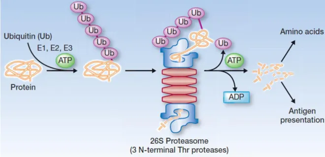
UBL domain comprises of classical alpha and beta ubiquitin folds. It exhibits three lysine on its surface (K26, K33, K52). K33 and K52 of UBL domain resembles in structure with K29 and K48 of ubiquitin. Poly-ubiquitination of K52 presents signal to proteasomal complex for protein degradation. This domain is present in all members of UHRF family except hUHRF3 and mUHRF3. It is composed of 76 amino acids and has 35% identity with ubiquitin. It has many functions including protein-protein interaction, proteasomal degradation, transcription
21 and cell cycle progression. It interacts with UIM (ubiquitin-interacting motif) of S5a proteasomal unit and helps in transport of aggresomes (Bronner et al., 2007a). UBL domain deletion cannot assist DNA methylation transfer (Smets et al., 2017). Recently it has been reported that UBL domain binds directly with DNMT1. Interestingly, although UBL has resemblance with ubiquitin but it binds with DNMT1 in a different way in contrast to ubiquitin. Along with RING domain, UBL domain is necessary for nuclear localization of DNMT1 and faithful transfer of methylation patterns (Li et al., 2018). Structural and biochemical study has revealed a bifunctional role of UBL domain. UBL binds with backside of E2 and coordinates with UHRF1 domains to read epigenetic marks and directs ubiquitin to H3. This is important in DNMT1 recruitment to newly synthesized chromatin (DaRosa et al., 2018). Another study has reported that UBL is necessary for stimulation of UHRF1 E3 ligase activity. Hydrophobic chunk present on UBL surface plays a role in establishment of E2/E3/chromatin network and indeed in DNA methylation (Foster et al., 2018).
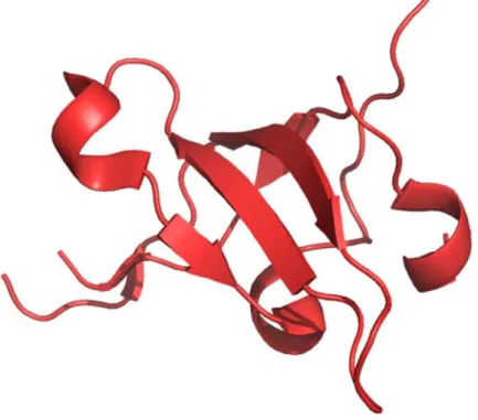
Figure 15: Crystal structure of UBL (Ubiquitin-like) domain of UHRF1 representing peculiar α-helix and
β-sheet folds. Adapted from structure deposited at RCSB protein data bank. PDB ID: 2FAZ
b. Tandem Tudor domain (TTD)
TTD mediates a variety of protein-protein interactions which are required in biological processes. This domain helps in reading of various histone marks (Lu & Wang, 2013). It is comprised of two subdomains: TTDN and TTDC. Each subdomain exhibits 5-stranded β-barrel folds. Crystal structure of TTD and H3K9me3 complex has validated that aromatic cage (Phe-152, Tyr-188 and Tyr-191) of TTDN interacts with H3K9’s triethylammonium moiety with
22 the help of two residues (Asn-194 and Asp-145) and thus provides charge for a stable interaction.
Figure 16: Crystal structure of tandem Tudor domain (TTD). A, Subdomains of TTD (TTDN and TTDC are
illustrated in yellow and green color respectively) with trimethylated lysine (marked with dark green). B, Aromatic cage of TTDN Tyr-188, Tyr-191 and Phe-152 (demonstrated in raspberry color) along with Asp-145
and Asn-194 (illustrated in blue) serve as binding pocket for trimethylated lysine residue. Adapted from structure deposited at RCSB protein data bank. PDB ID: 3DB3 (Nady et al., 2011)
Through its conserved aromatic cage in first Tudor subdomain, TTD identifies H3K9me3 while at the same time identifies H3K4 present in groove between two tandem subdomains. In addition, with SRA domain, TTD participates in subnuclear localization of UHRF1 (Nady et
al., 2011). TTD interacts with DNA ligase (LIG1) and this interaction enhances recruitment
of UHRF1 to replication foci to maintain DNA methylation (Ferry et al., 2017). Another structural study has shown that TTD binds with LIG1K126me3 and this interaction switches closed conformation of UHRF1 into open conformation (Jeltsch, 2019; Kori et al., 2019). Interaction of TTD with poly basic region (PBR) of UHRF1 interferes with H3K9me3 binding with TTD-PHD, and this leads to closed conformation of UHRF1.
23 Interruption of TTD-PBR interaction with the help of UHRF1 binding with hemi-methylated DNA or USP7 changes closed conformation of UHRF1 into open and facilitates its binding with chromatin (H3K9me3) (Zhang et al., 2015; Fang et al., 2016; Gao et al., 2018). It is well studied that UHRF1 expression levels are high in most of the cancers (Ashraf et al., 2017b) and TTD is essential for UHRF1 functioning. A screening study has reported small molecule antagonists against UHRF1-TTD-H3K9me2/3 interaction which can be further explored to synthesize new probe and inhibitors for anticancer therapy (Senisterra et al., 2018).
c. Plant homeodomain (PHD)
PHD is Zinc-finger domain which reads H3 tail and it has preference for unmethylated H3K4 as compared to methylated H3K4. This interaction is not affected by H3K9 methylation status and needs histone’s first two residues H3A1 and H3R2. Recognition of H3R2 by PHD is the main reason for this interaction. Any modification on H3R2 reduces its binding with PHD. Interestingly, PHD can entertain methylated H3K4 without disturbing the complex formation (Lallous et al., 2011; Rajakumara et al., 2011). Previously it was identified that UHRF1 is concentrated to PHC (pericentromeric heterochromatin) (Papait et al., 2007) but now it is known that UHRF1 is also localized to euchromatin playing role in transcription repression (Kim et al., 2009; Daskalos et al., 2011). UHRF1’s ability to repress transcription of targeted genes depends on interaction of PHD domain with unmodified arginine and it also indicates a crosstalk between UHRF1 function and histone arginine methylation (Rajakumara et al., 2011; Bronner et al., 2019). Houliston et al identified an inhibitor BPC (4-benzylpiperdine-1-carboximidamide) which can target TTD-PHD module to harmonize histone reading function for therapeutic purpose (Houliston et al., 2017).
Figure 17: Crystal structure representing pre PHD motif linked to canonical PHD domain of UHRF1. Adapted
24
d. Set and ring associated (SRA) domain
Among vertebrates, SRA domain has been found only in UHRF family (Bronner et al., 2007a). It plays an important role in DNA methylation patterns transfer by perceiving the hemi-methylated DNA (Arita et al., 2008; Avvakumov et al., 2008; Hashimoto et al., 2008), recruiting DNMT1(Bostick et al., 2007; Felle et al., 2011) and interacting with various epigenetic proteins like HDAC1 (Unoki et al., 2004) and G9a (Kim et al., 2009). SRA domain structure is composed of twisted and flared β-barrel which is covered by α-helicals. At one side of this barrel, α2 helical connects β5 and β6 strands while on other side another helical connects β6 and β7 strands.
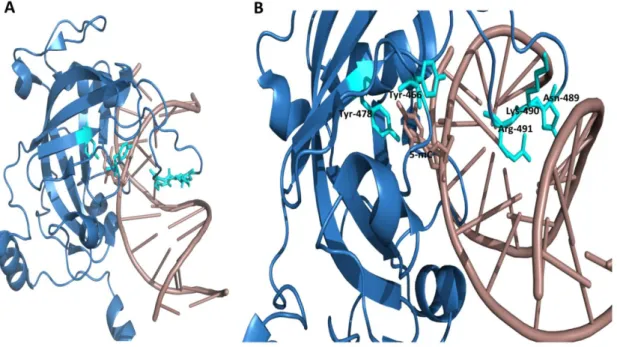
Overall DNA-SRA interactions looks like a hand grasping the hemi-methylated DNA while having a binding pocket for 5-methylcytosine in its palm. Two loops representing finger and thumb where finger projects into the major groove while thumb into the minor groove of DNA duplex. Binding pocket of SRA is very specialized which recognizes only 5-methylcytosine which is flipped out of DNA duplex. Tyrosine 478 and Tyrosine 466 stabilize 5-mC into binding pocket through π-stacking interaction (Avvakumov et al., 2008). Finger called as NKR (N489, K490 and R491) identifies 5-mC and flips out methylated cytosine. NKR finger not only recognizes CpG sequence but also differentiates methylation of complementary strand.
Figure 18: A, Crystal structure showing SRA domain (blue in color) with hemi-methylated DNA (dirty violet
Tyr-25 466 and Tyr-478 (Cyan color). NKR finger (Cyan color) is projected into major groove of DNA. Adapted from structure deposited at RSCB protein data bank. PDB ID: 3CLZ (Avvakumov et al., 2008)
Another study has revealed that SRA domain performs dual functions: recognition of DNA and recruitment of DNMT1 to proper place at DNA (Arita et al., 2008). Through recognition and flipping of 5-mC, SRA-DNA interaction supports UHRF1 to hold a place over hemi-methylated CpG sites and to recruit DNMT1 and perhaps some other proteins (Hashimoto et
al., 2008). A previous study has suggested a model which depicts that UHRF1 interacts with
HM CpG site through SRA domain and followed by DNMT1 recruitment (Vaughan et al., 2018). This model shows that UHRF1 first binds with DNA through its SRA domain which is in accordance with previous studies reporting that SRA moves over DNA in search of hemi-methylated DNA (Bronner et al., 2010; Greiner et al., 2015; Bronner et al., 2019). A recent study has reported that functional SRA domain and its finger loop is very essential for E3 ligase activity of UHRF1 towards H3 which is important for recruitment of DNMT1. This finger loop regulates conformation and activity of UHRF1 (Vaughan et al., 2019).
e. Really interesting new gene (RING) domain
At its C-terminus UHRF1 has a RING domain which has E3 ubiquitin ligase activity. RING domain structure is composed of two zinc-fingers and a special α-helix bundle (Tauber & Fischle, 2015).
Figure 19: Crystal structure of RING domain. Cysteine residues (blue color) interacting with zinc atoms (gray
color) to form zinc fingers which is necessary for interaction with substrates. Adapted from structure deposited at RSCB protein data bank. PDB ID: 3FL2
26 Due to its intrinsic E3 ligase activity, UHRF1 can either ubiquitinate itself or can ubiquitinate many other proteins including histones and non-histone proteins (Qin et al., 2015; Tauber & Fischle, 2015). A study on mouse embryonic stem cells has shown that RING domain-mediated monoubiquitination of H3K23 is essential for DNMT1 recruitment at replication site (Nishiyama et al., 2013). UHRF1 interacts with DNMT1 and regulates expression and stability of DNMT1 through ubiquitination of DNMT1 (Du et al., 2010). On the other hand, PARP1 can regulate the interaction between DNMT1 and UHRF1. It prevents UHRF1-mediated ubiquitination of DNMT1 and stabilizes DNMT1 levels (De Vos et al., 2014). Through complementation assay it has been shown that ligase activity of RING domain is required for H3K18 ubiquitination and finally for DNA methylation maintenance (Qin et al., 2015). As already described, RING domain in connection with UBL domain is essential for DNMT1 nuclear localization (Li et al., 2018). UHRF1 RING domain and its E3 ligase activity are crucial in survival during the presence of cytotoxic agents and also in suppression of promyelocytic leukemia (PML) tumor suppressor protein. Therefore, E3 ligases can be potential targets in anticancer therapy (Jenkins et al., 2005; Kirkin & Dikic, 2011; Guan et al., 2013; Popovic et al., 2014).
4.1.C. Roles of UHRF1
a. Role in DNA and histone methylation
UHRF1 is a member of epigenetic network which plays a decisive role in faithful transmission and maintenance of DNA methylation patterns (Bostick et al., 2007; Bronner et
al., 2007a; Bronner et al., 2010). UHRF1 co-localizes and directly interacts with DNMT1
during replication phase and recruits DNMT1 on chromatin. Knockdown of UHRF1 reduced the DNMT1 recruitment on chromatin which shows its crucial role in methylation maintenance (Bostick et al., 2007). SRA domain of UHRF1 showed higher binding affinity for hemi-methylated (HM) DNA and it recognizes hemi-methylated CpG sites which are preferential substrate for DNMT1 (Bostick et al., 2007; Sharif et al., 2007). Recruitment of DNMT1 on replication foci involves PCNA (proliferating cell nuclear antigen) but it is not required absolutely (Sharif et al., 2007).
Structural studies have provided the proof that SRA domain recognizes 5-mC and flips it out from DNA duplex. This base flipping is a signal to engage DNMT1 for methylation of the newly formed daughter strand (Arita et al., 2008; Avvakumov et al., 2008; Hashimoto et al.,