HAL Id: tel-01745671
https://tel.archives-ouvertes.fr/tel-01745671
Submitted on 28 Mar 2018HAL is a multi-disciplinary open access archive for the deposit and dissemination of sci-entific research documents, whether they are pub-lished or not. The documents may come from teaching and research institutions in France or abroad, or from public or private research centers.
L’archive ouverte pluridisciplinaire HAL, est destinée au dépôt et à la diffusion de documents scientifiques de niveau recherche, publiés ou non, émanant des établissements d’enseignement et de recherche français ou étrangers, des laboratoires publics ou privés.
A functionalizable nerve graft design based on an
organized electrospun silk fibroin nanofiber biomaterial
for peripheral nerve regeneration
Kayla Ann Belanger
To cite this version:
Kayla Ann Belanger. A functionalizable nerve graft design based on an organized electrospun silk fibroin nanofiber biomaterial for peripheral nerve regeneration. Biomechanics [physics.med-ph]. Uni-versité de Technologie de Compiègne, 2017. English. �NNT : 2017COMP2410�. �tel-01745671�
Par Kayla Ann BELANGER
Thèse présentée
pour l’obtention du grade
de Docteur de l’UTC
A functionalizable nerve graft design based on an
organized electrospun silk fibroin
nanofiber
biomaterial for peripheral nerve regeneration
Soutenue le 6 novembre 2017
Spécialité : Biomécanique et Bio-ingénierie : Unité de
Recherche Biomécanique et Bio-ingénierie (UMR-7338)
Université de Technologie de Compiègne
BioMécanique et BioIngénierie (BMBI)
Laboratoire CNRS UMR 7338 : Cellules Biomatériaux Bioréacteurs
Thèse
Pour obtenir le grade de
Docteur de l’Université de Technologie de Compiègne
A FUNCTIONALIZABLE NERVE GRAFT DESIGN BASED ON
AN ORGANIZED ELECTROSPUN SILK FIBROIN NANOFIBER
BIOMATERIAL FOR PERIPHERAL NERVE REGENERATION
Présentée et Soutenue par
Kayla Ann Belanger
le 6 Novembre, 2017
Jury :
Directeur de Thèse :
Pr. Christophe EGLES
Université de Compiègne
Rapporteurs :
Pr. Emmanuel PAUTHE
Université de Cergy Pontoise
Pr. Thomas CLAUDEPIERRE
Université de Lorraine
Professeur UTC :
Pr. Marion RISBET
Université de Compiègne
Examinateurs :
Pr. Catherine PICART
Grenoble INP
Pr. Bernard DEVAUCHELLE
CHU d’Amiens
2 | P a g e
Acknowledgements
I would like to thank, first and foremost, my PhD advisor, Professor Christophe Egles, for having accepted me for this PhD opportunity in the Biomechanics and Bioengineering laboratory at the Université de Technologie de Compiègne. I would like to thank you for your support and mentorship from the beginning until the end of my PhD not only pertaining to the research subject of this thesis, but also for your advice and guidance in respect to orienting myself in order to reach future career goals both in academia and in industry. I would like to thank you for your availability throughout my thesis, providing counseling on the progress of the research project whenever needed. I would especially like to thank your open-mindedness during our conversations always evoking progressive discussions for the advancement of the research project and your optimistic perspective encouraging the freedom to explore many different interesting research paths. I appreciate greatly the role you have played during my time as a PhD student at UTC.
I would like to thank Professor Bernard Devauchelle for his collaboration and his availability despite his very busy schedule. It was an honor to work with such a renowned and talented surgeon and this project would not have been able to advance as quickly or as successfully without your experience and advice.
I would like to thank Professor Frédéric Marin and Khalil Ben-Mansour for their enriching collaboration and assistance with everything pertaining to motion capture analysis. You both have helped advance this project much further than it could have gone without your expertise. In addition, I want to thank you for your help in interpreting results obtained from the mechanical tests done during my thesis.
I would like to thank Professor Guy Schlatter and Professor Anne Hébraud for their collaboration and availability during a very short but compact week spent as a guest in their lab. You both took a lot of time to collaborate on the creation of a new material that became the basis of the implants used in this work. I am very grateful for the time spent and experience gained during this week in your university under your guidance.
I would like to thank Professor David Kaplan and Dr. James White for the warm welcome into your lab during the first year of my thesis. This month was a great learning experience where I was able to acquire experience needed for the rest of my PhD. I was able to collaborate with a few of the PhD students and post docs which made this a very enriching experience. I want to especially thank Jimmy for his time and his guidance during the whole month I was at Tufts.
I would like to thank Professor Didier Gamet for his collaboration on electrophysiological analyses in this study. You gave much of your time in order to share your knowledge and expertise
3 | P a g e
on this subject which allowed me to present a more comprehensive evaluation on the results of my in vivo study.
I would like to thank Dr. Kamelia Ghazi-Naiji for all of her guidance during the time she was in BMBI C2B. You helped me immensely with all the DRG extractions we had to do together and always were there to give me advice when I needed it. I appreciate so much the time we were able to work together.
I would like to thank Pascale Vigneron for all the help you have given me throughout my time in Compiègne. Not only did you give me infinite advice and help with things involving my project and the lab (including staying with me during August vacation last year to help me take care of the animals), but you also were always there to guide me through confusion with French administration and any other issues I ran into during my time here!
I would like to thank everyone in the C2B team for their kindness and assistance throughout my PhD. From the engineer assistants to professors, you all helped me at different moments throughout my stay in Compiègne and I appreciate that a lot. I want to thank especially the other doctorate students in C2B including several from other BMBI teams who created a great ambiance in lab and who became a support system in and out of the lab!
I want to thank specifically Risa for being one of the first people I met at UTC and who has been a consistent support throughout our respective PhD’s. I would also like to thank Delphine for being exactly what I needed in an office mate and in consequence became a very good friend. I would like to thank all the jury members who have accepted to be a part of my PhD defense. I am extremely grateful for the time you all have dedicated in order to assist in my defense in addition to the travel most of you will make to be present for my presentation.
Most importantly, I would like to thank my family, specifically my parents for always supporting me and believing in me. When I told you both I may want to move to France for 3 years to do my PhD, you were always supportive even knowing how seldom you would be able to see me during this period. And I want to especially thank you for buying my plane tickets home for Christmas every year so I could spend the holiday with the whole family.
Finally, I would like to thank the Université de Technologie de Compiègne, La Région Hauts-de-France, FEDER, and CNRS for the financial support throughout this PhD.
4 | P a g e
Summary (English)
Injury to a peripheral nerve can cause loss of sensory and motor function, and if the injury is very severe where the nerve undergoes neurotmesis, unassisted nerve regeneration may not occur. In this case, where the gap between nerve segments is too large to carry out a direct end to end suture, a graft is sutured to bridge the gap between sectioned nerve segments. The autologous nerve graft, where a portion of a less important nerve from the same patient is removed and grafted between nerve segments, continues to be the gold standard procedure for nerve repair. However, there are several drawbacks of this technique including a second surgical procedure, loss of function at the donor site, possibility of developing a painful neuroma at the donor site, and the 50% success rate of autografts used in large gaps. There is therefore a need for a tissue engineered nerve graft that can replace the autograft, and this study aims to advance toward an effective autograft alternative.
This PhD is presented as a three part study consisting first of the development of a novel nerve guidance conduit based on a tri-layered silk fibroin nanofiber material comprised of a complex organization including two aligned fiber surfaces and a randomly deposited fiber interior to improve the mechanical properties of the material while not compromising the guidance capabilities of aligned nanofibers for nerve regeneration. The material is then used to fabricate a multi-channeled tube with an additional “jacket layer” in order to facilitate surgical implantation. This NGC has been submitted to be patented on July 12, 2017 and is the subject of the second article submitted for review for publication.
The second part of this study explores the different possibilities of the functionalization of the material in order to improve the effectiveness for nerve regeneration. This study explores functionalizing the silk fibroin with a second protein, several growth factors, and nanoparticles that all have potential to add favorable properties to the natural biocompatible silk fibroin material.
The final part of this study tests the effectiveness of growth factor-embedded silk fibroin NGCs for peripheral nerve regeneration in comparison with non-functionalized silk fibroin devices and a direct suture to simulate results obtained with an autograft. Three different techniques for
5 | P a g e
the evaluation of nerve regeneration were used in order to produce a more comprehensive analysis. As there are many mechanisms involved in nerve regeneration, only one or two analysis techniques cannot paint a complete picture of the success of nerve regeneration. Therefore, histological analyses, electromyography analyses, and motion capture analyses were carried out and considered together in order to make a conclusion on the level of nerve regeneration success during this study. The conclusions from this study were that a NGC functionalized with a combination of growth factors appeared to exhibit the most successful nerve regeneration and functional recovery.
6 | P a g e
Résumé (Français)
Une lésion au niveau d’un nerf périphérique peut provoquer la perte de fonction sensorielle et motrice, et dans le cas de neurotmésis, la régénération spontanée ne se produira pas. De plus, si l’espace entre les deux segments de nerf est trop important, une suture directe n’est pas possible et l’implantation d’une greffe est nécessaire afin de créer une liaison entre les deux segments de nerf. L’autogreffe de nerf est le « gold standard » pour des procédés de réparation nerveuse : une portion d’un nerf sein (qui est considéré comme un nerf moins important) est prise du même patient et implantée au site de la lésion. Cependant, il existe plusieurs désavantages avec ce procédé comme une deuxième chirurgie, la perte de fonction au site du don, la possibilité de développer un neurome sur ce même site, ainsi qu’un taux de réussite de 50% dans les cas où l’espace entre les deux segments de nerf est très important. Il reste donc, un besoin de trouver un procédé alternatif afin d’augmenter le taux de réussite et d’éliminer les désavantages de l’autogreffe. L’objectif de cette étude est d’avancer vers une solution alternative de l’autogreffon en utilisant des biomatériaux.
Cette thèse se divise en trois parties. La première se focalise sur le développement d’un modèle de guide nerveux basé sur des nanofibres de fibroïne de soie. Ce matériau est composé d’une organisation complexe qui inclut deux surfaces de nanofibres alignées avec une couche de nanofibres aléatoires à l’intérieur afin d’améliorer des propriétés mécaniques du matériau sans la perte d’orientation des fibres pour la régénération nerveuse. Le matériau est ensuite manipulé pour fabriquer un tube, multi-canaux avec une « enveloppe » supplémentaire afin de faciliter le procédé d’implantation chirurgicale. Ce guide nerveux a été soumis pour l’obtention d’un brevet européen le 12 juillet 2017 et cela est le sujet d’un deuxième article qui a été soumis pour publication.
La deuxième partie de cette étude explore des possibilités d’une fonctionnalisation du matériau afin d’améliorer son efficacité pour la régénération nerveuse. Cette étude explore la fonctionnalisation de la fibroïne de soie avec une deuxième protéine, plusieurs facteurs de croissance, et des nanoparticules. Chacune de ces fonctionnalisations donne une possibilité d’ajouter des propriétés favorable à la fibroïne de soie, un matériau naturel et biocompatible.
7 | P a g e
La troisième partie de cette étude examine l’efficacité d’un guide nerveux composé de la fibroïne de soie fonctionnalisée avec des facteurs de croissance pour la régénération nerveuse périphérique en comparaison avec un guide nerveux composé de la fibroïne de soie sans aucune fonctionnalisation et une suture direct (qui simule une autogreffe). Trois techniques d’évaluation différentes de la régénération nerveuse ont été réalisées afin d’obtenir une analyse plus complète. Il y a de nombreux mécanismes impliqués dans la régénération nerveuse, il est donc nécessaire d’étudier différents paramètres pour analyser l’efficacité de régénération. Les résultats d’analyses histologiques, d’électromyographie, et de capture de mouvement, ont été considérées ensemble afin d’arriver à une conclusion sur la réussite d’une régénération nerveuse pendant cette étude. Pour conclure cette étude, les guides nerveux fonctionnalisés avec une combinaison de facteurs de croissance démontrent une meilleure régénération nerveuse et une récupération de fonction supérieure.
8 | P a g e
Table of Contents
Acknowledgements 2 Summary (English) 4 Résumé (Français) 6 List of Figures 12 List of Tables 15 List of Abbreviations 16 Chapter 1, Introduction 17 1. Context 182. The Nervous System 19
2.1. The central nervous system 19
2.2. The peripheral nervous system 19
2.2.1. The autonomic nervous system 20
2.2.2. The somatic nervous system 21
2.3. Cells of the nervous system 22
2.3.1. Glial cells 22
2.3.1.1. Gila of the central nervous system 22
2.3.1.2. Glia of the peripheral nervous system 24
2.3.2. Neurons 29
2.4. Action potential and synapse 33
2.4.1. Action potential 33
2.4.2. Synapse 34
3. Nerve Injury and Repair Strategies 37
“Recent strategies in tissue engineering for guided peripheral nerve regeneration” 37
Abstract 38
1. Nerve Injury and Nerve Degeneration 39
1.1. Wallerian Degeneration 40
1.2. Natural Nerve Regeneration 41
2. Autograft and Allograft 41
3. Autologous Tissue Graft 42
3.1. Blood Vessels 42 3.2. Muscles 43 3.3. Tendons 43 4. Biomaterials 44 4.1. Material Properties 44 4.2. Synthetic Materials 45 4.3. Natural Materials 45
4.4. Commercialized Nerve Conduits 46
5. Biofunctionalization by Neurotrophic Factors 49
9 | P a g e
5.2. Neurotrophin-3 50
5.3. Multi-functionalization 50
5.4. Other Neurotrophic Factors 51
6. Conclusion 53
4. Silk Fibroin 60
5. Electrospinning 64
Chapter 2, Materials and Methods 69
1. Silk fibroin-based material fabrication 70
1.1. Silk fibroin extraction and solution preparation 70
1.2. Silk fibroin electrospinning solutions preparation 70
1.2.1. Functionalization 70
1.3. Electrospinning: fabrication of nanofiber-based materials 72 1.3.1. Fabrication of randomly deposited fiber-coated coverslips for in vitro testing 72 1.3.2. Fabrication of aligned fiber-coated coverslips for in vitro testing 72 1.3.3. Fabrication of randomly-deposited, aligned, and tri-layered fiber materials 73 For in vivo testing
1.4. Primary to secondary structure transition 73
1.5. Sample preparation for in vitro studies 74
1.5.1. Fiber-coated coverslips from 1.3.1 and1.3.2. 74
1.5.2. Material samples for 1.3.3 74
1.6. Sample preparation for in vivo studies 75
1.6.1. Multi-channeled tube 75
1.6.2. Tri-layered jacket 75
2. Nanoparticles solution preparation 75
2.1. Gold nanoparticles 76
2.2. Iron oxide nanoparticles 76
3. Fabrication of Alexa Fluor-tagged fibronectin 77
4. Mechanical strength testing 77
4.1. Tensile strength testing 77
4.2. Tear resistance testing 77
5. In vitro studies 78
5.1. Fetal rat dorsal root ganglia extraction and culture 78
5.1.1. Fetal rat dorsal root ganglia culture 78
5.1.2. Dissociated fetal rat dorsal root ganglia culture 79
5.2. Rat Schwann cell culture on functionalized silk fibroin fiber-coated samples 80 5.3. L929 cell culture on functionalized silk fibroin fiber-coated samples 80 5.4. PC12 cell culture on functionalized silk fibroin fiber-coated samples 80
5.5. MTS cytotoxicity assay 80
5.6. Immunostaining (in vitro) 81
6. In vivo studies 82
6.1. Surgery 82
10 | P a g e
6.3. Swimming sessions 83
6.4. Motion capture analyses 83
6.5. EMG tests 84
6.6. Nerve sample retrieval and block preparation 84
6.6.1. Nerve segment for cryostat sectioning 84
6.6.2. Nerve segment for paraffin embedded sectioning 85
6.7. Immunostaining (in vivo) 85
6.7.1. Immunostaining with DAPI, anti-β-tubulin III, and Alexa Fluor 488 85 Phalloidin
6.7.2. Immunostaining with DAPI and anti-myelin protein zero antibody 85
6.8. HE staining 86 6.9. CE activity stain 86 7. Sample imaging 86 7.1. SEM 86 7.2. TEM/STEM 87 7.3. Light microscopy 87 7.4. Epifluorescence microscopy 87 7.5. Confocal microscopy 87 7.6. ImageJ software 87
Chapter 3, Results: Part 1 – Nerve Guidance Conduit Design 89
Introduction 90
“A Multi-layered Nerve Guidance Conduit Design Adapted to Facilitate Surgical 91 Implantation”
Abstract 92
1. Introduction 93
2. Results 94
2.1. Nerve Guidance Conduit Design 94
2.2. Mechanical Tests 96
2.3. Surgery 97
3. Discussion 102
4. Conclusion 106
5. Experimental Section 107
5.1. Preparation of Silk Fibroin Solution 107
5.2. Electrospinning 107
5.3. Implant Fabrication – Jacketed, Multi-channel Design 108
5.4. Scanning Electron Microscope Imaging 109
5.5. Mechanical Strength Testing 109
5.6. Surgery 110
5.7. Fiber diameter and angle analysis 110
Acknowledgements 111
References 111
11 | P a g e
Introduction 114
Silk and growth factors 114
Silk and fibronectin 121
Silk and nanoparticles 122
Chapter 3, Results: Part 3 – In Vivo Study 130
Introduction 131 Histochemical Analyses 132 Electromyography evaluations 143 Locomotive evaluations 145 Chapter 4, Discussion 157 Part 1 158 Part 2 160 Part 3 168 Chapter 5, Conclusions 175
Publications and Communications 177
12 | P a g e
List of Figures
Chapter 1
Figure 1: Autonomic nervous system pathways Figure 2: CNS glial cells and neuron interactions Figure 3: Schwann cell differentiation
Figure 4: Conduction velocity vs axon diameter Figure 5: Schwann cell myelination of an axon Figure 6: Myelin sheath composition (PNS & CNS) Figure 7: A perisynaptic Schwann cell
Figure 8: Structural components of a neuron Figure 9: Neuron morphological classification
Figure 10: Diagram of dorsal and ventral roots of the nervous system Figure 11: Diagram of a growth cone
Figure 12: Node of Ranvier: ion channels Figure 13: Chemical and electrical synapse Figure 14: Diagram of a gap junction Figure 15: Wallerian degeneration Figure 16: Sericin and fibroin from silk Figure 17: Primary structure of fibroin Figure 18: Secondary structure of fibroin Figure 19: Simple electrospinning setup
Figure 20: Electrospinning setup with rotating drum collector
Figure 21: Electrospinning setup with micro-patterned honeycomb collector Figure 22: Electrospinning setup with coaxial spinneret
13 | P a g e
Chapter 2
Figure 1: Degumming a solubilization of silk fibroin Figure 2: Enzymatic reaction of MTS
Figure 3: Motion capture analysis setup
Chapter 3
Part 1
Figure 1.1: SEM images of tri-layered material Figure 1.2: Cross section of implant
Figure 1.3: Stress-elongation curves of materials
Figure 1.4: Average stress-strain measurements of materials Figure 1.5: Average Young’s modulus of materials
Figure 1.6: Average displacement and tensile force measurements of materials Figure 1.7: Implantation process
Part 2
Figure 2.1: Primary sensory neurons aligned on SF-based fibers
Figure 2.2: Primary sensory neurons in culture on functionalized materials Figure 2.3: Distributions of neurite length from cultured sensory neurons
Figure 2.4: Images from fluorescence staining of Schwann cells of functionalized SF-based fibers Figure 2.5: Migration paths of Schwann cells on aligned functionalized SF-based fibers
Figure 2.6: Fluorescence of FN in silk fibers
Figure 2.7: Images from fluorescence staining of PC12 cells on FN functionalized SF fibers Figure 2.8: TEM images of gold nanoparticles and iron oxide nanoparticles
Figure 2.9: TEM images of SF fibers functionalized with gold nanoparticles
Figure 2.10: TEM images of SF fibers functionalized with iron oxide nanoparticles Figure 2.11: SEM, TEM, and STEM images of functionalized SF fibers
14 | P a g e
Figure 2.12: Average fiber diameter of functionalized SF-based nanofibers
Figure 2.13: Images of stained L929 cells on SF fibers functionalized with nanoparticles Figure 2.14: MTS assay results of functionalized SF fibers
Figure 2.15: 48 hour rat Schwann cell culture on functionalized SF fibers
Part 3
Figure 3.1: HE stain of rat sciatic nerve after 4 months in vivo Figure 3.2: HE stain of rat sciatic nerve after 8 months in vivo
Figure 3.3: Cholinesterase activity stain of rat sciatic nerve after 4 months in vivo Figure 3.4: Cholinesterase activity stain of rat sciatic nerve after 8 months in vivo
Figure 3.5: Immunostained rat sciatic nerve cross sections after 4 and 8 months in vivo for axons Figure 3.6: Immunostained rat sciatic nerve cross sections after 4 and 8 months in vivo for axons Figure 3.7: Immunostained rat sciatic nerve cross section of control
Figure 3.8: Immunostained rat sciatic nerve cross sections after 4 and 8 months in vivo for myelin
Figure 3.9: Immunostained rat sciatic nerve cross sections after 8 months in vivo for myelin Figure 3.10: Needle electromyography setup
Figure 3.11: Needle electromyography results from the gastrocnemius muscle after 8 months in vivo
Figure 3.12: Foot displacement (x, y) results from motion capture tests after 6 months in vivo Figure 3.13: Vertical foot displacement in respect to time from motion capture tests after 6 months in vivo
Figure 3.14: Setup for motion capture analyses
Figure 3.15: Hip angle patterns from motion capture tests after 6 months in vivo Figure 3.16: Knee angle patterns from motion capture tests after 6 months in vivo Figure 3.17: Ankle angle patterns from motion capture tests after 6 months in vivo
15 | P a g e
List of Tables
Chapter 1
Table 1: Synthetic materials used for NGCs Table 2: Natural materials used for NGCs
Table 3: FDA approved and commercialized NGCs Table 4: Growth factors for nerve repair
Table 5: Properties of silk from the Bombyx mori Table 6: Silk degradation properties
Table 7: Biomedical applications of silk fibroin
Chapter 3
Part 3
Table 3.1: Average measurements during walking analysis cycle.
Table 3.2: Average measurements of hip angle patterns during a walking cycle. Table 3.3: Average measurements of knee angle patterns during a walking cycle. Table 3.4: Average measurements of ankle angle patterns during a walking cycle.
16 | P a g e
List of Abbreviations
BPheptyne 1-hydroxy-1-phosphonohept-6-ynyl)phosphonic acid
CA Carbonic anhydrase
CE Cholinesterase
CNS Central nervous system CNPase Cyclic nucleotide protease CNTF Ciliary neurotrophic factor DMSO Dimethyl sulfoxide
DRG Dorsal root ganglion
FN Fibronectin
FTIR Fourier-transform infrared spectroscopy
GF Growth factor
GNP Gold nanoparticle
HBSS Hank’s balanced salt solution
HE Hematoxylin and eosin
IONP Iron oxide nanoparticle L929 Mouse fibroblast cell line
LiBr Lithium bromide
MAG Myelin associated glycoprotein MBP Myelin basic protein
Na2CO3 Sodium carbonate
NCAM Neural cell adhesion molecule NGC Nerve guidance conduit
NGF Nerve growth factor
NT-3 Neurotrophin-3
P0 Protein zero
PCL Poly(ε-caprolactone) PBS Phosphate buffer saline PEO Poly(ethylene oxide) PLP Myelin proteolipid protein PMP22 Myelin protein 22
PNS Peripheral nervous system SEM Scanning electronic microscope
SF Silk fibroin
17 | P a g e
Chapter 1
Introduction
18 | P a g e
1. Context
Injuries to the peripheral nervous system affect a large population globally each year. Vehicular crashes are the most frequent cause of peripheral nerve injuries, with other common physical injuries including construction accidents, sports injuries, and combat-related trauma (Gu et al., 2011; Koppes & Thompson, 2015). Motorcycle accidents often result in an injury to the brachial plexus due to shoulder dislocations from falls at high speeds, while injuries from combat often affect the radial and ulnar nerves (Koppes & Thompson, 2015). However, approximately 75% of extremity trauma patients needing surgery sustain injuries at the median or digital nerves (in the wrist or the arm) (Koppes & Thompson, 2015). Lower extremity trauma are much less common, but most often affects the peroneal nerve (Kaiser, 2016). Millions of people are affected by nerve trauma and over 200,000 surgical procedures are performed annually in the United States for peripheral nerve injuries (Kehoe et al., 2012; Gaudin et al., 2016) as nerve gaps measuring at least 5 mm are unlikely to result in successful repair without a support (Kehoe et al., 2012; Cunha et al., 2011).
Since the 17th century, when the first end-to-end nerve suture technique was reported (Little et al., 2004; Gu et al., 2011), attempts to improve the efficacy of clinical nerve repair have continued. Throughout World War I and World War II, allografts and tubulation techniques were being extensively studied for nerve repair with disappointing results (Little et al., 2004). The autograft was first reported in the late 19th century by Ferrara (Dellon & Dellon, 1993), but it wasn’t until 1947 that Seddon published results of satisfactory results of approximately 39-52% of patients who underwent autograft procedures (Little et al., 2004). Nerve autograft procedures gained popularity in the 1960s and became the generally preferred method in the 1970s (Grinsell & Keating, 2014). The nerve autograft is known to yield satisfactory results in only about 50% of patients (Safa & Buncke, 2016) and, despite continuous research effort, is still considered the gold standard technique for nerve repair today (Grinsell & Keating, 2014; Liu et al., 2015).
The need for a superior alternative to the nerve autograft is evident due to the drawbacks of this procedure such as the need for an additional surgical procedure, the loss of function at the donor site, and the risk of developing a painful neuroma at the donor site. In response, tissue engineered nerve grafts have recently been extensively studied with the goal to provide a
19 | P a g e
replacement to the nerve autograft yielding greater success in nerve regeneration and fewer disadvantages concerning the clinical technique. Therefore, the goal of this work was to develop an effective alternative to the nerve autograft using a natural biomaterial-based tissue engineered nerve graft.
2. The Nervous System
The mammalian nervous system is made up of the central nervous system (CNS) and the peripheral nervous system (PNS) that work together directly through chemical and electrical communication networks. The CNS comprises the brain and the spinal cord while the PNS consists of all cranial and spinal nerves that branch out from the CNS to the periphery. The CNS and PNS each contain several types of neurons and glial cells specific to each system that are able to communicate with each other and support the proper functioning of the nervous system as a whole.
2.1. The central nervous system
Six components of the brain exist as well as several components of the spinal cord, which together make up the human CNS. The subdivisions of the brain include the cerebrum, the diencephalon, the cerebellum, the midbrain, the pons, and the medulla oblongata. Among the components of the spinal cord, the ventral and dorsal roots contain portions of motor and sensory neurons respectively. Neurons found in the CNS are almost exclusively interneurons which communicate with each other as well as between afferent and efferent neurons located mainly in the PNS. In addition to neurons, the CNS contains several different types of neuroglia including astrocytes, oligodendrocytes, microglia, and ependymal cells which serve to support and nourish the neurons aiding in proper function.
2.2. The peripheral nervous system
The PNS includes all cranial and spinal nerves allowing communication between the CNS and the receptors, muscles, and glands throughout the body. Similar to the CNS, the PNS contains certain types of glial cells, including Schwann cells and satellite cells, and neurons, including
20 | P a g e
efferent and afferent neurons. The PNS is divided into two subdivisions: the autonomic nervous system and the somatic nervous system.
2.2.1. The autonomic nervous system
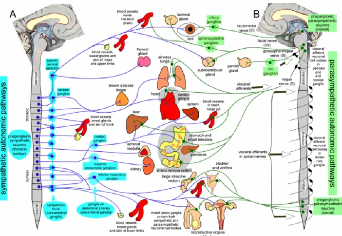
The autonomic nervous system is responsible for regulating involuntary bodily processes. Stimuli from sensory receptors are communicated to the CNS by sensory nerves were the information is processed and the response is sent to target organs by autonomic motor nerve cells. Examples of target organs in the autonomic nervous system include smooth muscle, cardiac muscles, blood vessels, and glands (Farley et al., 2014). A few functions of the autonomic nervous system include digestion, regulation of body temperature, urination, regulation of heart rate, and production of bodily fluids. The autonomic system is comprised of two subdivisions: the sympathetic nervous system and the parasympathetic nervous system (Figure 1).
Figure 1. The sympathetic and the parasympathetic pathways in the autonomic system of the peripheral nervous system (Blessing & Gibbins, 2008).
21 | P a g e
Sympathetic system pathways are triggered in situations of stress which result in what is commonly referred to as the fight or flight response. Sympathetic nervous responses may include bronchodilation (allowing increased airflow to and from the lungs), increased sweat gland activation, pupil dilation, and elevation of heart rate and blood pressure (Farley et al., 2014; Blessing & Gibbins, 2008). The sympathetic division is most attributed to be responsible for stressful situational responses, but there are also many pathways that are continuously being activated in order for the body to function properly day to day. Examples of common sympathetic activity includes control over central blood pressure, excessive water loss prevention in the gastrointestinal tract, and sweating to control body temperature (Blessing & Gibbins, 2008).
The parasympathetic nervous system, in contrast to, yet working in harmony with the sympathetic nervous system, is predominantly activated during rest. Examples of parasympathetic activation include control over secretory glands in order to regulate saliva, tears, and mucus, pupil diameter regulation, bronchoconstriction, decreasing heart rate, and stimulated secretion of digestive enzymes (Farley et al., 2014; Blessing & Gibbins, 2008).
2.2.2. The somatic nervous system
The somatic nervous system therefore is responsible for voluntary movements through the efferent neurons innervating skeletal muscles as well as afferent nerve cells that communicate interpreting external stimuli. There are four modalities of the sensory somatic system that together (1) permit the sensation of external objects in contact with the body and (2) provide bodily self-awareness. The first is discriminative touch, allowing a person to recognize pressure, vibration, shape, and the texture of an object. The second modality is nociception which is the ability to feel itching and pains such as sharp pains, dull burning pains, and deep aching pains. The third modality is proprioception which allows a person to recognize position and movement of the body such as muscle tension, joint pressure, and joint angle. The fourth modality is temperature allowing a person to distinguish between cold, cool, warm, and hot temperatures.
22 | P a g e
2.3. Cells of the nervous system
There are two principal cell types that are critical for the proper development and function of the nervous system: neurons, also known as nerve cells, and neuroglia, or glial cells. There are an estimated 100 billion neurons in the human body and about ten times as many glial cells (Azevedo et al., 2009). Neurons are the only cells in the nervous system responsible for the processing of information through electrical connections, while glial cells hold important roles not only assisting in in the support of neurons, but also maintaining efficiency in normal neural function (Birch, 2013; Jessen, 2004).
2.3.1. Glial cells
Once thought only to provide support, protection, and nutrition for the neurons within the nervous system (Ndubaku & de Bellard, 2008), glial cells are now understood to be responsible for performing important functions during neural development, assisting in healthy, developed function, and reacting to trauma or disease (Jessen, 2004). Glial cells are most commonly known for their formation of myelin that surrounds a nerve fiber allowing for more efficient conduction and faster transport of information. In more recent years, it has been discovered that glial cells are also essential elements to the formation and maintenance of synapses, the key functional unit in the nervous system allowing neurons to communicate with each other and to other cells (Verkhratsky & Butt, 2007). During development, glial cells are the support units creating a cellular framework for the formation of a functional nervous system (Jessen, 2004). Given the specific needs of the CNS and PNS, different types of glial cells are found in each system. Glia occupying the CNS include macroglia (astrocytes, oligodendrocytes, and ependymal cells) and microglia, and glia found in the PNS include Schwann cells, satellite cells, and enteric glial cells (Verkhrasty & Butt, 2007; Jessen, 2004).
2.3.1.1. Glia of the central nervous system
Astrocytes are the most abundant glial cells found in the CNS (~80%), accounting for around half of the volume of the CNS (Jessen, 2004). They received their name in 1893 by Michael von Lenhossek because of their star-like shape formed by the many cytoplasmic extensions from
23 | P a g e
the cell body that intertwine between nerve fibers and neuron cell bodies (Figure 2) (Ndubaku & de Bellard, 2008). The two main types of astrocytes are the protoplasmic astrocytes which are found in CNS grey matter and have many processes measuring about 50 µm on average and the fibrous astrocytes found in white matter with longer extensions measuring up to 300 µm (Verkhrasty & Butt, 2007). Astrocytes are known to connect blood vessels and neurons at synapse, but more recently have also been found to control the activity of surrounding non-neuronal cells (Volterra & Meldolesi, 2005). They also proliferate in proximity to sites of trauma for healing in the CNS (Silver & Miller, 2004).
Oligodendrocytes are highly specialized cells, accounting for about 5% of all glia in the CNS, that produce myelin, a lipid-rich insulation layer surrounding nerve fibers which are responsible for speeding up electrical impulse conduction. The myelin isolates the ionic channels of the enveloped axons and is therefore able to accelerate action potential propagation by saltatory conduction (Verkhrasty & Butt, 2007). Oligodendrocytes have fewer and shorter processes than astrocytes (often between ten and twenty) which are each capable of myelinating a separate nerve fiber (Ndubaku & de Bellard, 2008).
Ependymal cells constitute about 5% of glia in the CNS. This macroglia subtype forms a barrier between cells and cerebrospinal fluid acting as the intermediary for nutrient exchange. Ependymal cells are multiciliated and are responsible for the production and circulation of the cerebrospinal fluid through the brain and the spinal cord (Spassky et al., 2005; Verkhrasty & Butt, 2007).
Microglia (10-15% of CNS glia) originate from macrophages during early brain development and make up the brain immune system. These cells have many thin, branched, motile processes and are present throughout the brain each with their own domain. The processes are continually extending and contracting constantly scanning their territory for abnormalities. In fact, the entire brain can be fully surveyed by its microglia in several hours (Verkhrasty & Butt, 2007). When introduced to trauma or disease, microglia become activated as the brain’s defense system and gain the capability to migrate to the damaged area, proliferate, and phagocytose (Kettenmann et al., 2011).
24 | P a g e Figure 2. Interactions of glial cells, tissue, and neurons in the central nervous system (Allen & Barres, 2009).
2.3.1.2. Glia of the peripheral nervous system
Glial cells similar to the oligodendrocytes of the CNS due to their capability of myelinating neurons were discovered in the PNS by Theodore Schwann in 1839 (Bhatheja & Field, 2006) and the term Schwann cell was coined by Louis Ranvier in 1871 (Ndubaki & de Bellard, 2008). Schwann cells are the most abundant glial cells found in the PNS accounting for about 80% of all cell nuclei within the endoneurium, the smallest structural pathways of peripheral nerves and nerve roots (Birch, 2013). Like the neuroglia found in the CNS, Schwann cells are not only responsible for support and trophic properties, but also directing developing and regenerating neurons, regulating the microenvironment, signaling, and eliminating cellular debris (Verkhratsky & Butt, 2007; Bhatheja & Field, 2006).
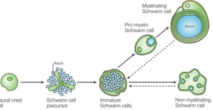
Schwann cells develop from the neural crest, a group of cells originating in the dorsal section of the neural tube (Birch, 2013). In the beginning of development, neural crest cells form precursor Schwann cells. Precursor Schwann cells have not yet ensured their survival and depend entirely on signals from axons in order to avoid apoptosis (Jessen & Mirsky, 2005). Precursor Schwann cells then form immature Schwann cells around the time of birth that hold the potential
25 | P a g e
to further differentiate into four different types of Schwann cells: myelinating Schwann cells, non-myelinating Schwann cells, perisynaptic Schwann cells of neuromuscular junctions, and terminal Schwann cells of sensory neurites (Verkhratsky & Butt, 2007).
Immature Schwann cells start to ensheath the nerve fibers of the PNS before differentiation into mature Schwann cells; the Schwann cell lineage is highly dependent on the size of the axons with which it is in contact (Verkhratsky & Butt, 2007). Immature Schwann cells in contact with large axons (axons with a diameter larger than the critical diameter of about 0.7 µm) will differentiate into myelinating Schwann cells (Figure 3).
Figure 3. The development and differentiation of myelinating and non-myelinating Schwann cells from a neural crest cell. (Jessen & Mirsky, 2005)
Immature Schwann cells initially envelop several nerve fibers, but then migrate to myelinate a single axon as the cells differentiate into mature myelinating Schwann cells (Ndubaku & de Bellard, 2008). Mature myelinating Schwann cells eventually line the length of an axon and, similar to oligodendrocytes in the CNS, form a myelin sheath around the axon increasing the conduction velocity of electrical impulses as seen in Figure 4. Conduction velocity of an unmyelinated axon is directly related to the diameter, therefore the transport speed of
26 | P a g e
information in large diameter axon would be inefficiently slow without the insulation of the myelin sheath (Allen & Barres, 2009).
Figure 4. Relationship between axon diameter and conduction velocity of myelinated and non-myelinated axons (Verkhratsky & Butt, 2008).
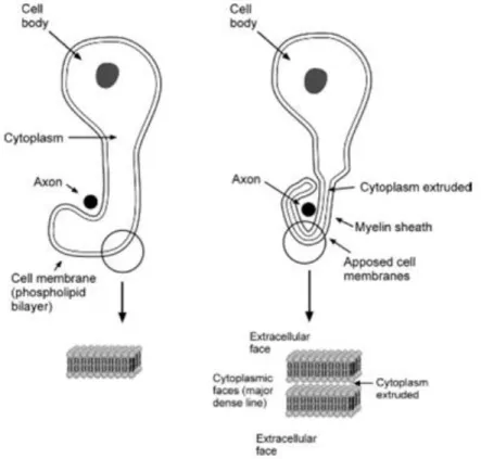
In order to produce the myelin sheath, Schwann cells wrap its membrane around the axon in a spiraling movement (as depicted in Figure 5), extruding nearly all cytoplasm while compacting the stacked membrane bilayers, and increasing the membrane area several thousand fold (Jessen, 2004). The layers of compacted membrane of myelin is referred to as lamellae, and the quantity of lamellae along with the length of myelin along the axon, or internode, are determined by the diameter of the axon and determine conduction properties including the speed of conduction. There can be between 10 and 100 lamellae and internode lengths between 100 and 1000 µm (Verkhratsky & Butt, 2007).
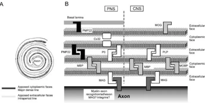
The myelin sheath is made up of layers of Schwann cell membrane and is therefore composed mostly of lipids (around 70%) in addition to several different membrane proteins. The primary membrane protein of myelin produced by Schwann cells, accounting for between 50 and 60% of membrane proteins in PNS myelin, is protein zero (P0) which has essential adhesive properties that facilitate myelin formation. Other membrane proteins found in PNS myelin include myelin basic protein (MBP) (5-15% of myelin proteins), myelin protein 22 (PMP22) (2-5% of myelin proteins), myelin associated glycoprotein (MAG) (~0.1% of myelin proteins), myelin
27 | P a g e
proteolipid protein (PLP), and cyclic nucleotide protease (CNPase) and are also essential for the compaction of the cell membrane forming lamellae (Birch, 2013; Verkhratsky & Butt, 2007).
Figure 5. Myelination of an axon in the peripheral nervous system by a Schwann cell (Verkhratsky & Butt, 2008).
Immature Schwann cells connected with axons smaller than the critical diameter (~0.7 µm) will not produce myelin and undergo ensheathment of several nerve fibers in the PNS creating a structure called a Remak bundle (Bhatheja & Field, 2006). This type of non-myelinating Schwann exhibits several surface characteristics identical to immature Schwann cells such as the neural cell adhesion molecule (NCAM) which provides support to unmyelinated axons while also providing a physical boundary preventing ephaptic transmission between the unmyelinated axons (Verkhratsky & Butt, 2007).
The final two types of mature Schwann cells are terminal Schwann cells found at motor and sensory terminals. The processes of terminal Schwann cells are integrated closely at nerve terminals and are essential regulators of synapses as seen in Figure 7. (Verkhratsky & Butt, 2007).
28 | P a g e Figure 6. Composition of the myelin sheath of a Schwann cell in the peripheral nervous system and an oligodendrocyte in the central nervous system (Verkhratsky & Butt, 2008).
29 | P a g e
Dissimilar to glial cells in the CNS, Schwann cells have an exceptional response to the injury of the nerve. Once Schwann cells lose contact with the axon after trauma, they will de-differentiate back to immature Schwann cells in order to prepare for nerve regeneration. Unlike precursor Schwann cells, immature and mature Schwann cells have the capacity to survive without the contact of a nerve fiber. Schwann cells begin to clear cellular debris and proliferate guiding the regenerating axons by signaling and providing a physical support (Jessen & Mirsky, 2005).
Satellite cells and enteric glial cells are secondary types of glial cells found in the PNS and are much less abundant that Schwann cells. Satellite cells are glial cell that surround the neuron cell bodies in dorsal root ganglia and autonomic ganglia providing support and nourishment (Birch, 2013; Jessen & Mirsky, 2005). Enteric glial cells are found in the autonomic ganglia in the gastrointestinal tract and have a form and biochemistry similar to astrocytes of the CNS (Jessen, 2004).
2.3.2. Neurons
Neurons are the most highly specialized cells in the human body pertaining to intercellular communication and are the functional foundation of the nervous system. These cells are capable of receiving, processing, and communicating information through electrical and chemical signals between themselves and innervated tissue throughout the body. Neurons are non-dividing cells, and therefore possess extreme longevity. Depending on the type and position in the nervous system, the size of a neuron can be between around 20 µm in diameter to up to 2 meters long (Bear et al., 2007).
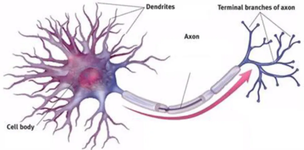
The three main structural segments of the neuron are the soma, the dendrites, and the axon (Figure 8.). The soma, or the cell body, of neurons are either located in the CNS (including all CNS neurons and PNS somatic efferent neurons), or within ganglia located in the PNS. Found in the cytoplasm of the soma are all organelles commonly found in animal cells with the exception of centrioles which are organelles specific for assisting in cell division. Rough endoplasmic reticulum and mitochondria are particularly important in proper neuronal function and are in
30 | P a g e
abundance in the neuron cell body compared glial cells and other non-neuronal cells (Bear et al., 2007).
Figure 8. Diagram of a multipolar neuron and its structural components.
One of the types of processes of a neuron is the dendrite. Neurons often have several or many of these extensions with branches called dendritic branches covered in thousands of synapses facilitating the retrieval of information. The second type of neuronal process is the axon. There is only one axon for each nerve cell and it is a highly specialized structure of the neuron that allows the transportation of electrical impulses throughout the body in order for the nervous system to communicate. Axons no not contain any rough endoplasmic reticulum and virtually no free ribosomes which means that there is no protein synthesis in the axon. Information is therefore relayed over the distance of the axon by membrane proteins (Bear et al., 2007). The point at which the axon joins the cell body is called the axon hillock and is where action potential usually occurs.
Neurons can be classified in a few different groups depending on the number of neurites they have. For example, a neuron contained a single neurite is classified as a unipolar neuron, a neuron containing two neurites is classified as bipolar, and a neuron containing 3 or more neurites is classified at multipolar (Figure 9) (Bear et al., 2007).
31 | P a g e Figure 9. Diagram of the classification of neurons by morphology and number of neurites.
Neurons can also be classified by their function. In the peripheral nervous system, there are sensory neurons and motor neurons. Sensory neurons are unipolar neurons that form the afferent signaling route which propagates from the peripheral nerve terminals toward the CNS. The cell bodies of sensory neurons are located in the dorsal root ganglia as seen in Figure 10. Motor neurons are multipolar neurons make up the efferent pathways driving information from the brain toward muscles and signaling glands (Kendel and Schwartz, 2013). Motor neuron cell bodies are located in the ventral horn of the spinal cord (Figure 10). Other neurons can interneurons serve as liaisons between peripheral neurons and central neurons.
When either motor or sensory neurons are regenerating, growth cones can be found at the tip of the axonal extensions and they are finger-like extensions searching for the most favorable environment towards which to grow (Figure 11). The phenomenon of this guidance factor is called chemotropism and has been proven true for the orientation of nerve extensions (Fuh et al., 2013). At the end of these growth cones are filopodia that extend and retract under the influence of the environment. There are made up of mainly cytoskeleton molecules but also membrane proteins specialized in adhesion and environmental exploration. These structures contain specific receptors that induce significant growth while interacting with guidance molecules (Claudepierre et al., 2008). When a growth cone meets an unfavorable environment it has the tendency to retract to find a more favorable environment toward which to orient itself.
32 | P a g e
The four mechanisms that guide a growth cone are attraction mediated by contact, repulsion mediated by contact, chemoattraction, and chemorepulsion (Goodman, 1996).
Figure 10. Diagram of the cross section of the spinal cord depicting the location and paths of motor and sensory neurons in the peripheral nerve regeneration (Birch, 2013).
Finally, neurons contain axon terminals that intervene in cellular communication. These terminals are composed of terminal buttons containing synaptic vesicles. These structures enclose neurotransmitters that influence the activity of other cells by their release.
33 | P a g e Figure 11. Depiction of a growth cone in the peripheral nerve regeneration.
2.4. Action Potential and Synapse 2.4.1. Action potential
Neural communication is carried out through electrical impulses which is a consequence of external stimuli. The plasma membrane of a neuron has a resting electrical potential of about -70 mV which represents the difference in charge between interior and the exterior of the cell (the exterior of the cell is positively charged and the interior of the cell is negatively charged) (Marieb, 2006). This difference in charge is maintained by membrane pumps and a selective permeability of specific ions at the plasma membrane. After an external stimulus, a large influx of sodium and potassium ions occurs as voltage-gated sodium and potassium channels open. A change in ionic concentration depolarizes the neuron and may allow the membrane potential to reach about +30 mV which will produce an action potential (Purves, 2005).
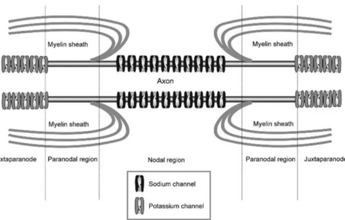
The moment an action potential is produced, a chain reaction throughout the axon occurs. The action potential causes a changes in the structures of voltage-gated ion channels which travels down the length of the axon. The propagation speed of this reaction depends on the myelination of an axons since one the channels found at the Nodes of Ranvier (located between myelinating Schwann cells along the length of the nerve fiber as depicted in Figure 12) (Marieb, 2006). The increased rate of action potential transmission is due to salutatory conduction which
34 | P a g e
refers to the action potentials jumping between Nodes of Ranvier. The result of an action potential is the release of neurotransmitters at a synapse.
Figure 12. Representation of ion channels at the Node of Ranvier (Verkhratsky & Butt, 2008).
2.4.2. Synapse
Synapses are the junctions with other neurons or innervated tissue that permit a transfer of information from one cell to another. This junction was discovered by Charles Scott Sherrington in 1897 who named this site a synapse (Sherrington, 1906). A synapse involves a unidirectional current of neurotransmitters which are intermediaries of cell-to-cell communication consisting of presynaptic and postsynaptic elements (Bear, et al., 2007). The space between these two elements is called the synaptic cleft.
The presynaptic side at synapse is usually the axon terminal of a neuron which contains mitochondria and cytoskeletal filaments for sufficient energy and the transport of necessary synaptic elements for the transmission of information (Tansey, 2006). The postsynaptic element contains numerous receptors and channels on the surface of the cell membrane which assist in
35 | P a g e
the transfer of information. The synaptic cleft is about 30 nm wide and consists of an interstitial liquid where the physicochemical exchanges take place (Pereda, 2014).
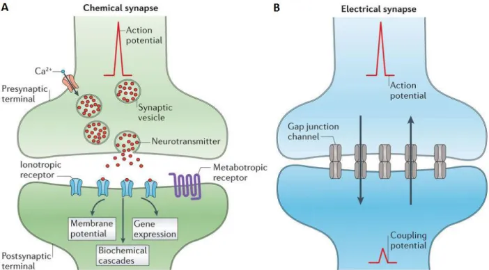
Both chemical and electrical synapse occur within the nervous system (Figure 13). The most common synaptic transmission of information is assisted by chemical synapse, an indirect process that uses chemical messengers called neurotransmitters transported by synaptic vesicles. The production of an action potential eventually arrives at the axon terminal and causes calcium channels to open allowing calcium ions to enter the cell. The rapid increase of the concentration of calcium activates the production of synaptic vesicles containing neurotransmitters. The neurotransmitters then diffuse across the synaptic cleft and attach to receptor channels on the postsynaptic element. The neurotransmitters signal the opening or closing of membrane channels on the postsynaptic cell membrane. This communication may excite or inhibit the postsynaptic cell depending on the receptors involved during synapse (Pereda, 2014; Bear et al., 2007).
Electrical synapse are much less common than chemical synapse in the nervous system and contain a direct transmission to the postsynaptic element. Information is passed from cell to cell with the assistance of direct and passive ionic transport from gap junction channels (Figure 14) (Purves, 2001). The gap junctions are highly specialized and align with paired channels on the presynaptic and postsynaptic cell membranes forming a pore that is larger than the pores of voltage-gated ion channels. There, there is a simple and direct diffusion of substances to the interior of the postsynaptic cell from the interior of the presynaptic neuron. The main purpose of electrical synapses is to allow synchronization of electrical activity between connected neurons (Purves, 2001).
36 | P a g e Figure 13. Depictions of chemical and electrical synapse of the nervous system (Pereda, 2014).
Figure 14. Illustration of gap junctions at an electrical synapse with membrane potential behavior graphics (Purves et al., 2001).
37 | P a g e
3. Nerve Injury and Repair Strategies
Nerve injury and recent repair strategies have been outlined in a review entitled “Recent strategies in tissue engineering for guided peripheral nerve regeneration” published in
Macromolecular Bioscience in April, 2016 (doi: 10.1002/mabi.201500367).
Recent strategies in tissue engineering for guided peripheral
nerve regeneration
Kayla Belanger*, Tony M. Dinis, Sami Taourirt, Guillaume Vidal, David L. Kaplan, and Christophe Egles
K. Belanger, Dr. T. M. Dinis, S. Taourirt, Dr. G. Vidal, Prof. C. Egles
Sorbonne University, Université de Technologie de Compiègne, CNRS, UMR 7338 Biomechanics and Bioengineering, Centre de Recherches Royallieur – CS 60 3019, 60203 Compiègne cedex, France
E-mail: [email protected] Prof. D. L. Kaplan
Department of Biomedical Engineering, Tufts University, 4 Colby Street, Medford, MA, 02155, United States of America
Prof. C. Egles
Department of Oral and Maxillofacial Pathology, Tufts University, School of Dental Medicine, 55 Kneeland Street, Boston, MA, 02111, United States of America
38 | P a g e
Abstract
The repair of large crushed or sectioned segments of peripheral nerves remains a challenge in regenerative medicine due to the complexity of the biological environment and the lack of proper biomaterials and architecture to foster reconstruction. Traditionally such reconstruction was only achieved by using fresh human tissue as a surrogate for the absence of the nerve. However, recent focus in the field has been on new polymer structures and specific
biofunctionalization to achieve the goal of peripheral nerve regeneration by developing artificial nerve prostheses. This review presents various tested approaches as well their effectiveness for nerve regrowth and functional recovery.
Keywords:
bioengineering, biomaterials, peripheral nervous system, polymers, regenerative medicine39 | P a g e
1. Nerve Injury and Nerve Degeneration
Injury to a peripheral nerve, known as peripheral neuropathy, often leads to an impairment of motor and sensory functions within the PNS due to an interference in nerve conduction. Motor vehicle accidents are the primary cause of traumatic peripheral neuropathy and more than half of nerve injuries affect the lower limbs (such as deriving from the peroneal nerve). Peripheral neuropathies are mainly caused by elongation, laceration, or compression. Elongation is the most frequent form of traumatic neuropathy. The connective tissue making up the nerve presents a certain elasticity, however when tension is greater than its capacity of resistance, the result is a neuropathy. In the case of nerve laceration, which accounts for about 30% of severe peripheral nerve injuries, a complete tear may occur, but generally nerve continuity is maintained by at least several fragments of the nerve.[1,2] Conversely, nerve compression does not involve severing of
the nerve and continuity is maintained. However, the mechanical stress acting on the conductive membrane can still cause loss of motor and sensory function as a result of several mechanisms including mechanical compression, ischemia, and mechanical deformation. Prolonged mechanical compression, whether singular or repetitive events, can damage endoneurial channels resulting in increased permeability and result in endoneurial oedema. An oedema consequently alters the ionic balance of the endoneurium and alters fascicular microcirculation thus causing an ischemia due to the increase in endoneurial fluid pressure.[3]
There are two widely used grading systems to classify the severity of peripheral nerve injuries. The simpler of the two, the Seddon classification, was developed in 1943 by Sir Herbert Seddon and includes three grades of nerve injury: neuropraxia, axonotmesis, and neurotmesis.[4]
However, the Sunderland classification, proposed in 1951 by Sir Sydney Sunderland, is a more comprehensive grading system which provides more detailed information related to surgical intervention needs after nerve injury.[5] The Sunderland grading system includes five degrees of
nerve injury. The first degree is equivalent to neuropraxia in the Seddon system and involves a temporary disruption of conduction resulting in local demyelination and short-term sensory impairment. The second degree is equivalent to axonotmesis in the Seddon system, which refers to axonal rupture while neural connective tissue remains undamaged. The third, fourth, and fifth degrees further delineate axonal rupture by damage of the endoneurium, perineurium, and
40 | P a g e
epineurium, respectively.[4,5] Injuries qualifying as second, third, fourth, or fifth degree will lead
to Wallerian degeneration, and injuries of the fifth degree, completely sectioned nerves, may require a nerve graft.[5]
1.1. Wallerian Degeneration
A severe injury to a nerve fiber leads to its degeneration, and in the case of axotomy, the distal segment will progressively degenerate whereas the proximal axon and perikaryon undergo physiological and metabolic modifications to prepare for regeneration. This process was first studied by Augustus Volney Waller in 1850[6] who gave it the named Wallerian degeneration
(Figure 1). When the axon is damaged, the myelin sheath, which provides necessary contact with the axon in order to maintain its integrity, forces the Schwann cells to split and retract their sheaths. The loss of contact with the axon stimulates and activates macrophages that phagocytose the myelin debris and axon fibers over three to six weeks. The lack of contact also acts as a stimulant for the proliferation of Schwann cells to migrate toward the damaged site. Schwann cells and fibroblasts orient themselves in columns known as Bands of Bünger, or tubes of regeneration. These bands function to attract neurites by producing neurotrophic factors. During the degeneration process, Schwann cells overexpress the class II receptors which link the free nerve growth factors to the extracellular matrix in order to offer a substrate with neurotrophic support and a chemotactic guide for axonal regrowth.
Figure 1*. Schematic representation of Wallerian Degeneration
41 | P a g e
1.2. Natural Nerve Regeneration
In the hour following a nerve lesion, metabolic disruption within the nerve cells takes place. The focus turns to the manufacture of structural elements necessary for nerve regeneration in contrast to the usual priority to transmit nervous influx. This is a biological process that puts in place a permissive environment in order to restore the architecture, metabolism, and functions of the damaged tissue.[7] Genomic regulations are also modified, particularly those involved in
synthesizing proteins found at the surface of Schwann cells and the axolemma.[8]
Regeneration begins with a phase of neural reorganization intended to reestablish cellular integrity. The process then promotes and guides the elongation of axons in the direction of the target at a rate of about 2-5mm per day.[9] Finally, new synaptogenesis is established. Upstream
from the axotomy, the plasma membrane fuses shut in order to close off the axon. This terminal then begins to structure itself in a budding manner forming a growth cone[10] which contain
filopodia that extend and retract under the influence of the environment. When the growth cone meets an unfavorable environment, it has the tendency to retract to search for a more favorable one towards which it then orients itself. The four mechanisms guiding a growth cone are attraction mediated by contact, repulsion mediated by contact, chemoattraction, and chemorepulsion.
2. Autograft and Allograft
The autograft is currently considered the gold standard for the treatment of nerve lesions, consisting of harvesting a peripheral nerve directly from the patient. The first clinical trials for autografts date back to the 1870’s when Phillipeaux and Vulpian grafted optic nerve segments within the same dog.[11] The greatest disadvantage of using an autograft is inevitably the necessity
of creating an additional nerve injury that can result in loss of function near the donor site. In addition, the supplementary deficiency can potentially induce the formation of a painful neuroma at the site of the secondary injured nerve.[12]
Due to advances in medical research, an alternative to the autograft, the allograft, has been developed which consists of using a nerve sample from a different patient. Immunology research has led to the development of methods to limit the risks of rejection of these allografts by
42 | P a g e
irradiation, successive freezing and defrosting.[13] The immunosuppressive treatments
administered to the recipient are more efficient; however they compromise the immune system rendering it more vulnerable to other complications.
3. Autologous Tissue Graft
In the past several years, regenerative medicine has oriented itself toward decellularized autologous tissue grafts in order to overcome the disadvantages of autologous nerves. The biological tissues that have received the most attention include blood vessels, skeletal muscles, and tendons.
3.1. Blood Vessels
The concept of using vascular tissue has been studied since the end of the 19th century. Arterial conduits were first used by Bünger in 1891.[14] The exploration of nerve regrowth via
arterial conduit grafts between two separated peripheral nerve segments after lesion has long been considered less promising compared to venous conduit grafts. Yet recently, a rodent study comparing three biological material conduits (nerve, artery, dermis) demonstrated encouraging use with the artery as a biological conduit.[15] The regeneration of myelinated axons and the
rescue from muscular atrophy following an arterial conduit graft led to nerve repair to the same extent as that from nerve autografts.
Throughout the years, venous conduits have been the object of many experimental and clinical studies. Using a venous conduit graft for the reparation of damaged human nerves was reported for the first time in 1909 by Wrede.[14] In 1990, Chie and Strauch described a case in
which 15 patients received autologous venous conduit grafts for the treatment of painful neuromas or nerve segment injuries of 3 cm in length.[16] This study demonstrates that venous
conduits can be used for lesions inferior to 3 cm and is comparable to nerve regeneration obtained from autografts. Later, a clinical study on the repair of the digital nerve with a venous conduit embedded with one or more nerve slices was performed.[17] The study demonstrated that
more than 65% of patients presented good results following surgical intervention. Two years later, another study using the same method for 16 patients was reported and the results
43 | P a g e
suggested that the venous conduit graft with the interposition of nervous tissue in the lumen of the conduit is a reliable alternative for lesion distances between 2 and 4.5 cm.[18] In addition, it
was recently demonstrated that replacing the venous conduit lumen with type I collagen amplifies axonal regeneration.[19] However, for segments greater than 4 cm, vein constriction preventing
regrowth as a result of scarring was observed.
3.2. Muscles
Skeletal muscles are composed of a tubular basal lamina similar to that of nerve conduits. The basal lamina is also oriented longitudinally and contains constituents in its extracellular matrix, like collagen type IV and laminin, that direct and advance the regeneration of nerve fibers.[20-22] In the human body, muscle tissue is present in significant quantities which prompts
its use for autologous tissue grafts. The first muscle tissue grafts for nerve repair date back to the 1940’s.[14] Graft conduits from decellularized muscle tissue are described in two clinical studies
by Pereira et al. in 1991. The first concerned peripheral nerve guides in 10 patients suffering from leprosy,[23] and following surgery, 70% of the patients presented better perception and improved
sensation from the affected limb. In addition, the same team reported 24 cases of digital nerve repair from decellularized muscle grafts.[24] Monitoring 6 to 40 months after the procedures
indicated better outcomes compared to patients who underwent nerve autografts. Nevertheless, the use of decellularized muscles presents some disadvantages such as the possibility of the nerve fibers growing outside of the graft and creating a neuroma.[25]
3.3. Tendons
Tendons can be used as nerve conduits to fill gaps with maximum distances of 1 cm. The architecture of the tendon, a longitudinal arrangement of the collagen bundles, provides favorable grafts for nerve regeneration in the rat sciatic nerve.[26] The axons align themselves on
the collagen fibers of the tendon for nerve regrowth. In order to verify the validity of this model, tendon tissue grafts have been compared to muscle tissue with functional and morphological similarities.[27] Tendons can therefore support nerve regeneration in a similar manner to