RESEARCH OUTPUTS / RÉSULTATS DE RECHERCHE
Author(s) - Auteur(s) :
Publication date - Date de publication :
Permanent link - Permalien :
Rights / License - Licence de droit d’auteur :
Bibliothèque Universitaire Moretus Plantin
Institutional Repository - Research Portal
Dépôt Institutionnel - Portail de la Recherche
researchportal.unamur.be
University of Namur
Synthesis and Crystallographic Characterization of a Maleimide Derivative of
Tryptamine
Dubois, Jean; Colaço, Melwin; Rondelet, Grégoire; Wouters, Johan
Published in: Crystals DOI: 10.3390/cryst6110153 Publication date: 2016 Document VersionPublisher's PDF, also known as Version of record Link to publication
Citation for pulished version (HARVARD):
Dubois, J, Colaço, M, Rondelet, G & Wouters, J 2016, 'Synthesis and Crystallographic Characterization of a Maleimide Derivative of Tryptamine', Crystals, vol. 6. https://doi.org/10.3390/cryst6110153
General rights
Copyright and moral rights for the publications made accessible in the public portal are retained by the authors and/or other copyright owners and it is a condition of accessing publications that users recognise and abide by the legal requirements associated with these rights. • Users may download and print one copy of any publication from the public portal for the purpose of private study or research. • You may not further distribute the material or use it for any profit-making activity or commercial gain
• You may freely distribute the URL identifying the publication in the public portal ? Take down policy
If you believe that this document breaches copyright please contact us providing details, and we will remove access to the work immediately and investigate your claim.
Technical Note
Synthesis and Crystallographic Characterization of
a Maleimide Derivative of Tryptamine
Jean Dubois1,†, Melwin Colaço2,†, Grégoire Rondelet1and Johan Wouters1,*
1 Department Chemistry, University of Namur, 61 rue de Bruxelles, B-5000 Namur, Belgium;
jean.dubois@unamur.be (J.D.); gregoire.rondelet@unamur.be (G.R.)
2 Department Chemistry, St Joseph’s College, P O Box 27094, Bangalore 560 027, India; mocolaco@hotmail.com * Correspondence: johan.wouters@unamur.be; Tel.: +32-81-724550
† Both authors contributed equally. Academic Editor: Helmut Cölfen
Received: 4 October 2016; Accepted: 16 November 2016; Published: 21 November 2016
Abstract:While mechanosynthesis of the target compound, 1-[2-(1H-indol-3-yl)-ethyl]-pyrrole-2,5-dione, C14H12N2O2, did not yield the desired product, it instead resulted in an open intermediate.
On the other hand, synthesis starting from the activated maleic anhydride yielded the final maleimide compound. The outcome of the mechanosynthesis has been evaluated by powder X-ray diffraction, and structures of both the final product and open intermediate have been confirmed using single-crystal crystallography.
Keywords:mechanosynthesis; grinding; single-crystal structure; powder X-ray diffraction; maleic anhydride; tryptamine
1. Introduction
This work forms a part of our study on epigenetic modulation by DNA methyltransferases (Mtases) [1,2]. It was a consequence of attempting to synthesize analogues of N-phthalyl-L-tryptophan (RG108), which are non-nucleosidic inhibitors of Mtases [3,4].
1-[2-(1H-Indol-3-yl)-ethyl]-pyrrole-2,5-dione, C14H12N2O2(Scheme1, compound 1) is a potential
Mtase inhibitor [4]. We report here our attempt to synthesize this compound by mechanosynthesis based on a successful approach adopted for phthalimides using grinding of corresponding anhydrides and amines [5].
This approach led to the corresponding open product, 1o.
Crystals 2016, 6, 153; doi:10.3390/cryst6110153 www.mdpi.com/journal/crystals
Technical Note
Synthesis and Crystallographic Characterization of a
Maleimide Derivative of Tryptamine
Jean Dubois 1,†, Melwin Colaço 2,†, Grégoire Rondelet 1 and Johan Wouters 1,*
1 Department Chemistry, University of Namur, 61 rue de Bruxelles, B-5000 Namur, Belgium;
jean.dubois@unamur.be (J.D.); gregoire.rondelet@unamur.be (G.R.)
2 Department Chemistry, St Joseph’s College, P O Box 27094, Bangalore 560 027, India;
mocolaco@hotmail.com
* Correspondence: johan.wouters@unamur.be; Tel.: +32-81-724550
† Both authors contributed equally.
Academic Editor: Helmut Cölfen
Received: 4 October 2016; Accepted: 16 November 2016; Published: date
Abstract: While mechanosynthesis of the target compound,
1-[2-(1H-indol-3-yl)-ethyl]-pyrrole-2,5-dione, C14 H12 N2 O2, did not yield the desired product, it instead resulted in an open intermediate.
On the other hand, synthesis starting from the activated maleic anhydride yielded the final maleimide compound. The outcome of the mechanosynthesis has been evaluated by powder X-ray diffraction, and structures of both the final product and open intermediate have been confirmed using single-crystal crystallography.
Keywords: mechanosynthesis; grinding; single-crystal structure; powder X-ray diffraction; maleic
anhydride; tryptamine
1. Introduction
This work forms a part of our study on epigenetic modulation by DNA methyltransferases (Mtases) [1,2]. It was a consequence of attempting to synthesize analogues of N-phthalyl-L-tryptophan (RG108), which are non-nucleosidic inhibitors of Mtases [3,4].
1-[2-(1H-Indol-3-yl)-ethyl]-pyrrole-2,5-dione, C14H12N2O2 (Scheme 1, compound 1) is a potential
Mtase inhibitor [4]. We report here our attempt to synthesize this compound by mechanosynthesis based on a successful approach adopted for phthalimides using grinding of corresponding anhydrides and amines [5].
This approach led to the corresponding open product, 1o.
Scheme 1. Chemical diagram of compounds under study.
The crystal structure of N-phthalyl-L-tryptophan (RG108) has been obtained alone (CSD [6] refcode OZITAT [7]) or as a salt with dicyclohexylamine (CSD refcode WADRAW [8]). The crystal structure of a nitro-substituted analog of RG108 has also been determined (CSD refcode EVIWEM
Scheme 1.Chemical diagram of compounds under study.
The crystal structure of N-phthalyl-L-tryptophan (RG108) has been obtained alone (CSD [6] refcode OZITAT [7]) or as a salt with dicyclohexylamine (CSD refcode WADRAW [8]). The crystal structure of a nitro-substituted analog of RG108 has also been determined (CSD refcode EVIWEM [9]).
Crystals 2016, 6, 153 2 of 8
To the best of our knowledge, these are the only deposited crystal structures for this, otherwise well documented, family of compounds.
2. Results and Discussion 2.1. Synthesis and Crystallization 2.1.1. Mechanosynthesis
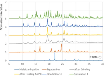
Initial attempts to obtain target maleimide 1 were based on our previous work where mechanosynthesis was used to prepare phthalimide compounds [5]. In brief, the starting solid reactants, tryptamine (2) and maleic anhydride (3) (Figure1), were ground in an equimolar ratio, either manually using a mortar and pestle or mechanically using a vibration ball mill Retsch MM400 (Retsch GmbH, Haan, Germany ) (60 min at 30 Hz, 2–10 balls added to a 2 mL plastic vial). Progress of reaction was monitored by powder X-ray diffraction (Panalytical ProX’pres, Almelo, the Netherlands, Cu radiation) (Figure2).
Crystals 2016, 6, 153 2 of 8
[9]). To the best of our knowledge, these are the only deposited crystal structures for this, otherwise well documented, family of compounds.
2. Results and Discussion
2.1. Synthesis and Crystallization
2.1.1. Mechanosynthesis
Initial attempts to obtain target maleimide 1 were based on our previous work where mechanosynthesis was used to prepare phthalimide compounds [5]. In brief, the starting solid reactants, tryptamine (2) and maleic anhydride (3) (Figure 1), were ground in an equimolar ratio, either manually using a mortar and pestle or mechanically using a vibration ball mill Retsch MM400 (Retsch GmbH, Haan, Germany ) (60 min at 30 Hz, 2–10 balls added to a 2 mL plastic vial). Progress of reaction was monitored by powder X-ray diffraction (Panalytical ProX’pres, Almelo, the Netherlands, Cu radiation) (Figure 2).
Figure 1. Synthetic route to 1, using a solvent-free mechanosynthetic approach.
Figure 2. Powder X-ray diffractograms showing the outcome of the grinding experiment. Starting
solids (maleic anhydrate and tryptamine), solid obtained after grinding (60 min at 30 Hz), solid obtained after heating (30 min at 140 °C). Diffractograms simulated on the basis of the single crystal structures of 1 and 1o are presented for comparison.
Figure 1.Synthetic route to 1, using a solvent-free mechanosynthetic approach.
Crystals 2016, 6, 153 2 of 8
[9]). To the best of our knowledge, these are the only deposited crystal structures for this, otherwise well documented, family of compounds.
2. Results and Discussion
2.1. Synthesis and Crystallization
2.1.1. Mechanosynthesis
Initial attempts to obtain target maleimide 1 were based on our previous work where mechanosynthesis was used to prepare phthalimide compounds [5]. In brief, the starting solid reactants, tryptamine (2) and maleic anhydride (3) (Figure 1), were ground in an equimolar ratio, either manually using a mortar and pestle or mechanically using a vibration ball mill Retsch MM400 (Retsch GmbH, Haan, Germany ) (60 min at 30 Hz, 2–10 balls added to a 2 mL plastic vial). Progress of reaction was monitored by powder X-ray diffraction (Panalytical ProX’pres, Almelo, the Netherlands, Cu radiation) (Figure 2).
Figure 1. Synthetic route to 1, using a solvent-free mechanosynthetic approach.
Figure 2. Powder X-ray diffractograms showing the outcome of the grinding experiment. Starting
solids (maleic anhydrate and tryptamine), solid obtained after grinding (60 min at 30 Hz), solid obtained after heating (30 min at 140 °C). Diffractograms simulated on the basis of the single crystal structures of 1 and 1o are presented for comparison.
Figure 2.Powder X-ray diffractograms showing the outcome of the grinding experiment. Starting solids (maleic anhydrate and tryptamine), solid obtained after grinding (60 min at 30 Hz), solid obtained after heating (30 min at 140◦C). Diffractograms simulated on the basis of the single crystal structures of 1 and 1o are presented for comparison.
Upon grinding, characteristic diffraction peaks of the reactants were replaced by peaks associated to the new ground product (Figure2). Recrystallization of the ground solid from a saturated solution in a MeOH/toluene mixture led to small single crystals useful for crystal structure determination. X-ray diffraction analysis revealed that the compound obtained upon grinding is the open maleamide intermediate 1o (Section 2.2). This result is consistent with similar observations that we made for phthalimides [5]. Powder diffractogram simulated on the basis of the single crystal structure coordinates (Figure2) corresponds to the experimental data recorded on the solid obtained after grinding, confirming that the bulk powder corresponds to 1o. Conversion in almost quantitative (>95%) as judged from NMR and powder X-ray diffraction data.
We expected to get the final, closed maleimide 1 on heating the intermediate [5]. Despite several attempts, we were unable to convert intermediate 1o into the target compound 1.
A reasonable explanation for the failure of the formation of the desired product using unactivated reactants is probably linked to the reduced nucleophilicity of the conjugated amide intermediate 1o. Indeed, thermal and uncatalyzed amide formation has been scarcely reported [10,11] especially when the nucleophile is conjugated amide.
2.1.2. Synthesis Using Activated Maleic Acid
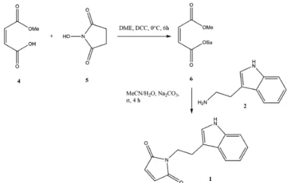
As we were unsuccessful in mechanosynthesis of 1, we developed a procedure (Figure3) based on a similar approach described in the literature [12].
Crystals 2016, 6, 153 3 of 8
Upon grinding, characteristic diffraction peaks of the reactants were replaced by peaks associated to the new ground product (Figure 2). Recrystallization of the ground solid from a saturated solution in a MeOH/toluene mixture led to small single crystals useful for crystal structure determination. X-ray diffraction analysis revealed that the compound obtained upon grinding is the open maleamide intermediate 1o (Section 2.2). This result is consistent with similar observations that we made for phthalimides [5]. Powder diffractogram simulated on the basis of the single crystal structure coordinates (Figure 2) corresponds to the experimental data recorded on the solid obtained after grinding, confirming that the bulk powder corresponds to 1o. Conversion in almost quantitative (>95%) as judged from NMR and powder X-ray diffraction data.
We expected to get the final, closed maleimide 1 on heating the intermediate [5]. Despite several attempts, we were unable to convert intermediate 1o into the target compound 1.
A reasonable explanation for the failure of the formation of the desired product using unactivated reactants is probably linked to the reduced nucleophilicity of the conjugated amide intermediate 1o. Indeed, thermal and uncatalyzed amide formation has been scarcely reported [10,11] especially when the nucleophile is conjugated amide.
2.1.2. Synthesis Using Activated Maleic Acid
As we were unsuccessful in mechanosynthesis of 1, we developed a procedure (Figure 3) based on a similar approach described in the literature [12].
Figure 3. Synthetic route to 1 using a solvent-based approach via activated maleic acid reactant. DME:
dimethylether; DCC: dicyclohexylcarbodiimide; OSu: N-oxysuccinimidyl group.
In brief, a solution of monomethylmaleate (4) (5.204 g, 40 mmol) and N-hydroxysuccinimide (5) (4.61 g, 40 mmol) in DME (15 mL) was cooled to 0 °C. Dicyclohexylcarbodiimide (DCC, 8.25 g, 40 mmol) was added and stirring was continued at this temperature for 4 h. The reaction mixture was allowed to stand for 2 h in a refrigerator and then filtered. The solution was concentrated under reduced pressure. The residue was triturated in Et2O/hexane, filtered, and then dried in vacuo to
afford solid methyl succinimidylmaleate (6). Tryptamine (2) (0.32 g, 2.0 mmol) and sodium carbonate (1.06 g, 10 mmol) were dissolved in water (15 mL), and then in acetonitrile (25 mL). Methyl succinimidylmaleate (6) (0.45 g, 2 mmol) was added, and the mixture was stirred for 4 h. The solution was acidified to pH 1 with 2N HCl, diluted with EtOAc (100 mL), and washed with 1N HCl (2 × 100 mL) and water (2 × 100 mL). The organic phase was dried with MgSO4, filtered, and concentrated in
vacuo to give final maleimide 1 (Figure 3). Final product was recrystallized from a concentrated
solution in acetonitrile at room temperature. The yield of this reaction has not been optimized but was already very good (>80%), consistent with data from the literature [12]. Single crystals suitable for crystallography were obtained and led to the structure described in the following section.
Figure 3. Synthetic route to 1 using a solvent-based approach via activated maleic acid reactant. DME: dimethylether; DCC: dicyclohexylcarbodiimide; OSu: N-oxysuccinimidyl group.
In brief, a solution of monomethylmaleate (4) (5.204 g, 40 mmol) and N-hydroxysuccinimide (5) (4.61 g, 40 mmol) in DME (15 mL) was cooled to 0◦C. Dicyclohexylcarbodiimide (DCC, 8.25 g, 40 mmol) was added and stirring was continued at this temperature for 4 h. The reaction mixture was allowed to stand for 2 h in a refrigerator and then filtered. The solution was concentrated under reduced pressure. The residue was triturated in Et2O/hexane, filtered, and then dried in vacuo to afford solid methyl
succinimidylmaleate (6). Tryptamine (2) (0.32 g, 2.0 mmol) and sodium carbonate (1.06 g, 10 mmol) were dissolved in water (15 mL), and then in acetonitrile (25 mL). Methyl succinimidylmaleate (6) (0.45 g, 2 mmol) was added, and the mixture was stirred for 4 h. The solution was acidified to pH 1 with 2N HCl, diluted with EtOAc (100 mL), and washed with 1N HCl (2×100 mL) and water (2×100 mL). The organic phase was dried with MgSO4, filtered, and concentrated in vacuo to give final
maleimide 1 (Figure3). Final product was recrystallized from a concentrated solution in acetonitrile at room temperature. The yield of this reaction has not been optimized but was already very good (>80%), consistent with data from the literature [12]. Single crystals suitable for crystallography were obtained and led to the structure described in the following section.
Crystals 2016, 6, 153 4 of 8
2.2. Structural Commentary
The open intermediate 1o (Figure4a, Table1) was obtained by grinding maleic anhydride and tryptamine (Figure1). The structure of the final target molecule 1 (Figure 4b, Table 1) was obtained using activated methyl succinimidylmaleate (Figure3) as confirmed by determination of its crystal structure.
Crystals 2016, 6, 153 4 of 8
2.2. Structural Commentary
The open intermediate 1o (Figure 4a, Table 1) was obtained by grinding maleic anhydride and tryptamine (Figure 1). The structure of the final target molecule 1 (Figure 4b, Table 1) was obtained using activated methyl succinimidylmaleate (Figure 3) as confirmed by determination of its crystal structure.
(a)
(b)
Figure 4. Displacement ellipsoid plots (50% probability) for (a)
1-[2-(1H-indol-3-yl)-ethyl]-pyrrole-2,5-dione, C14 H12 N2 O2, 1 and (b) its open analogue, 1o. Atom numbering of non-H atoms is presented.
Table 1. Experimental details of the crystal structure determination.
(1) (1o)
Chemical formula C14H12N2O2 C14H14N2O3
Mr 240.26 258.27
Crystal system, space group Orthorhombic, P212121 Monoclinic, P21/n
a, b, c (Å) 6.2720 (3), 8.9037 (6), 12.8304 (6), 12.0034 (10), 15.0252 (6) 12.1047 (8) α, β, γ (°) 90, 90, 90 90, 92.357 (6), 90 V (Å3) 1209.11 (9) 1292.59 (16) Z 4 4 Radiation type Mo Kα Cu Kα µ (mm−1) 0.09 0.78 Crystal size (mm) 1.0 × 0.8 × 0.5 0.13 × 0.07 × 0.05 Tmin, Tmax 0.917, 0.956 0.930, 0.960
No. of ref. measured, independent, and
observed 4460, 2144, 1789 5570, 2181, 1524
Rint 0.036 0.069
θmax (°) 25.0 64.0
R[F2 > 2σ(F2)], wR(F2), S 0.041, 0.097, 1.05 0.060, 0.185, 1.02
No. of reflections 2144 2011
Figure 4.Displacement ellipsoid plots (50% probability) for (a) 1-[2-(1H-indol-3-yl)-ethyl]-pyrrole-2,5-dione, C14H12N2O2, 1 and (b) its open analogue, 1o. Atom numbering of non-H atoms is presented.
Table 1.Experimental details of the crystal structure determination. (1) (1o)
Chemical formula C14H12N2O2 C14H14N2O3
Mr 240.26 258.27
Crystal system, space group Orthorhombic, P212121 Monoclinic, P21/n
a, b, c (Å) 6.2720 (3), 8.9037 (6), 12.8304 (6), 12.0034 (10), 15.0252 (6) 12.1047 (8) α, β, γ (◦) 90, 90, 90 90, 92.357 (6), 90 V (Å3) 1209.11 (9) 1292.59 (16) Z 4 4 Radiation type Mo Kα Cu Kα µ(mm−1) 0.09 0.78 Crystal size (mm) 1.0 × 0.8 × 0.5 0.13 × 0.07 × 0.05 Tmin, Tmax 0.917, 0.956 0.930, 0.960
No. of ref. measured, independent, and observed 4460, 2144, 1789 5570, 2181, 1524
Rint 0.036 0.069 θmax(◦) 25.0 64.0 R[F2> 2σ(F2)], wR(F2), S 0.041, 0.097, 1.05 0.060, 0.185, 1.02 No. of reflections 2144 2011 No. of parameters 163 172 ∆ρmax,∆ρmin(e Å−3) 0.11, −0.16 0.23, −23
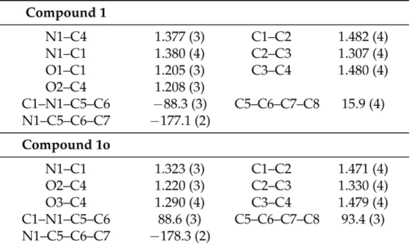
As expected, nitrogen atom N1 of the maleimide ring in 1 is sp2hybridized (sum of valence angles close to 360◦, C1–N1–C5 = 125.9(2)◦, C4–N1–C5 = 124.4(2)◦, C1–N1–C4 = 109.7(2)◦), and C–C and
C–N bond lengths (Table2) are intermediate between single and double bonds, suggesting electronic delocalization within this ring.
In the open intermediate 1o, nitrogen atom N1 binds to C1=O and forms an amide. This group is conjugated to the C2=C3–C4OOH part, as deduced from the bond lengths (Table2). Within the maleamide moiety, a strong O3–H3O···O1 intra-molecular hydrogen bond is observed (Table3).
Table 2.Selected geometric features. Bond lengths (Å), valence angles (◦), and torsion angles (◦).
Compound 1 N1–C4 1.377 (3) C1–C2 1.482 (4) N1–C1 1.380 (4) C2–C3 1.307 (4) O1–C1 1.205 (3) C3–C4 1.480 (4) O2–C4 1.208 (3) C1–N1–C5–C6 −88.3 (3) C5–C6–C7–C8 15.9 (4) N1–C5–C6–C7 −177.1 (2) Compound 1o N1–C1 1.323 (3) C1–C2 1.471 (4) O2–C4 1.220 (3) C2–C3 1.330 (4) O3–C4 1.290 (4) C3–C4 1.479 (4) C1–N1–C5–C6 88.6 (3) C5–C6–C7–C8 93.4 (3) N1–C5–C6–C7 −178.3 (2)
Table 3.Hydrogen bond geometry (Å,◦).
D–H···A D–H H···A D···A D–H···A Compound 1 N2–H2···O2i 0.86 2.30 2.952 (3) 133 Compound 1o O3–H3O···O1 0.82 1.68 2.490 (3) 171 N2–H2···O1ii 0.86 2.09 2.938 (3) 169 N1–H1···O2iii 0.86 2.18 2.941 (3) 147
Symmetry code: (i) –x−1
2,−y + 1, z−12; (ii) –x + 1,−y + 1,−z; (iii)−12+ x,12−y,12+ z.
Conformation of the two compounds is distinct. In molecule 1, the planar indole ring is almost perpendicular to the maleimide ring (acute angle between planes = 68.6(2)◦). In the open intermediate 1o, the planar indole heterocycle is almost parallel to the maleamide moiety (acute angle between planes = 6.9(2)◦). This is also reflected in the values of the torsions angles defining the conformation of the molecules (Table2).
In both compounds, nitrogen N2 of the indole ring serves as a H-bond donor (Table 3). For compound 1o, the position of H atom on oxygen atom O3 of the carboxylic acid was unambiguously determined from the residual electron density. This H atom is involved in a strong intramolecular H bond involving O3–H and the carbonyl oxygen atom (O1) of the amide moiety (Table3).
In the crystal packing of 1 (Figure5a), in addition to the H-bonds involving N2 and O2 (Table3), T-shaped π–π contacts are observed between the delocalized indole ring and the maleimide ring (distance between centroids Cg(1)···Cg(2): 4.421(2)Å, and dihedral angle between planes: 41.5◦, Cg(1) being the centroid of the five-membered ring N1–C1–C2–C3–C4 and Cg(2) the centroid of the six-membered ring C9–C10–C11–C12–C13–C14). This leads to C–H···πinteraction (C3–H3···Cg(2)i: 3.631(3)Å with i = 12 −x, 1−y, 12 + z). In the crystal packing of 1o (Figure5b), in addition to the intermolecular H–bonds that involve N1, N2, O1, and O2 (Table3), parallel π–π stacking is observed between the indole ring and the maleamide pseudo-cyclic.
Crystals 2016, 6, 153 6 of 8
Crystals 2016, 6, 153 6 of 8
membered ring C9–C10–C11–C12–C13–C14). This leads to C–H···π interaction (C3–H3···Cg(2)i:
3.631(3)Å with i = ½ − x, 1 − y, ½ + z). In the crystal packing of 1o (Figure 5b), in addition to the intermolecular H–bonds that involve N1, N2, O1, and O2 (Table 3), parallel π–π stacking is observed between the indole ring and the maleamide pseudo-cyclic.
(a)
(b)
Figure 5. Packing diagram for (a) 1-[2-(1H-indol-3-yl)-ethyl]-pyrrole-2,5-dione, C14 H12 N2 O2, 1 and
(b) its open analogue, 1o.
In conclusion, 1-[2-(1H-Indol-3-yl)-ethyl]-pyrrole-2,5-dione (1), a potential DNA methyltransferase inhibitor, has been successfully synthesized using activated reactants and synthesis in solution. Direct mechanosynthesis, starting from the unactivated reactant and using dry grinding, led to the amide intermediate 1o. This molecule did not convert into the cyclized final product in contrast with results obtained for phthalimides using dry grinding of corresponding unactivated anhydrides and amines. Reduced nucleophilicity of the conjugated amide intermediate is probably part of the explanation. Restricted conformation of molecule 1o, in the solid, and crystal packing further play a role in the stability of the intermediate product.
As a perspective to this work, liquid-assisted grinding could be tested by addition of a few drops of solvent during grinding. Mechanosynthesis starting from activated reactants is another possible alternative.
Figure 5.Packing diagram for (a) 1-[2-(1H-indol-3-yl)-ethyl]-pyrrole-2,5-dione, C14H12N2O2, 1 and
(b) its open analogue, 1o.
In conclusion, 1-[2-(1H-Indol-3-yl)-ethyl]-pyrrole-2,5-dione (1), a potential DNA methyltransferase inhibitor, has been successfully synthesized using activated reactants and synthesis in solution. Direct mechanosynthesis, starting from the unactivated reactant and using dry grinding, led to the amide intermediate 1o. This molecule did not convert into the cyclized final product in contrast with results obtained for phthalimides using dry grinding of corresponding unactivated anhydrides and amines. Reduced nucleophilicity of the conjugated amide intermediate is probably part of the explanation. Restricted conformation of molecule 1o, in the solid, and crystal packing further play a role in the stability of the intermediate product.
As a perspective to this work, liquid-assisted grinding could be tested by addition of a few drops of solvent during grinding. Mechanosynthesis starting from activated reactants is another possible alternative.
3. Materials and Methods 3.1. NMR Data 1o:1H NMR (400 MHz DMSO) 10.82 (br s, 1H), 9.20 (br s, 1H), 7.50 (d, 1H, J = 7.79 Hz), 7.31 (d, 1H, J = 8.01 Hz), 7.14 (s, 1H), 7.03 (t, 1H, J = 7.44 Hz), 6.95 (t, 1H, J = 7.44 Hz), 6.38 (dd, 1H, J1 = 12.59 Hz, J2 = 1.14 Hz), 6.22 (dd, 1H, J1 = 12.59 Hz, J2 = 1.37 Hz), 3.44 (dd, 2H, J1 = 6.64 Hz, J2 = 6.64 Hz), 2.87 (t, 2H, J = 7.33 Hz); 13C NMR (400 MHz, CDCl 3) 167.89, 166.04, 136.78, 134.16, 132.13, 127.59, 123.34, 121.49, 118.84, 118.64, 111.95, 111.72, 40.06, 24.96; 1:1H NMR (400 MHz CDCl3) 8.03 (br s, 1H), 7.66 (d, 1H, J = 7.79 Hz), 7.34 (d, 1H, J = 8.01 Hz), 7.19 (t, 1H, J = 6.98 Hz), 7.13 (t, 1H, J = 6.98 Hz), 7.03 (s, 1H), 6.64 (s, 2H), 3.84 (t, 2H, J = 7.67 Hz), 3.06 (t, 2H, 7.67 Hz); 13C NMR (400 MHz, CDCl 3) 170.94, 136.34, 134.13, 127.38, 122.28, 121.96, 119.36, 118.69, 111.91, 111.28, 38.55, 24.40.
3.2. Crystal Data Collection and Refinement
Crystal data, data collection, and structure refinement details are summarized in Table 1. Crystal data used for the refinement were collected on a Gemini R Ultra diffractometer at room temperature. Data were treated (cell determination and data reduction) using CrysAlis PRO (Rigaku Oxford Diffraction, Oxford, UK). Structures were solved using SIR2004 [13] and refined with SHELXL2014/8 [14].
For compound 1o, data had to be collected using an enhanced Cu radiation source, as only small crystals could be obtained. In both structures, non-hydrogen atoms have been refined anisotropically. Positions of H atoms were observed in the Fourier difference maps, then calculated at their ideal position and refined using a riding model (C–H and N–H bond distances fixed to 0.93 and 0.86 Å, respectively, thermal factors fixed to 1.2 times the value of the parent (C or N) atom). In structure 1o, H atom on O3, forming a strong intra-molecular H bond, was found in the∆F map and its position was refined using a riding model: the thermal factor was fixed (1.5 times) on the basis of the value of the oxygen atom (O3) to which it was covalently bound.
Final coordinates and structure factors have been deposited to CSD (CCDC 1508084-1508085).
Acknowledgments:Crystal structure determinations were performed using equipment available at the Plateforme de Caractérisation (PC2) at UNamur and calculation were done on the Plateforme de Calculs Intensif of UNamur. G.Rondelet benefited from a Télévie Grant (F.R.S.—FNRS—Télévie 7.4.532.15.F).
Author Contributions:Jean Dubois, Melwin Colaço and Johan Wouters conceived and designed the experiments; Melwin Colaço, Jean Dubois, and Grégoire Rondelet performed the experiments; Johan Wouters and Melwin Colaço analyzed the data.
Conflicts of Interest:The authors declare no conflict of interest. The founding sponsors had no role in the design of the study; in the collection, analyses, or interpretation of data; in the writing of the manuscript, or in the decision to publish the results.
References
1. Rondelet, G.; Dal Maso, T.; Willems, L.; Wouters, J. Structural basis for recognition of histone H3K36me3 nucleosome by human de novo DNA methyltransferases 3A and 3B. J. Struct. Biol. 2016, 194, 357–367. [CrossRef] [PubMed]
2. Gillet, N.; Vandermeers, F.; de Brogniez, A.; Florins, A.; Nigro, A.; François, C.; Bouzar, A.-B.; Verlaeten, O.; Stern, E.; Lambert, D.; et al. Chemoresistance to valproate treatment of bovine leukemia virus-infected sheep; identification of improved HDAC inhibitors. Pathogens 2012, 1, 65–82. [CrossRef] [PubMed]
3. Brueckner, B.; Garcia Boy, R.; Siedlecki, P.; Musch, T.; Kliem, H.; Zielenkiewicz, P.; Suhai, S.; Wiessler, M.; Lyko, F. Epigenetic reactivation of tumor suppressor genes by a novel small-molecule inhibitor of human DNA methyltransferases. Cancer Res. 2005, 65, 6305–6311. [CrossRef] [PubMed]
Crystals 2016, 6, 153 8 of 8
4. Suzuki, T.; Tanaka, R.; Hamada, S.; Nakagawa, H.; Miyata, N. Design, synthesis, inhibitory activity, and binding mode study of novel DNA methyltransferase 1 inhibitors. Bioorg. Med. Chem. Lett. 2010, 20, 1124–1127. [CrossRef] [PubMed]
5. Colaço, M.O.; Dubois, J.; Wouters, J. Mechanochemical synthesis of phthalimides with crystal structures of intermediates and products. CystEngComm 2015, 17, 2523–2528. [CrossRef]
6. Groom, C.R.; Bruno, I.J.; Lightfoot, M.P.; Ward, S.C. The Cambridge Structural Database. Acta Cryst. 2016, 72, 171–179. [CrossRef] [PubMed]
7. Griesbeck, A.G.; Neudorfl, J.; de Kiff, A. Photoinduced electron-transfer chemistry of the bielectrophoric N-phthaloyl derivatives of the amino acids tyrosine, histidine and tryptophan. Beilstein J. Org. Chem. 2011, 7, 518–524. [CrossRef] [PubMed]
8. Braun, J.; Boittiaux, I.; Tilborg, A.; Lambert, D.; Wouters, J. The dicyclohexylamine salt of RG108 (N-phthalyl-L-tryptophan), a potential epigenetic modulator. Acta Cryst. 2010, 66, o3175–o3176. [CrossRef] [PubMed]
9. Tilborg, A.; Boittiaux, I.; Norberg, B.; Lambert, D.; Wouters, J. 4-Nitro-N-phthalyl-L-tryptophan. Acta Cryst.
2011, 67, o2116. [CrossRef] [PubMed]
10. Allen, C.L.; Chhatwal, A.R.; Williams, J.M. Direct amide formation from unactivated carboxylic acids and amines. Chem. Commun. 2012, 48, 666–668. [CrossRef] [PubMed]
11. Lundberg, H.; Tinnis, F.; Selander, N.; Adolfsson, H. Catalytic amide formation from non-activated carboxylic acids and amines. Chem. Soc. Rev. 2014, 43, 2714–2742. [CrossRef] [PubMed]
12. Casimir, J.R.; Guichard, G.; Briand, J.-P. Methyl 2-((succinimidooxy)carbonyl)benzoate (MSB): A new, efficient reagent for N-phthaloylation of amino acid and peptide derivatives. J. Org. Chem. 2002, 67, 3764–3768. [CrossRef] [PubMed]
13. Burla, M.C.; Caliandro, R.; Camalli, M.; Carrozzini, B.; Cascarano, G.L.; Caro, L.D.; Giacovazzo, C.; Polidori, G.; Spagna, R. SIR2004: An improved tool for crystal structure determination and refinement. J. Appl. Cryst. 2005, 38, 381–388. [CrossRef]
14. Sheldrick, G.M. Crystal structure refinement with SHELXL. Acta Cryst. 2015, 71, 3–8.
© 2016 by the authors; licensee MDPI, Basel, Switzerland. This article is an open access article distributed under the terms and conditions of the Creative Commons Attribution (CC-BY) license (http://creativecommons.org/licenses/by/4.0/).
![Figure 4. Displacement ellipsoid plots (50% probability) for (a) 1-[2-(1H-indol-3-yl)-ethyl]-pyrrole-2,5- 1-[2-(1H-indol-3-yl)-ethyl]-pyrrole-2,5-dione, C 14 H 12 N 2 O 2 , 1 and (b) its open analogue, 1o](https://thumb-eu.123doks.com/thumbv2/123doknet/14509823.720734/5.892.252.647.264.650/figure-displacement-ellipsoid-plots-probability-pyrrole-pyrrole-analogue.webp)
![Figure 5. Packing diagram for (a) 1-[2-(1H-indol-3-yl)-ethyl]-pyrrole-2,5-dione, C 14 H 12 N 2 O 2 , 1 and (b) its open analogue, 1o](https://thumb-eu.123doks.com/thumbv2/123doknet/14509823.720734/7.892.245.646.128.778/figure-packing-diagram-indol-ethyl-pyrrole-dione-analogue.webp)
