HAL Id: tel-03214206
https://tel.archives-ouvertes.fr/tel-03214206
Submitted on 1 May 2021HAL is a multi-disciplinary open access
archive for the deposit and dissemination of sci-entific research documents, whether they are pub-lished or not. The documents may come from teaching and research institutions in France or abroad, or from public or private research centers.
L’archive ouverte pluridisciplinaire HAL, est destinée au dépôt et à la diffusion de documents scientifiques de niveau recherche, publiés ou non, émanant des établissements d’enseignement et de recherche français ou étrangers, des laboratoires publics ou privés.
Corinna Bode
To cite this version:
Corinna Bode. Implants à base de PLGA pour la libération oculaire. Human health and pathology. Université de Lille, 2019. English. �NNT : 2019LILUS008�. �tel-03214206�
Faculté des Sciences Pharmaceutiques et Biologiques Ecole Doctorale Biologie-Santé
PLGA IMPLANTS FOR OCULAR DRUG DELIVERY
___
IMPLANTS A BASE DE PLGA POUR
LA LIBERATION OCULAIRE
THESE DE DOCTORAT Soutenu le 30 avril 2019
Par Corinna BODE
Dirigée par Prof. Juergen SIEPMANN
Laboratoire INSERM U1008, Médicaments et Biomatériaux à Libération Contrôlée
Composition du Jury :
Monsieur Juergen SIEPMANN Directeur de thèse
Professeur à l’Université de Lille
Madame Aurélie MALZERT-FREON Président du jury/rapporteur
Professeur à l’Université de Caen
Madame Béatrice HEURTAULT Rapporteur
Maître de Conférences à l’Université de Strasbourg
Monsieur Heiko KRANZ Examinateur
I want to express my gratitude to those who contributed to the success of this thesis.
To begin with, I would like to thank Professor Juergen SIEPMANN, director of the Inserm U1008 unit, for welcoming me in the laboratory for Controlled Drug Delivery Systems and Biomaterials and for supervising this thesis. Thank you for all the valuable advice, the constant availability and kind encouragement over the last years. Vielen Dank!
My sincere gratitude goes to Dr. Heiko KRANZ who made this cooperation with Bayer possible in the first place. Thank you so much for the commitment and support!
Je remercie Professeur Florence SIEPMANN pour son aide, son optimisme et le soutien de nature scientifique et administrative.
J’aimerais aussi remercier mes deux rapporteurs, Professeur Aurélie MALZERT-FREON de l’Université de Caen et Dr. Béatrice HEURTAULT de l’Université de Strasbourg, d’avoir pris le temps nécessaire d’évaluer ma thèse et de faire partie du jury.
Merci encore à mes stagiaires, Astrid FIVES et Aleksandra KRUSZKA, qui m’ont soutenu durant ma thèse par leur travail de valeur et leur bonne humeur.
Je voudrais remercier toute l’équipe du laboratoire : Jérémy VERIN pour le SEM ; Susanne
MUSCHERT, Mounira HAMOUDI-BENYELLES et Youness KARROUT pour leur
contri-bution aux bonnes conditions de travail dans le laboratoire ; Alexandra MACHADO pour les commandes et l’administration et Hugues FLORIN pour la maintenance des machines au la-boratoire.
Les remerciements particuliers à
Maria pour m’avoir fait me sentir bienvenu en France, son aide et sa créativité ; Ting pour
m’avoir toujours remonté le moral ; Doha pour sa fiabilité et son écoute et Youcef pour son aide au laboratoire.
toire : Fahima, Martin, Adam, Kévimy, Lauranne, Céline, Thomas, Julie, Oriane, Ester,
Elisa, Federica, Fabiana, Nourhène, Saliha et Sine. Cela m‘a fait plaisir de travailler avec
vous !
Ich bin dankbar für meine Freunde Julia und Franzi. Vielen Dank für die moralische Unter-stützung zu jeder Tages- und Nachtzeit!
Ein ganz besonderer Dank gilt meiner Familie: meinen Eltern für ihre unbegrenzte Unterstüt-zung in jeglicher Hinsicht und meinem Bruder Thomas für seine Hilfe und Zuverlässigkeit.
Page List of abbreviations………... iv INTRODUCTION GENERALE……… 1 Contexte de la recherche……… 2 Objectifs de la recherche……… 4 Présentation de ce travail……… 5 GENERAL INTRODUCTION………... 6 Research Context………... 7 Research Objectives………... 9
Presentation of the work………. 10
CHAPTER 1: INTRODUCTION………... 11
1.1 The eye………. 12
1.1.1 Anatomy of the eye……… 12
1.1.2 Dynamic and static ocular barriers………. 13
1.1.3 Blood-ocular barriers………. 15
1.1.4 Ocular clearance……… 16
1.2 Diseases of the eye……….………... 17
1.2.1 Diabetic retinopathy………... 17
1.2.2 Age-related macular degeneration……….. 18
1.2.3 Uveitis……… 20
1.3 Therapy of the eye……… 21
1.3.1 Laser treatment………... 21
1.3.2 Anti-VEGF factors………. 22
1.3.3 Corticosteroid………. 24
1.4 Ocular drug delivery………. 25
1.4.1 Topical………... 26
1.4.2 Periocular………... 27
1.4.3 Suprachoroidal………... 27
1.4.4 Systemic/oral………. 28
1.4.5 Intravitreal……….. 28
1.5 Ocular implants for sustained drug release………... 30
1.6 Poly(D,L-lactic-co-glycolic)acid………. 32
1.6.1 Physico-chemical properties……….. 32
1.6.2 Biodegradation and biocompatibility……… 34
1.6.3 Sterilization……… 37
1.7 Techniques prolonging drug release using PLGA……… 38
1.7.1 Microparticles……… 38
1.7.2 In-situ forming implants……… 39
1.7.3 Pre-formed implants……….. 42
References……….. 44
CHAPTER 2: MATERIALS AND METHODS……… 66
2.1 Materials………... 67
2.2 Methods……… 68
2.2.1 In-situ forming PLGA implants for intraocular drug delivery……… 68
2.2.1.1 Preparation of the liquid formulations……… 68
2.2.1.2 In-situ formation of implants……….. 68
2.2.1.3 Characterization of in-situ formed implants………... 68
2.2.1.4 Determination of the drug solubility………... 70
2.2.2 Often neglected: PLGA/PLA swelling orchestrates drug release - HME implants 71 2.2.2.1 Implant formation………... 71
2.2.2.2 Implant characterization………. 72
2.2.2.3 Determination of drug solubility……… 74
2.2.2 Coloring of PLGA implants to better understand drug release mechanisms…... 75
2.2.3.1 Implant preparation……… 75
2.2.3.2 Implant characterization………. 75
2.2.3.3 Determination of the solubility of riboflavin……….. 77
2.2.4 In-situ forming PLGA implants: How additives affect swelling and drug release. 78 2.2.4.1 Preparation of the liquid formulation……….. 78
2.2.4.2 In-situ implant formation……… 78
Part 1: In-situ forming PLGA implants for intraocular drug delivery……… 82
3.1.1 Importance of the volume of the release medium………. 82
3.1.2 Impact of the drug loading………. 85
3.1.3 Impact of the PLGA molecular weight……….. 88
3.1.4 Impact oft he polymer concentration………. 90
3.1.5 Conclusion………. 97
Part 2: Often neglected: PLGA/PLA swelling orchestrates drug release – HME implants……… 98
3.2.1 Morphology……… 98
3.2.2 PLGA (RG 502H)-based implants……… 101
3.2.3 PLGA (RG 752H)-based implants……… 105
3.2.4 PLA (R 202H)-based implants……… 108
3.2.5 The orchestrating role of PLGA/PLA swelling for drug release………. 111
3.2.6 Conclusion………... 113
Part 3: Coloring of PLGA implants to better understand drug release mecha-nisms……….. 114
3.3.1 Pre-formed (HME) implants……….. 114
3.3.2 In-situ forming implants……… 121
3.3.3 Conclusion………. 126
Part 4: In-situ forming PLGA implants: How additives affect swelling and drug release……… 127 3.4.1 Carbopol……… 127 3.4.2 PEG 400………. 132 3.4.3 HPMC……… 134 3.4.4 Stearic acid………. 137 3.4.5 ATBC………. 139 3.4.6 Conclusion………. 141 References……….. 142
GENERAL CONCLUSION AND FURTURE PERSPECTIVES………... 144
General Conclusion……… 145
Abbreviations
AMD age-related macular degeneration
AREDS age-related disease study
ATBC acetyltributyl citrate
BA bioavailability
BAB blood-aqueous barrier
BRB blood-retinal barrier
CME cystoid macular edema
CNV choroidal neovascularization
DME diabetic macular edema
DMSO dimethyl sulfoxide
DR diabetic retinopathy
EVA ethylene vinyl acetate
FA fluocinolone acetonide
GA geographic atrophy
HME hot melt extrudate
HPMC hydroxy methylcellulose
ISFI in-situ forming implant
Mw molecular weight
PEG poly (ethylene glycol)
PGA poly (glycolic acid)
PGF placental growth factor
PLA poly (lactic acid)
PLGA poly(lactic-co-glycolic acid)
PVA polyvinyl alcohol
RPE retinal pigment epithelium
RVO retinal vein occlusion
TA triamcinolone acetonide
Tg glass transition temperature
VEGF vascular endothelial growth factor
Contexte de la recherche
Les causes principales de déficience visuelle et de perte de vision sont la dégénérescence ma-culaire liée à l'âge, la rétinopathie diabétique et l'uvéite. La dégénérescence mama-culaire liée à l'âge (DMLA) existe sous deux formes : la DMLA "sèche" ou atrophique et la DMLA "humide" ou néovasculaire. La première est caractérisée par la présence d'un drusen mou entre la mem-brane de Bruch et l'épithélium pigmentaire rétinien (EPR). Environ 10 à 15 % des patients souffrant de DMLA sèche développent une DMLA humide qui est déclenchée par des facteurs de croissance endothéliale vasculaire (VEGF) provoquant une croissance anormale des vais-seaux sanguins dans la membrane de Bruch. Cela peut endommager l'EPR et les photorécep-teurs et provoquer des fuites de sang et de protéines, endommageant ainsi la vision. Dans la rétinopathie diabétique (RD), l'hyperglycémie persistante peut causer des microanévrysmes qui finissent par briser les jonctions endothéliales serrées dans la barrière hématorétinienne (BHR) et entraîner une fuite de protéines dans le vitré. L'œdème maculaire diabétique (OMD) est une conséquence de la RD. La perméabilité capillaire rétinienne anormale peut entraîner un gonfle-ment extravasculaire de la macula et finalegonfle-ment une distorsion de la vision ou même une perte de vision. L'uvéite est une inflammation interne de l'œil qui peut survenir dans différentes par-ties de l'œil (antérieure, intermédiaire et postérieure). Dans l'uvéite postérieure, la rétine et/ou la choroïde sont affectées. Une inflammation chronique de l'uvée peut causer des dommages structurels dans l'œil et peut entraîner des complications comme l'œdème maculaire avec un risque élevé de perte irréversible de la vision. Pour ces trois maladies, les corticostéroïdes peu-vent être utilisés soit comme traitement de première intention, soit comme traitement complé-mentaire.
Les corticostéroïdes ont des propriétés anti-inflammatoires et inhibent la synthèse des facteurs de croissance endothéliale vasculaire, des prostaglandines et des cytokines inflammatoires. Ils peuvent également stabiliser les jonctions endothéliales serrées et diminuer la rupture du BRB. Dexaméthasone est cinq fois plus puissant que l'acétonide de triamcinolone. Il a également une solubilité plus élevée. Ainsi, des concentrations intravitréennes plus élevées peuvent être at-teintes. De plus, en raison de ses propriétés moins lipophiles, l'accumulation dans le réseau trabéculaire et le cristallin est réduite, ce qui pourrait diminuer le risque de pression intraocu-laire élevée souvent associé aux stéroïdes. Malgré ces propriétés favorables de la dexamétha-sone, la courte demi-vie vitreuse d'environ 5,5 heures (comparativement à environ 18 jours d'acétonide de triamcinolone) restreint la thérapie. Pour obtenir des concentrations thérapeu-tiques de dexaméthasone sur de longues périodes de temps, le médicament doit être administré
fréquemment. Cependant, en utilisant des gouttes ophtalmiques, seule une très petite portion (environ 0,001 à 0,0004 %) du médicament administré se trouve à l'intérieur du vitré, ce qui entraîne des concentrations inférieures à la concentration minimale efficace. D'autre part, la barrière rétinienne sanguine limite le transport du médicament dans le vitré après son adminis-tration systémique, ce qui nécessite de fortes doses de médicament, ce qui peut entraîner des effets secondaires non désirés. Pour surmonter cet obstacle, la dexaméthasone peut être injectée directement dans l'œil, mais chaque administration comporte le risque d'introduire des infec-tions et d'autres effets secondaires tels que décollement de la rétine, cataracte, hémorragies vi-trées et rétiniennes, endophtalmie et pression intraoculaire accrue. Une libération prolongée du médicament réduit la fréquence des injections intravitréennes et peut améliorer l'efficacité du traitement grâce à une libération constante du médicament.
L'acide poly(acide lactique-co-glycolique) (PLGA) est un polymère qui est souvent utilisé pour une administration prolongée de médicaments. C'est l'un des polymères biodégradables les plus courants et il a été utilisé pour la première fois dans les années 1960 comme suture biorésor-bable. Il est disponible sur le marché dans de nombreux produits approuvés par la FDA pour la libération contrôlée parentérale et d'autres techniques d'administration. Il est possible de fabri-quer différentes formes telles que des membranes, des tiges et des disques en utilisant des tech-niques de moulage, d'extrusion et autres. Le PLGA est également soluble dans une large gamme de solvants tels que le tétrahydrofuranne, l'acétone, les solvants chlorés et l'acétate d'éthyle, ce qui le rend adapté à une large gamme de techniques de préparation. Le PLGA s'est révélé bien toléré et biocompatible dans les tissus oculaires et non oculaires. Par exemple, l'implant oculaire biodégradable Ozurdex contient de la dexaméthasone dans une matrice PLGA qui n'a pas be-soin d'être enlevée chirurgicalement une fois la libération du médicament terminée.
Objectifs de la recherche
Les implants de formation in situ peuvent être injectés avec une petite taille d'aiguille, tandis que les implants préformés évitent l'utilisation de solvants potentiellement toxiques. Afin d'adapter la libération du médicament pour les deux systèmes, les différents mécanismes de libération et le comportement de biodégradation doivent être mieux compris. Ils peuvent dé-pendre de divers facteurs, tels que la surface totale, la charge de médicament, la solubilité du médicament, le gonflement des polymères, l'autocatalyse, par exemple, les mécanismes exacts qui se trouvent derrière la libération et la dégradation du médicament sont très complexes et ne sont pas entièrement compris.
Le but de cette thèse était le développement et la caractérisation de différents systèmes implan-taires contenant de la dexaméthasone et du PLGA pour une libération prolongée du médicament pour administration intravitréenne. Les principaux objectifs sont les suivants :
(i) Préparation et caractérisation des implants de formation in situ en mettant l'accent sur l'impact du volume du milieu de libération, du type et de la concentration du polymère ainsi que de la teneur en médicament des formulations.
(ii) Préparation et caractérisation des implants préformés en mettant l'accent sur la charge de médicament, le type de polymère et l'importance du comportement de gonflement pour la libération du médicament.
(iii) Visualisation de la libération du médicament et de l'absorption d'eau des implants pré-formés et pré-formés in situ à l'aide de médicaments modèles colorés pour mieux com-prendre les mécanismes sous-jacents.
(iv) Étude de l'effet de différentes quantités d'additifs sur les principales caractéristiques des implants formés in situ, en particulier la libération de médicaments, le gonflement des implants, la température de transition vitreuse et la morphologie.
Présentation de ce travail
Le présent ouvrage est composé de quatre chapitres :
I. Le premier chapitre donne un aperçu de la structure de l'œil et des défis qui en résultent pour le traitement des maladies affectant le segment postérieur. Il montre différentes approches d'application de médicaments sur le vitré et met l'accent sur les systèmes d'administration prolongée de médicaments potentiels pour une injection intravitréenne. II. Le deuxième chapitre énumère les matériaux et les méthodes utilisés pour la recherche
de ces travaux.
III. Le troisième chapitre présente les résultats et la discussion. Il est divisé en quatre parties. Chaque partie correspond à l'un des objectifs de recherche décrits ci-dessus. Bientôt, les principaux sujets sont :
1. Implants PLGA de formation in situ pour l'administration intraoculaire de dexa-méthasone
2. Souvent négligé : Le gonflement PLGA/PLA orchestre la libération du médica-ment - HME implants
3. Coloration des implants PLGA pour mieux comprendre les mécanismes de libé-ration du médicament
4. Implants PLGA de formation in situ : comment les additifs influent le gonfle-ment et la liberation du médicagonfle-ments
IV. Enfin, la quatrième partie donne une conclusion générale et résume les résultats de cette thèse.
Research Context
The major causes for vision impairment and vision loss are age-related macular degeneration, diabetic retinopathy and uveitis. Age-related macular degeneration (AMD) exists in two forms: the “dry” or atrophic AMD and the “wet” or neovascular AMD. The first one is characterized by an occurrence of soft drusen between the Bruch’s membrane and retinal pigment epithelium (RPE). Around 10-15% of patients with a dry AMD develop a wet AMD which is triggered by vascular endothelial growth factors (VEGF) causing an abnormal growth of blood vessels into the Bruch’s membrane. This can damage the RPE and photoreceptors and lead to blood and protein leakage, ultimately damaging the vision. In diabetic retinopathy (DR) continuous hy-perglycemia can cause microaneurysms which eventually break down the endothelial tight junctions in the blood retinal barrier (BRB) and lead to a protein leakage into the vitreous. Diabetic macular edema (DME) is a consequence of DR. The abnormal retinal capillary per-meability can lead to extravascular swelling in the macula and finally a distortion of vision or even vision loss. Uveitis is an internal inflammation of the eye and can occur in different parts of the eye (anterior, intermediate and posterior). In the posterior uveitis, the retina and/or cho-roid are affected. A chronic inflammation of the uvea can cause structural damage in the eye and can lead to complications like macular edema with a high risk of irreversible vision loss. For all three diseases corticosteroids can be either used as a first-line therapy or as an add-on therapy.
Corticosteroids have anti-inflammatory properties and they inhibit the synthesis of vascular endothelial growth factors, prostaglandins and inflammatory cytokines. They can also stabilize endothelial tight junctions and decrease the breakdown of the BRB. Dexamethasone is five times more potent than triamcinolone acetonide. It also has a higher solubility. Thus, higher intravitreal concentrations can be achieved. Additionally, due to its less lipophilic properties, the accumulation in the trabecular meshwork and lens is reduced which might decrease the risk of elevated intraocular pressure often associated with steroids. Despite these favorable proper-ties of dexamethasone, the short vitreous half-life of approximately 5.5 hours (compared to approximately 18 days of triamcinolone acetonide) restricts the therapy. To achieve therapeutic dexamethasone concentrations over prolonged periods of time, the drug has to be administered frequently. However, using eye drops only a very small portion (approximately 0.001 to 0.0004 %) of the administered drug is found inside the vitreous, leading to concentrations below the minimum effective concentration. On the other hand, the blood-retinal-barrier limits drug transport into the vitreous after systemic administration, requiring high drug doses, which can lead to undesired side effects. To overcome this hurdle, dexamethasone can be directly injected
into the eye, but each administration bears the risk of introducing infections and other side effects such as retinal detachment, cataract, vitreous and retinal hemorrhages, endophthalmitis and increased intraocular pressure. A prolonged drug release reduces the frequency of intravi-treal injections and can improve the efficacy of the therapy due to a constant drug release. Poly(lactic-co-glycolic acid) (PLGA) is a polymer that is often used for a sustained drug deliv-ery. It is one of the most common biodegradable polymers and was first used in the 1960s as bioresorbable sutures. It is commercially available in many FDA-approved products for paren-teral controlled release and other administration techniques. It is possible to fabricate various forms such as membranes, rods and disc using molding, extrusion and other techniques. PLGA is also soluble in a broad range of solvents such as tetrahydrofuran, acetone, chlorinated sol-vents and ethyl acetate, which makes it suitable for a wide range of preparation techniques. PLGA was shown to be well tolerated and biocompatible in ocular and non-ocular tissues. For example, the biodegradable, ocular implant Ozurdex contains dexamethasone in a PLGA matrix that does not have to be surgically removed after the drug release is completed.
Research Objectives
In-situ forming implants and pre-formed implants using PLGA can both sustain the drug release and biodegrade over time to avoid any surgical removal after complete drug liberation. In-situ forming implants can be injected with a small needle size, whereas pre-formed implants avoid the use of potentially toxic solvents. In order to adapt the drug release for both systems, the different release mechanisms and biodegradation behavior have to be better understood. They can depend on various factors, such as the total surface area, drug loading, solubility of the drug, polymer swelling, autocatalysis, e.g. The exact mechanisms behind the drug release and degradation are very complex and are not fully understood.
The aim of this thesis was the development and characterization of different implant systems containing dexamethasone and PLGA for a sustained drug release for intravitreal administra-tion. Main objectives include:
(i) Preparation and characterization of in-situ forming implants with emphasis on the im-pact of the volume of the release medium, polymer type and concentration as well as drug content of the formulations.
(ii) Preparation and characterization of pre-formed implants with focus on the drug load-ing, polymer type and the importance of swelling behavior to the drug release.
(iii) Visualization of drug release and water uptake of in-situ forming and pre-formed im-plants using colored model drugs to better understand the underlying mechanisms.
(iv) Investigation of the effect of different amounts of additives on the key features of in-situ forming implants in particular drug release, implant swelling, glass transition tem-perature and morphology.
Presentation of the work
The present work is composed of four chapters:
I. The first chapter gives an overview of the structure of the eye and its resulting chal-lenges for the treatment of diseases affecting the posterior segment. It shows different approaches to apply drugs to the vitreous and focuses on potential sustained drug deliv-ery systems for an intravitreal injection.
II. The second chapter lists the materials and methods used for the research of this work.
III. The third chapter contains the results and discussion. It is divided into four parts. Each part corresponds to one of the research objectives described above. Shortly, the main subjects are:
1. In-situ forming PLGA implants for intraocular dexamethasone delivery
2. Often neglected: PLGA/PLA swelling orchestrates drug release - HME implants
3. Coloring of PLGA implants to better understand drug release mechanisms
4. In-situ forming PLGA implants: How additives affect swelling and drug release
IV. Finally, the fourth part gives a general conclusion and summarizes the findings of this thesis.
CHAPTER 1:
INTRODUCTION
1.1 The eye
1.1.1 Anatomy of the eye
The eye can be divided into two segments – the anterior segment including the cornea, con-junctiva, iris, ciliary body and lens and the posterior segment including the sclera, choroid, retina and vitreous (Figure 1.1) [1,2]. The transparent, avascular cornea is directly exposed to the environment and is comprised of five layers: the epithelium, Bowman’s membrane, stroma, Descernet’s membrane and endothelium. The Bowman’s and Descernet’s membrane are inter-face layers, whereas the other three are cellular layers. The cellular layers differ in polarity and act as a mechanical barrier from exogenous substances [2,3]. The conjunctiva is a thin layer that covers the eyelids and sclera. It produces mucus and tears to lubricate the eye. The iris is the colored part of the eye. The circular membrane has an opening in the center, the pupil. Through contraction the pupil can alter the amount of light that enters the eye. The ciliary body produces the aqueous humor and its ciliary muscles control the shape of the lens to focus the vision. The aqueous humor is responsible for supplying nutrients and oxygen to avascular tis-sues (cornea and lens) and also removes macrophages, blood and waste products found between the cornea and lens. The entire aqueous humor is renewed in approximately 100 min [4]. The aqueous humor leaves the anterior chamber via the trabecular meshwork and canal of Schlemm. The sclera is commonly called the “white of the eye”. It is connected with the cornea and covers around 80% of the eyeball. The middle layer of the posterior segment is the choroid and is comprised of three distinguished layers: suprachoroid, vascular layer and Bruch’s membrane (from out to in). The blood vessels in the choroid are lined by endothelial cells, through which molecules can be exchanged between the blood and choroid. The Bruch’s membrane acts as a diffusional barrier to macromolecules [5]. The innermost layer is the retina containing the neu-ral retina, which is involved in signal transduction, and the retinal pigment epithelium (RPE). The RPE cells are polarized and the tight junctions in between those cells hinder the diffusion of small molecules from the choroid to the retina. The retina contains photoreceptor cells in-cluding rods and cones. These cells convert light into a nerve signal. The macular is part of the retina and is located close to the optic nerve. With its high concentration of photoreceptor cells in the center of the macula (called fovea), it is responsible for high-acuity vision. The posterior segment of the eye is filled with approximately 4 mL vitreous humor. Even though it consists to 98% of water, it has a high viscosity due to collagen type II and hyaluronic acid [6–8].
Figure 1.1: Schematic structure of the eye. From [9].
1.1.2 Dynamic and static ocular barriers
The eye is protected by various static (cornea, conjunctiva, sclera, blood-ocular barriers) and dynamic (tear turnover, nasolacrimal drainage, reflexive blinking, choroidal blood flow) barri-ers. They effectively protect the eye from external influences and limits the access of drugs into the eye (Figure 1.2) [10].
The first defense mechanism of the eye is the tear-film barrier. The tear fluid consists of three layers: a lipid, an aqueous and a mucin layer. The lipid layer protects the aqueous layer from evaporation. The aqueous layer makes for 90% of the tear film volume and keeps the ocular surface moist and lubricated. Through tear drainage and fluid turnover, it dilutes and washes away foreign particles and any substances administered topically. It also protects the eye against infection due to antibacterial lysozymes contained in the aqueous layer. The mucin layer pro-vides the cornea with nutrients and also promotes adherence of the aqueous layer to the ocular surface [11–13].
The avascular cornea acts as a mechanical barrier which protects the ocular tissue and prevents exogenous substances from entering the eye due to its different layers and polarities. The outer-most layer, the corneal epithelium, is comprised of 5-6 layers of cells with tight junctions. It is hydrophobic and, thus, the major barrier for hydrophilic compounds. Depending on the molec-ular weight and polarity, the drug can pass via two different routes: paracellmolec-ular for ions and hydrophilic drugs with a molecular weight under 350 Da, or intracellular for lipophilic drugs. The stroma, which accounts for 90% of the corneal thickness, persists of collagen, proteogly-cans, keratocytes and 70-80% of water. With its hydrophilicity, it prevents very lipophilic com-pounds from entering the eye. The endothelium is a thin cellular layer with limited permeability for hydrophilic molecules. Small lipophilic molecules may pass this layer. With its combination of different layers with different polarities, the cornea is the main barrier for topically applied substances. Only small molecules with an optimal lipophilicity may diffuse through the cornea [2,3,14,15]. Estimates indicate that only 5% of a topically administered drug actually passes the cornea and reaches the anterior chamber [16].
The conjunctiva is a thin transparent mucous epithelial barrier that covers the exposed part of the eyeball and the inner lining of the eye lids. It is comprised of two layers: the conjunctival epithelium and the stroma. The epithelium contains mucus-producing Goblet cells. The space between the epithelial cells are wider compared to the cornea and, thus, the permeability of the conjunctiva is higher. Additionally, its surface is around 17 times bigger than the cornea which increases the area for drug absorption. However, due to the blood capillaries in the stroma, a big amount of the absorbed drug gets lost into the systemic circulation [17–19].
The sclera mainly consists of glycoproteins and collagen fibrils and the permeability is higher than for the cornea and conjunctiva. The permeability is more dependent on the molecular ra-dius than the molecular weight, as it has been shown that spherical molecules can permeate better than linear ones. Additionally, the negatively charged proteoglycan matrix hinders posi-tively charged molecules from diffusing through the sclera [19–22].
Figure 1.2: Physiological barriers in ocular drug delivery. From [19].
1.1.3 Blood-ocular barriers
The blood-ocular barrier consists of the blood-aqueous barrier (BAB) and the blood-retinal bar-rier (BRB) (Figure 1.2).
The blood-aqueous barrier (BAB) is located in the anterior segment of the eye. It is formed by the endothelium of the blood vessels of the iris, ciliary blood vessels and the nonpigmented epithelium of the ciliary body. Both cell layers contain tight junctions that control the non-specific drug-entry between the blood and aqueous humor. Additionally, they maintain the chemical composition of the humor and the transparency because they hinder blood proteins from entering. Even when intact, the BAB is incomplete and bigger molecules like horseradish peroxidase with a molecular weight of 40 kDa can access the aqueous humor through the fe-nestrated ciliary capillaries. Nevertheless, the BAB is efficient to prevent bigger proteins and
hydrophilic drugs from entering the aqueous humor under normal conditions. The integrity of the BAB might be disrupted during an inflammation [23–26].
The blood-retinal barrier (BRB) is formed by the retinal pigment epithelium (RPE) and retinal vascular endothelium (also called outer and inner blood-retinal barrier). It regulates the home-ostasis of the retina and visual cells and restricts access of molecules from the blood into the posterior segment. The RPE is a monolayer with tight junctions and their function is supported by astrocytes and Müller cells. The RPE selectively transports nutrients between the choroid and retina and prevents intercellular diffusion of hydrophilic molecules. As a result, the passive diffusion of molecules from the choroid to the retina is restricted and the passage of nutrients regulated. The inner BRB (the endothelial cells of the retinal vessels) also has narrow tight junctions and prevents larger or hydrophilic compounds from entering the vitreous. Facilitated diffusion across the BRB is only possible for small molecules (e.g. glucose, sodium, potassium, phosphate), whereas active transport occurs with amino acids, prostaglandins and others [2,5,25,27–29].
1.1.4 Ocular clearance
Nearly every compound can be cleared from the vitreous via the anterior pathway (also called aqueous clearance), independently of their size or lipophilicity. The molecules diffuse from the vitreous through the vitreous membrane into the aqueous humor of the posterior chamber (be-tween the iris, ciliary body and lens). From there they are eliminated through the canal of Schlemm via the trabecular pathway or the uveoscleral pathway (Figure 1.3). The aqueous hu-mor is secreted at a rate of 2.0-2.5 µL/min and is estimated to be entirely replaced after approx-imately 100 min [4,30,31].
Figure 1.3: Schematic diagram of aqueous humor flow pathway. From [32].
Small, lipophilic drugs can be eliminated via the anterior and posterior pathway. Molecules with a molecular weight of <350 Da can passively diffuse through the retina and the blood-ocular barriers from where the choroidal blood flow transport them away. Due to the large surface of the retina compared to the anterior membrane and the fact that they can be eliminated via two pathways, small molecules have a drastically lower intravitreal half-life of several hours than bigger molecules such as proteins [28,33–35].
1.2 Diseases of the eye
Three of the most common diseases that can significantly impact the vision and lead to legal blindness in the western world are diabetic retinopathy, age-related macular degeneration and uveitis.
1.2.1 Diabetic retinopathy
In working-aged people diabetic retinopathy (DR) is the leading cause of preventable blindness [36,37]. The main risk factors for this disease are diabetes duration, level of HbA1c, blood pressure and hypertension [36,38]. With a growing incidence of diabetes mellitus, the number of people suffering from DR is expected to rise from 126.6 million people in 2010 to 191.0 million by 2030 with an increase from 37.3 to 56.3 million for vision-threatening DR [39]. DR can be divided into non-proliferative and proliferative DR [40]. The initial stages are asymptomatic. In non-proliferative DR, a sustained hyperglycemia can increase the permeabil-ity of retinal capillaries which in turn leads to a loss of elasticpermeabil-ity of the endothelial capillary
wall. The number of pericytes, a layer of smooth muscle cells protecting the capillaries, de-creases [41,42] and microaneurysm, small bulges filled with blood, form in the retina. Hemor-rhages develop in the retina when those microaneurysms rupture. Eventually, the endothelial tight junctions that form the blood retinal barrier break down and fluids containing proteins leak into the retina. This subretinal fluid build-up leads to a swelling of the macula which is the typical characteristic of the diabetic macular edema (DME) [43,44]. In DME, the thickening of the macular area causes blurred vision and occurs in 10% of diabetics [45]. If DME is not treated it can lead to a proliferative DR. [41,42]
As the disease progresses, retinal capillary vascular lumen can get clogged by white blood cells or platelets causing a hypoxia. In low oxygen conditions, more vascular endothelial growth factor (VEGF) is excreted and new retinal vessels are formed. This process is called neovascu-larization and is a main characteristic of the proliferative DR. The newly formed vessels are fragile and can bleed easily, causing hemorrhage and retinal detachment ultimately leading to vision loss [46].
There are three main treatment options in DR, laser photocoagulation, vitrectomy, and pharma-cotherapy. Only the latter is used in early stages. In proliferative DR, laser photocoagulation is used to prevent sever vision loss [47]. Vitrectomy is a surgical treatment that involves the re-moval of the vitreous humor (partly or completely). It reduces retinal neovascularization and macular edema [36]. Several drugs can be administered to treat the disease: Inflammatory cy-tokines have been shown to be elevated in aqueous and vitreous samples in DME and prolifer-ative DR. The concentration of those cytokines correlate with the severity of the disease. [43,48–50] Ozurdex® (Allergan Inc.) and Iluvien® (Alimera Sciences) both contain glucocor-ticoids which inhibit the formation of certain inflammatory cytokines. They are approved for the use in DME and increase the efficacy of the treatment of persistent DME.
1.2.2 Age-related macular degeneration
Age- related macular degeneration (AMD) is the most common cause of irreversible blindness in people over 65 years [27]. In the United States over 1.75 million people suffer from this disease. Due to rapid aging, this number is expected to increase to almost 3 million by 2020 [51]. It can lead to vision impairment and vision loss. Common risk factors are smoking, hy-pertension, advanced age and race [52,53].
AMD can be divided into the “dry” or atrophic AMD and “wet” or neovascular AMD. The most prevalent form is the atrophic AMD and causes only mild vision impairment. In the be-ginning it is often asymptomatic. A typical characteristic of the atrophic AMD is the appearance of soft drusen (yellow aggregates containing extracellular material) between the Bruch’s mem-brane and the retinal pigment epithelium (RPE). Drusen formation is a normal process in aging people but the presence of more than five small drusen or a few intermediate drusen is catego-rized as mild AMD (Table 1.1).
Table 1.1: Classification of AMD, according to AREDS. From [54].
Classification Category Clinical signs
No AMD 1 0-5 small drusen (<63 µm in diameter)
Early AMD 2 Multiple small drusen or a few
intermediate-sized drusen (63-124 µm in diameter), or mac-ular pigmentary changes
Intermediate AMD 3 Extensive intermediate drusen or at least one large drusen (≥125 µm), or GA not involving the foveal center
Advanced AMD 4 GA involving the foveal center or any
evi-dence of choroidal neovascularization
Abbreviations: AMD, age-related macular degeneration; AREDS, Age-related disease study; GA, geographic atrophy
The RPE can be damaged by excess drusen. A retinal atrophy (also geographic atrophy, GA) can result from an inflammatory response and a hypoxic state can cause an over-expression of VEGF. The latter is associated with neovascular AMD and appears in about 15% of the patients. A high amount of VEGF causes an erratic growth of blood vessels from the choroid in the retina, also called choroidal neovascularization (CNV). Eventually, those newly formed blood vessels break through the Bruch’s membrane, damaging the RPE and photoreceptors. In con-sequence, blood and proteins leak into the vitreous, leading to a blurry vision and even sudden vision loss [55].
Until today, no treatment for atrophic AMD is approved by the FDA. To reduce oxidative stress, a supplement of antioxidants, a reduced intake of fat and no smoking might be indicated. The first-line therapy for neovascular AMD is the intravitreal injection of anti-VEGF such as ranibi-zumab (Lucentis, Genentech), bevaciranibi-zumab (Avastin, Genentech), and aflibercept (Eylea, Bayer). They inhibit the VEGF and, thus, decrease the choroidal neovascularization. Another
line of therapy is photodynamic therapy in combination with verteporfin. Verteporfin is injected intravenously and then activated with a laser in the eye, leading to a selective damage of ocular tissue. Steroids are a possible adjunctive to the standard therapy since inflammatory cells were found in CNV tissue. They are not suitable as a monotherapy but in combination with anti-VEGF therapy they may reduce injection frequency and improve long-term efficacy [56–59].
1.2.3 Uveitis
Uveitis, an ocular inflammation, is another very common cause of visual impairment in the developed world. The incidence is highest in the working age population [60,61]. It can be categorized anatomically based on the location of the inflammation into anterior (iris), interme-diate (ciliary body, pars plana) and posterior (choroid) uveitis. It is classified in acute or chronic depending on if the inflammation lasts more than three months [62]. Both, the acute and chronic uveitis, are often associated with underlying systemic diseases such as tuberculosis, HIV, sar-coidosis and Behçet’s syndrome, but also by exogenous factors like infections and trauma [63,64].
Different inflammatory mediators, for example T-cell cytokines, interleukins and tumor necro-sis factor-α, are found to be involved in the disease. Probably caused by T-cells entering the eye, the BRB gets damaged and blood vessels start to leak into the retina [65]. This causes a cystoid macular edema (CME). In the beginning, this damage is reversible and the BRB can seal up again. However, a chronic or severe edema can permanently damage the retina and impair the vision [66].
Corticosteroids are a common treatment for uveitis. They inhibit certain inflammatory path-ways, reduce retinal swelling (macular edema) and are also immunosuppressant [67]. Caused by the limited penetration of steroids into the posterior vitreous, a local administration of eye drops or ointments is only effective in anterior uveitis. For posterior uveitis a systemical/oral, periocular or intravitreal administration is necessary. When corticosteroids are administered to patients systemically over a prolonged period of time, they have to be monitored closely for side effects, such as diabetes, hypertension and osteoporosis [68]. After a periocular injection of triamcinolone acetonide high concentrations are found in the anterior and posterior part of the eye with low systemic concentrations. However, for long-term treatment, the injection has to be repeated every 2 to 4 months, thus, increasing the risks of side effects such as globe per-foration, endophthalmitis, hemorrhage and ptosis (drooping lid) [6,69]. Nowadays, intravitreal
implants containing corticosteroids are approved by the FDA for the treatment of Uveitis, such as Retisert® (Bausch + Lomb) and Ozurdex® (Allergan Inc.) [64,70]. In some cases, the pa-tient’s treatment with corticosteroids is not sufficient and other immunosuppressive drugs are being administered systemically, including T-cell inhibitors, alkylating agents, and antimetab-olites [71].
1.3 Therapy of the Eye 1.3.1 Laser Treatment
To prevent further vision loss, patients with DR and AMD can be treated with a focused laser beam (Light Amplification by the Stimulated Emission of Radiation).
Since the 1980s, laser photocoagulation is used as a standard treatment for DME. The Early Treatment of Diabetic Retinopathy Study (ETDRS) has shown that a focal laser photocoagula-tion of microaneurysms can decrease the probability of ongoing vision loss. In this process, a laser with a discrete wavelength is trained on affected parts of the retina. The light gets absorbed by the pigmented retinal layers, leading to a localized temperature increase. The elevated tem-perature denatures tissue proteins and induces necrosis; the hypoxia of the retina decreases. The exact mechanism of action is unknown. One hypothesis is that an improved oxygenation of the retina reduces the VEGF production and, thus, the neovascularization and edema. Possible side effects of the laser coagulation include, for example, retinal damage, inflammation, reduced night vision, decreased color vision and retinal hemorrhages. In contrast to the pharmacological treatment, the laser photocoagulation only prevents further vision loss and does not decrease vision impairment [47,72–77].
In neovascular AMD photodynamic therapy can be used. Before the approval of anti-VEGF factors, it was first line therapy. It involves a photosensitive compound called photosensitizer, a light with a wavelength absorbed by the photosensitizer and oxygen radicals. The photosen-sitizer approved for the use in AMD is verteporfin (Visudyne®, Bausch + Lomb). Approxi-mately 10 min before the light treatment, an intravenously infusion is given to the patient. Ver-teporfin is a lipophilic compound with a high affinity to neovascular tissue that accumulates in the mitochondria. Without light-activation the drug is inactive, limiting systemic side effects. When a light with a specific wavelength is then directed on the retina, the verteporfin in the abnormal blood vessels absorbs the light and the ground state elevates into an excited triplet state. The energy, that gets released when the molecule falls back into its initial ground state,
gets transferred to other molecules in the surrounding area. During this energy transfer, oxygen radicals are generated which can damage proteins and lipid membranes in the surrounding tis-sue. The cytotoxicity caused by the oxygen only has a restricted area due to a reactive distance of only 0.1 µm which in turn limits the damage to other tissues. As with the laser photocoagu-lation, the vision is not improved, only the progress of vision loss is decreased [73,78–80].
1.3.2 Anti-VEGF Factors
Vascular endothelial growth-factors (VEGF) play an important role in the pathophysiology of AMD and DR. In mammals, the VEGF family is comprised of five different kinds: VEGF-A, -B, -C, -D and placental growth factor (PGF). The main mediator in the angiogenesis of the retina and choroid is the VEGF-A that interacts with various growth factor receptors. It activates the intracellular kinase domains via autophosphorylation and promotes a proliferative signal and angiogenesis [81–83].
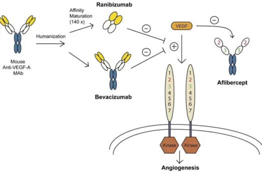
Figure 1.4: A representation of the three various types of anti-VEGF therapy and how they differ in development and mechanism of action. While ranibizumab and bevacizumab are both humanized monoclonal antibodies, ranibizumab has further affinity purification and decreased size. In contrast, aflibercept replicates two portions of the VEGF receptor (VEGFR) attached to an antibody Fc region, effectively working as a “trap receptor”. From [52].
To inhibit or reduce the effect of VEGF-A in the retina and choroid, different anti-VEGF ther-apies have been developed. Most commonly used are bevacizumab (Avastin®, Roche), ranibi-zumab (Lucentis®, Novartis), aflibercept (Eylea®, Bayer) and pegaptanib (Macugen®, Bausch+Lomb) (Figure 1.4). They inhibit the pathological neovascularization, prevent visual impairment and optimally reverse the on-set of vision loss [81,84].
Bevacizumab was developed and approved by the FDA for the systemic use in metastatic can-cers in 2004. It inhibits all VEGF-A isoforms and is used off-label intravitreally to treat neo-vascular AMD and DME. It is a humanized monoclonal IgG antibody with a molecular weight of 149 kDa. Due to its large size the retinal penetration is limited. In order to increase the effi-cacy, ranibizumab was developed. It is a recombinant humanized Fab fragment with a molecu-lar weight of only 48 kDa. Since 2006, it is FDA approved for the treatment of neovascumolecu-lar AMD, DME and macular edema secondary to retinal vein occlusion (RVO). Due to its reduced size compared to bevacizumab the ability to penetrate the retina should be increased. Also, after diffusion from the ocular tissue into the blood stream, the elimination half-live is lower, limiting systemic side effects [81,85,86].
Aflibercept is a recombinant fusion protein where the binding domains of VEGFR-1 and -2 are fused to an IgG Fc fragment. The 115 kDa big molecule has stronger affinity to bind VEGF-A isoforms than the receptors (“trap receptor”). In contrast to bevacizumab and ranibizumab it also inhibits VEGF-B and PLGF. It gained FDA approval for neovascular AMD in 2011 and for RVO in 2015. It is also approved and available as a systemic formulation to treat metastatic colorectal cancer [84,87–89].
In contrast, pegaptanib is an RNA aptamer that has a high selectivity for the isoform of VEGF-A165. With a molecular weight of approximately 50 kDa, it is the smallest of the compounds mentioned here. Compared to the other antibodies, the aptamer can be easily manufactured in a large scale and is cost-effective. It was approved for neovascular AMD in 2004 and is also used off-label in Branch RVO (occlusion of one of the branches of the retinal vein) and DME [90–92].
Due to the permeability of the anti-VEGF agents through the BRB, part of the injected drugs can reach the systemic circulation and decrease VEGF plasma levels. One of the functions of systemic VEGF is the up-regulation of NOS and protection of the vascular patency and integ-rity. When the plasma levels are reduced, the risk of hypertension and thromboembolic events increases. Due to the increase in prevalence of AMD and DR in age, many patients are elderly
and already an increased risk for hypertension and other cardiovascular diseases, which are the most common comorbidities with wet AMD [84,93–95].
Even though anti-VEGF therapy has shown a high efficacy in various studies, when injected over a long term, the efficacy can be reduced. Also, some patients seem to not respond to the treatment with no benefit to the intravitreal injection [96]. Additionally, every intravitreal in-jection bears risks such as hemorrhage, retinal detachment and endophthalmitis. Therefore, ad-ministration frequency should be limited.
1.3.3 Corticosteroids
Since the 1950s, corticosteroids are used to treat ocular inflammation such as uveitis. They can also reduce diabetic and cystoid macular edema and exudative AMD. They target different in-flammatory pathways and angiogenic cascades. Their main mechanism of action is the binding to cystolic glucocorticoid receptors. When activated, these receptors decrease the production of pro-inflammatory proteins and increase the expression of anti-inflammatory proteins such as cytokines [97–100]. They also inhibit macrophages (and thus inhibiting the release of angio-genic growth factors) and leukocytes, which are pro-inflammatory, and suppress the VEGF expression. The latter might make them suitable as an adjunctive to treat CNV and could reduce the administration frequency of anti-VEGF injections. They also have been shown reduce the paracellular permeability and enhance the integrity of tight junctions, explaining their use in macular edema [97,99,101–103].
Corticosteroids are able to penetrate the eye through the BRB and are found in the choroid, retina and sclera after systemic administration in an even higher concentration than topical ap-plication [104–106]. However, a continuously high systemic concentration may lead to many side effects such as Cushing’s syndrome, osteoporosis and diabetes [68]. These side effects can be limited with an intravitreal injection, where the corticosteroid is delivered directly to the side of action. In the eyes they can cause cataract and an elevated intraocular pressure leading to glaucoma [97,107]. Commonly used corticoids are triamcinolone acetonide, dexamethasone and fluocinolone acetonide (Figure 1.5).
Figure 1.5: Chemical structure of triamcinolone acetonide (A), dexamethasone (B) and fluocinolone acetonide (C). From [108].
Triamcinolone acetonide (TA) is highly insoluble in water. When injected intravitreally, the drug crystals can act as a depot. It has an intravitreal half-life of approximately 18 days and can be detected for up to three months in the vitreous. It is commonly used as an intravitreal injec-tion containing 4 mg TA in 0.1 mL [98,99].
Dexamethasone is one of the most prescribed corticosteroids for the treatment of ocular diseases and is five times more potent than triamcinolone acetonide. Due to its higher solubility, it has no depot formulation and only a very short intravitreal half-life of three hours. However, the lower lipophilicity could lead to a lesser accumulation in the trabecular meshwork and lens, resulting in lower risk of steroid-related side-effects, such as increased intraocular pressure and cataract. The biodegradable intravitreal implant Ozurdex® (Allergan Inc.) contains 0.7 mg dex-amethasone that is released over a period of six months [34,97,109–111].
The potency of fluocinolone acetonide (FA) is comparable with that of dexamethasone and only has 1/24 of the solubility of dexamethasone in water. It is incorporated in the non-biodegradable implant Iluvien® (Alimera Sciences) which releases FA over a period of up to three years. The most common side effect of FA is increased intraocular pressure [97,112,113].
1.4 Ocular drug delivery
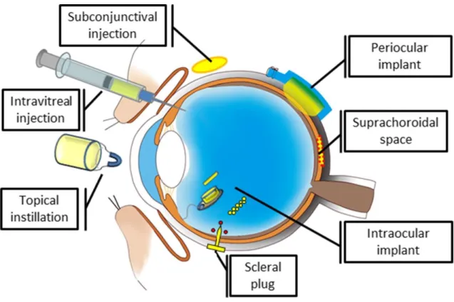
Due to its unique anatomy and physiology, ocular drug delivery remains challenging. The var-ious ocular barriers significantly impede the drug delivery to the ocular tissues. In order to reach the retina in the back of the eye, several different methods are being investigated (Figure 1.6).
Figure 1.6: Examples of drug delivery systems and devices for the posterior segment of the eye. From [114].
1.4.1 Topical
A topical treatment (e.g. eye drops and ointments) is usually indicated for diseases affecting the front of the eye, such as conjunctivitis, keratitis, dry eye, elevated intraocular pressure and an-terior uveitis. Eye drops are noninvasive and self-administrable which leads do a high patient compliance short-term. The challenge here is the small volume that can be administered (ap-proximately 30 µL) and a tear volume of around 7 µL. Due to reflexive blinking, tear dilution and tear turnover, the residence time in the eye is limited and after 15-30 s most of the admin-istered solution is washed away via the naso-lachrymal duct [3,115]. The part of the drug that gets absorbed can enter the eye via the corneal or non-corneal route. In the corneal route the molecules diffuse through the cornea into the aqueous humor and then in the intraocular tissue. Molecules that only have a poor corneal permeability (mostly hydrophilic and larger molecules) can get absorbed from the conjunctiva, passing the sclera, into the choroid or RPE of the pos-terior segment [24,27,116]. It is generally estimated that only about 5% of the applied dosage reaches the anterior segment and an even smaller amount (0.001%) passes through the aqueous humor and lens into the posterior segment [5,117–119]. To achieve therapeutic concentrations
eye drops have to be instilled frequently or in high doses [9]. Studies have shown that a frequent administration over a long period of time leads to poor patient adherence to the therapy. For example, 50% of patients suffering with glaucoma stopped their topical treatment within six months [120].
1.4.2 Periocular
In periocular administration, the drug gets injected into the surrounding area of the eye and includes sub-Tenon’s, peribulbar, retrobulbar and subconjunctival injections. Small amounts of the drug could reach the vitreous through the choroid via systemic circulation or the anterior route (cornea aqueous vitreous) [1]. However, due to the high choroidal blood flow and the limited permeability of the cornea, these routes are secondary. The main route by which the drug can reach the vitreous is transscleral. The sclera covers approximately 95% of the ocular surface and, thus, offers a large region for absorption [121]. It is permeable for molecules up to 70 kDa (compared to < 1 kDa of the cornea) which makes it a more suitable administration technique especially for hydrophilic drugs compared to a topical application [122]. Interest-ingly, the scleral permeability mainly depends on size and not the lipophilicity of the drug [35,123]. In order to reach the aqueous and vitreous humor, the drugs have to permeate different layers of ocular tissue (sclera, choroid, Bruch’s membrane, RPE). Additionally, the high cho-roidal blood flow limits the bioavailability. After a subconjunctival injection roughly 80-95% of the drug gets absorbed into the systemic circulation and only a small amount is able to pen-etrate the sclera. This leads to a short duration of action and a bioavailability of up to 10% in the aqueous and 0.1% in the vitreous [124,125]. However, the relatively high administration volume of up to 0.5 mL compensates for the low bioavailability. Studies have shown that the concentration of dexamethasone in the subretinal fluid is higher after subconjunctival injection than after peribulbar or oral administration [126]. Nevertheless, same as with a topical applica-tion, a frequent administration is necessary in order to maintain therapeutic concentrations in the ocular tissue, and every injection bears the risk of side effects such as subconjunctival hem-orrhage, hyperemia and irritation of the conjunctiva [127,128].
1.4.3 Suprachoroidal
A relatively new method is the suprachoroidal injection and was first introduced by Einmahl et al. in 2002 [129]. The drug is administered into the suprachoroidal space between the sclera
and the choroid using a microneedle and an administration volume of up to 50 µL. Due to the circumvention of the sclera and the injection site being closer to the retina, the bioavailability is with 0.2-4% higher than after subconjunctival injections, especially for lipophilic drugs and macromolecules that do not have to pass the scleral barrier. The bioavailability mainly depends on the drug elimination by choroidal blood flow and the permeability across the RPE. Supra-choroidal injections do not penetrate the inner eye or influence visual acuity as opposed to intravitreal injections. They already have been tested for administration of bevacizumab for AMD and triamcinolone acetonide with high concentrations measured in the vitreous and retina [130–132]. However, each injection poses a small risk for subconjunctival and suprachoroidal hemorrhage [128,132–134].
1.4.4 Systemic/oral
A systemic or oral administration can be used for front- and back-of-the eye diseases. It is the most noninvasive route of administration and, thus, has a high patient compliance. However, the bioavailability is very low due to the BAB and BRB. Only about 2% of the administered drug can be found in the vitreous cavity. To reach efficient drug concentrations in the eye, high doses have to be given, which increases the risk of unwanted side effects. [5,115,135]
There are two possible pathways for a drug to reach the eye. The drug can penetrate through the leaky vessels of the ciliary body and the iris into the aqueous humor, passing the BAB, or through penetration of the RPE passing the BRB. The tight junctions limit this process consid-erably. For small hydrophilic compounds the permeability through the tight junctions is in-versely correlated with the molecular weight, hindering big molecules from entering. For lipo-philic drugs it is easier to penetrate the ocular barriers even through transcellular diffusion. Due to the limited permeability through the blood-ocular barriers only small or extremely lipophilic drugs can be administered systemically to treat ophthalmic conditions, e.g. steroids or antibiot-ics are given systemically to treat inflammatory conditions [2,135,136].
1.4.5 Intravitreal
The highest concentrations in the vitreous can be achieved with intravitreal injections. They directly bypass the BRB and have the highest bioavailability, but it is also the most invasive
method. The duration of action after an intravitreal injection strongly depends on the character-istics of the drug. The anterior elimination is available for all compounds, independent of size and hydrophilicity, and includes drug diffusion from the vitreous into the aqueous humor via the trabecular or uveoscleral pathway. The posterior way is mostly used by small, lipophilic drugs and includes a passive diffusion or active transport across the BRB. Since the retina has a rather big surface, drugs that get eliminated via both pathways (e.g. corticosteroids) have a drastically lower intravitreal life than larger compounds (e.g. anti-VEGF) [3,24]. The half-life of low-molecular weight drugs can be as small as 2 h to 6 h. In order to maintain a thera-peutically effective concentration in the vitreous, frequent injections would be necessary which decreases patient compliance and increases the probability of serious side-effects. Those serious side effects have a low incidence rate but can be potentially sight-threatening and include retinal detachment, endophthalmitis, vitreous hemorrhage and cataract formation [1,2,5,9,115]. To re-duce the incidence of these side effects, the drug release in the vitreous should be prolonged.
Table 1.2. Summary of the different administration techniques. Adapted from [13,128]. Administration
technique
Advantages Disadvantages BA in the
vitreous
Side effects Topical Easy
self-administra-tion, non-invasive
Frequent applica-tion necessary, poor compliance
0.001% Conjunctival redness, irritation
Periocular High administration volume
Frequent applica-tion necessary
0.1% Hemorrhage, hyperemia, irrita-tion of conjunctiva Suprachoroidal Minimal injection
risk
Drug elimination via choroidal blood
flow
0.2-4% Subconjunctival and supracho-roidal hemorrhage
Systemic/oral High compliance, non-invasive
Systemic toxicity 2% Systemic side effects depending on the mechanism of action of
the administered drug Intravitreal Targeted delivery,
most direct
Invasive, poor compliance
100% Retinal detachment, hemorrhage, cataract, endophthalmitis Abbreviation: BA, bioavailability
1.5 Ocular implants for sustained drug release
Due to the limited ocular bioavailability of the different drug administrations and the increased side effects connected with multiple intraocular injections, implants for a prolonged drug re-lease are an interesting possibility. Therapeutic agents are encapsulated in a biocompatible ma-trix that is then implanted in the eye. Depending on the system, the implants can release constant drug amounts over a period of days up to multiple years. The implants can be divided into non-biodegradable and non-biodegradable [115,137,138].
1.5.1 Non-biodegradable implants
Non-biodegradable implants are often made from polyvinyl alcohol (PVA) and ethylene vinyl acetate (EVA). The PVA is permeable for water which after implantation diffuses into the im-plant where it partially dissolves the drug contained inside, whereas EVA is impermeable and limits water penetration and drug diffusion. The dissolved drug can then diffuse into the rounding tissue. Often, these implants are inserted via invasive surgery and usually require sur-gical removal upon completion of the drug release [9,97,115].
Retisert® (Bausch + Lomb) contains 0.59 mg fluocinolone acetonide in a PVA/EVA-matrix. It is disc-shaped with a size of 3x2 mm (see Figure. 7). To insert the implant, a 3-4 mm long incision through the pars plana is made during a surgical procedure. Initially it releases 0.6 µg/day and 0.3-0.4 µg/day in a steady state for about 30 months. It is FDA approved for chronic noninfectious uveitis affecting the posterior segment. It has been shown to reduce the recurrence rate of sever posterior uveitis, improve the visual acuity and decrease the need for adjunctive immunosuppressive therapy [9,97].
Figure 1.7: The size of Iluvien® (left) and Retisert® (right) in comparison. Adapted from [139].
Iluvien® (Alimera sciences) is a rod-shaped device with a size of 3.5x0.37 mm containing 0.19 mg fluocinolone acetonide (Figure 1.7). A polyimide tube with fluocinolone acetonide is embedded in a PVA matrix and injected with a 25-gauge inserter in a surgical procedure. It releases a low dose of fluocinolone acetonide over 13-36 months and was approved by the FDA in 2014 for diabetic macular edema [97,140].
Figure 1.8: I-vation implant. A: structure, B: size, C: implantation. From [141].
A different approach is I-vation containing 0.925 mg triamcinolone acetonide. Its titanium, hel-ical coil measures 0.5-0.21 mm and gets anchored in the sclera (Figure 1.8). Due to its shape, the surface area is maximized. It has no FDA approval yet. It was tested in a phase I clinical trial for the treatment of diabetic macular edema. A phase II trial was terminated leaving the current status of the implant unclear [97,142,143].
1.5.2 Biodegradable implants
Biodegradable implants consist of a biocompatible polymer that produces nontoxic degradation products. They can be eliminated safely and do not cause chronic-foreign body reactions. They often contain synthetic aliphatic polyesters of the poly-α-hydroxy family, such as PGA, PLA and PLGA [97,115].
Surodex® (Oculex Pharmaceuticals, Inc.) is a rod-shaped device with a size of 1.0x0.5 mm. It contains 0.060 mg dexamethasone in a PLGA matrix with HPMC. It gets inserted into the an-terior chamber after cataract surgery and controls the postoperative inflammation. In a random-ized controlled study for the control of postcataract surgery inflammation, Surodex® was found
to be more effective as 0.1% dexamethasone eyedrops. However, due to its limited release over 7-10 days, the application of the implant is limited. It is only approved in China and Singapore [135,144–146].
Figure 1.9: Ozurdex® intravitreal injection device. From [64].
Ozurdex® (Allergan Inc.) contains 0.7 mg dexamethasone within a PLGA copolymer (Nova-dur™, Allergan Inc.). The implant is rod shaped and releases the drug over a period of up to six months. The release is biphasic: the initial release within the first two months is higher than the release in the following (up to six) months. It gets injected with a specially designed applicator through the pars plana in an office-based procedure without the need for sutures (Figure 1.9). Upon injection in the eye, water diffuses into the implant and dexamethasone gets released. Over time the PLGA degrades into lactic acid and glycolic acid which are metabolized in the Krebs cycle to water and carbon dioxide. It got FDA approval for macular edema following central and branch retinal vein occlusion in 2009, followed by approval for the treatment of noninfectious uveitis involving the posterior segment in 2010 and for the treatment of diabetic macular edema in 2014 [64,97,147].
1.6 Poly(D, L-lactic-co-glycolic)acid 1.6.1 Physico-chemical properties
Poly(lactic-co-glycolic)acid (PLGA) is a biodegradable and biocompatible polymer that is syn0thesized via ring opening polymerization of the cyclic diesters lactide and glycolide. The dimers are linked together by ester linkages (Figure 1.10). PLGA is one of the most common biodegradable polymers and was first used in the 1960s as bioresorbable sutures. It is also pos-sible to fabricate various forms such as membranes, rods and disc using molding, extrusion and
other techniques. They are commercially available in many FDA-approved products for paren-teral controlled release and other administration techniques. PLGA is soluble in a wide range of solvents such as tetrahydrofuran, acetone, chlorinated solvents and ethyl acetate, which makes it suitable for a wide range of preparation techniques [115,148–151].
Figure 1.10: Principle of PLGA synthesis through ring-opening copolymerization of lactide and glycolide. Adapted from [152].
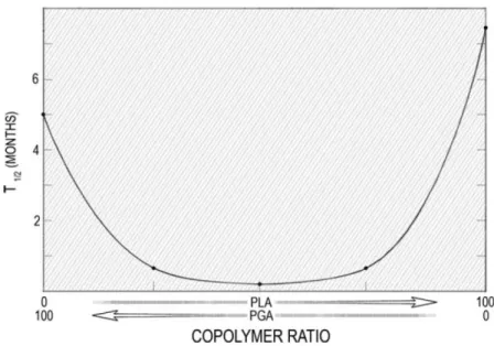
Due to the additional methyl group of lactic acid, the molecule is chiral. It exists in two enanti-omers: D- and L-lactic acid. Normally, a racemic mixture is used for the polymerization, lead-ing to equal amounts of D- and lactic acid in the polymer chain. The polymer structure of L-polylactic-acid (L-PLA) is highly regular and, thus, it is semi crystalline, whereas D,L-PLA is amorphous due to various irregularities of the polymer chain. Poly-glycolic acid (PGA) is highly crystalline. PLGAs containing less than 70% glycolide are amorphous [153,154]. Also, the additional methyl group of the lactic acid makes the molecule more hydrophobic compared to glycolic acid. Thus, by varying the composition of the PLGA (different ratios of lactide : glycolide used during polymerization, e.g. 50:50, 75:25, 85:15,…) the hydrophobicity of the polymer can be adjusted. More hydrophobic polymers take up less water and degrade slower. Figure 1.11 shows the relation between the lactide/glycolide ratio and its resulting degradation time. The fastest degrading PLGA contains a ratio of 50:50. The molecular weight of the PLGA can also influence the mechanical strength of the polymer and might influence the biodegrada-tion rate and hydrolysis [154–157].
m + n
Lactide Glycolide Poly(lactic-co-glycolic) acid
![Figure 1.2: Physiological barriers in ocular drug delivery. From [19].](https://thumb-eu.123doks.com/thumbv2/123doknet/14557411.726192/24.892.114.781.103.662/figure-physiological-barriers-ocular-drug-delivery.webp)
![Figure 1.3: Schematic diagram of aqueous humor flow pathway. From [32].](https://thumb-eu.123doks.com/thumbv2/123doknet/14557411.726192/26.892.110.787.110.356/figure-schematic-diagram-aqueous-humor-flow-pathway.webp)
![Figure 1.8: I-vation implant. A: structure, B: size, C: implantation. From [141].](https://thumb-eu.123doks.com/thumbv2/123doknet/14557411.726192/40.892.212.674.278.582/figure-vation-implant-a-structure-size-implantation-from.webp)
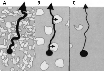
![Figure 1.12: Drug-release mechanisms and biodegradation of matrix implant. Adapted from [115]](https://thumb-eu.123doks.com/thumbv2/123doknet/14557411.726192/44.892.190.757.283.1075/figure-drug-release-mechanisms-biodegradation-matrix-implant-adapted.webp)