RESEARCH OUTPUTS / RÉSULTATS DE RECHERCHE
Author(s) - Auteur(s) :
Publication date - Date de publication :
Permanent link - Permalien :
Rights / License - Licence de droit d’auteur :
Bibliothèque Universitaire Moretus Plantin
Institutional Repository - Research Portal
Dépôt Institutionnel - Portail de la Recherche
researchportal.unamur.be
University of Namur
The impact of instilled carbide nanoparticles on rat lungs: an in vivo perspective on
acute intratracheal instillation.
Lozano Garcia, Omar; Lison, Dominique; Escamilla-Rivera, Vicente; Mejia Mendoza, Jorge
Humberto; Toussaint, Olivier; Dogne, Jean-Michel; Lucas, Stéphane
Published in:
Journal of Physics : Conference Series
DOI:
DOI:10.1088/1742-6596/617/1/012017 Publication date:
2015
Document Version
Publisher's PDF, also known as Version of record
Link to publication
Citation for pulished version (HARVARD):
Lozano Garcia, O, Lison, D, Escamilla-Rivera, V, Mejia Mendoza, JH, Toussaint, O, Dogne, J-M & Lucas, S 2015, 'The impact of instilled carbide nanoparticles on rat lungs: an in vivo perspective on acute intratracheal instillation.', Journal of Physics : Conference Series, vol. 617, no. 1, 012017. https://doi.org/DOI:10.1088/1742-6596/617/1/012017
General rights
Copyright and moral rights for the publications made accessible in the public portal are retained by the authors and/or other copyright owners and it is a condition of accessing publications that users recognise and abide by the legal requirements associated with these rights. • Users may download and print one copy of any publication from the public portal for the purpose of private study or research. • You may not further distribute the material or use it for any profit-making activity or commercial gain
• You may freely distribute the URL identifying the publication in the public portal ? Take down policy
If you believe that this document breaches copyright please contact us providing details, and we will remove access to the work immediately and investigate your claim.
This content has been downloaded from IOPscience. Please scroll down to see the full text.
Download details:
IP Address: 138.48.76.65
This content was downloaded on 02/09/2015 at 15:46
Please note that terms and conditions apply.
The impact of instilled carbide nanoparticles on rat lungs: an in vivo perspective on acute
intratracheal instillation
View the table of contents for this issue, or go to the journal homepage for more 2015 J. Phys.: Conf. Ser. 617 012017
(http://iopscience.iop.org/1742-6596/617/1/012017)
The impact of instilled carbide nanoparticles on rat lungs: an
in vivo perspective on acute intratracheal instillation
O Lozano1,†, D Lison2, V. Escamilla-Rivera3, J Mejía1, O Toussaint4, JM Dogné5, and S Lucas1
1
Research Centre for the Physics of Matter and Radiation (PMR), Namur Nanosafety Center (NNC), NAmur Research Institute for LIfe Sciences (NARILIS), University of Namur (UNamur), Namur, Belgium
2
Louvain centre for Toxicology and Applied Pharmacology, Université Catholique de Louvain, Brussels, Belgium.
3
Department of Toxicology, Center of Research and Advanced Studies of IPN (CINVESTAV-IPN), Mexico City, D.F., Mexico. Department
4
Laboratory of Biochemistry and Cellular Biology (URBC), Namur Nanosafety Center (NNC), NAmur Research Institute for LIfe Sciences (NARILIS), University of Namur (UNamur), Namur, Belgium
5
Department of Pharmacy, Namur Nanosafety Center (NNC), NAmur Research Institute for LIfe Sciences (NARILIS), University of Namur (UNamur), Namur, Belgium
†
Corresponding author’s e-mail: [email protected]
Abstract. In order to study a scenario of acute high concentration exposure via the pulmonary
pathway of silicon carbide and titanium carbide nanoparticles, female Wistar rats were administered by intratracheal instillation doses of 0.5 and 5 mg/rat of each nanomaterial. Inflammatory parameters were studied: protein concentration, lactate dehydrogenase activity, total cell count and differentiated cell count (macrophages, neutrophils, oesonophils, lymphocytes). The genotoxicity potential was assessed by the formation of micronuclei from pneumocytes type II. It was found that silicon carbide nanoparticles induce an inflammatory response and a dose dependent genotoxicity, although the genotoxicity levels are comparably lower to the inflammatory response.
1. Introduction
The principal route of exposure to nanomaterials (NMs) is the pulmonary pathway [1]. Carbide nanomaterials, like Silicon Carbide (SiC) and Titanium Carbide (TiC) are widely used in several industries requiring high performance products [2, 3], yet they have not been widely studied in comparison of other material families such as their oxide versions, silica and titania. Given the exposure hazard of such materials at the nanoscale could be due to wear, breakdown or decommissioning, our research group has dedicated several efforts to map the impact of exposure on rats to such NMs in terms: toxicity[4] and biopersistence [5] by whole body exposure (WBE), biopersistence from acute intratracheal instillation [6], oral administration in acute and subacute exposures [7, 8], and in vitro [9]. An unexplored subject has been the toxicological impact of SiC and TiC nanoparticles (NPs) via intratracheal instillation at doses simulating an acute high concentration
4th International Conference on Safe Production and Use of Nanomaterials (Nanosafe2014) IOP Publishing Journal of Physics: Conference Series 617 (2015) 012017 doi:10.1088/1742-6596/617/1/012017
Content from this work may be used under the terms of theCreative Commons Attribution 3.0 licence. Any further distribution of this work must maintain attribution to the author(s) and the title of the work, journal citation and DOI.
exposure scenario. This study presents addresses such question using rats, focusing on the inflammatory parameters from broncheoalveolar lavages (BAL) and the genotoxicity potential of SiC and TiC NPs.
2. Materials and methods
2.1. Nanoparticles
SiC and TiC NPs were provided by Sirris (www.sirris.be). They were heated at 200 °C during 2h to eliminate possible endotoxin traces. Their hydrodynamic diamters are 31 and 25 nm for SiC and TiC NPs, respectively. Their full physicochemical characterization has been reported elsewhere [6].
2.2. Animals
Wistar female rats, 200-220 g, were used in this study.
2.3. Exposure protocol
The NPs were prepared in suspensions with a physiologically sterile solution (0.9% NaCl) with 1 % Tween 20 and sonicated. Suspensions were prepared in two doses: 0.5 (low dose) and 5 (high dose) mg/rat. The exposure was acute, delivering the suspensions, 300 µL/rat, to the rat lung via intratracheal instillation. During this procedure rats were anesthetized, exposing surgically the trachea. After the exposure procedure the incision was closed with a suture point.
2.4. Genotoxicity studies
The genotoxicity potential of SiC and TiC NPs was studied in vivo in the rats pneumocytes type II. The inflammatory response was assessed 3 days after instillation by sacrificing the rats and measuring different parameters in a broncheoalveolar lavage (BAL) with a saline solution: total protein concentration, enzymatic activity of lactate dehydrogenase (LDH), and the total and differential number of cells. Pneumocytes type II were recovered after several BAL to eliminate inflammatory cells and a lung perfusion to eliminate the erythrocytes. Lung enzymatic digestion was carried out by an elastase solution flowing through the lung trachea for 30 minutes at 37 °C. Then the lung was cut in small sections and the cell suspension was filtered and then incubated in Petri boxes covered with rat IgG for 1 hour at 37 °C. The cells possessing the Fc receptor (macrophages) will adhere while the pneumocytes type II will rest in suspension and were recovered for culture. After two days, the culture chambers are washed and fixed with methanol. The plates are colored with orange acridine and counted with a fluorescent microscope.
The negative control was NaCl and the positive control was WC-Co (94:6 mass ratio, 5 mg) which generates micronuclei [10].
BAL was centrifuged, the supernatant was used to quantify the total proteins and LDH activity, and the cellular sediment was resuspended in NaCl for the cellular count. Protein concentration was measured by spectrometry after complexation with pyrogallol red-molybdate. LDH activity was measured by spectrometry after reduction of the NAD+ catalysed by the enzyme. Total cell counting was done with a Neubauer plate and differential cells were counted from colored cytospins with Diff-Quick [11].
3. Results and discussion
3.1. BAL analysis
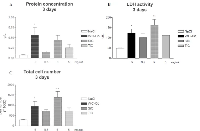
The protein concentration reflects the permeability of the broncheo-alveolar barrier. It is observed that for the high dose of SiC there is an elevated, even if not significant, concentration of proteins when compared to those of TiC. LDH activity, a marker of cytotoxicity, is found the highest for the high dose of SiC, noting that it is statistically significant with a value higher than even the positive control. The LDH activity of the low dose of SiC and the high dose of TiC is close to the positive control. The
4th International Conference on Safe Production and Use of Nanomaterials (Nanosafe2014) IOP Publishing Journal of Physics: Conference Series 617 (2015) 012017 doi:10.1088/1742-6596/617/1/012017
total cell number follow the same trend as the LDC activity: high dose of SiC has the highest statistically significant number of cells, higher than the positive
TiC high dose having similar levels close to the positive control.
Figure 1. Inflammatory parameters in BAL
C) Cell total number. Nar represent the mean
negative control (NaCl) and exposed sample, evaluated by Student p<0.5 and ** for p<0.01.
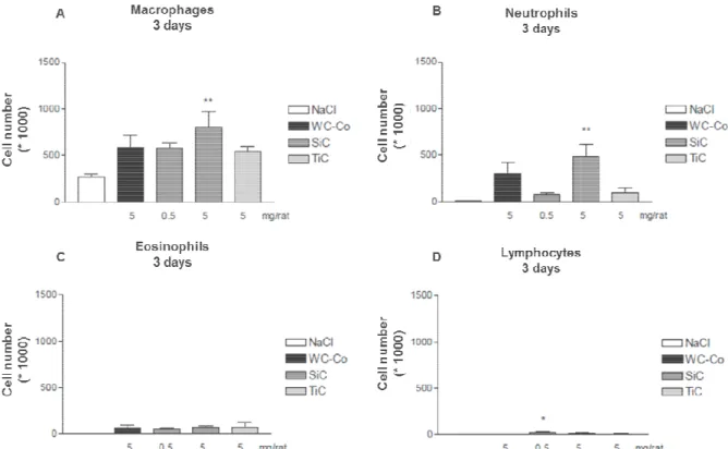
The cell count for macrophages and neutrophils has a significant increase for the high dose of SiC NPs, and lymphocytes had an increase for the low dose of SiC NPs. Eosinophils had similar cell counts for all doses, although showed no significant difference versus the negative control. Exposure to TiC NP showed higher levels (not significant) of macrophages and neutrophils with respect to the negative control.
3.2. Pneumocyte type II analysis
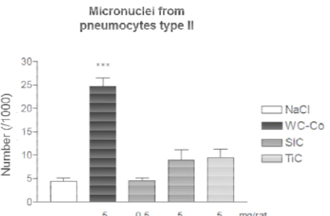
The number of micronuclei formed from pneumocytes type II for SiC and TiC NPs exposure were not significantly different from those formed on the negative control
genotoxicity potential for the high dose of SiC
cytotoxicity and a higher presence of macrophages and neutrophils
neutrophils response. It is also contrasting that a similar level of micronuclei is generated for both nanoparticle doses even if the biopersistence of TiC NPs in the lung is 2 times lower than SiC NPs This level of micronuclei production is quite lower than those produced by WC
clear that the production of micronuclei is dose dependent as evidenced by the low and high dose of SiC NPs.
As previously reported, pulmonary toxicity of WC reactive oxygen species and transient acute infla
particles no cellular infiltration was observed in BAL, however micronuclei formation in type II follow the same trend as the LDC activity: high dose of SiC has the highest
number of cells, higher than the positive control; with both SiC low dose and TiC high dose having similar levels close to the positive control.
parameters in BAL after exposure. A) Total protein count. B) LDH activity. C) Cell total number. Nar represent the mean ± SEM (n = 4-6). Statistical significance, between negative control (NaCl) and exposed sample, evaluated by Student-Newman-Keuls test wi
The cell count for macrophages and neutrophils has a significant increase for the high dose of SiC NPs, and lymphocytes had an increase for the low dose of SiC NPs. Eosinophils had similar cell ough showed no significant difference versus the negative control. Exposure to TiC NP showed higher levels (not significant) of macrophages and neutrophils with respect to the
Pneumocyte type II analysis
The number of micronuclei formed from pneumocytes type II for SiC and TiC NPs exposure were not significantly different from those formed on the negative control. In fact there seems to be a similar genotoxicity potential for the high dose of SiC and TiC NP, even if the high dose of SiC generated cytotoxicity and a higher presence of macrophages and neutrophils, while TiC did not generate a
It is also contrasting that a similar level of micronuclei is generated for both oses even if the biopersistence of TiC NPs in the lung is 2 times lower than SiC NPs This level of micronuclei production is quite lower than those produced by
WC-the production of micronuclei is dose dependent as evidenced by WC-the low and high dose of As previously reported, pulmonary toxicity of WC-Co is related to the capacity of induce both, reactive oxygen species and transient acute inflammation [12]. Here, after 72 h exposure to WC
no cellular infiltration was observed in BAL, however micronuclei formation in type II follow the same trend as the LDC activity: high dose of SiC has the highest control; with both SiC low dose and
. A) Total protein count. B) LDH activity. 6). Statistical significance, between Keuls test with * for
The cell count for macrophages and neutrophils has a significant increase for the high dose of SiC NPs, and lymphocytes had an increase for the low dose of SiC NPs. Eosinophils had similar cell ough showed no significant difference versus the negative control. Exposure to TiC NP showed higher levels (not significant) of macrophages and neutrophils with respect to the
The number of micronuclei formed from pneumocytes type II for SiC and TiC NPs exposure were not In fact there seems to be a similar P, even if the high dose of SiC generated , while TiC did not generate a It is also contrasting that a similar level of micronuclei is generated for both oses even if the biopersistence of TiC NPs in the lung is 2 times lower than SiC NPs [6]. -Co. In addition, it is the production of micronuclei is dose dependent as evidenced by the low and high dose of Co is related to the capacity of induce both, . Here, after 72 h exposure to WC-Co no cellular infiltration was observed in BAL, however micronuclei formation in type II
4th International Conference on Safe Production and Use of Nanomaterials (Nanosafe2014) IOP Publishing Journal of Physics: Conference Series 617 (2015) 012017 doi:10.1088/1742-6596/617/1/012017
epithelial cells was present. Meanwhile, SiC induced a pronounced inflammatory response in the lung and no signs of genotoxicity were observed. From this differences, we
substantial lung toxicity through the damage of resident cells in the alveoli, as demonstrated by the significant increase of LDH concentration in BAL. When cellular integrity is compromised, several molecules are liberated from cytosol, among them the damage
molecules (such as Ca++, ATP, RNA, DNA, etc.) could trigger inflammation and promote recruitment of innate inflammatory cells such as macrophages and neutrophils towards the alveoli, even absence of pathogens [13].
Figure 2. Number of (A) macrophages, (B) neutrophils, (C) eosinophils, and (D) lymphocytes
exposure. Statistical significance,
Student-Newman-Keuls test with * for p<0.5 and ** for p<0.01.
Mechanistically, production of reactive oxygen species (ROS) is considered as the major player in NP induced toxicity [14]. It is possibly that SiC, but not TiC, induce free radicals that arise from redox reactions between their highly oxidized surface
aforementioned process is more relevant because their increased surface/volume ratio make it more capable to produce in ROS in higher amounts compared to TiC and WC
relatively similar hydrodynamic diameter of both SiC and TiC NPs, mediated effect.
Depending on the severity, ROS could elicit different
in proliferation and antioxidant defense is promoted, in mild levels DNA damage and release of inflammation mediators, whereas at high concentration, ROS provoke excessiv
damaged leading to cell death [
damage to type II cells is due to an overwhelming ROS production and subsequent cell death, since micronuclei formation could only
DNA damage allowed them progress to cell division.
epithelial cells was present. Meanwhile, SiC induced a pronounced inflammatory response in the lung and no signs of genotoxicity were observed. From this differences, we propose that SiC produce substantial lung toxicity through the damage of resident cells in the alveoli, as demonstrated by the significant increase of LDH concentration in BAL. When cellular integrity is compromised, several
cytosol, among them the damage-associated molecular
, ATP, RNA, DNA, etc.) could trigger inflammation and promote recruitment of innate inflammatory cells such as macrophages and neutrophils towards the alveoli, even
. Number of (A) macrophages, (B) neutrophils, (C) eosinophils, and (D) lymphocytes . Statistical significance, between negative control (NaCl) and exposed sample, evaluated by
Keuls test with * for p<0.5 and ** for p<0.01.
Mechanistically, production of reactive oxygen species (ROS) is considered as the major player in . It is possibly that SiC, but not TiC, induce free radicals that arise from redox heir highly oxidized surface [6] and the biological milieu. For SiC, the aforementioned process is more relevant because their increased surface/volume ratio make it more capable to produce in ROS in higher amounts compared to TiC and WC-Co.
relatively similar hydrodynamic diameter of both SiC and TiC NPs, this is a
everity, ROS could elicit different cellular responses: at low levels, an increase in proliferation and antioxidant defense is promoted, in mild levels DNA damage and release of inflammation mediators, whereas at high concentration, ROS provoke excessive organelle and DNA [15]. Therefore, one possibility by which SiC did not shown DNA damage to type II cells is due to an overwhelming ROS production and subsequent cell death, since micronuclei formation could only be detected in cells without compromised viability, in which mild DNA damage allowed them progress to cell division.
epithelial cells was present. Meanwhile, SiC induced a pronounced inflammatory response in the lung propose that SiC produces a substantial lung toxicity through the damage of resident cells in the alveoli, as demonstrated by the significant increase of LDH concentration in BAL. When cellular integrity is compromised, several associated molecular-pattern (DAMP) , ATP, RNA, DNA, etc.) could trigger inflammation and promote recruitment of innate inflammatory cells such as macrophages and neutrophils towards the alveoli, even in the
. Number of (A) macrophages, (B) neutrophils, (C) eosinophils, and (D) lymphocytes after between negative control (NaCl) and exposed sample, evaluated by
Mechanistically, production of reactive oxygen species (ROS) is considered as the major player in . It is possibly that SiC, but not TiC, induce free radicals that arise from redox and the biological milieu. For SiC, the aforementioned process is more relevant because their increased surface/volume ratio make it more Therefore, given the NP surface-chemical cellular responses: at low levels, an increase in proliferation and antioxidant defense is promoted, in mild levels DNA damage and release of e organelle and DNA , one possibility by which SiC did not shown DNA damage to type II cells is due to an overwhelming ROS production and subsequent cell death, since cells without compromised viability, in which mild
4th International Conference on Safe Production and Use of Nanomaterials (Nanosafe2014) IOP Publishing Journal of Physics: Conference Series 617 (2015) 012017 doi:10.1088/1742-6596/617/1/012017
The impact of SiC NPs was studie
evaluating the impact of a SiC nanoaerosol when the whole respiratory pathway is taken into account. The toxicological analysis was found very
response was evidenced by a elevated presence of macrophages and neutrophils 24 increment in the LDH activity.
Figure 3. Number of micronuclei from pneumocytes type II
after exposure. Statistical significance, between negative control (NaCl) and exposed sample, evaluated by Student Newman-Keuls test with *** for p<0.01.
4. Summary
There is a dose dependent production of micronuclei with both SiC and TiC NPs exposed by intratracheal instillation. This genotoxicity, however seems not rela
response and more specially the relation with the neutrophils presence. The high dose, 5 mg, of SiC NPs produced the most important inflammatory response. TiC NPs, while not induced a inflammatory response, produced a similar level of micronuclei like SiC NPs.
Acknowledgements. This work was supported by the Service Public de Wallonie (SPW)
générale opérationnelle – Economie, Emploi et Recherche (DGO6), Départément des Programmes de Recherche (Nanotoxico Project, SPW/FUNDP research convention No. 516252).
Georgina B. Vega for her insightful dialogues about the inflammatory response.
References
1. Oberdörster, G., V. Stone, and K. Donaldson,
perspective. Nanotoxicology, 2007.
2. SCP. Silicon Carbide Products
http://www.scprobond.com/
3. Ferro-TiC. Titanium Carbide Applications from: http://www.ferro-tic.com/
4. Laloy, J., et al., Acute inflammatory response in rats after exposure to silicon carbide
nanoaerosol in a whole-body exposure model.
5. Lozano, O., et al., What is the global and local biopersistence dyna
nanoparticles in rat lungs after chronic inhalation exposure? A general biopersistence descriptive model. In preparation, 2015.
The impact of SiC NPs was studied recently in a Whole Body Exposure (WBE) model ting the impact of a SiC nanoaerosol when the whole respiratory pathway is taken into account. The toxicological analysis was found very similar to this study, where a lim
a elevated presence of macrophages and neutrophils 24
. Number of micronuclei from pneumocytes type II after exposure. Statistical significance, between negative (NaCl) and exposed sample, evaluated by Student
Keuls test with *** for p<0.01.
There is a dose dependent production of micronuclei with both SiC and TiC NPs exposed by This genotoxicity, however seems not related to the prominent inflammatory response and more specially the relation with the neutrophils presence. The high dose, 5 mg, of SiC NPs produced the most important inflammatory response. TiC NPs, while not induced a inflammatory
lar level of micronuclei like SiC NPs.
This work was supported by the Service Public de Wallonie (SPW)
Economie, Emploi et Recherche (DGO6), Départément des Programmes de t, SPW/FUNDP research convention No. 516252).
her insightful dialogues about the inflammatory response.
Oberdörster, G., V. Stone, and K. Donaldson, Toxicology of nanoparticles: A historical Nanotoxicology, 2007. 1(1): p. 2-25.
Silicon Carbide Products. [cited 2014 January 8, 2014]; Available from:
http://www.scprobond.com/.
Titanium Carbide Applications. 2013 [cited 2013 December 12, 2013]; Available
tic.com/.
Acute inflammatory response in rats after exposure to silicon carbide body exposure model. J Nanopar Res. Submitted, 2014.
What is the global and local biopersistence dynamics of silicon carbide nanoparticles in rat lungs after chronic inhalation exposure? A general biopersistence
In preparation, 2015.
d recently in a Whole Body Exposure (WBE) model [4], ting the impact of a SiC nanoaerosol when the whole respiratory pathway is taken into account. a limited inflammatory a elevated presence of macrophages and neutrophils 24 h after an
. Number of micronuclei from pneumocytes type II after exposure. Statistical significance, between negative (NaCl) and exposed sample, evaluated by
Student-There is a dose dependent production of micronuclei with both SiC and TiC NPs exposed by ted to the prominent inflammatory response and more specially the relation with the neutrophils presence. The high dose, 5 mg, of SiC NPs produced the most important inflammatory response. TiC NPs, while not induced a inflammatory
This work was supported by the Service Public de Wallonie (SPW) – Direction Economie, Emploi et Recherche (DGO6), Départément des Programmes de t, SPW/FUNDP research convention No. 516252). Thanks to Dr.
Toxicology of nanoparticles: A historical
. [cited 2014 January 8, 2014]; Available from:
. 2013 [cited 2013 December 12, 2013]; Available
Acute inflammatory response in rats after exposure to silicon carbide
J Nanopar Res. Submitted, 2014.
mics of silicon carbide nanoparticles in rat lungs after chronic inhalation exposure? A general biopersistence
4th International Conference on Safe Production and Use of Nanomaterials (Nanosafe2014) IOP Publishing Journal of Physics: Conference Series 617 (2015) 012017 doi:10.1088/1742-6596/617/1/012017
6. Lozano, O., et al., Development of a PIXE analysis method for the determination of the
biopersistence of SiC and TiC nanoparticles in rat lungs. Nanotoxicology, 2012. 6 (3): p.
263-271 (doi:10.3109/17435390.2011.572301).
7. Lozano, O., et al., Effects of SiC nanoparticles orally administered in a rat model:
biodistribution, toxicity and elemental composition changes in feces and organs. Toxicol Appl
Pharmacol, 2012. 264(2): p. 232-245. doi: 10.1016/j.taap.2012.08.004
8. Laloy, J., et al., Can TiC nanoparticles produce toxicity in oral administrationto rats? Toxicology Reports, 2014. 1: p. 172-187.
9. Mejia, J., et al., Are stirring and sonication pre-dispersion methods equivalent for in vitro
toxicology evaluation of SiC and TiC? J Nanopart Res, 2012. 14: p. 815-832.
10. M, D.B., et al., In vivo genotoxicity of hard metal dust: induction of micronuclei in rat type II
epithelial lung cells. Carcinogenesis, 2003. 24(11): p. 1793-1800.
11. J, M., et al., Respiratory toxicity of multi-wall carbon nanotubes. Toxicol Appl Pharmacol, 2005. 207(3): p. 221-31.
12. De Boeck, M., et al., In vivo genotoxicity of hard metal dust: induction of micronuclei in rat
type II epithelial lung cells. Carcinogenesis, 2003. 24(11): p. 1793-1800.
13. Wynn, T.A., Integrating mechanisms of pulmonary fibrosis. The Journal of Experimental Medicine, 2011. 208(7): p. 1339-1350.
14. Nel, A., et al., Toxic potential of materials at the nanolevel. Science (New York, N.Y.), 2006.
311(5761): p. 622-7.
15. Xia, T., et al., Comparison of the Abilities of Ambient and Manufactured Nanoparticles To
Induce Cellular Toxicity According to an Oxidative Stress Paradigm. Nano Lett, 2006. 6: p.
1794 - 1807.
4th International Conference on Safe Production and Use of Nanomaterials (Nanosafe2014) IOP Publishing Journal of Physics: Conference Series 617 (2015) 012017 doi:10.1088/1742-6596/617/1/012017