Received: 8 January 2003 Revised: 7 April 2003 Accepted: 2 May 2003
Published online: 27 August 2003 © Springer-Verlag 2003
Abstract The aim of this study was to test the efficacy of four different radio-frequency ablation (RFA) sys-tems in normal hepatic parenchyma in large animals. The RFA was ap-plied to pig livers in vivo and to calf livers ex vivo using the Radionics cluster needle, RITA starburst XL needle, Radiotherapeutics Le Veen 4.0 needle, and the Berchtold 14-G saline-perfused 15-mm active-tip needle based on constructor specifi-cations. The volume of tissue coagu-lation from RF was calculated from measurements of the vertical diame-ter (Dv) and transverse diamediame-ter (Dt). Lesion shape was characterized using the ratio between Dt/Dv. Ra-diotherapeutics and RITA produced in vivo lesion volume of 42±10, 39±4 cm3with a reproducible spheri-cal shape (Dt/Dv of 1.01±0.16 and 0.97±0.1, respectively). Radionics produced in vivo RF lesions volume of 29±11 cm3with an ovoid shape (Dt/Dv 0.88±0.09). The RF lesions with the Berchtold device could not
be assessed in vivo as 5 of 8 animals died during treatment. Ex vivo RF lesions had similar volumes with each system; however, the Radio-therapeutics device produced more reproducible shaped lesions than the other systems. In our experimental study, we found no difference be-tween expandable needle systems in vivo. Cooled needles produced slightly smaller and ovoid shape in vivo lesions.
Keywords Experimental study · Radio-frequency ablation · Liver · Electrode Alban L. Denys Thierry De Baere Viseth Kuoch Benoit Dupas Patrick Chevallier David C. Madoff Pierre Schnyder Francesco Doenz
Radio-frequency tissue ablation of the liver:
in vivo and ex vivo experiments with four
different systems
Introduction
Image-guided tumor ablation has become increasingly popular in recent years. While various percutaneous ablative techniques currently exist for the treatment of selected patients with hepatic malignancy, RFA is cur-rently the most widely used technique [1]. To perform RFA, a needle electrode is inserted into the tumor under imaging guidance. This probe emits alternative current with frequencies ranging from 450 to 500 KHz that
in-duce ionic agitation, thus producing heat. At tempera-tures above 60°C, cells die instantly from protein coagu-lation and dehydration.
The first RF needle electrodes available produced small volumes of necrosis with destruction in the liver limited to the maximal diameter perpendicular to the needle axis less than 1.6 cm after a single focus of RF delivery [2]. For this reason, complete tumor destruction usually necessitated multiple, overlapping RF deliveries, limiting the use of this technique. To avoid this problem, A. L. Denys (
✉
) · P. ChevallierP. Schnyder · F. Doenz Department of Radiology and Interventional Radiology,
Centre Hospitalier Universitaire Vaudois, 1011 Lausanne, Switzerland
e-mail: [email protected] Tel.: +41-21-3144470
Fax: +41-21-3144443 T. D. Baere · V. Kuoch
Department of Interventional Radiology, Institut Gustave Roussy,
39 Rue Camille Desmoulins, 94805 Villejuif, France B. Dupas
Department of Radiology, CHU Hotel Dieu, Place Alexis Ricordeau, 44093 Nantes Cedex 1, France D. C. Madoff
Department of Interventional Radiology, University of Texas,
M.D. Anderson Cancer Center, Houston, TX, 77030, USA
with regard to their own RF device. The systems were operated during the experiment by experts experienced in running the ma-chines who were chosen by each company.
For this study, a Berchtold Elektrotom 60 W 106 HITT device was used in association with a 14-G single needle electrode. The needle has an exposed tip 15 mm long in which there are three groups of two side holes each, placed at 120° angle from each oth-er. The needle was connected to an infusing pump that was set to deliver 100 ml/h of normal saline at room temperature through the needle side holes. Care was taken to begin the infusion immediate-ly prior to the liver puncture in order to avoid obstruction of the needle electrode side holes during placement. According to com-pany recommendations, three separate 5-min RF deliveries (total RF time: 15 min) with a power of 50 W was performed at each needle position. Between each 5-min RF delivery, the RF was turned off for 2-min intervals.
The Radionics RF system used for this study was a 3CC1 Rad-ionics generator that produces a maximum power of 200 W through a 17-G triple cluster-cooled needle electrode. The triple cluster needle is composed of three parallel 2.5-cm active-cooled needles spaced 0.5 cm apart and arranged in a triangular pattern. Needle cooling is ensured by peristaltic perfusion of chilled saline, which allows the electrode to maintain tip temperatures below 25°C during RF delivery. The generator was used in the auto-con-trol mode generating a pulsed RF delivery for 15 min for each needle position. The auto-control mode allowed for the maximum power to be delivered until the impedance rose to 10Ωabove the baseline value. When the current reached this level, it was auto-matically switched off for 15 s; thereafter, it was switched on again generating pulsed RF known to increase size of lesions [9].
The Radiotherapeutics RF system used for these procedures in-cluded a Radiotherapeutics RF3000 generator with a maximal 200-W power output that was linked to a 4-cm Le Veen needle. Following deployment within the liver, we used the recommended algorithm that started with a RF power of 80 W that was further increased by 10 W per minute up to 130 W. After 10 min, if im-pedance had not increased, the RF power was again increased by 10-W installments. The procedure was terminated when a marked increase in impedance, called “roll off,” occurred.
The RITA RF system chosen for the experiment was the RITA RF model 1500 combined with 14-G Starburst XL needles. The needle was flushed before puncture by injecting saline through the lateral hub. After adequate needle positioning within the liver, the prongs were first deployed at 2-cm length with the power set at 50 W until a temperature of 80°C was reached. Subsequently, the prongs were advanced forward until they were deployed at 3 cm. At this length, a power of 70 W was used until a temperature of 105°C was reached. After further deployment of 4 cm, the energy was set at 90 W until reaching 110°C. Finally, after 5-cm deploy-ment, RF energy was set at 110 W until a mean temperature of 110°C was reached and maintained for 7 min. The generator was then turned off. If a temperature of 70°C was maintained for at least 30 s after switch off, the procedure was considered complet-ed. If this temperature was not maintained, an additional 5-min RF delivery was performed with the temperature maintained at 110°. In addition, if the temperature of one prong remained low, the prongs were pulled back into pre-deployment position, rotated by 45°, then deployed again. Careful attention was paid during de-ployment to avoid pulling back of the needle.
Statistical evaluation
The volume of tissue destruction obtained by RF was evaluated by approximating the lesion to a sphere using the following formula: π(DvxDtxDt)/6. Because only one transverse diameter Dt to the enhanced or wet electrode, and some combined forms
such as cooled-wet or expandable-wet electrodes, in an attempt to increase the area of tissue destruction ob-tained with one RF delivery [3, 4, 5, 6, 7, 8, 9, 10, 11]. Furthermore, the control of RF delivery according to im-pedance, time, and/or temperature is different from one system to another.
The purpose of this study was to compare the efficacy in normal liver parenchyma in vivo and ex vivo of four commercially available RF delivery systems (needle and generator; time of study: from June to November 2001) from Bertchtold (Tuttlingen, Germany), Radionics (Burlington, Mass.), Radiotherapeutics (Sunnyvale, Calif.), and RITA (Mountain View, Calif.). The RF pro-tocols of energy deposits were made according to manu-facturers’ specifications.
Materials and methods
Study designExperiments were performed in the interventional radiology research center of the national center for agronomic research. Large White Swine weighing between 80 and 100 kg were used in accordance with legislation governing animal care. After pre-med-ication with Ketamine, 21 animals were intubated and anesthe-tized with methoxyflurane (1–2%). Cardiac and respiratory param-eters were monitored and maintained throughout the procedures. After careful skin preparation, grounding pads were placed on the thigh according to manufacturer’s specifications. Median lapar-otomies with intra-operative ultrasound guidance (7.5-MHz probe) were performed in all animals to allow for precise positioning of the RF electrodes. The RF electrodes were placed by one of the two investigators (A.D. or T.D.B.), into the thicker portion of the liver and away from large vessels, as previously described [3]. Due to the relatively large size of the RF lesions produced when compared with the remainder of the liver, only one to three lesions could be created per animal liver. Radio-frequency units and the order in which each lesion was produced were randomly assigned.
The animals were killed and the livers harvested immediately after the experiments. Specimens were cut along the needle axis into 5-mm-thick slices. The RF areas of destruction were mea-sured in two perpendicular directions, one along the needle axis called vertical diameter (Dv) and the other perpendicular to it called transverse diameter (Dt) These measurements were per-formed in consensus of two investigators (A.D., T.D.B.). Macro-scopic changes in specimens have been demonstrated to correlate well with coagulation necrosis at histopathological examination [2].
The ex vivo study was conducted on explanted calf livers at room temperature. One grounding pad was positioned more than 20 cm away from the distal tip of the RF electrode. The tip of the needle electrode was at least 5 cm deep within the liver parenchy-ma (lesions were created for each system, 5–8 lesions were creat-ed in each calf liver). Lesion size measurements were performcreat-ed in the same manner as in the in vivo experiment.
Protocols of RF delivery
Four RF systems were evaluated during this study which included Bertchtold Elektrotom (Tuttlingen, Germany), Radionics
(Burling-electrode could be measured, we used it twice in the formula as-suming that the second transverse diameter measurement was sim-ilar to the first. Volume differences of in vivo lesions obtained with the multiple RF systems were compared using a one-way analysis of variance (ANOVA) test followed by an unpaired Student’s t test for group to group comparison. The same statisti-cal evaluation was applied for ex vivo lesions. Lesion variability was compared between groups using crossed variance component analysis by F test of variance. Evaluation of the volume variation in each group was given by a variation coefficient calculated as follows: standard deviation/mean value.
Radio-frequency lesion shape was characterized by the ratio between the transverse and vertical diameter Dt/Dv. This ratio was compared between groups using one way ANOVA test, unpaired
t test was used for group-to-group comparison. Variability was
compared between groups using crossed variance component anal-ysis by F test of variance. Evaluation of the variation of volume in
each group was given by a variation coefficient calculated as fol-lows: standard deviation/mean value. Statistics were performed using the JMP statistical software (Statistical package 4.0, SAS Institute, Cary, N.C.)
Results
In vivo RF lesions
A total of 27 lesions could be completed in 21 pigs. Eight lesions each were created with the Radionics, Ra-diotherapeutics, and RITA systems. In three cases, when using the RITA device, the target temperature could not be reached in one of the thermocouples. This was thought to be due a large vessel in the vicinity of this ar-ray; therefore, the needle was re-deployed after a 45° ro-tation of the device. No complications were observed us-ing Radionics, RITA, and Radiotherapeutics system.
Regarding RF delivery time, RFA lesion creation took 15 min for Radionics and 19 min for Berchtold. The Fig. 1a–d Radio-frequency systems tested. a The tip of the RITA
14G Starburst XL needle tested with nine prongs deployed at the extremity. b The Radionics cooled-tip triple-cluster needle and single cooled-tip needle. c The Radiotherapeutics 4 cm Le Veen needle. d The Berchtold needle tips with side holes at the needle extremity
mean RF delivery times for Radiotherapeutics was 17 min (standard deviation 2.2 min, range 12–20 min) and 25 min for RITA lesions (standard deviation 3.4 min, range 15–32 min).
With the Bertchtold system, only three lesions could be completed as 5 of 8 animals used with this unit died during the RF delivery. Analysis of RFA lesions in the 5 dead animals showed extension of the tissue coagulation along the vascular (portal or hepatic venous) pedicles (Fig. 2) with fresh clots in the hepatic veins in two cases. Because of this, RFA lesions created with this system did not allow for statistical analysis. Furthermore, one lesion created with the Berchtold system had extensive coagu-lation diffusing along the liver capsule (Fig. 3).
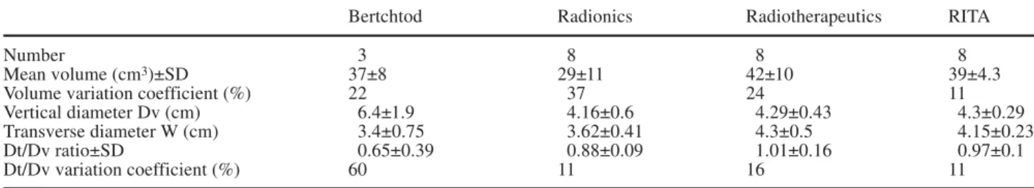
Lesions volumes obtained with RITA, Radiotherapeu-tics, and Radionics, were 39±4.3, 42±10, and 29±11 cm3, respectively (Table 1). Statistical differences were ob-served between these three groups using ANOVA test (p<0.05). Group-by-group statistical comparison demon-strated statistical differences between Radiotherapeutics and Radionics (p<0.05) and nearly significant difference between RITA and Radionics (p=0.056). No statistical
difference existed when RITA was compared with Ra-diotherapeutics using an unpaired t test. F test analysis of variance found significant differences in variance be-tween RITA, Radiotherapeutics, and Radionics (p<0.05). Transverse diameter was statistically significantly differ-ent between RITA and Radiotherapeutics (4.15±0.23 and 4.3±0.5 cm, respectively) and Radionics (3.62±0.41; p<0.05).
The Dt/Dv ratio for Radiotherapeutics and RITA RF systems were 1.01±0.16 and 0.97±0.1, respectively, cor-responding to near spherical shapes of the volume of co-agulation (Fig. 4). More elongated lesions were demon-strated by Radionics and Bertchtold RF systems with Dt/Dvratios of 0.88±0.09 and 0.65±0.39, respectively. There was not significant difference and there was not significant variance of Dv/Dt ratio using F test.
Fig. 2a, b Radio-frequency (RF) in vivo lesions created with the Berchtold system. a The RF lesion extending along the hepatic vein wall away from the RF lesion with fresh clots inside of the vein. b Another Berchtold in vivo lesion showing coagulation fol-lowing vascular branches away from the RF lesion
Fig. 3a, b The RF in vivo lesion created with the Berchtold system. a The anterior aspect of the liver shows a large lesions ex-tended all along the liver surface. b The liver shows this ovoid le-sion extending along the liver capsule
Ex vivo RF lesions
Thirty-two lesions could be performed in explanted calf liver (8 lesions for each system) Ex vivo lesion volumes ranged between 55 and 68 cm3without statistical differ-ences found between the four devices using the ANOVA test and Student’s t test (Table 2). Variation coefficients were also greater for lesion volume in ex vivo lesions as compared with in vivo lesions. Comparison of variance between groups of ex vivo lesion volume did not show any statistical significance, whereas comparison of vari-ance of the Dv/Dt ratio in the same groups was highly significant (p<0.00000001).
Discussion
Percutaneous RF tumor ablation has gained significant popularity during recent years. In particular, RFA has been advocated by many physicians as a primary treat-ment for selected patients with hepatocellular carcinoma. Recent reports have demonstrated that RFA is more effi-cacious than percutaneous ethanol injection (PEI) allow-ing for equivalent tumor control in fewer treatment ses-sions [14]; however, one limitation of this therapy is the size of RF lesions that can be obtained with one RF de-livery. Although the efficacy of RFA is close to 100% for tumors less than 2 cm in diameter, the local recurrence rates are ≥20% for tumors larger than 2.5 or 3 cm, and
≥50% for lesions larger than 5 cm [10]. In the same way, destruction of a lesion of 2 cm in size with a 1-cm safety margin corresponds to a RFA size in volume of 33.49 cm3. For a larger lesion of 3 cm in diameter with the same 1-cm safety margin, this RFA volume should be 65.42 cm3. In our study the technological improve-ments allowed a theoretical destruction of lesions be-tween 2 and 3 cm in diameter. One must consider that these improvements are important, but they are still far from what we may expect from a clinical point of view. New experimental developments recently demonstrated in experimental studies, such as combined wet cooled or wet expandable, may further improve RFA lesion size [9, 10, 11].
Both RITA and Radiotherapeutics developed multi-probe needles that are able to deploy nine (Christmas-tree shaped) or ten (umbrella shaped) prongs, respective-ly. In our experimental study, these two systems provid-ed the largest volume of tissue destruction. In addition, these systems created near-spherical lesions in vivo with Table 1 Mean volume of radio-frequency ablation (RFA) lesions created in pig liver in vivo
Bertchtod Radionics Radiotherapeutics RITA
Number 3 8 8 8
Mean volume (cm3)±SD 37±8 29±11 42±10 39±4.3
Volume variation coefficient (%) 22 37 24 11
Vertical diameter Dv (cm) 6.4±1.9 4.16±0.6 4.29±0.43 4.3±0.29
Transverse diameter W (cm) 3.4±0.75 3.62±0.41 4.3±0.5 4.15±0.23
Dt/Dv ratio±SD 0.65±0.39 0.88±0.09 1.01±0.16 0.97±0.1
Dt/Dv variation coefficient (%) 60 11 16 11
Fig. 4 The cut liver shows a 4.7-cm almost spherical in vivo le-sion created with the RITA system
Table 2 Mean volume and di-ameter ratio of ex vivo RFA le-sions
Bertchtold Radionics Radiotherapeutics RITA
Number 8 8 8 8
Mean volume (cm3)±SD 67±24 66±20 55±13 68±20
Volume variation coefficient (%) 35 31 24 29
Vertical diameter Dv (cm) 5.42±1.2 5.56±0.72 4.35±0.34 5.94±0.62 Transverse diameter Dt (cm) 4.63±0.62 4.69±0.44 4.48±0.47 4.61±0.45 Dt/Dv ratio±SD 0.89±0.16 0.84±0.06 1.12±0.08 0.78±0.03
hepatic tumors have a spherical shape. We did not observe any difference in terms of RF lesion volume between these two devices; however, repositioning of the needle was necessary in 3 cases with the RITA device, increasing the lesion creation time. The RITA RF system monitors intra-lesional temperature with five thermocou-ples. When a needle is positioned close to a vessel, the temperature does not increase in this area. As a conse-quence, the needle position can be modified to avoid this limitation. This may explain, in part, the reproducible volumes obtained with this device as compared with the Radiotherapeutics system which monitors treatment with an impedance control mechanism.
Another strategy that can be used to increase RF lesion size is to minimize carbonization (“charring”) of tissue around needle tip. This can be done through a con-tinuous perfusion of cold saline within the needle lumen as proposed by Radionics; however, our study shows that in vivo lesions produced by Radionics were slightly smaller and more ovoid in shape than the lesions pro-duced by RITA or Radiotherapeutics devices.
Bertchtold applies another concept to increase lesion size with RF. Isotonic saline can be infused into the tis-sue through holes at the extremity of the needle, thereby increasing electrical conductivity in the needle vincinity [10]. The protocol used in our investigation and recom-mended by the Berchtold company killed 5 of 8 animals within the first 3 min of RF delivery with perfusion. At present, we have no definite explanation for these deaths; however, livers removed from 4 of the 8 pigs treated with this device show coagulation running along the large hepatic veins and away from the RF lesion. This finding suggested diffusion of the heated saline along the outside wall of these veins. Moreover, in 2 cases, clots were found in the hepatic veins. For this rea-son, we considered pulmonary embolism as a possible explanation for their deaths; however, in clinical series, such complications have never been reported, to our knowledge in the literature [11]; however, the shape of the lesions were irregular as previously observed [12, 13].
Our study supports the recent finding of Goldberg et al. who evaluated the consequences of hyperosmolar sa-line injection [14], mentioning that shape of lesions were irregular and hardly predictable, especially when large amounts of saline (>25 ml) were injected. In our study, a total amount of 25 ml was used as recommended by the manufacturer. This large amount of saline may explain for spread of coagulation distally to the RF lesions along the vascular bed or under the capsule.
This study also demonstrates that using ex vivo com-parisons of RF systems brings no significant valuable in-formation to the in vivo efficacy of these systems. In-deed, despite statistical differences that were observed in
higher for ex vivo than for in vivo lesions for RITA and Radiotherapeutics systems.
This study does have some limitations. Firstly, it is not known whether the results of this animal liver model accurately predict the results of treating liver tumors in patients [15]. Differences in tissue vascularization, im-pedance, and heterogeneity in tumors may modify the re-sults that we observed. Secondly, we have no definite ex-planation for the deaths of 5 animals using Bertchtold device. We hypothesized that the deaths were due to massive pulmonary emboli on the basis of hepatic vascu-lar thrombosis. Since that hypothesis was retrospectively supposed on the basis of liver pathological studies, we did not take the opportunity to check the lung at autopsy. This complication has never been described in human studies with this unit; however, literature regarding use of the device is limited. Thirdly, we did not use the most recent expandable needles developed by RITA and Ra-diotherapeutics (Starburst Xli and Le Veen 6.0, respec-tively) since they were not yet available. Fourth is the size of pig liver. Even when using the largest pigs avail-able in our institute, the thickness of the livers rarely ex-ceeded 6 cm due to their leafy anatomy. For this reason, it was possible to perform only one to three RF lesions per animal resulting in the relatively low number of le-sions in our study. Fifthly, systems were run according to each manufacturer’s specifications. Their recommenda-tions may or may not have represented the greatest de-gree of optimization possible. Superiority of one system over another only reflects the situation in the experimen-tal conditions described at the moment of the study. This may rapidly change in the future due to new needle or protocol improvements.
Conclusion
In this experimental study, we did not observe differ-ences between expandable needles systems in vivo. Cooled needles produced slightly smaller lesions with a more ovoid shape. Improvements in needles and delivery protocols are still mandatory to treat with safety margins tumors larger than 3 cm in diameter.
Acknowledgement We thank P. Frascarolo for his precious help in the statistical evaluation of our results.
References
1. Goldberg SN, Dupuy DE (2001) Image-guided radiofrequency tumor ablation: challenges and opportunities, part I. J Vasc Interv Radiol
12:1021–1032
2. McGahan JP, Griffey SM, Budenz RW, Brock JM (1995) Percutaneous ultrasound-guided radiofrequency elec-trocautery ablation of prostate tissue in dogs. Acad Radiol 2:61–65
3. de Baere T, Denys A, Johns-Wood B et al. (2001) Radiofrequency liver ablation: experimental comparative study of water—cooled vs expandable systems. Am J Roentgenol
176:187–192
4. Goldberg SN, Gazelle GS, Solbiati L, Rittman WJ, Mueller PR (1996) Radiofrequency tissue ablation: in-creased lesion diameter with a perfu-sion electrode. Acad Radiol 3:636–644 5. Goldberg SN, Hahn PF, Tanabe KK et
al. (1998) Percutaneous radiofrequency tissue ablation: Does perfusion-mediat-ed tissue cooling limit coagulation ne-crosis? J Vasc Interv Radiol 9:101–111
6. Goldberg SN, Solbiati L, Hahn PF et al. (1998) Large-volume tissue ablation with radiofrequency by using a clus-tered, internally-cooled electrode technique: laboratory and clinical ex-perience in liver metastases. Radiology 209:371–379
7. Curley SA, Izzo F, Delrio P et al. (1999) Radiofrequency ablation of unresectable primary and metastatic hepatic malignancies: results in 123 patients (see comments). Ann Surg 230:1–8
8. Curley SA, Izzo F, Ellis LM, Nicolas-Vauthey J, Vallone P (2000) Radiofre-quency ablation of hepatocellular cancer in 110 patients with cirrhosis. Ann Surg 232:381–391
9. Goldberg SN, Stein MC, Gazelle GS, Sheiman RG, Kruskal JB, Clouse ME (1999) Percutaneous radiofrequency tissue ablation: optimization of pulsed radiofrequency technique to increase coagulation necrosis. J Vasc Interv Radiol 10:907–916
10. Livraghi T, Goldberg SN, Lazzaroni S et al. (2000) Hepatocellular carcinoma: radiofrequency ablation of medium and large lesions. Radiology 214:761–768 11. Gangi A, Dupas B, Guth S et al. (2001)
Application des électrodes humides de radiofréquence dans le traitement des tumeurs hépatiques. J Radiol 82:1430
12. Livraghi T, Goldberg SN, Monti F et al. (1997) Saline-enhanced radio-frequency tissue ablation in the treat-ment of liver metastases. Radiology 202:205–210
13. Miao Y, Ni Y, Mulier S et al. (1997) Ex-vivo experiment on radiofrequency liver ablation with saline infusion through a screw-tip cannulated elec-trode. J Surg Res 71:19–24
14. Goldberg SN, Ahmed M, Gazelle GS et al. (2001) Radio-frequency thermal ablation with NaCl solution injection: effect of electrical conductivity on tissue heating and coagulation-phan-tom and porcine liver study. Radiology 219:157–165
15. Denys AL, de Baere T, Mahe C et al. (2001) Radiofrequency tissue ablation of the liver: effects of vascular occlu-sion on leocclu-sion diameter and biliary and portal damages in a pig model. Eur Radiol 11:2102–2108
16. Goldberg S, Hahn P, Halpern E, Fogle R, Gazelle G (1998) Radiofrequency tissue ablation: effect of pharmacologic modulation of blood flow on coagula-tion diameter. Radiology 209:761–767