Sequential Multiple-Target Sensor: In
3+
, Fe
2+
, and Fe
3+
Discrimination by an Anthracene-Based Probe
Alba Finelli,
†Valentin Chabert,
†Nelly Hérault,
†Aurélien Crochet,
‡Cheal Kim,
§and Katharina M. Fromm
*
,††Department of Chemistry, University of Fribourg, Ch. du Musée 9, 1700 Fribourg, Switzerland ‡FriMat, Department of Chemistry, University of Fribourg, Ch. du Musée 9, 1700 Fribourg, Switzerland
§Department of Fine Chemistry, Seoul National University of Science and Technology (SeoulTech), Seoul 139-743, Republic of
Korea
*
S Supporting InformationABSTRACT: Indium is a nonphysiological toxic metal widely used in industry. While misunderstood, its toxicity is proposed to be linked to a perturbation of Fe3+homeostasis through the
binding of In3+ ions to essential iron metalloproteins such as transferrins. Therefore, the monitoring of In3+ and Fe3+ in
biological environments is of prime interest for both basic research and diagnosis. Here we report the design of a salen-type anthracene-based probe able to selectively sense and discrim-inate In3+ and Fe2+/3+ions by fluoro-colorimetry.
■
INTRODUCTIONPhysiological metal ions play key roles in all kingdoms of life, and their homeostasis is finely regulated. Being the most abundant transition metal on earth, iron is involved in many biological processes such as DNA synthesis, oxygen transport, or electron transfer. Hence, slight perturbations of its cellular homeostasis can lead to severe diseases, such as Parkinson or Alzheimer diseases.1,2On the other hand, the industrial use of metals leads to a release of nonphysiological metal ions into the environment, increasing their bioavailability and hence the frequency of associated diseases. Nowadays, the industrial use of indium is globally increasing due to the growth of photovoltaic and optoelectronic industries3−5 with the global indium market estimated at 810 tons in 2016.6While toxicity and exposure data are limited for this metal,7−9recent reports highlighted the involvement of indium in lung diseases of exposed workers.10−15 Indeed, indium has been proposed to perturb alveolar macrophage functions, resulting in the so-called indium lung disease.16 Furthermore, the biochemical similarities between In3+ and Fe3+ions (ionic radii of 94 pm and 78.5 pm (high spin)/69 pm (low spin), in octahedral environment, respectively)17 may be responsible for a disruption of the iron metabolism by indium, resulting in imbalances at iron absorption sites, transportation, usage, and storage in cells.18 For instance, the iron transport has been proposed to be inhibited by indium ions, due to their tight binding to transferrins.19Indeed, possessing the same charge, In3+ions can accommodate in Fe3+binding sites of transferrins
with a high affinity. For Fe3+detection, numerous probes have been recently reported.20−22 For instance, Lee et al. have reported a selective rhodamine-based sensor showing a sensitive and selective detection of intracellular Fe3+ ions in
hepatocytes.23,24To the best of our knowledge, Kim and co-workers were thefirst in synthesizing an indium sensor, based on pyrene moieties.25Due to the high interest of monitoring In3+concentrations in biological environments, the number of optical probes for In3+ detection is also increasing.26−35 Nevertheless, the design of new chemosensors able to quantify the cellular concentrations of iron and indium ions is still needed for the fundamental understanding of indium toxicity as well as for the diagnosis of associated diseases.
Lately, considerable efforts have been made in the conception of new chemosensors for the selective detection of multiple metal ions.36−42 Among the available sensing methods, probes based on metal ion-induced fluorescence changes are principally attractive and offer many advantages such as the selectivity, the fast responses, and the high sensitivity.43−45However, due to the fluorescence quenching effects by certain metal ions,46 the development of turn-on fluorescence chemosensors remains very challenging. On the other hand, the colorimetric method represents an easy and fast technique allowing detection of targets by a color change, ideally even visible with the naked-eye.47−50
http://doc.rero.ch
Published in "Inorganic Chemistry 58(20): 13796–13806, 2019"
which should be cited to refer to this work.
In order to design a versatile probe especially able to discriminate In3+among other cations, Schiff base ligands have
caught our attention.51−56Azomethine ligands, such as salen and its derivatives, which are commonly used as chelating ligands in coordination chemistry, are of crucial interest in multiple areas such as supramolecular chemistry,57,58material sciences,59−61 or catalysis.62−64 Furthermore, salen based ligands are well-known to coordinate In3+ cations with their
N2O2 chelating site, producing complexes which are widely
used as efficient catalysts for e.g. isoselective lactide polymer-ization.65,66 Such compounds are easy to prepare and are readily tunable.
Building on this framework, we developed the first fluorescent and colorimetric multitarget anthracene-based sensor S1 able to distinguish indium and iron through distinct optical methods. This multisensing probe is able to selectively detect In3+ ions with a strong excimer fluorescence emission and selectively senses Fe2+/Fe3+ ions by the formation of a
broad charge transfer (CT) band involving the inner O2O2and
outer N2O2coordination spheres of S1 and giving rise to a red
color of the complex.
■
EXPERIMENTAL SECTIONMaterials and Methods. All experiments were performed in air and at RT. Ligand S1 was prepared based on the procedure reported previously by Mandolini and co-workers.67 All chemicals were
commercial products of reagent grade and were used without further purification.1H and13C NMR measurements were carried out with a
Bruker 400 MHz spectrometer at ambient temperature, and chemical shifts are given in ppm with respect to the residual solvent peak. Mass spectra (ESI-TOF, positive mode) were recorded with a Bruker esquire HCT spectrometer with a DMF/ACN mixture as solvent. The UV-vis spectra were recorded with a Perkin−Elmer Lambda 40 spectrometer. The spectrofluorimetric studies have been conducted on a Varian Cary Eclipse spectrofluorimeter.
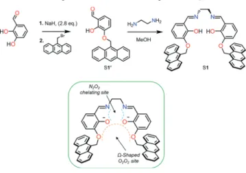
Sensor Synthesis. Synthesis of Anthracene-Based Aldehyde S1′. To a suspension of pure NaH (0.73 mg, 2.8 equiv) in dry DMSO (3 mL) was added a solution of 2, 3-dihydroxybenzaldehyde (152 mg, 1.1 mmol) in DMSO (2 mL) at 20−25 °C. After stirring for 90 min, a solution of 9-bromomethylanthracene (300 mg, 1.1 mmol) in DMSO (2 mL) was added. Stirring was continued for 20−24 h in the dark, whereupon the mixture was poured into water (25 mL) and extracted with CHCl3(3× 20 mL). The aqueous layer was acidified with 6 M
HCl to adjust the pH to 4 and again extracted with CHCl3(3× 50
mL). The combined CHCl3layers were washed with 1 M HCl (2×
20 mL). The solvent was evaporated, and the residue was purified by column chromatography (silica gel, DCM/hexane (8:2)) to give the pure aldehyde S1′ as a yellow solid (187.8 mg, 52%) (Figure S1).1H NMR (400 MHz, DMSO-d6):δ 6.10 (s, 2H), 6.93−6.99 (t, J = 8 Hz, 1H), 7.27−7.30 (d, J = 4 Hz, 1H), 7.36−7.39 (d, J = 12 Hz, 1H), 7.46−7.58 (m, 4H), 8.02−8.05 (d, J = 8 Hz, 2H), 8.39−8.49 (d, J = 12 Hz, 2H), 8.52 (s, 1H), 9.93 (s, 1H), 11.04 (s, 1H).13C NMR (100 MHz, DMSO-d6):δ 193.81, 151.61, 148.28, 131.45, 131.20, 129.37, 129.24, 127.54, 127.14, 125.75, 124.79, 123.00, 122.35, 122.08, 120.62, 120.32, 119,83, 64.01. ESI-mass: m/z calculated for C22H16O3+Na+([M + Na]+), 351.09; found, 351.09.
Synthesis of the Anthracene-Based ReceptorS1. The suspension of the corresponding aldehyde S1′ (0.2 g, 0. 609 mmol, 2 equiv) was solubilized in MeOH (10 mL), and ethylenediamine was added slowly (0.0203 mL, 0.304 mmol). After letting the reaction stir at reflux overnight in the dark, the light-yellow product S1 was collected byfiltration and dried under vacuum (163.7 mg, 79%) (Figure S2).
1H NMR (400 MHz, DMSO-d 6):δ 3.97 (s, 4H), 6.07 (s, 4H), 6.76− 6.81 (t, J = 8 Hz, 2H), 6.93−6.96 (d, J = 8 Hz, 2H), 7.17−7.20 (d, J = 12 Hz, 2H), 7.54−7.44 (m, 8H), 8.00−8.03 (d, J = 12 Hz, 4H), 8.38 (s, 2H), 8.42−8.45 (d, J = 16 Hz, 4H), 8.50 (s, 2H), 13.67 (s, 2H). 13C NMR (100 MHz, DMSO-d 6):δ 161.35, 157.60, 151.47, 150.09, 131.08, 129,31, 128.26, 126.58, 125.88, 125.70, 125.40, 124.40, 118.00, 115.94, 70.80, 67.7. ESI-mass: m/z calculated for C46H36N2O4+H+([M + H]+), 681.21; found, 681.27.
Sensing Tests of S1. Stock solutions (1 mM) of sensor S1 (4.765 mg, 0.007 mmol) were prepared in DMF (7 mL). In order to obtain a solution with afinal concentration of 40 μM, 24 μL of the previous solution (1 mM) was then diluted into 2.976 mL of DMF (3 mL in the cuvette). Metal ion salts were added from 0 to 3 equiv using 20 mM solutions of M(NO3) (M = Na, K, Ag), M(NO3)2(M = Mn, Ni,
Cu, Zn, Cd, Mg, Ca, Pb), M(ClO4)2(M = Fe), and M(NO3)3(M =
Al, Fe, Cr, Ga, In) in DMF (addition of 9μL, corresponding to 1.5 equiv), respectively. UV−vis and fluorescence spectra were recorded after mixing of the samples for a few seconds at ambient temperature. Fluorescence Titrations ofS1. For In3+, a solution of 40μM of
sensor S1 was prepared in a cuvette of 3 mL. Then, 6−90 μL of a solution of In(NO3)3 in DMF (5 mL, 20 mM) was added to the
solutions of S1 (40μM) prepared previously. After mixing them for a few seconds,fluorescence spectra were recorded at 30 °C.
UV−Vis Titrations of S1. For Fe3+, a solution of 40μM of sensor
S1was prepared in a cuvette of 3 mL. Then, 0.6−18 μL of a solution of Fe(NO3)3in DMF (5 mL, 20 mM) was added to the solutions of
S1(40μM) prepared previously. After mixing them for a few seconds, UV−vis spectra were recorded at room temperature.
NMR Titration ofS1. For In3+, a solution of 0.5 mM in CDCl
3of
sensor S1 was prepared in an NMR tube. Then, 1.7−10.2 μL of a solution of In(NO3)3in MeOD (1.5 mM) was added to the solutions
of S1. After mixing them for a few seconds, the1H NMR spectra were recorded at room temperature.
Job Plot Measurements. For In3+, a stock solution (1 mM) of
sensor S1 (4.765 mg, 0.007 mmol) was prepared in DMF (7 mL). Volumes of 0, 18, 36, 54, 72, 90, 108, 126, 144, 162, and 180μL of the chemosensor S1 solutions (1 mM) were transferred to independent vials. In the same way, the volumes of 9, 8.1, 7.2, 6.3, 5.4, 4.5, 3.6, 2.7, 1.8, 0.9, and 0μL of In3+solutions (20 mM) were
added to the correspondent diluted S1 solution. Each vial is thenfilled with the latter solution to a total volume of 3 mL and after shaking them for a few seconds,fluorescence spectra were recorded at 30 °C. For Fe2+and Fe3+, the same procedure has been adopted and the
UV−vis spectra were recorded at room temperature.
Competition with Other Metal Ions. For Fe2+/Fe3+ and In3+, a
stock solution (1 mM) of sensor S1 (4.765 mg, 0.007 mmol) was prepared in DMF (7 mL). In order to obtain a solution with afinal concentration of 40μM, 24 μL of the previous solution (1 mM) was then diluted into 2.967 mL of DMF (3 mL in the cuvette). Metal ions salts were added (addition of 18μL, corresponding to 3 equiv) using 20 mM solutions of M(NO3) (M = Na, K, Ag), M(NO3)2(M = Mn,
Ni, Cu, Zn, Cd, Mg, Ca, Pb), M(ClO4)2(M = Fe), and M(NO3)3(M
= Al, Fe, Cr, Ga, In) in DMF, respectively. Then, 9μL (corresponding to 3 equiv) of the analyzed metal ion, namely Fe2+/Fe3+and In3+(40
mM solution), was added and the UV−vis or fluorescence spectra were recorded after mixing the samples for a few seconds at room temperature, respectively 30°C.
■
RESULTS AND DISCUSSIONLigand Synthesis and Characteristics. A salen-type compound has been modified via the introduction of two anthracene moieties, employed as chromophores for their efficient fluorogenic behavior. The anthracene derivative of the o-vanillin, S1′, was synthesized by a nucleophilic substitution reaction between 2,3-dihydroxybenzaldehyde and 9-bromome-thylanthracene. The final anthracene ligand S1 has been produced by a nucleophilic addition forming a hemiaminal, reacting ethylenediamine as starting material with the anthracene corresponding aldehyde S1′. The subsequent dehydration provides the resulting imine-based ligand S1 (Scheme 1). The latter has been fully characterized by1H and
13
C NMR, UV−vis, ESI-MS spectrometry and by single crystal and powder X-ray diffraction.
The self-assembling of the two anthracene moieties upon the coordination of certain metal cations is expected to induce a strong A−A* excimer fluorescence.68 Indeed, containing a N2O2 imine-based coordinating site, the ligand S1 can bind
transition metal ions upon deprotonation. The coordination of the N2O2moiety preorganizes the ligand in aΩ-shape, offering
thus a second recognition site, O2O2, for the coordination of a
second metal ion (Scheme 1). The coordination of a second metal ion at the O2O2site becomes favorable and is expected
to further modify the previous architecture, altering in turn the luminescence by bringing thefluorophores close to each other. Indeed, changing the degrees of freedom (mobility) of the molecular scaffold, receptor S1 could have the ability to produce an excimer emission by metal ion-induced fluo-rescence.
Colorimetric Sensing of Fe2+/3+. The coordination of
Fe2+and Fe3+ions by S1 (40μM) leads to a coloration of the
solution in red brown. This feature is the result of the establishment of broad CT bands in the 450−700 nm spectral window. One of them was assessed to be a metal-to-ligand charge transfer (MLCT) from a t2gd-orbital of Fe2+to theπ*
antibonding orbital on the N2O2site, leading to the oxidation
of the metallic center.69 The other band was described as a ligand-to-metal charge transfer (LMCT) from a π-orbital of N2O2 to the eg unoccupied d-orbital of Fe3+, leading to the
reduction of the metal.70−72 The absorption coefficients (ε) were determined to be 2900 and 4300 M−1cm−1, respectively. Although the spectral features are similar for these two metal ions, the CT bands appeared to be more intense for Fe3+. To exclude any interference in Fe2+ sensing by Fe3+, the
experiments were additionally carried out under anaerobic conditions, avoiding thus the potential oxidation of Fe2+ into Fe3+by O
2(Figure S3). Similar UV−vis spectral features have
been observed under these conditions.
The Fe2+/Fe3+ binding properties of S1 were then investigated by UV−vis titrations. Upon the addition of increasing amounts of Fe3+(Fe(NO3)3) or Fe2+(Fe(ClO4)2),
the characteristic absorption band at 366 nm is slightly red-shifted by 3 nm, giving rise to two isosbestic points at 385 and 389 nm (Figure 1A).
The corresponding binding isotherm curve shows a plateau after 1 equiv of Fe2+/Fe3+was added, indicating the formation
of a 1:1 complex. This binding stoichiometry is then confirmed by a Job plot analysis which exhibits a maximum at 0.5 attesting the formation of a 1:1 complex in both cases (Figure S4−S5). The stoichiometry is supported by mass spectrometry for the Fe3+compound with m/z: 734.19 [M− NO3]+where
M([(FeS1− 2H) + NO3]) = 796.12, Figure S6. (The Fe2+
-complex oxidizes rapidly in air). Moreover, a1H NMR titration of S1 by Fe3+ and Fe2+ salts has been performed, showing a complete disappearance of the signals for the OH-group upon the addition of 1 equiv of Fe3+or Fe2+ions. This confirms the participation of O atoms (Figure S7−S8) of the N2O2
chelating site in the iron coordination. For both Fe2+- and
Fe3+-S1 complexes, the proton signal becomes broader upon metal ion addition, preventing an accurate titration of the complexes. This phenomenon indicates that the Fe2+-complex
is high spin, while in the case of Fe3+, no conclusion can be drawn from NMR data as both high and low spin states give paramagnetic species.
Scheme 1. Synthetic Route of the Ligand S1 and Its Potential Recognition Sites Generating aΩ-Shape
Figure 1.(A) UV−vis titration of S1 (40 μM) by Fe(NO3)3(0 to 80μM) in DMF. The insets depict a zoom of the 370−420 nm window and the
corresponding binding isotherm curve (λ = 530 nm). (B) UV−vis spectrum and color of S1 solutions (100 μM in DMF) upon the addition of 1.2 equiv (120μM) of different metal ions.
The suggested coordination mechanism involves a twisted arrangement of the anthracene moieties impeding sterically the coordination of the O2O2recognition site by a second cation
(Scheme 2A). Therefore, the sensing ability of other metal ions has been determined by monitoring the potential CT transitions in the visible range after the addition of each metal ion. The absence of the latter broad band confirms that sensor S1 can be used as a colorimetric chemosensor able to selectively sense Fe3+or Fe2+among other cations (Ag+, Al3+, Ga3+, In3+, Zn2+, Cu2+, Mg2+, Cr3+, Co2+, Ni2+, Na+, K+, Ca2+,
and Pb2+) by colorimetry (Figure 1B). The calculation of
detection limit (LOD) was then performed through standard deviations and linearfittings, giving a result of 0.44 μM for Fe3+
and 0.69μM for Fe2+ (Figure S9−S10).
Fluorimetric Sensing of In3+. Initially, after an excitation
at 365 nm, receptor S1 exhibits a weak monomer emission in
the 375−475 nm range with a maximum at 420 nm. This might be explained by a photoinduced electron transfer (PET) which takes place from the nitrogen atoms of the imine groups to the fluorophore moieties, leading to fluorescence quench-ing.73The subsequent coordination of In3+by S1 generates an intense excimer emission in the 475−600 nm range with a maximum at 508 nm (λex = 365 nm), probably via a parallel
self-assembly of the two anthracene moieties through π−π stacked dimer interactions, leading to a bright green fluorescence (Figure 2A).74−76 Concomitantly, a strong decrease of the typical monomer emission band is observed.
The binding mode was then analyzed by performing a spectrofluorimetric titration of S1 by In3+. This titration
showed an increase of the excimer emission until 2 equiv of In3+ added. In the corresponding binding isotherm curve
obtained by plotting the relative fluorescence intensity (I/I0) Scheme 2. Suggested Coordination Modes of the Sensor S1. (A) Coordination of Fe2+ or Fe3+Ions by S1 Leading to the Establishment of a Broad CT Band, Giving Rise to a Red Brown Color. (B) Coordination of In3+Ions by S1 Leading to the
Self-Assembling of the Anthracene Moieties, Giving Rise to a Strong Excimer Emission
Figure 2. (A) Spectrofluorimetric titration of S1 (40 μM) by In(NO3)3 (0 to 240 μM) in DMF (λex= 365 nm). The inset depicts the
corresponding binding isotherm curve by plotting the relativefluorescence intensity (I/I0) changes (λem= 508 nm). (B) Selective detection of In3+
by sensor S1. Fluorescence emission spectra (λex= 365 nm) of S1 (40μM in DMF) upon the addition of 1.2 equiv (48 μM) of different metal ions
(detailed labeling of thefluorescence intensity depending on the used metal ion can be found in theSupporting Information, Figure S11).
changes as a function of the metal concentration, a plateau is reached after the addition of 2 equiv of metal ion, revealing the formation of a 2:1 complex (Figure S12). This result also indicates the formation of the excimer conformation already after the coordination of thefirst In3+ion within the N
2O2site
(Scheme 2B) and displays thus a ratiometry with an isoemissive point at 428 nm (λex= 365 nm). The calculation
of detection limit (LOD) was performed through standard deviations and linearfittings yielding 0.53 μM (Figure S13).
Together with the Job plot result fromfluorescence titration, which presents a maximum at around 0.35, the ESI-MS analysis also indicates the formation of a 2:1 complex (m/z: 1095.03 [M-NO3]+ where M([(In2S1 − 2H) + 4NO3]) =
1156.03,Figure S14). Additionally, a1H NMR titration of S1
by In3+ions in CDCl3has been carried out in order to confirm
the complex stoichiometry. The perturbation of the S1 1H
NMR spectrum upon the addition of In3+ suggests a drastic reorganization and distortion of the ligand backbone during the complex formation, probably similar to what is observed in
single crystal structures of 1:1 salen-type indium complexes.66 For instance, the initial aromatic and methylene bridge signals disappear in favor of new shifted signals that appear during the titration, showing the formation of a new species in slow exchange regime (Figures 3A and 3B). The splitting of the anthracene external mutliplet signal at 7.5 ppm reflects the formation of an asymmetrical complex which is stabilized after the addition of 2 equiv of In3+ added. The shift of the
methylene bridge-CH2 singlet from 6.07 to 6.12 ppm also
follows this tendency and confirms the 2:1 stoichiometry of the complex. Indeed, the binding isotherm of the complex formation established based on the methylene bridge-CH2
signal appearing at 6.12 ppm, taking tetramethylsilan (TMS) as reference, shows a plateau after the addition of 2 equiv of In3+
ions (Figure 3C) (More details of the1H NMR titration can be found in theSupporting Information, Figures S15−S16). In addition, a 1H NMR titration of S1 by In3+ ions has been performed in DMSO displaying the complete deprotonation of the−OH groups, validating the contribution of oxygen atoms
Figure 3.(A) Aromatic region of the1H NMR titration of S1 with In(NO
3)3(in MeOD) forming the corresponding In3+-S1 complex at room
temperature (0.5 mM) in CDCl3. (B)1H NMR proton signals assignation of the complex assignation. (C)1H NMR titration binding isotherm of
the In3+-S1 complex formation focused on the methylene-CH
2bridge integrals, taking TMS as reference.
(from the N2O2 chelating site) in the complex formation
(Figure S17).
Altogether, these results suggest that the chelation of one In3+ion in thefirst N
2O2recognition site of the ligand brings
the anthracene moieties into close enough proximity to start the formation of the excimer A−A* in the sensing mechanism. However, the overall structure is subsequently rigidified upon complexation of the second In3+ ion, amplifying thus the excimer emission in solution (Scheme 2B).
The lack of a single crystal X-ray structure prevents the exact characterization of the coordination compound. Nevertheless, several previously described salen-based indium complexes provide valuable examples of different possible coordination modes. Indeed, some reported indium structures65,77,78 allow us to state that the reaction of S1 with one In3+could give rise
to a distorted square pyramidal coordination of In3+ by the
N2O2 chelating moiety of S1 (formed by two phenolate
moieties and two imine groups) with an axial oxygen donor atom from a nitrate ion. This will likely enhance the nonplanarity of the ligand as it wraps around a metal ion that is too large to perfectlyfit into the N2O2cavity. For the
second indium ion, it is suggested to be coordinated by all four oxygen atoms of the O2O2 compartment of the ligand.
Similarly to a few similar structures reported earlier,79 it is proposed that the coordination sphere of the second metal ion is completed by two O atoms of two nitrate ions in axial positions. The presence of a complex dimer or 1D coordination polymers can be discussed via bridging nitrate or solvent ligands; however, this phenomenon seems to be less probable due to the steric hindrance of the two anthracene moieties and the fact that no evidence of such species or oligomers was observed by ESI-MS analysis. In total, one 2−-charged ligand and three nitrate anions would thus be coordinated directly to the indium ions, while an additional anion may not directly be coordinated to the complex. This situation would perfectlyfit to the observed mass spectra.
The binding capacity of sensor S1 for diverse metal ions was also studied by spectrofluorimetry, showing however no excimer formation. This specific behavior allows sensor S1 to detect and discriminate In3+ from all other metal ions tested (Ag+, Al3+, Ga3+, Zn2+, Cu2+, Fe3+, Fe2+, Mg2+, Cr3+, Co2+, Ni2+,
Na+, K+, Ca2+, and Pb2+) by fluorometry (Figure 2B). The
excimer emission is not produced by the coordination of other cations due to the lack of an efficient distortion of the S1’s backbone, avoiding thus the excimer formation.
Compared to other 3+ charged, yet smaller, metal ions such as Al3+(ionic radius 67.5 pm) or Ga3+(76 pm), In3+is the only
one generating an excimer emission. Furthermore, the interaction of S1 with lanthanide ions such as Er3+(103 pm)
or Eu3+ (108 pm), which are bigger than In3+, has been
qualitatively studied and also no optical response has been observed. This phenomenon is likely due to the “optimum” size of the indium ion to induce the excimer formation from the two anthracene moieties of the ligand.66Crystal structures of salen-type ligands with smaller metal ions do not lead to a strong deformation of the salen backbone on one hand,80while larger ones may notfit well into the N2O2chelating site.
81,82 This may then possibly lead to sandwich-type complexes with two ligands per cation, binding via N2O2-entities like in the
case of Tb3+83,84 or protonation of the N-donor atoms and binding via the O2O2entity only, as for Ln3+,85preventing thus
the excimer formation, respectively offering quenching path-ways. This allows us to argue that indium possesses the “optimal size”, resulting in a strong excimer emission generating from the perfect arrangement of the anthracene moieties.
Competition Studies. In order to challenge the robustness of the sensor, potential interference from other ions in the detection of Fe2+/Fe3+ and In3+ was investigated in the
presence of other competitive ions, in particular Ag+, Al3+,
Ga3+, Zn2+, Cu2+, Fe2+, Fe3+, In3+, Mg2+, Cr3+, Co2+, Ni2+, Na+,
K+, Ca2+, and Pb2+. These cations were added to a solution of
S1 (40 μM) before the addition of equivalent amounts of In3+or Fe2+/Fe3+ (2 and 1 equiv, 88 μM and 48 μM,
respectively) (Figures S18−S20).
In the case of In3+, the experiment was performed by
monitoring the excimer emission of the In3+-S1 complex. Most
of the present cations did not interfere with the detection of In3+except three of them, Cu2+, Fe2+, and Fe3+, which inhibited
the indium sensing considerably (Figure 4A). Indeed, after the coordination of these three metal ions by S1, no subsequent excimer emission band was observed upon In3+addition even though a decrease of the monomer emission has been observed. Cu2+ and Fe3+ are well-known paramagnetic ions with an incompletelyfilled d shell. These cations have been described to strongly quench the fluorescence of the fluorophore by electron or energy transfer.86−88
Moreover, the coordination of Cu2+, Fe2+, or Fe3+ions by S1 would also
prevent the assembly of the anthracene moieties, avoiding the In3+-induced excimer emission. Therefore, the formation of
these three different complexes decreases the monomer emission fluorescence of S1 and impedes the formation of the excimer upon the addition of In3+ions (Figure S20).
Figure 4.(A) Selectivity of S1 (40μM) toward In3+(88μM, 2.2 equiv) in the presence of other metal ions (88 μM, 2.2 equiv) in DMF (λ ex= 365
nm,λem= 508 nm). (B) Selectivity of S1 (40μM) toward Fe3+(48μM, 1.2 equiv) in the presence of other metal ions (48 μM, 1.2 equiv) in DMF
(λ = 500 nm).
For Fe3+ and Fe2+, similar competition experiments were performed by monitoring the typical broad absorption band of the Fe2+- or Fe3+-S1 complex upon the addition of the
challenging cations. In this case, Cu2+ ions were the only cations able to interfere with the detection of Fe2+/Fe3+
(Figures S19 and 4B). Indeed, when Cu2+ was preliminarily added to the solution, no color change and respectively no appearance of the broad CT band were observed upon the subsequent addition of Fe2+or Fe3+(Figure S18−S19).
These results suggest a stronger binding of Cu2+compared
to the other cations, suggesting a binding strength tendency: Cu2+ > Fe3+ > Fe2+ > In3+. However, the binding constant
could not be accurately determined, due to a too high affinity of S1 for these cations. A study of Cu2+complexation by sensor S1 revealed a 1:1 stoichiometry (Figure S21). Although this higher affinity for Cu2+limits the sensing of In3+and Fe2+/Fe3+ to copper-free media, it also expands the sensing ability of the probe, able to detect and discriminate Fe2+/Fe3+and Cu2+ions when used in the form of the In3+-S1 complex as “on−off” sensor. Indeed, the In3+-S1 complex can be hence used as a
switch-off fluorescent probe for Cu2+and Fe2+/Fe3+detection, which subsequently discriminates these metal ions by color-imetry upon the formation of the Fe2+- or Fe3+-S1 complex (Scheme 3).
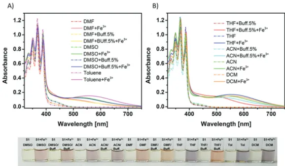
Solvent Effect Studies. The behavior of S1 has been investigated in common solvents by UV−vis and spectro-fluorimetric measurements. The fluorescence emission spec-trum of the ligand S1 upon excitation at 365 nm exhibits the characteristic monomer emission of the anthracene moiety in all tested conditions. However, the excimer emission appeared to be strongly dependent on the solvent system used.89
Figure 5 shows a clear enhancement of the excimer formation in less polar solvents such as toluene, THF, or DCM. By increasing the polarity of the solvents, the In3+-S1
complex displays a decrease of the emission presumably due to nonradiative relaxation and solvent interactions inducing a lack of flexibility within the sensor and inhibiting the excimer formation.90,91 For instance, in pure DMSO as well as in a mixture of DMSO with 5% of buffer (Bis-Tris, 10 mM, pH 7), the addition of In3+ to the solution of S1 resulted only in a slight excimer emission. In contrast, thefluorescence emission
spectra of the In3+-S1 complex in the other solvents and their respective buffer mixtures exhibit a broad excimer emission band, reducing drastically the monomer emission band. In DCM, THF, or toluene, this effect is even more pronounced, increasing the excimer emission band intensity and decreasing almost totally the monomer emission band. Therefore, the In3+
sensing is less effective in DMSO than in other solvent systems, even though a slight excimer emission band can be detected. These results showed a clear dependency of the In3+-S1 excimer formation as a function of the solvent used (Figure 5). Moreover, the presence of the buffer leads in general to a weaker emission than in the absence of buffer. This might be due to the interaction of the buffer with the ligand, e.g. via H-bonds, and hence additional quenching pathways.
The Fe2+/Fe3+ detection is however not dependent on the solvent system. Indeed, the characteristic broad CT band appearing upon Fe2+/Fe3+ addition has been observed in all tested solvents (Figure 6).
These results revealed that the use of different solvents does not interfere with the detection of Fe2+/Fe3+and In3+, despite
the low efficiency of the In3+ detection in DMSO and in its buffer mixtures. These measurements also showed that the use Scheme 3. Suggested Recognition Mechanism of Fe2+, Fe3+, and Cu2+ Ions by the In3+-S1 Complex
Figure 5.Solvent dependency of thefluorogenic detection of In3+(40
μM) in DCM, THF, DMF, DMSO, ACN, toluene, and their corresponding buffer (5%, Bis-Tris, 10 mM, pH 7) mixtures.
of buffer in diverse solvents is also possible in order to target potential biological assays.
Crystallography. In order to gain further insights into the coordination mechanisms of metal ions by S1, considerable efforts were undertaken to obtain good quality single crystals of the host−guest complexes. This was however only possible for compound S1′, the ligand S1, and the 1:1 Cu2+-S1 complex.
For S1, having a C2-axis running through the geometrical
middle of the central C−C bond of the molecule, the two anthracene moieties point outward with the chromophore planes nearly perpendicular to the dioxo-benzene rings with an angle of 85° (Figure 7). These latter two dioxo-benzene rings
are arranged in an antiparallel fashion forming an angle of 70°, probably due to the steric hindrance of the two chromophores. This arrangement is stabilized by the H-bonds between the alcohol proton H1 and the N atom N1 (and their symmetry equivalents), with a distance of 1.947 Å.
In the Cu2+-S1 complex, the molecule of S1 is now deprotonated and wraps around the Cu2+ cation, which
occupies the N2O2 chelating site of the ligand in a
quasi-perfect square planar fashion (Figure 8). Having a bond valence sum92of nearly 2.0 provided by the N2O2cavity of the
sensor, Cu2+does not need to complete its coordination sphere
using additional solvent or anion entities. The angle sum around the metal ion within the N2O2 recognition moiety is
360.85°, indicating the quasi planarity of this coordination. Compared to the structure of S1 alone, the orientation of one side arm of the complex is almost preserved with an angle of 79.40° between the external anthracene moiety and the main plane formed by the dioxo-benzene ring. For the other extremity of the ligand, the anthracene moiety points toward the benzene ring with an angle of 64.44°, inducing a slight distortion of the planarity of the N2O2coordination of 20.03°
based on the mean aromatic ring planes. The two anthracene moieties are in parallel planes; however, they do not stack on top of each other, showing closest contacts via C32̵ π and C10 ̵C33 at 3.584 and 3.534 Å, respectively. Access to the O2O2
cavity seems thereby hindered by the large hydrophobic groups, one of which points above, and the other below that binding site. A detailed description of all single crystal structures and their corresponding figures can be found in theSupporting Information (Figures S22−S24 and Table S1). The structural aspects of this Cu complex of S1 allow us to conclude that the binding of a metal ion to the first coordination site, N2O2, apparently induces a steric hindrance
of the anthracene moieties within the Cu2+ complex. This binding restricts thus the access to the second recognition site, O2O2, inhibiting the coordination of a potential second cation
and impeding the correct self-assembly of the anthracene moieties. From the above solution studies, we assume that the ligand will adopt a similar behavior in the presence of iron,
Figure 6.Solvent dependency of the colorimetric detection of Fe3+(40μM) in (A) DMF, DMSO, and toluene and in (B) THF, ACN, DCM, and
their correspondent buffer (5%, Bis-Tris, 10 mM, pH 7) mixtures.
Figure 7.Crystal structure of sensor S1, (#2(1/2 − x, 1/2 − y, z)); H atoms are omitted except for O1−H and O1#2-H (thermal ellipsoids at 50% probability level).
Figure 8.Two views of the crystal structure of the complex Cu2+-S1,
all H atoms are omitted for clarity (thermal ellipsoids at 50% probability level).
while a distinct arrangement of the probe in contact with indium seems to occur. Indeed, solid state structures of Schiff base derived ligands with indium show a ca. 10° greater folding deformation of the ligand scaffold than for transition metal ions such as Cu2+.65,66Therefore, the coordination of thefirst indium ion to the probe S1 might lead to a parallel arrangement of the anthracene moieties on one side of the O2O2 cavity, as indicated by the excimer formation, thereby
giving access to the O2O2 cavity and thereby rendering the
coordination of a second indium ion possible.
■
CONCLUSIONA highly versatile sensor has been successfully synthesized and introduced as first selective chromogenic and fluorogenic sensor for iron and indium ions. Probe S1 is able to detect In3+ ions exhibiting strong excimer emission in various common solvents. Moreover, the designed compound can also be employed as selective sensor to detect the presence of Fe2+or
Fe3+ ions. In both cases, competition experiments in the presence of other cations demonstrated that the Cu2+ ion
prevents the excimer emission of the two anthracene moieties of the sensor S1 and impedes the color change of the solution. This effect may lead to the formation of a potential new sensor, the In3+-S1 complex, which could be used as “On−Off”
fluorogenic sensor for the detection of Cu2+
and Fe2+/Fe3+, subsequently distinguishable by colorimetry. Moreover, the obtainment of the ligand crystal structure and its Cu-complex helped to interpret the different detection mechanisms. This new anthracene-functionalized Schiff-base probe S1, which exhibits very low detection limits for the sensing of In3+ and
Fe2+/Fe3+ions in various solvent systems, could be used in the tracking of environmental In3+and Fe2+/Fe3+ions and in vitro
biological samples. It could then also provide inspiration for the design and development of new hydrosoluble sensors for such detection in vivo.
■
ASSOCIATED CONTENT*
S Supporting InformationThe Supporting Information is available free of charge on the ACS Publications website at DOI: 10.1021/acs.inorg-chem.9b01478.
General experimental information; NMR spectrum of the aldehyde based starting material S1′ and of the ligand S1; NMR titration of the probe S1; UV−vis and fluorescent titrations of sensor S1; ESI mass analysis for Fe2+, Fe3+, and In3+complex; Competition experiments;
Detection limits of Fe2+, Fe3+, and In3+; Fe2+ and Fe3+ oxidation test; Crystallographic data (PDF)
Accession Codes
CCDC 1860984−1860986 contain the supplementary crys-tallographic data for this paper. These data can be obtained free of charge viawww.ccdc.cam.ac.uk/data_request/cif, or by emailing [email protected], or by contacting The Cambridge Crystallographic Data Centre, 12 Union Road, Cambridge CB2 1EZ, UK; fax: +44 1223 336033.
■
AUTHOR INFORMATION Corresponding Author *E-mail:[email protected]. ORCID Alba Finelli:0000-0002-0273-9613 Nelly Hérault:0000-0001-8986-7759 Cheal Kim: 0000-0002-7580-0374 Katharina M. Fromm:0000-0002-1168-0123 Author ContributionsThe manuscript was written through contributions of all authors. All authors have given approval to thefinal version of the manuscript.
Notes
The authors declare no competingfinancial interest.
■
ACKNOWLEDGMENTSA.F. is grateful to Noémie Voutier for fruitful discussions. This work was supported by the Swiss National Science Foundation (grant number 200020_152777) and the National Research Foundation of Korea through a “Bilateral Korean-Swiss Science and Technology Program”, by Frimat and the University of Fribourg.
■
REFERENCES(1) Oakley, A. E.; Collingwood, J. F.; Dobson, J.; Love, G.; Perrott, H. R.; Edwardson, J. A.; Elstner, M.; Morris, C. M. Individual dopaminergic neurons show raised iron levels in Parkinson disease. Neurology 2007, 68, 1820−1825.
(2) Bishop, G. M.; Robinson, S. R.; Liu, Q.; Perry, G.; Atwood, C. S.; Smith, M. A. Iron: a pathological mediator of Alzheimer disease? Dev. Neurosci. 2002, 24, 184−187.
(3) Rapp, R. A. The transition from internal to external oxidation and the formation of interruption bands in silver-indium alloys. Acta Metall. 1961, 9, 730−741.
(4) Stanbery, B. J. Copper indium selenides and related materials for photovoltaic devices. Crit. Rev. Solid State Mater. Sci. 2002, 27, 73− 117.
(5) Nagano, K.; Nishizawa, T.; Umeda, Y.; Kasai, T.; Noguchi, T.; Gotoh, K.; Ikawa, N.; Eitaki, Y.; Kawasumi, Y.; Yamauchi, T. Inhalation carcinogenicity and chronic toxicity of indium-tin oxide in rats and mice. J. Occup. Health 2011, 53, 175−187.
(6) Lokanc, M.; R. Eggert, M. R. The Availability of Indium: The Present, Medium Term, and Long Term; 2015.
(7) Lim, C. H.; Han, J.-H.; Cho, H.-W.; Kang, M. Studies on the toxicity and distribution of indium compounds according to particle size in Sprague-Dawley rats. Toxicol. Res. 2014, 30, 55.
(8) Castronovo, F. P.; Wagner, H. N. Factors affecting the toxicity of the element indium. Br. J. Exp. Pathol. 1971, 52, 543.
(9) Clarkson, T. W.; Friberg, L.; Nordberg, G. F.; Sager, P. R. Biological monitoring of toxic metals; Springer Science & Business Media, 2012; ISBN 1461309611.
(10) Harvey, R. R.; Virji, M. A.; Cummings, K. J. Assessing risk of indium lung disease to workers in downstream industries. Am. J. Ind. Med. 2017, 60, 310.
(11) Choi, S.; Won, Y. L.; Kim, D.; Lee, M.; Choi, Y. j.; Park, J.; Kim, H.; Jung, J. I.; Lee, S.; Kim, E. Interstitial lung disorders in the indium workers of Korea: an update study for the relationship with biological exposure indices. Am. J. Ind. Med. 2015, 58, 61−68.
(12) Suzuki, Y.; Matsushita, H. Interaction of metal ions with phospholipid monolayer and their acute toxicity. Ind. Health 1969, 7, 143−154.
(13) Bi, X.; Westerhoff, P. Adsorption of III/V ions (In(III), Ga(III) and As(V)) onto SiO2, CeO2and Al2O3 nanoparticles used in the
semiconductor industry. Environ. Sci.: Nano 2016, 3, 1014−1026. (14) Stellman, J. M. Encyclopaedia of occupational health and safety; International Labour Organization, 1998; Vol. 1; ISBN 9221092038. (15) Chapin, R. E.; Harris, M. W.; Hunter, E. S., III; Davis, B. J.; Collins, B. J.; Lockhart, A. C. The reproductive and developmental toxicity of indium in the Swiss mouse. Toxicol. Sci. 1995, 27, 140− 148.
(16) Cummings, K. J.; Nakano, M.; Omae, K.; Takeuchi, K.; Chonan, T.; Xiao, Y.; Harley, R. A.; Roggli, V. L.; Hebisawa, A.; Tallaksen, R. J. Indium lung disease. Chest 2012, 141, 1512−1521.
(17) Shannon, R. D. Revised effective ionic radii and systematic studies of interatomic distances in halides and chalcogenides. Acta Crystallogr., Sect. A: Cryst. Phys., Diffr., Theor. Gen. Crystallogr. 1976, 32, 751−767.
(18) McIntyre, P. A.; Larson, S. M.; Eikman, E. A.; Colman, M.; Scheffel, U.; Hodkinson, B. A. Comparison of the metabolism of iron-labeled transferrin (Fe· TF) and indium-iron-labeled transferrin (In· TF) by the erythropoietic marrow. J. Nucl. Med. 1974, 15, 856−862.
(19) Moshtaghie, A. A.; Ghaffari, M. A. Study of the binding of iron and indium to human serum apo-transferrin. Iran. Biomed. J. 2003, 7, 73−77.
(20) Wu, S.-P.; Chen, Y.-P.; Sung, Y.-M. Colorimetric detection of Fe3+ ions using pyrophosphate functionalized gold nanoparticles.
Analyst 2011, 136, 1887−1891.
(21) Narayanaswamy, N.; Govindaraju, T. Aldazine-based colori-metric sensors for Cu2+and Fe3+. Sens. Actuators, B 2012, 161, 304−
310.
(22) Jung, J. M.; Lee, S. Y.; Kim, C. A novel colorimetric chemosensor for multiple target metal ions Fe2+, Co2+, and Cu2+ in
a near-perfect aqueous solution: Experimental and theoretical studies. Sens. Actuators, B 2017, 251, 291−301.
(23) Lee, M. H.; Van Giap, T.; Kim, S. H.; Lee, Y. H.; Kang, C.; Kim, J. S. A novel strategy to selectively detect Fe(III) in aqueous media driven by hydrolysis of a rhodamine 6G Schiff base. Chem. Commun. 2010, 46, 1407−1409.
(24) Wang, B.; Hai, J.; Liu, Z.; Wang, Q.; Yang, Z.; Sun, S. Selective Detection of Iron (III) by Rhodamine-Modified Fe3O4Nanoparticles.
Angew. Chem., Int. Ed. 2010, 49, 4576−4579.
(25) Kim, S. K.; Kim, S. H.; Kim, H. J.; Lee, S. H.; Lee, S. W.; Ko, J.; Bartsch, R. A.; Kim, J. S. Indium(III)-Induced Fluorescent Excimer Formation and Extinction in Calix[4]arene−Fluoroionophores. Inorg. Chem. 2005, 44, 7866−7875.
(26) Cho, H.; Chae, J. B.; Kim, C. A thiophene-based blue-fluorescent emitting chemosensor for detecting indium (III) ion. Inorg. Chem. Commun. 2018, 97, 171−175.
(27) Kim, C.; Chae, J. B. A Highly Selective Fluorescent Chemosensor for Detecting Indium (III) with a Low Detection Limit and its Application. J. Fluoresc. 2018, 28, 1363−1370.
(28) Wu, Y.-C.; Li, H.-J.; Yang, H.-Z. A sensitive and highly selective fluorescent sensor for In3+. Org. Biomol. Chem. 2010, 8, 3394−3397.
(29) Jang, H. J.; Kang, J. H.; Yun, D.; Kim, C. A multifunctional selective“turn-on” fluorescent chemosensor for detection of Group IIIA ions Al3+, Ga3+and In3+. Photochem. Photobiol. Sci. 2018, 17,
1247−1255.
(30) Okda, H. E.; El Sayed, S.; Otri, I.; Ferreira, R. C. M.; Costa, S. P. G.; Raposo, M. M. M.; Martínez-Máñez, R.; Sancenón, F. A simple and easy-to-prepare imidazole-based probe for the selective chromo-fluorogenic recognition of biothiols and Cu (II) in aqueous environments. Dyes Pigm. 2019, 162, 303−308.
(31) Lee, S. Y.; Yang, M.; Kim, C. A dual target chemosensor for the fluorometric detection of In3+ and colorimetric detection of Fe3+.
Spectrochim. Acta, Part A 2018, 205, 622−629.
(32) Kim, H.; Kim, K. B.; Song, E. J.; Hwang, I. H.; Noh, J. Y.; Kim, P.-G.; Jeong, K.-D.; Kim, C. Turn-on selective fluorescent probe for trivalent cations. Inorg. Chem. Commun. 2013, 36, 72−76.
(33) Han, D. Y.; Kim, J. M.; Kim, J.; Jung, H. S.; Lee, Y. H.; Zhang, J. F.; Kim, J. S. ESIPT-based anthraquinonylcalix[4]crown chemosensor for In3+. Tetrahedron Lett. 2010, 51, 1947−1951.
(34) Mehta, P. K.; Hwang, G. W.; Park, J.; Lee, K.-H. Highly Sensitive Ratiometric Fluorescent Detection of Indium (III) Using Fluorescent Probe Based on Phosphoserine as a Receptor. Anal. Chem. 2018, 90, 11256−11264.
(35) Santos-Figueroa, L. E.; Llopís-Lorente, A.; Royo, S.; Sancenón, F.; Martínez-Máñez, R.; Costero, A. M.; S, G.; M, P. A Chalcone-Based Highly Selective and Sensitive Chromofluorogenic Probe for Trivalent Metal Cations. ChemPlusChem 2015, 80, 800−804.
(36) Wu, D.; Sedgwick, A. C.; Gunnlaugsson, T.; Akkaya, E. U.; Yoon, J.; James, T. D. Fluorescent chemosensors: the past, present and future. Chem. Soc. Rev. 2017, 46, 7105−7123.
(37) Zhou, X.; Lee, S.; Xu, Z.; Yoon, J. Recent progress on the development of chemosensors for gases. Chem. Rev. 2015, 115, 7944− 8000.
(38) Formica, M.; Fusi, V.; Giorgi, L.; Micheloni, M. New fluorescent chemosensors for metal ions in solution. Coord. Chem. Rev. 2012, 256, 170−192.
(39) Gale, P. A.; Caltagirone, C. Fluorescent and colorimetric sensors for anionic species. Coord. Chem. Rev. 2018, 354, 2−27.
(40) Zhang, Z.; Lu, S.; Sha, C.; Xu, D. A single thiourea-appended 1, 8-naphthalimide chemosensor for three heavy metal ions: Fe3+, Pb2+,
and Hg2+. Sens. Actuators, B 2015, 208, 258−266.
(41) Hwang, S. M.; Kim, M. S.; Lee, M.; Lim, M. H.; Kim, C. Single fluorescent chemosensor for multiple targets: sequential detection of Al3+and pyrophosphate and selective detection of F−in near-perfect aqueous solution. New J. Chem. 2017, 41, 15590−15600.
(42) Song, E. J.; Kang, J.; You, G. R.; Park, G. J.; Kim, Y.; Kim, S.-J.; Kim, C.; Harrison, R. G. A single molecule that acts as a fluorescence sensor for zinc and cadmium and a colorimetric sensor for cobalt. Dalt. Trans. 2013, 42, 15514−15520.
(43) Xu, Z.; Xiao, Y.; Qian, X.; Cui, J.; Cui, D. Ratiometric and selective fluorescent sensor for Cu(II) based on internal charge transfer (ICT). Org. Lett. 2005, 7, 889−892.
(44) Zeng, L.; Miller, E. W.; Pralle, A.; Isacoff, E. Y.; Chang, C. J. A selective turn-on fluorescent sensor for imaging copper in living cells. J. Am. Chem. Soc. 2006, 128, 10−11.
(45) Yoon, S.; Miller, E. W.; He, Q.; Do, P. H.; Chang, C. J. A bright and specific fluorescent sensor for mercury in water, cells, and tissue. Angew. Chem. 2007, 119, 6778−6781.
(46) Chang, J. H.; Choi, Y. M.; Shin, Y. A significant fluorescence quenching of anthrylaminobenzocrown ethers by paramagnetic metal cations. Bull. Korean Chem. Soc. 2001, 22, 527−530.
(47) Palomares, E.; Vilar, R.; Durrant, J. R. Heterogeneous colorimetric sensor for mercuric salts. Chem. Commun. 2004, 40, 362−363.
(48) Kim, K. B.; Kim, H.; Song, E. J.; Kim, S.; Noh, I.; Kim, C. A cap-type Schiff base acting as a fluorescence sensor for zinc (II) and a colorimetric sensor for iron (II), copper (II), and zinc (II) in aqueous media. Dalt. Trans. 2013, 42, 16569−16577.
(49) Seth, P.; Ghosh, S.; Figuerola, A.; Ghosh, A. Trinuclear heterometallic Cu(II)-Mn(II) complexes of a salen type Schiff base ligand: anion dependent variation of phenoxido bridging angles and magnetic coupling. Dalt. Trans. 2014, 43, 990−998.
(50) Helal, A.; Rashid, M. H. O.; Choi, C.-H.; Kim, H.-S. Chromogenic and fluorogenic sensing of Cu2+ based on coumarin. Tetrahedron 2011, 67, 2794−2802.
(51) Hui, J. K.; Yu, Z.; MacLachlan, M. J. Supramolecular Assembly of Zinc Salphen Complexes: Access to Metal-Containing Gels and Nanofibers. Angew. Chem., Int. Ed. 2007, 46, 7980−7983.
(52) Kleij, A. W. Zinc-centred salen complexes: versatile and accessible supramolecular building motifs. Dalt. Trans. 2009, 38, 4635−4639.
(53) Bermejo, M. R.; Carballido, R.; Fernández-García, M. I.; González-Noya, A. M.; González-Riopedre, G.; Maneiro, M.; Rodríguez-Silva, L. Synthesis, Characterization, and Catalytic Studies of Mn(III)-Schiff Base-Dicyanamide Complexes: Checking the Rhombicity Effect in Peroxidase Studies. J. Chem. 2017, 2017, 10 pages. .
(54) Nakamura, T.; Kimura, H.; Okuhara, T.; Yamamura, M.; Nabeshima, T. A Hierarchical Self-Assembly System Built Up from Preorganized Tripodal Helical Metal Complexes. J. Am. Chem. Soc. 2016, 138, 794−797.
(55) Akine, S.; Taniguchi, T.; Nabeshima, T. Helical metallohost-guest complexes via site-selective transmetalation of homotrinuclear complexes. J. Am. Chem. Soc. 2006, 128, 15765−15774.
(56) Finelli, A.; Hérault, N.; Crochet, A.; Fromm, K. M. Threading salen-type Cu-and Ni-complexes into one-dimensional coordination
polymers: Solution versus solid state, and the size effect of the alkali metal ion. Cryst. Growth Des. 2018, 18, 1215−1226.
(57) Akine, S.; Taniguchi, T.; Nabeshima, T. Acyclic bis (N2O2
chelate) ligand for trinuclear d-block homo-and heterometal complexes. Inorg. Chem. 2008, 47, 3255−3264.
(58) Mousavi, M.; Béreau, V.; Costes, P.; Duhayon, C.; Sutter, J.-P. Oligomeric and polymeric organizations of potassium salts with compartmental Schiff-base complexes as ligands. CrystEngComm 2011, 13, 5908−5914.
(59) Lü, X.; Wong, W.; Wong, W. Self-Assembly of Luminescent Platinum-Salen Schiff-Base Complexes. Eur. J. Inorg. Chem. 2008, 2008, 523−528.
(60) Cheng, J.; Ma, X.; Zhang, Y.; Liu, J.; Zhou, X.; Xiang, H. Optical chemosensors based on transmetalation of salen-based Schiff base complexes. Inorg. Chem. 2014, 53, 3210−3219.
(61) Shoora, S. K.; Jain, A. K.; Gupta, V. K. A simple Schiff base based novel optical probe for aluminium (III) ions. Sens. Actuators, B 2015, 216, 86−104.
(62) Cozzi, P. G. Metal−Salen Schiff base complexes in catalysis: practical aspects. Chem. Soc. Rev. 2004, 33, 410−421.
(63) Baleizão, C.; Gigante, B.; Sabater, M. J.; Garcia, H.; Corma, A. On the activity of chiral chromium salen complexes covalently bound to solid silicates for the enantioselective epoxide ring opening. Appl. Catal., A 2002, 228, 279−288.
(64) Canali, L.; Sherrington, D. C. Utilisation of homogeneous and supported chiral metal (salen) complexes in asymmetric catalysis. Chem. Soc. Rev. 1999, 28, 85−93.
(65) Aluthge, D. C.; Ahn, J. M.; Mehrkhodavandi, P. Overcoming aggregation in indium salen catalysts for isoselective lactide polymerization. Chem. Sci. 2015, 6, 5284−5292.
(66) Aluthge, D. C.; Patrick, B. O.; Mehrkhodavandi, P. A highly active and site selective indium catalyst for lactide polymerization. Chem. Commun. 2013, 49, 4295−4297.
(67) Cametti, M.; Nissinen, M.; Dalla Cort, A.; Mandolini, L.; Rissanen, K. Ion Pair Recognition of Quaternary Ammonium and Iminium Salts by Uranyl− Salophen Compounds in Solution and in the Solid State. J. Am. Chem. Soc. 2007, 129, 3641−3648.
(68) Shellaiah, M.; Wu, Y.-H.; Singh, A.; Raju, M. V. R.; Lin, H.-C. Novel pyrene-and anthracene-based Schiff base derivatives as Cu2+
and Fe3+ fluorescence turn-on sensors and for aggregation induced
emissions. J. Mater. Chem. A 2013, 1, 1310−1318.
(69) Tyagi, N.; Singh, O.; Singh, U. P.; Ghosh, K. Nitric oxide (NO) reactivity studies on mononuclear iron (II) complexes supported by a tetradentate Schiff base ligand. RSC Adv. 2016, 6, 115326−115333.
(70) Chiang, L.; Savard, D.; Shimazaki, Y.; Thomas, F.; Storr, T. FeIII Bipyrrolidine Phenoxide Complexes and Their Oxidized Analogues. Inorg. Chem. 2014, 53, 5810−5819.
(71) Hasan, K.; Fowler, C.; Kwong, P.; Crane, A. K.; Collins, J. L.; Kozak, C. M. Synthesis and structure of iron (III) diamine-bis (phenolate) complexes. Dalt. Trans. 2008, 22, 2991−2998.
(72) Mayilmurugan, R.; Sankaralingam, M.; Suresh, E.; Palaniandavar, M. Novel square pyramidal iron (III) complexes of linear tetradentate bis (phenolate) ligands as structural and reactive models for intradiol-cleaving 3, 4-PCD enzymes: Quinone formation vs. intradiol cleavage. Dalt. Trans. 2010, 39, 9611−9625.
(73) Simon, T.; Shellaiah, M.; Srinivasadesikan, V.; Lin, C.-C.; Ko, F.-H.; Sun, K. W.; Lin, M.-C. A simple pyrene based AIEE active schiff base probe for selective naked eye and fluoresence off−on detection of trivalent cations with live cell application. Sens. Actuators, B 2016, 231, 18−29.
(74) Liu, H.; Yao, L.; Li, B.; Chen, X.; Gao, Y.; Zhang, S.; Li, W.; Lu, P.; Yang, B.; Ma, Y. Excimer-induced high-efficiency fluorescence due to pairwise anthracene stacking in a crystal with long lifetime. Chem. Commun. 2016, 52, 7356−7359.
(75) Erdemir, S.; Kocyigit, O. Anthracene excimer-based“turn on” fluorescent sensor for Cr3+and Fe3+ions: its application to living cells.
Talanta 2016, 158, 63−69.
(76) Gao, Y.; Liu, H.; Zhang, S.; Gu, Q.; Shen, Y.; Ge, Y.; Yang, B. Excimer formation and evolution of excited state properties in discrete
dimeric stacking of an anthracene derivative: a computational investigation. Phys. Chem. Chem. Phys. 2018, 20, 12129−12137.
(77) Maudoux, N.; Roisnel, T.; Dorcet, V.; Carpentier, J.; Sarazin, Y. Chiral (1, 2)-Diphenylethylene-Salen Complexes of Triel Metals: Coordination Patterns and Mechanistic Considerations in the Isoselective ROP of Lactide. Chem. - Eur. J. 2014, 20, 6131−6147.
(78) Jones, C.; Junk, P. C.; Black, S. J.; Lewis, J. The molecular structure of [InBr(Salen)(OSMe2)]. Main Group Met. Chem. 2001,
24, 123−124.
(79) Bhattacharya, S.; Mohanta, S. Heterometallic copper (II)−lead (II), nickel (II)−lead (II) and copper (II)−indium (III) compounds derived from an acyclic double-compartment Schiff base ligand. Inorg. Chim. Acta 2015, 432, 169−175.
(80) Mei, Y.; Borger, J. E.; Wu, D.-J.; Grützmacher, H. Salen supported Al−O−CP and Ga−PCO complexes. Dalt. Trans. 2019, 48, 4370−4374.
(81) Liao, S.; Yang, X.; Jones, R. A. Self-assembly of luminescent hexanuclear lanthanide salen complexes. Cryst. Growth Des. 2012, 12, 970−974.
(82) Du, J.; Wang, X.; Zou, X.; Li, Y.; Li, W.; Yao, X.; Li, G. Structures and luminescent sensors of mixed-counterions based salen-type lanthanide coordination polymers. Luminescence 2018, 33, 1040− 1047.
(83) Ren, M.; Xu, Z.-L.; Bao, S.-S.; Wang, T.-T.; Zheng, Z.-H.; Ferreira, R. A. S.; Zheng, L.-M.; Carlos, L. D. Lanthanide salen-type complexes exhibiting single ion magnet and photoluminescent properties. Dalt. Trans. 2016, 45, 2974−2982.
(84) Li, Q.; Yan, P.; Chen, P.; Hou, G.; Li, G. Salen type sandwich triple-decker tri-and di-nuclear lanthanide complexes. J. Inorg. Organomet. Polym. Mater. 2012, 22, 1174−1181.
(85) Yang, Y.; Yan, P.-F.; Gao, P.; Gao, T.; Hou, G.-F.; Li, G.-M. Salen-type lanthanide complexes with luminescence and near-infrared (NIR) properties. J. Inorg. Organomet. Polym. Mater. 2013, 23, 1211− 1218.
(86) Rurack, K. Flipping the light switch‘ON’−the design of sensor molecules that show cation-induced fluorescence enhancement with heavy and transition metal ions. Spectrochim. Acta, Part A 2001, 57, 2161−2195.
(87) Datta, A.; Das, K.; Massera, C.; Clegg, J. K.; Sinha, C.; Huang, J.-H.; Garribba, E. A mixed valent heterometallic Cu(II)/Na(I) coordination polymer with sodium−phenyl bonds. Dalt. Trans. 2014, 43, 5558−5563.
(88) He, W.; Liu, Z. A fluorescent sensor for Cu2+and Fe3+based on
multiple mechanisms. RSC Adv. 2016, 6, 59073−59080.
(89) Puangsamlee, T.; Tachapermpon, Y.; Kammalun, P.; Sukrat, K.; Wainiphithapong, C.; Sirirak, J.; Wanichacheva, N. Solvent control bifunctional fluorescence probe for selective detection of Cu2+ and
Hg2+via the excimer of pyrenylacetamide subunits. J. Lumin. 2018,
196, 227−235.
(90) Choi, Y.-G.; Kwak, G. Solvatochromic fluorescence in an immiscible two-phase system of alcohols and hydrophobic polymers: Distinction between light alcohol and water. Polymer 2018, 141, 194− 201.
(91) Nagata, Y.; Nishikawa, T.; Suginome, M. Solvent-dependent fluorescence and circular dichroism properties of poly(quinoxaline-2, 3-diyl)s bearing pyrene pendants. Chem. Commun. 2012, 48, 11193− 11195.
(92) Liu, W.; Thorp, H. H. Bond valence sum analysis of metal-ligand bond lengths in metalloenzymes and model complexes. 2. Refined distances and other enzymes. Inorg. Chem. 1993, 32, 4102− 4105.