Laparoscopic ventral hernia repair is safe and cost effective
G. Beldi, R. Ipaktchi, M. Wagner, B. Gloor, D. Candinas
Department of Visceral and Transplant Surgery, Inselspital University Bern, CH-3010 Bern, Switzerland Received: 20 June 2005/Accepted: 8 September 2005/Online publication: 7 December 2005
Abstract
Background: Ventral hernia repair is increasingly per-formed by laparoscopic means since the introduction of dual-layer meshes. This study aimed to compare the early complications and cost effectiveness of open hernia repair with those associated with laparoscopic repair. Methods:Open ventral hernia repair was performed for 92 consecutive patients using a Vypro mesh, followed by laparoscopic repair for 49 consecutive patients using a Parietene composite mesh.
Results: The rate of surgical-site infections was signifi-cantly higher with open ventral hernia repair (13 vs 1; p= 0.03). The median length of hospital stay was sig-nificantly shorter with laparoscopic surgery (7 vs 6 days; p= 0.02). For laparoscopic repair, the direct operative costs were higher (2,314 vs 2,853 euros; p = 0.03), and the overall hospital costs were lower (9,787 vs 7,654 euros; p = 0.02).
Conclusions:Laparoscopic ventral hernia repair leads to fewer surgical-site infections and a shorter hospital stay than open repair. Despite increased operative costs, overall hospital costs are lowered by laparoscopic ven-tral hernia repair.
Key words: Cost — Laparoscopy — Mesh — Poly-propylene — Ventral hernia
The use of mesh for ventral hernia repair has been proved superior to direct suturing methods in terms of long-term recurrence [11]. With the introduction of new mesh types, ventral hernia repair via laparoscopic means is gaining increasing acceptance. In the initial series, expanded polytetrafluoroethylene (ePTFE) meshes were used predominantly [7]. However, because of the small
pore size, these meshes are, without impregnation of disinfectants, more prone to infection [4, 6].
Polyester and polypropylene meshes were introduced in ventral hernia repair with the development of dual-layer technology. These meshes have a larger pore size and different biologic properties than ePTFE [12]. A marked inflammatory reaction leads to incorporation of the mesh into the abdominal wall. Synthesis of a neo-peritoneal layer combined with integration of the mesh minimizes the risk for infection [12].
No study comparing open and laparoscopic ventral hernia repair using polypropylene-based meshes had been performed. Therefore, we conducted a cohort study comparing these two methods in terms of postoperative morbidity and treatment costs in a single center.
Materials and methods
All patients with mesh implantation for ventral hernia repair who underwent surgery between March 2003 and March 2005 were in-cluded in the study. The indication for mesh implantation was a minimum hernia diameter of 2 cm. Five patients were excluded from laparoscopic surgery: four who had general contraindications for laparoscopy and one who was not willing to undergo laparoscopic hernia repair.
Patient data for open repair were reviewed retrospectively from the medical records. The data for patients who had undergone lapa-roscopic repair were collected prospectively. The collected data in-cluded the patientsÕ age, gender, body mass index (BMI), surgical history, risk factors, comorbidity, mesh size, complications, and fol-low-up evaluation.
Open repair technique
For open repair, patients were in the supine position with arms ab-ducted. Single-shot antibiotic prophylactics with amoxicillin/clavulanic acid 1.2 g (Augmentin; GlaxoSmithKline, Mu¨nchenbuchsee, Switzerland) was administered intravenously. The sac of the hernia was excised in most cases. A Vypro mesh (Ethicon Schweiz, Johnson & Johnson Medical, CH-8957 Spreitenbach, Switzerland) was used. The fascia was adapted with a running PDS 1suture. The mesh size was chosen so as to overlap the rectus sheath at least 5 cm. The mesh was placed on the dorsal rectus sheath and fixed with polypropylene su-tures. Closed suction drains were placed onto the mesh and also subcutaneously. The patient was not allowed to lift weights for 4 weeks postoperatively.
Presented at 13th European Association for Endoscopic Surgery (EAES) Congress, Venice, Italy, 1–4 June 2005
Correspondence to:B. Gloor Surg Endosc (2006) 20: 92–95 DOI: 10.1007/s00464-005-0442-9
Laparoscopic repair technique
For laparoscopic repair, the patients were in the supine position with arms abducted. Single-shot antibiotic prophylactics with amoxicillin/ clavulanic acid were administered. A pneumoperitoneum of 12 mmHg was established using a limited open technique. A minimum of two additional trocars were inserted in the left flank, and complete ad-hesiolysis of the abdominal wall was performed.
A Parietene composite mesh (Sofradim, Tre´voux, France) was used for all patients. The mesh selected was larger than the hernia defect, allowing at least 4 cm of mesh beyond the perimeter of the fascial defect. The mesh was prepared by placing two types of sutures alternatively (Prolene 0 and Ethilon 0) on the edge of the mesh every 3 to 4 cm. The mesh was rolled and inserted into the abdominal cavity. The sutures were lifted above the abdominal wall with a suture passer. After reduction of the intraabdominal pressure to 8 mmHg, the threads were knotted extracorporeally. The patient was not allowed to lift weights for 4 weeks postoperatively.
Cost analysis
Operative costs were calculated by assessing all re-sources, adding an average cost factor for operative time. In-hospital costs were provided by the adminis-tration of the hospital integrating personnel salaries, materials, and equipment. The results are presented in euros using values for the year 2005.
Statistical analysis
Results are expressed as median and range for age, BMI, operative time, mesh size, and length of stay. Costs are expressed as mean ± standard deviation. The Mann– Whitney U test was used to compare age, BMI, operative time, mesh size, and length of stay. The StudentÕs t-test was used to compare costs. FisherÕs exact test was performed to compare proportions. All p values less than 0.05 were considered statistically significant. Data were analyzed statistically with SPSS Software (SPSS Inc., Chicago, IL, USA).
Results
Open hernia repair was performed for 92 consecutive patients: 87 between March 2003 and July 2004 and 5 between July 2004 and March 2005. Laparoscopic ven-tral hernia repair was performed for 49 consecutive patients between July 2004 and March 2005. The pa-tientsÕ characteristics are outlined in Tables 1and 2. No significant differences in the patientsÕ general and sur-gical risk factors were identified. The operative time and mesh size were not significantly different (Table 3). The length of stay was significantly shorter for patients after laparoscopic ventral hernia repair than for patients after open repair. The follow-up period for the patients was 69 weeks (range, 6–115 weeks) after open hernia repair and 10 weeks (range, 6–25 weeks) after laparoscopic ventral hernia repair.
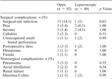
Surgical complications occurred in 25 cases (27.2%) after open ventral hernia repair and in 7 cases (14.3%) after laparoscopy (p = 0.09). Most of the complications involved infections (Table 4). Surgical-site infections
occurred significantly more often in patients after open hernia repair. Among the patients with surgical-site infections, the mesh had to be removed for one patient. For eight patients, the infection was treated using vacuum-assisted closure. Prolonged postoperative pain over 6 weeks was found in 5.4% of the patients who had open hernia repair and in 6.1% of those who had lapa-roscopic ventral repair. Among the open repair patients, two required repetitive nerve infiltrations.
Pain after laparoscopic ventral hernia repair was always localized at the sites of the transfascial sutures. A second laparoscopy was performed for one patient to rule out recurrence. In this patient and another patient, the pain was treated successfully by removing up to four transfascial sutures with the patient under local anes-thesia.
Nonsurgical complications were experienced by se-ven patients (7.6%) after open repair and one patient (2%) after laparoscopic repair (p = 0.26). One patient died 73 days after open repair of multiorgan failure attributable to surgical-site infection and pneumonia.
The mean cost of surgery was 2,314 ± 925 euros for open ventral hernia repair and 2,853 ± 1,147 euros for laparoscopic repair (p = 0.03). The hospital cost was 7,312 ± 7,697 euros for open repair and 4,902 ± 2,514 euros for laparoscopic repair (p = 0.04). The overall costs were 9,787 ± 8,021euros for open repair and 7,654 ± 3,204 for laparoscopic repair (p = 0.02).
Discussion
In the current series, laparoscopic ventral hernia repair using a dual-layer polypropylene mesh and transfascial suturing significantly reduced surgical-site infections, length of hospital stay, and costs as compared with open mesh repair.
Surgical-site infections after open mesh repair oc-curred for 14% of the patients. This rate is comparable with the 4% to 18% rate reported in published large series [5]. In our series, only one mesh had to be re-moved in each group because of infection.
In contrast to the use of ePTFE meshes, conservative treatment of infected polypropylene-based meshes was possible for four patients after open repair [5, 6]. The incidence of seroma formation was markedly lower after laparoscopic repair than in series using ePTFE mesh [3, 7]. This low incidence of seroma formation may be attributable to the fact that the large pores of the dual-layer polypropylene mesh allows a more efficient resorption of wound secretion into the abdominal cavity than afforded by ePTFE meshes.
The rates of fistula formation (1for open vs 0 for laparoscopic surgery) and unrecognized bowel injuries (1in each group) did not differ between the two groups. These complications were well within the range up to 3% for fistula formation and up to 6% for bowel injury in other published series [1, 2, 7, 9, 13].
Transfascial sutures result in a very rigid fixation of the mesh to the fasciae of the abdominal wall. In vivo experiments showed that the tension of the mesh with transfascial sutures is 2.5 times greater than fixation
with metallic staples [14]. The use of fixation by sutures has only the advantage of being applicable in all kinds of hernias independently of their size, and of avoiding the exposure of metallic materials to the intestines with their potential sequelae (adhesions, small bowel obstruction and perforation, and hernia formation) [8, 10].
Pain attributable to nerve entrapment and tight suturing was relieved by suture removal for two patients under local anesthesia.
This study has shown decreased overall hospital costs for laparoscopic hernia repair despite higher operative costs. The types of fixation device and mesh are important factors contributing to direct operative costs. Whether the use of staplers may markedly de-crease operative time and consequently costs remains to be proved. In addition, to prove an overall cost
reduc-tion, indirect and intangible costs will need to be as-sessed prospectively.
Although laparoscopic ventral hernia repair had advantages over open surgery in the short-term out-come, the rate of recurrence in the long term will ulti-mately define its impact. To answer this question with unbiased scientific evidence, large, controlled random-ized trials are required. Before initiation of these studies, the risks and advantages of various mesh materials and their fixation methods must be characterized.
References
1. Balen EM, Diez-Caballero A, Hernandez-Lizoain JL, Pardo F, Torramade JR, Regueira FM, Cienfuegos JA (1998) Repair of ventral hernias with expanded polytetrafluoroethylene patch. Br J Surg 85: 1415–1418
2. Bauer JJ, Harris MT, Kreel I, Gelernt IM (1999) Twelve-year experience with expanded polytetrafluoroethylene in the repair of abdominal wall defects. Mt Sinai J Med 66: 20–25
Table 1. Patient characteristics
Open (n = 92) Laparoscopic (n = 49) pValue Male/female 59/33 33/19 0.54
Age (years): n (range) 62 (28–84) 57 (20–84) 0.43
ASA (SD) 0.46
1–2 33 21
3–4 42 22
BMI (kg/m2): n (range) 27.5 (17–52) 29.0 (20–65) 0.53
Previous hernia repair: n (%) 18 (19.5) 9 (18.4) 1.00 Type of previous laparotomy: n (%)
Midline 57 (62) 28 (57.1) 0.59
Pfannenstiel 6 (6.5) 0 (0.0) 0.09
Transverse epigastric 10 (10.9) 7 (14.3) 0.59
Lumbotomy 3 (3.3) 3 (6.1) 0.42
Appendectomy 2 (2.2) 1(2) 1.00
No previous abdominal surgery 14 (15.2) 10 (20.4) 0.48 ASA, American Society of Anesthesiology; BMI, body mass index
Table 2. Patient risk factors for hernia formation
Risk factors Open (n = 92) n (%) Laparoscopic (n = 49) n (%) pValue Obesity 28 (30.4) 11 (22.4) 0.33 Cardiovascular disease 32 (34.8) 24 (49) 0.11 Immunosuppression 13 (14.1) 6 (12.2) 1.00 COPD 7 (7.6) 9 (18.4) 0.09 Smoking 7 (7.6) 8 (16.3) 0.15 Renal failure 6 (6.5) 4 (8.2) 0.74 COPD, chronic obstructive pulmonary disease
Table 3. Surgical details Open
(n = 92) n (range)
Laparoscopic
(n = 49) n (range) p Value Operative time (min) 155 (60–375) 158 (50–360) 0.28 Conversions — 3
Mesh size (cm2) 400 (40–1,000) 500 (144–1,100) 0.36
LOS (days) 7 (2–87) 6 (3–32) 0.02 LOS, length of hospital days
Table 4. Peri- and postoperative morbidity Open (n = 92) Laparoscopic (n = 49) pValue Surgical complications: n (%) Surgical-site infection 13 (14.1) 1 (2) 0.03 Pain 5 (5.4) 3 (6.1) 1.00 Seroma 5 (5.4) 2 (4.1) 1.00 Cellulitis 3 (3.3) 0 0.55 Unrecognized small bowel perforation 1 (1.1) 1 (2) 0.99 Postoperative ileus 3 (3.3) 1(2) 1.00 Hematoma 1 (1.1) 0 1.00 Fistula 1 (1.1) 0 1.00 Nonsurgical complications: n (%) Pneumonia 3 (3.3) 0 0.55 Atrial fibrillation 2 (2.2) 0 0.54 Renal failure 1 (1.1) 0 1.00 Diarrhea/Colitis 1 (1.1) 1 (2) 0.99 94
3. Carbajo MA, Martp del Olmo JC, Blanco JI, Toledano M, de la Cuesta C, Ferreras C, Vaquero C (2003) Laparoscopic approach to incisional hernia. Surg Endosc 17: 118–122
4. Carbonell AM, Matthews BD, Dreau D, Foster M, Austin CE, Kercher KW, Sing RF, Heniford BT (2005) The susceptibility of prosthetic biomaterials to infection. Surg Endosc 19: 430–435 5. Cassar K, Munro A (2002) Surgical treatment of incisional hernia.
Br J Surg 89: 534–545
6. Diaz JJ Jr, Gray BW, Dobson JM, Grogan EL, May AK, Miller R, Guy J, OÕNeill P, Morris JA Jr (2004) Repair of giant abdominal hernias: does the type of prosthesis matter? Am Surg 70: 396–401, discussion 401–402
7. Heniford BT, Park A, Ramshaw BJ, Voeller G (2003) Laparo-scopic repair of ventral hernias: nine yearsÕ experience with 850 consecutive hernias. Ann Surg 238: 391–399, discussion 399– 400
8. Karahasanoglu T, Onur E, Baca B, Hamzaoglu I, Pekmezci S, Boler DE, Kilic N, Altug T (2004) Spiral tacks may contribute to intraabdominal adhesion formation. Surg Today 34: 860–864 9. Koehler RH, Begos D, Berger D, Carey S, LeBlanc K, Park A,
Ramshaw B, Smoot R, Voeller G (2003) Minimal adhesions to
ePTFE mesh after laparoscopic ventral incisional hernia repair: reoperative findings in 65 cases. J Soc Lap Endosc Surg 7: 335–340 10. Ladurner R, Mussack T (2004) Small bowel perforation due to protruding spiral tackers: a rare complication in laparoscopic in-cisional hernia repair. Surg Endosc 18: 1001
11. Luijendijk RW, Hop WC, van den Tol MP, de Lange DC, Braaksma MM, JN IJ, Boelhouwer RU, de Vries BC, Salu MK, Wereldsma JC, Bruijninckx CM, Jeekel J (2000) A comparison of suture repair with mesh repair for incisional hernia. N Engl J Med 343: 392–398
12. McGinty JJ, Hogle NJ, McCarthy H, Fowler DL (2005) A com-parative study of adhesion formation and abdominal wall in-growth after laparoscopic ventral hernia repair in a porcine model using multiple types of mesh. Surg Endosc 19: 786–790
13. McLanahan D, King LT, Weems C, Novotney M, Gibson K (1997) Retrorectus prosthetic mesh repair of midline abdominal hernia. Am J Surg 173: 445–449
14. vanÕt Riet M, de van Vos Steenwijk PJ, Kleinrensink GJ, Steyerberg EW, Bonjer HJ (2002) Tensile strength of mesh fixation methods in laparoscopic incisional hernia repair. Surg Endosc 16: 1713–1716