Isomerization and aggregation of 2-(2-(2-hydroxy-4-nitrophenyl)
hydrazono)-1-phenylbutane-1,3-dione: Recent evidences from theory
and experiment
Silvia Hristova
a, Fadhil S. Kamounah
b,c, Aurelien Crochet
d, Poul Erik Hansen
c, Katharina M. Fromm
d,
Daniela Nedeltcheva
a, Liudmil Antonov
a,⁎
aInstitute of Organic Chemistry with Centre of Phytochemistry, Bulgarian Academy of Sciences, Acad. G. Bonchev str., bldg. 9, 1113 Sofia, Bulgaria b
Department of Chemistry, University of Copenhagen, Universitetsparken 5, DK-2100 Copenhagen Ø, Denmark
c
Department of Science and Environment, Roskilde University, DK-4000 Roskilde, Denmark
dDepartment of Chemistry, University of Fribourg, Chemin du Musée 9, CH-1700 Fribourg, Switzerland
a b s t r a c t
The title compound potentially can exist as four isomers in solution. Recently Lycka has proposed a protocol for distinguishing two of them based on15N NMR. This approach has been confirmed theoretically, in the current
study, and further developed into a logical scheme that allows the existence of each of the isomers to be proven in solution. The experimental data, obtained by NMR and UV–Vis spectroscopy, have shown that the studied compound exists as a mixture of two solvent stabilized isomers in diluted solutions of dimethyl sulfoxide. How-ever, the concentration effect also plays a substantial role allowing formation of linear aggregates as the X-ray analysis and theoretical calculations show.
Keywords: Rotary switch NMR DFT X-ray Aggregation 1. Introduction
The title compound, structure 1 inScheme 1, has been synthe-sized by Mahmudov et al. [1,2] and studied by using molecular spec-troscopy in relation to its potential tautomerism. The authors have stated that 1 exists as a mixture between azo and hydrazone tauto-mers in methanol and dimethyl sulfoxide (DMSO). Recently we have investigated the spectral behaviour of 1 in a variety of solvents, proving that the conclusions previously made for the existence of the azo tautomer are wrong [3]. The combined use of UV–Vis spectros-copy, NMR (1H and13C) and theoretical calculations has shown
that the investigated compound exists as a mixture of isomers of the hydrazone tautomer, generally assigned as E-oriented (major form) and Z-oriented (minor form) in Scheme 1. Very recently Lycka [4], using15N NMR, reached the same conclusion for 1 and 2
in DMSO and deuterochloroform.
However, the previous studies have left some questions open. On one side, according to the theoretical calculations, four isomers could exist in solution (Scheme 2). It was concluded that their stabilization depends on the solvent– structures 1E′ and 1Z′ are presented in
DMSO possibly as a result of a solute-solvent interaction. Compound 1 spontaneously deprotonates at low concentrations and aggregates at high concentrations (10−4M and higher) according to the UV–Vis investigations [3]. These two processes are linked and depend strongly on the solvent. Although the dimer existence was proven in-directly, its exact structure could not be established using molecular spectroscopy. Therefore, a theoretical guess was made, suggesting linear wire-like aggregates between E′ isomers (Scheme 3) with par-ticipation of the OH groups, which explains the deprotonation at low concentrations. This opens a general question about the reasons for stabilization of the E′ and Z′ isomers in which an OH group exists that is unengaged in intramolecular hydrogen bonding– either by formation of complexes with proton acceptor solvents or traces of water, or aggregation in all solvents. Due to the very low solubility of 1 in most of the solvents, this question is difficult to be answered purely experimentally.
Therefore, in the current communication, we will theoretically check the reliability of the approach proposed by Lycka [4] and will use his data to suggest a general logical scheme for distinguishing between all four isomers. A combination between theoretical and ex-perimental data will be used to prove the effects of the concentration and solvent in the stabilization of the E′ and Z′ isomers in dimethyl sulfoxide.
⁎ Corresponding author.
E-mail address:[email protected](L. Antonov).
http://doc.rero.ch
Published in "Journal of Molecular Liquids 283(): 242–248, 2019"
which should be cited to refer to this work.
2. Experimental part
2.1. Compounds
The synthesis of the compounds has been described previously [3].
2.2. Spectral measurements
Spectral measurements were performed on Jasco V-570 UV–Vis-NIR spectrophotometer, equipped with a thermostatic cell holder (using Huber MPC-K6 thermostat with precision 1 °C), in spectral grade sol-vents at ambient temperature.
The NMR spectra were recorded at 400 MHz for1H and at 100.6 MHz for13C at a Bruker 400 Avance III NMR spectrometer. TMS or the solvent
signal was used as reference. NOE and HMBC spectra are recorded using standard pulse programs and conditions.
2.3. X-ray measurements
Single crystals of C16H13N3O5(1) were obtained from methanol by
slow evaporation. A suitable crystal was selected and mounted on a loop with oil and measured on a STOE IPDS II diffractometer. The crystal was kept at 250(2) K during data collection. Using Olex2 [5], the struc-ture was solved with the ShelXT strucstruc-ture solution program [6] using in-trinsic phasing, and refined with the ShelXL refinement package [7] using least squares minimization.
Crystal Data for C16H13N3O5(M = 327.29 g/mol): monoclinic, space
group P21/n (no. 14), a = 10.3609(6) Å, b = 8.7949(3) Å, c = 16.8458
(10) Å,β = 93.153(5)°, V = 1532.72(14) Å3, Z = 4, T = 250(2) K,
μ(MoKα) = 0.108 mm−1, Dcalc= 1.418 g/cm3, 19,148 reflections
mea-sured (4.508°≤ 2Θ ≤ 50.218°), 2733 unique (Rint= 0.0358, Rsigma=
0.0166), which were used in all calculations. Thefinal R1was 0.0286 (I
N 2σ(I)), and wR2was 0.0786 (all data). Crystallographic data have
been deposited with the Cambridge Crystallographic Data Centre (CCDC-1858058 (1)). Copies may be obtained free of charge on applica-tion to the Director, CCDC, 12 Union Road, Cambridge CB2 1EZ, UK (e-mail:[email protected]).
Compound 1 crystallizes in the monoclinic space group P21/n (No.
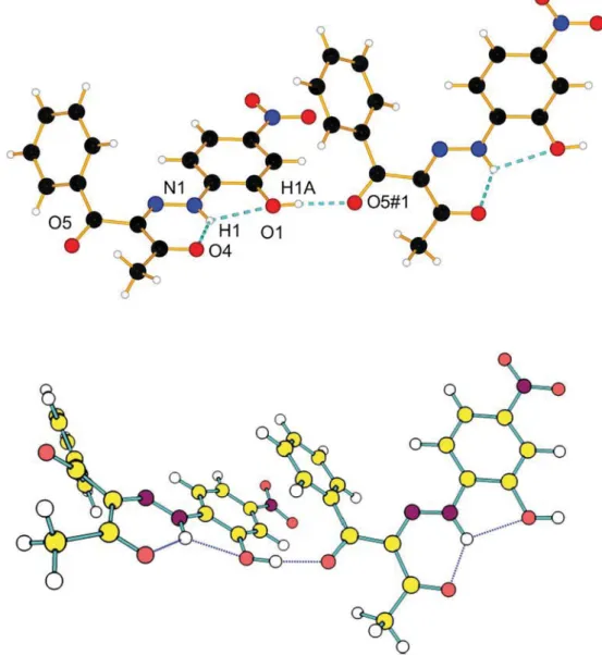
14) with one molecule per asymmetric unit. The phenyl moiety is twisted by 61° with respect to the mean plane formed by the rest of the atoms of 1, probably due to steric reasons. An intramolecular H-bond is found between O4 and N1–H1 with a distance of 1.888 Å and an angle of 130.7°. Intermolecular H-bonds are found between O1– H1A and O5 of a neighbor molecule with 1.884 Å and an angle of almost 176°. Molecules pack pairwise in a head-to-tail manner with short dis-tances involvingπ–π contacts between e.g. N2 and C6 (3.398 Å), C4 and C15 (3.441 Å) and C4 and C7 (3.541 Å),Fig. 1. With respect to one such pair, the next pair is parallel, but offset by ca. 4.3 Å, still involving π–π stacking between the carbonyl groups C15–O4 and its symmetry equivalent, at distances of 3.225 Å, as well as a short N1–C16 contact of 3.354 Å. The tilted phenyl ring is involved in an intermolecular H-bond between H10 and O4 with a distance of 2.681 and an C10–H10– O4 angle of 162°.
2.4. Quantum-chemical calculations
Quantum-chemical calculations were performed by using the Gaussian 09 program suite [8]. If not explicitly described, the M06-2X functional [9,10] was used with the TZVP basis set [11]. Thisfitted hy-brid meta-GGA functional with 54% HF exchange is especially devel-oped to describe main-group thermochemistry and the non-covalent interactions, showing very good results in the prediction of the position of the tautomeric equilibrium in azo naphthols possessing intramolecu-lar hydrogen bonds, [12] and in the description of the proton transfer re-actions in naphthols [13,14]. All structures were optimized without restrictions, using tight optimization criteria and ultrafine grid in the Scheme 1. Isomers of the structures under study.
Scheme 2. Possible isomers of the hydrazone form of 1.
Scheme 3. Sketch of the linear aggregate of 1E′.
computation of two-electron integrals and their derivatives, and the true minima were verified by performing frequency calculations in the corresponding environment. Solvent effects are described by using the Polarizable Continuum Model (the integral equation formalism variant, IEFPCM, as implemented in Gaussian 09) [15]. The absorption spectra of the compounds were predicted using the TD-DFT formalism. TD-DFT calculations were carried out at the same functional and basis set, which is in accordance with conclusions about the effect of the basis set size and the reliability of the spectral predictions [16–18].
The NMR chemical shieldings of selected tautomeric forms of the studied compounds were calculated using the GIAO approximation [19] at the B3LYP/6-311+G(2d,p) level of theory. This level of theory was recommended in the pioneering work by Cheeseman et al. [20], fo-cused on the comparison of different models for calculating nuclear magnetic resonance shielding tensors and shows very good results in predicting the NMR spectra of azo-hydrazone tautomerism [21]. The calculated absolute shieldings were transformed to chemical shifts using the reference compound tetramethylsilane, Si(CH3)4, for
hydro-gen, carbon and nitromethane, CH3NO2, for nitrogen atoms:δ = δcalc
(ref)− δcalc. Bothδcalc(ref) andδcalcwere evaluated at the same
compu-tational level. For the1H chemical shifts the experimental and calculated
shifts of compounds 1 and 3 in DMSO were plotted again each other to gives a linear equationδ(1
H)experimental =−0.956 × δ(1H)calculated
+ 30.516 (R2= 0.995, Supplementary information, Tables S1 and S2,
Fig. S1).
3. Results and discussion
As seen fromTable 1, where the theoretical results are summarized, in all solvents the E form should be more stable, followed by the E′ iso-mer, practically in the whole range (as dielectric constant) of used sol-vents when implicit solvation is applied. According to the calculations measurable amounts of E and E′ could be presented in solution and Z′ could be seen in polar solvents in amounts to less than a few percent. The availability of the OH group, not involved in intramolecular hydro-gen bonding, in E′ and Z′, allows their substantial stabilization when specific interaction with DMSO or water molecules is considered by adding explicit solvent molecules. The same type of stabilization can be achieved when homodimers (interaction between two molecules following the pattern inScheme 3) are formed. It is difficult to deter-mine which of these two processes is stronger, because the stabilization energies are almost the same. The stabilization energies1of the DMSO
complexes of E′ and Z′ are 13 and 12.9 kcal/mol respectively, while the stabilization through wire-like dimer2is 12.2 and 11.4 kcal/mol. Correspondingly, the closed forms E and Z, where neither strong solvent complexes nor stable dimers could be possible, are destabilized. Here specific solvation with proton donor solvents, involving the free C_O group could be expected, but the same could exists in E′ and Z′ as well, which nulls the effect. In acetonitrile no strong solvent complexes could be expected, while the aggregation cannot be fully excluded, tak-ing into account that the stabilization energies are almost the same as in DMSO.
Taking into account that all four isomers are almost identical from the viewpoint of UV–Vis spectroscopy, their identification in DMSO was made by careful analysis of the NMR spectra of 1 by using 3 as a reference compound [3]. It was shown on basis of1H and13C spectra that E′ and Z′
forms are presented in solution as major and minor component, respec-tively, which matches the theoretical predictions very well. The recent study of Lycka has brought additional,15N NMR based, evidences for
stronger stabilization of the E-oriented COMe group [4]. His conclusions are based on using2J(15N
β,13C) constants of 1 in DMSO‑d6. According to
Cheatham et al. [22] the differences in these constants could be large enough and can easily denote which carbonyl carbon is closer to the elec-tron lone pair of Nβnitrogen (seeScheme 4for the numbering). The
measured2J values (in Hz) for the major isomer are correspondingly
b1 for C-2 and 11.9 for C-4, showing that the Ph-linked carbonyl carbon is closer to the Nβnitrogen lone pair. The opposite is proven for the
minor component, which was assigned as Z-oriented. These trends have been reproduced theoretically as summarized inTable 2. The theo-retically predicted2J(15Nβ,13C-4) constants are huge in the case of E(E′)
Fig. 1. Above: Ortep representation of 1, ellipsoids are drawn with 50% of probability, H-bonds are draw as blue dash lines; Below: Stacking of 1, H-H-bonds are drawn as dash blue lines and short contact as dash orange lines; for clarity, only the shortest short contacts are represented.
1 Defined as ΔE = (E
isomer+ EDMSO)-Ecomplex. Positive value indicates that the complex
is favored.
2 Defined as ΔE = 2.E
monomer-Edimer. Positive value indicates that the dimer is favored.
forms and negligible for Z(Z′) ones. The exactly opposite is true for2J
(15N
β,13C-2). The obtained trends and values confirm reasonably well
the experimental data (Table 2). Although it is known that13C\\15N
cou-plings over two or three bonds are generally small, we have calculated2J
(15N
α,13C) couplings. The calculated results show for C-2′ in respect of
Nαvalues of 1.0 Hz corresponding to E′/Z′, while values for 2.9 Hz of
C-6′ are calculated for the E/Z isomers. In the paper of Lycka the value2J
(15N
α,13C-2′) was measured as 1.6 for the major isomer and 1.5 for the
minor one, and no value for2J(15Nα,13C-6′) is shown [4]. Consequently
the combined use of2J(15N
α,13C) and2J(15Nβ,13C) confirms the
availabil-ity of 1E′ as a major component and 1Z′, as a minor ones, in solution. In the general context of the study of isomerization of similar rotary switches, the use of the combination of these two2J constants gives a
good opportunity to distinguish between E and Z and their correspond-ing E′ and Z′ analogues:2J(15N
α,13C-2′) is defined by the Nα-C-1′
rota-tion, while2J(15N
β,13C-2) is a function of the Nβ-C-1 bond rotation. As
a result each of the four isomers, shown inScheme 2, can be presented as a unique logical combination (small/large) of the2J constants and the
assignment can be easily done based on the experimental data (Table 2).
The crystal structure of 1 is shown inFig. 2. Two conclusions can be drawn from it: the studied compound exists as E′ isomer in the solid state, which confirms previously made conclusions for its dominance in proton acceptor solvents [3] or in solvents with mixed nature like methanol or water (see relative stability in water as a guideline,
Table 1); as predicted by the theoretical calculations, 1E′ is stabilized by formation of linear aggregates (Fig. S3) and as can be seen from
Fig. 1there is a very good match between theory and experiment, again taking into account the fact that crystals were grown in methanol. This particular structure of the aggregate excludes effective overlapping of the electronic density of the monomers and, consequently, no sub-stantial spectral changes in accordance with the exciton theory [13,23–25] could be observed in the absorption spectra. The theoreti-cally predicted absorbance in both monomer and dimer, shown in
Fig. 1, is located in the range 352–365 nm. Most probably the experi-mental spectra of both species are also almost identical and strongly overlaps, which could not bring UV–Vis spectral evidences for the self-association. The evidences are indirect: in the experimentally measured UV–Vis spectra [3] the observed changes are related only to the process of deprotonation in proton acceptor solvents at low concentrations (10−5M and lower) as a result of the aggregates' destruction according to the general equilibrium scheme:
Aggregates⇔Monomers⇔Anions
The assumed scheme brings up the question of what actually hap-pens in proton acceptor solvents (like DMSO and DMF) at the concen-tration range used in the NMR measurements. As was proven, the deprotonation is a result of the formation of solute-solvent (mono-mer-solvent) complexes at low concentrations. Following the scheme, a rise of the concentration should thus reduce the monomers in favor of the aggregates, which brings a concern about the existence of the solute-solvent complexes as a whole at the NMR concentrations.
As seen from [3] the observed chemical shifts for H-6′ are rather sim-ilar for the major form in all three solvents suggesting the same molec-ular environment (Table 2). Furthermore, the predicted chemical shifts are almost the same for E and Z, while two different values were mea-sured for the major and minor forms and different values are predicted for E′ and Z′. The same pattern, based on structural similarities, is obvi-ous in the predicted values for the solute-solvent complexes and the ag-gregates. As seen in the case of DMSO it is less obvious to distinguish between E′ and the E′-E′ aggregate based on NH chemical shifts.
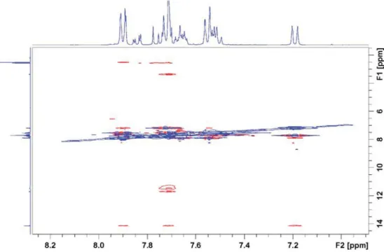
Looking at the X-ray structure (Fig. 2), the H-3′ to H-2″ or H-6″ (from the neighbor molecule in the aggregate) distances are 3.49 and 3.67 Å. In the calculated dimers, they are similar with 3.4 and 3.8 Å. These dis-tances should allow observation of NOESY cross peaks if the complex is also present in solution. The NOESY spectrum, presented inFig. 3, a cross-peak is seen between OH at 11.52 to H-3′ at 7.72 ppm. In the pre-vious report a cross peak with water was seen to H-3′ and it was sug-gested that the hydrogen bond partner was DMSO (see Supplementary material, Fig. S5). The observation of cross peaks from H2O to H-3′ suggests a complex with a water molecule. However, the
calculations suggest that the DMSO molecule is involved as shown in
Fig. 4. Actually the formation of this kind of associate in presence of Table 1
Relative energies (M06-2X/TZVP) of the isomers of 1a
in various solvents in kcal/mol units. Species Isomer Solvent (PCM
model) ΔE ΔE + ZPE ΔG Monomer E Chloroform 0.00 0.00 0.00 Z 2.44 (1.62) 1.83b 2.41 (1.38) 2.07 (1.03) E′ 0.94 1.26b 0.88 0.29 Z′ 2.54 2.72b 2.16 1.19 E Acetonitrile 0.00 0.00 0.00 Z 2.07 (1.13) 1.48b 2.07 (0.86) 1.83 (0.39) E′ 0.79 1.07b 0.84 0.54 Z′ 1.93 2.14b 1.48 0.71 E DMSO 0.00 0.00 0.00 Z 2.06 (1.11) 1.46b 2.05 (0.83) 1.83 (0.35) E′ 0.78 1.07b 0.84 0.57 Z′ 1.90 2.12b 1.53 0.71 Monomer-DMSO complexd E 4.65 7.96b 4.85 5.59 Z 2.30 7.85b 1.97 2.82 E′ 0.00 0.00 0.00 Z′ 1.15 0.89b 0.89 0.49 Dimer E′-E′ 0.00 0.00 0.00c Z′-Z′ 1.43 1.75b 0.96 0.05c a
Relative energies in the case of 2 are given in brackets.
b
Relative energies in the case of 1 using B3LYP/6-311+G(2d,p)//M06-2X/TZVP.
c
Less reliable due to very lowfirst positive frequency.
d The corresponding water complexes in water environment have the following
rela-tive energies (following the same order from E to Z′): 3.25, 3.59, 0.0, 1.07 kcal/mol, the predicted complexes are shown in Fig. S2.
Scheme 4. Numbering of carbonyl and nitrogen atoms in 1.
water explains very well why the deprotonation is suppressed in presence of water as it seen experimentally [3]. The deprotonation needs free monomers to interact directly with DMSO, which
deprotonates them, while the water molecules stabilize the aggregates. In dry DMSO as, shown in Fig. S6 the compound is substantially deprotonated.
Table 2
Predicted (B3LYP/6-311+G(2d,p)//M06-2X/TZVP) and experimental15
N NMR parameters of selected atoms of 1 in DMSO. Structure δ(1 H), ppm δ(15 N), ppm 2 J(15 Nβ,13C), Hz 2J(15Nα,13C), Hz OH NH H-6′ Nα Nβ C-2 C-4 C-2′ C-6′ E 9.15 14.54 7.34 −208.7 −35.9 −0.4 8.2 −0.3 2.9 Z 10.12 12.96 7.35 −217.4 −46.1 8.3 −0.6 −0.2 2.8 E′ 5.28 14.35 7.74 −219.3 −16.2 −0.1 9.0 1.0 −0.1 Z′ 5.24 12.73 8.34 −227.6 −26.1 9.3 −0.4 1.0 −0.1 E-DMSO complex 9.43 14.69 8.83 −200.7 −39.0 −0.3 8.5 −0.3 2.8 Z-DMSO complex 10.22 13.30 7.78 −215.2 −54.9 8.1 −1.2 −0.3 2.9 E′-DMSO complex 13.71 (10.94)a 14.35 (14.31)a 7.63 (7.65)a −215.0 −13.7 −0.1 9.0 1.1 −0.1 Z′-DMSO complex 13.71 (10.92)a 12.95 (12.84)a 8.24 (8.27)a −223.0 −23.4 9.3 −0.4 1.1 −0.1 E′-E′ aggregate 10.34 14.49 7.49 −216.5 −14.6 −0.2 10.0 1.1 −0.1 Z′-Z′ aggregate 8.95 12.82 8.16 −225.0 −24.3 9.7 −0.4 1.0 −0.1 Major form (E′)b,c
11.52 14.14 7.20 −220.9c −21.3c b1c 11.9c 1.6c Minor form (Z′)b,c 11.34 11.70 7.75 −229.7c −29.8c 12.5c b1c 1.5c a
Water complex in DMSO (see Fig. S2).
b Experimentally measured [3], also in Fig. S1. c
Taken from [4], relative error ± 0.3 Hz for2
J constants.
Fig. 2. H-bonds motifs of 1 (upper), H-bonds are draw as blue dash lines, Symm. op.:−1/2 + x, 3/2 − y, −1/2 + z; Theoretically predicted dimer of 1E′ (down).
The calculated structure as shown inFig. 4has reasonable distances between H-3′ and H-2″, H-6″ to enable the observation of a NOE cross peak and the OH group is in the vicinity of H-3′ and is also hydrogen bonded indirectly to DMSO. The complex is as seen asymmetric and re-quires motion to give a single set of NMR resonances. The observed chemical shift for the OH proton should near the predicted value for the water complex, which is observed inTable 2.
4. Conclusions
Based on the experimental data published by Lycka we have proven that it is possible to distinguish between the isomers of 1 using the2J constants in the15N NMR spectra. This approach confirms that the
major form in solution of 1 and 3 is E′, which in the case of 1 is stabilized by forming linear aggregate. In DMSO, in presence of water, an associate involving both water and DMSO is observed, which explains experi-mentally observed effect of water addition. The crystallographic data confirm perfectly the theoretically predicted structure of the E′ aggregates.
Acknowledgements
Thefinancial support from Bulgarian Ministry of Education by the project DFNP-17-66/26.07.2017 for support of young scientists is grate-fully acknowledged.
Appendix A. Supplementary data
Supplementary data to this article can be found online
References
[1] K.T. Mahmudov, R.A. Rahimov, M.B. Babanly, P.Q. Hasanov, F.G. Pashaev, A.G. Gasanov, M.N. Kopylovich, A.J.L. Pombeiro, Tautomery and acid–base properties of some azoderivatives of benzoylacetone, J. Mol. Liq. 162 (2011) 84–88,https://doi. org/10.1016/j.molliq.2011.06.005.
[2] W. Kuznik, M.N. Kopylovich, G.I. Amanullayeva, A.J.L. Pombeiro, A.H. Reshak, K.T. Mahmudov, I.V. Kityk, Role of tautomerism and solvatochromism in UV–VIS spectra of arylhydrazones ofβ-diketones, J. Mol. Liq. 171 (2012) 11–15,https://doi.org/10. 1016/j.molliq.2012.03.023.
Fig. 4. E′-E′ complex involving water molecule as a linker.
Fig. 3. Part of the NOESY spectrum of 1 in DMSO‑d6(the full spectrum is shown in Fig. S4).
[3] S. Hristova, F.S. Kamounah, N. Molla, P.E. Hansen, D. Nedeltcheva, L. Antonov, The possible tautomerism of the potential rotary switch 2-(2-(2-Hydroxy-4-nitrophe-nyl)hydrazono)-1-phenylbutane-1,3-dione, Dyes Pigments 144 (2017) 249–261,
https://doi.org/10.1016/j.dyepig.2017.05.021.
[4] A. Lyčka, 15 N NMR study of (E)- and (Z)-2-(2-(2-hydroxy-4-nitrophenyl) hydrazono)-1-phenylbutane-1,3-diones. A suitable method for analysis of hydrazone isomers, Dyes Pigments 150 (2018) 181–184,https://doi.org/10.1016/j. dyepig.2017.10.023.
[5] O.V. Dolomanov, L.J. Bourhis, R.J. Gildea, J.A.K. Howard, H. Puschmann, OLEX2: a complete structure solution, refinement and analysis program, J. Appl. Crystallogr. 42 (2009) 339–341,https://doi.org/10.1107/S0021889808042726.
[6] G.M. Sheldrick, SHELXT– integrated space-group and crystal-structure determina-tion, Acta Crystallographica Section A Foundations and Advances 71 (2015) 3–8,
https://doi.org/10.1107/S2053273314026370.
[7] G.M. Sheldrick, Crystal structure refinement with SHELXL, Acta Crystallographica Section C Structural Chemistry 71 (2015) 3–8, https://doi.org/10.1107/ S2053229614024218.
[8] M.J. Frisch, G.W. Trucks, H.B. Schlegel, G.E. Scuseria, M.A. Robb, J.R. Cheeseman, G. Scalmani, V. Barone, B. Mennucci, G.A. Petersson, H. Nakatsuji, M. Caricato, X. Li, H.P. Hratchian, A.F. Izmaylov, J. Bloino, G. Zheng, J.L. Sonnenberg, M. Hada, M. Ehara, K. Toyota, R. Fukuda, J. Hasegawa, M. Ishida, T. Nakajima, Y. Honda, O. Kitao, H. Nakai, T. Vreven, J.A. Montgomery Jr., J.E. Peralta, F. Ogliaro, M.J. Bearpark, J. Heyd, E.N. Brothers, K.N. Kudin, V.N. Staroverov, R. Kobayashi, J. Normand, K. Raghavachari, A.P. Rendell, J.C. Burant, S.S. Iyengar, J. Tomasi, M. Cossi, N. Rega, N.J. Millam, M. Klene, J.E. Knox, J.B. Cross, V. Bakken, C. Adamo, J. Jaramillo, R. Gomperts, R.E. Stratmann, O. Yazyev, A.J. Austin, R. Cammi, C. Pomelli, J.W. Ochterski, R.L. Martin, K. Morokuma, V.G. Zakrzewski, G.A. Voth, P. Salvador, J.J. Dannenberg, S. Dapprich, A.D. Daniels, Ã. Farkas, J.B. Foresman, J.V. Ortiz, J. Cioslowski, D.J. Fox, Gaussian 09 Revision D.01, Gaussian, Inc., Wallingford, CT, USA, 2013.
[9] Y. Zhao, D.G. Truhlar, Density functionals with broad applicability in chemistry, Acc. Chem. Res. 41 (2008) 157–167,https://doi.org/10.1021/ar700111a.
[10] Y. Zhao, D.G. Truhlar, The M06 suite of density functionals for main group thermo-chemistry, thermochemical kinetics, noncovalent interactions, excited states, and transition elements: two new functionals and systematic testing of four M06-class functionals and 12 other functionals, Theor. Chem. Accounts 120 (2008) 215–241,
https://doi.org/10.1007/s00214-007-0310-x.
[11] F. Weigend, R. Ahlrichs, Balanced basis sets of split valence, triple zeta valence and quadruple zeta valence quality for H to Rn: design and assessment of accuracy, Phys. Chem. Chem. Phys. 7 (2005) 3297,https://doi.org/10.1039/b508541a. [12] S. Kawauchi, L. Antonov, Description of the tautomerism in some azonaphthols, J.
Phys. Org. Chem. 26 (2013) 643–652,https://doi.org/10.1002/poc.3143.
[13] Y. Manolova, V. Kurteva, L. Antonov, H. Marciniak, S. Lochbrunner, A. Crochet, K.M. Fromm, F.S. Kamounah, P.E. Hansen, 4-Hydroxy-1-naphthaldehydes: proton trans-fer or deprotonation, Phys. Chem. Chem. Phys. 17 (2015) 10238–10249,https:// doi.org/10.1039/C5CP00870K.
[14] Y. Manolova, H. Marciniak, S. Tschierlei, F. Fennel, F.S. Kamounah, S. Lochbrunner, L. Antonov, Solvent control of intramolecular proton transfer: is 4-hydroxy-3-(piperidin-1-ylmethyl)-1-naphthaldehyde a proton crane? Phys. Chem. Chem. Phys. 19 (2017) 7316–7325,https://doi.org/10.1039/C7CP00220C.
[15] J. Tomasi, B. Mennucci, R. Cammi, Quantum mechanical continuum solvation models, Chem. Rev. 105 (2005) 2999–3094,https://doi.org/10.1021/cr9904009. [16] R. Improta, UV-visible absorption and emission energies in condensed phase by
PCM/TD-DFT methods, in: V. Barone (Ed.), Computational Strategies for Spectros-copy, John Wiley & Sons, Inc., Hoboken, NJ, USA 2011, pp. 37–75,https://doi.org/ 10.1002/9781118008720.ch1.
[17] L. Antonov, S. Kawauchi, Y. Okuno, Prediction of the color of dyes by using time-dependent density functional theory, Bulg. Chem. Commun. 46 (2014) 228–237.
[18] D. Jacquemin, B. Mennucci, C. Adamo, Excited-state calculations with TD-DFT: from benchmarks to simulations in complex environments, Phys. Chem. Chem. Phys. 13 (2011), 16987.https://doi.org/10.1039/c1cp22144b.
[19] K. Wolinski, J.F. Hinton, P. Pulay, Efficient implementation of the gauge-independent atomic orbital method for NMR chemical shift calculations, J. Am. Chem. Soc. 112 (1990) 8251–8260,https://doi.org/10.1021/ja00179a005.
[20] J.R. Cheeseman, G.W. Trucks, T.A. Keith, M.J. Frisch, A comparison of models for cal-culating nuclear magnetic resonance shielding tensors, J. Chem. Phys. 104 (1996) 5497–5509,https://doi.org/10.1063/1.471789.
[21] S. Hristova, S. Angelova, P.E. Hansen, L. Antonov, 4-Carboxyl-2,6-dinitrophenylazohydroxynaphthalenes tautomerism NMR re-explained and other methods verified, Dyes Pigments 142 (2017) 226–229,https://doi.org/10.1016/j. dyepig.2017.03.037.
[22] M. Kline, D. Pierce, S. Cheatham, Assignment of oxime and hydrazone configuration using1H-5N and13C-15N coupling measurements at natural abundance: assignment
of oxime and hydrazone configuration, Magn. Reson. Chem. 55 (2017) 154–156,
https://doi.org/10.1002/mrc.4536.
[23] M. Kasha, Energy transfer mechanisms and the molecular exciton model for molec-ular aggregates, Radiat. Res. 20 (1963) 55–70.
[24] M. Kasha, H.R. Rawls, M. Ashraf El-Bayoumi, The exciton model in molecular spec-troscopy, Pure Appl. Chem. 11 (1965)https://doi.org/10.1351/pac196511030371. [25] L. Antonov, G. Gergov, V. Petrov, M. Kubista, J. Nygren, UV–Vis spectroscopic and
chemometric study on the aggregation of ionic dyes in water, Talanta. 49 (1999) 99–106,https://doi.org/10.1016/S0039-9140(98)00348-8.