ORIGINAL ARTICLE
First imaging results of an intraindividual comparison
of
11
C-acetate and
18
F-fluorocholine PET/CT in patients
with prostate cancer at early biochemical first or second
relapse after prostatectomy or radiotherapy
Franz Buchegger&Valentina Garibotto&Thomas Zilli&Laurent Allainmat&
Sandra Jorcano&Hansjörg Vees&Olivier Rager&Charles Steiner&Habib Zaidi&
Yann Seimbille&Osman Ratib&Raymond Miralbell
Received: 8 May 2013 / Accepted: 6 August 2013 / Published online: 9 October 2013 # Springer-Verlag Berlin Heidelberg 2013
Abstract
Purpose18F-Fluorocholine (FCH) and 11C-acetate (ACE) PET are widely used for detection of recurrent prostate cancer (PC). We present the first results of a comparative, prospective PET/CT study of both tracers evaluated in the same patients presenting with recurrence and low PSA to compare the diagnostic information provided by the two tracers.
Methods The study group comprised 23 patients studied for a rising PSA level after radical prostatectomy (RP, 7 patients, PSA≤3 ng/ml), curative radiotherapy (RT, 7 patients, PSA ≤5 ng/ml) or RP and salvage RT (9 patients, PSA ≤5 ng/ml). Both FCH and ACE PET/CT scans were performed in a random sequence a median of 4 days (range 0 to 11 days) apart. FCH PET/CT was started at injection (307±16 MBq) with a 10-min dynamic acquisition of the prostate bed, follow-ed by a whole-body PET scan and late (45 min) imaging of the
pelvis. ACE PET/CT was performed as a double whole-body PET scan starting 5 and 22 min after injection (994±72 MBq), and a late view (45 min) of the prostate bed. PET/CT scans were blindly reviewed by two independent pairs of two expe-rienced nuclear medicine physicians, discordant subgroup results being discussed to reach a consensus for positive, negative end equivocal results.
Results PET results were concordant in 88 out of 92 local, regional and distant findings (Cohen’s kappa 0.929). In par-ticular, results were concordant in all patients concerning local status, bone metastases and distant findings. Lymph-node results were concordant in 19 patients and different in 4 patients. On a per-patient basis results were concordant in 22 of 23 patients (14 positive, 5 negative and 3 equivocal). In only one patient was ACE PET/CT positive for nodal metastases while FCH PET/CT was overall negative; interestingly, the ACE-positive and FCH-negative lymph nodes became ACE-positive in a second FCH PET/CT scan performed a few months later. Conclusion Overall, ACE and FCH PET/CT showed excel-lent concordance, on both a per-lesion and a per-patient basis, suggesting that both tracers perform equally for recurrent prostate cancer staging.
Keywords 11C-Acetate .18F-Fluorocholine . PET/CT . Intraindividual comparison . Prostate cancer recurrence
Introduction
Promising results in the detection of recurrent prostate cancer (PC) have been obtained with different PET tracers such as11 C-acetate (ACE),11C-choline, and18F-fluorocholine (FCH) [1,2]. Prostate-specific antigen (PSA) is a sensitive early blood Franz Buchegger and Valentina Garibotto contributed equally to this
study.
F. Buchegger
:
V. Garibotto:
L. Allainmat:
O. Rager:
C. Steiner:
H. Zaidi:
Y. Seimbille:
O. RatibNuclear Medicine Division, University Hospital of Geneva, 1211 Geneva 14, Switzerland
T. Zilli
:
H. Vees:
R. MiralbellRadiation-Oncology Division, University Hospital of Geneva, 1211 Geneva 14, Switzerland
S. Jorcano
:
R. MiralbellRadiation Oncology Division, Teknon Oncologic Institute, 08022 Barcelona, Spain
F. Buchegger (*)
Service of Nuclear Medicine, University Hospital of Geneva, Rue Micheli-du-Crest 24, 1211 Geneva 14, Switzerland
marker for PC recurrence particularly after radical prostatecto-my (RP) when PSA is expected to become undetectable. After surgery recurrence of cancer can be suspected at the earliest rise in PSA [3,4]. PSA values of between 0.01 and 0.1 ng/ml after RP represent a probability for a further rise in PSA and tumour recurrence of 67 %, and this probability rises to 90 % with PSA values >0.1 ng/ml [5]. After primary radiation therapy (RT), a PSA value≥2 ng/ml above the nadir is a robust standard for recurrence reliably predicting distant metastases, and cause-specific and overall mortality [6].
In this study we compared the performance of ACE and FCH for diagnosing early recurrent PC by assessing the cor-relation between the blinded reading of the two imaging sets. Early localization of recurrences might allow a still curative salvage treatment to be undertaken [7], although a consensus has not yet been defined for the indication for PET/CT in this situation [8–10]. Our group favours PET/CT very early after the definition of recurrence. Having experience with both ACE [11] and FCH [12–14], we aimed to evaluate both tracers in a comparative assessment to determine if one is superior to the other in the setting of early biochemical recurrence of PC.
Materials and methods
This prospective feasibility study was approved by the Ethics Committee of the University Hospital of Geneva, Swissmedic and OFSP, Section of Radioprotection, and was performed in conformance with Swiss legislation and Good Clinical Practice (GCP) rules and with the ethical standards of the Declaration of Helsinki and later amendments. All patients provided written informed consent to the study protocol foreseeing two PET/CT scans, one with FCH and the other with ACE in random order. In case of positive findings, the patients were investigated by com-plementary imaging (MR, bone scintigraphy) and biopsying the suspected local relapse, when indicated and feasible.
Patients
Up to the time of this report, 23 patients had been studied. The patients’ characteristics are summarized in Table1.
Inclusion criteria were:
– biochemical relapse after RP (group A), curative RT with or without androgen deprivation (AD, group B) or RP and salvage RT (with or without AD, group C)
– PSA values ≤3 ng/ml for group A and C and ≤5 ng/ml for group B
– signed written informed consent Exclusion criteria were:
– patients under AD
– patients with active major inflammatory/infectious process
Compliance with further investigations (biopsy of suspected recurrence, complementary MR and bone scintig-raphy) was not an inclusion criterion. Seven patients were included in group A, 7 in group B and 9 in group C. Mean PSA values at study entry were lowest in group A (1.1± 0.9 ng/ml), highest in group B (3.5±0.9 ng/ml), and interme-diate in group C (2.4±1.0 ng/ml).
Dual PET/CT studies
FCH and ACE PET/CT studies were performed according to a random sequence in 21 patients, while 2 patients had both PET/CT scans performed on the same day (ACE in the morn-ing and FCH in the afternoon, the two studies separated by a minimum of 2.5 h). The median interval between the two scans was 4.0 days (range 0 to 11 days).
18
F-Fluorocholine PET/CT
FCH (N,N-dimethyl-N-[18F]fluoromethylethanolammonium) was prepared according to GMP rules at AAA (Advanced Accelerator Applications), St Genis-Poully, France.
Dual PET/CT studies were performed on either a Biograph 64 scanner (18 patients) or a Biograph 16 scanner (5 patients) (Siemens Medical Solutions, Erlangen, Germany). All patients fasted for at least 4 h before the FCH and ACE PET studies. After bladder voiding, patients underwent an initial low-dose CT scan from the mid thigh to the skull performed under a standard protocol (arms held above the head and shallow breathing) using 120 kVp, 60 mAs, pitch 1.5 and 1 s per rotation. Under these conditions, the mean effective radiation dose for an adult patient was estimated at 4.5 mSv, as calculated using the IMPACT CT patient dosimetry calculator (http://www.impactscan.org/ ctdosimetry.htm).
After the CT scan, patients received a standard activity of 307 ± 16 MBq18F-FCH injected intravenously, and a contin-uous list-mode PET acquisition centred on the prostate bed was recorded over 10 min starting immediately with the tracer injection. The 10-min list mode data were used for generating 3 × 3-min time frames corresponding to 0 to 3 min, 3 to 6 min and 6 to 9 min after injection. Following the list-mode acquisi-tion, a standard whole-body PET study was performed from the mid thigh to the skull over seven to eight bed positions of 3 to 4 min each depending on patient size and weight. Two addi-tional late images of 5 min each of the pelvis were acquired immediately after the whole-body PET scan (about 45 min after tracer injection).
Following Fourier rebinning and model-based scatter correction, PET images were reconstructed using two-dimensional iterative normalized attenuation-weighted
ordered subsets expectation maximization (NAW-OSEM) [15]. The CT-based attenuation map was used to correct emission data. The default reconstruction parameters of four iterations and eight subsets followed by a postprocessing gaussian filter (kernel size 5 mm, full-width at half-maximum) were applied. Images of the three phases of FCH PET/CT are shown in Fig.1.
11
C-Acetate PET/CT
ACE was prepared at the Cyclotron Unit, University Hospital of Geneva, according to GMP rules. For the ACE PET/CT scan, which was identical to the FCH PET scan performed either on the Biograph 64 (18 patients) or the Biograph 16 (5 patients), patients fasted for at least 4 h before the study. After bladder voiding, patients received a standard activity of 994+ 72 MBq ACE injected intravenously. After ACE injection they underwent a low-dose CT scan from the mid thigh to the skull performed under identical conditions to those used for the FCH PET scan using 120 kVp, 60 mAs, pitch 1.5 and 1 s per rotation.
A first whole-body PET study was performed 5 min after tracer injection from the mid thigh to the skull over seven to
eight bed positions of 2 min each, followed immediately (about 22 min after injection) by a second whole-body PET scan of 3 min per bed position and finally a late PET scan of the prostate bed was recorded about 45 min after injection. ACE PET images were reconstructed using the same proce-dure described above for FCH PET. Images of the three phases of ACE PET/CT are shown in Fig.1.
PET/CT interpretation
Each patient's PET/CT scans were interpreted blindly by two pairs of two experienced nuclear medicine physicians who grad-ed either the FCH or ACE PET scans for the presence of recurrent tumour as positive, equivocal or negative. FCH PET scans were graded according to the typical time-course of activity in malignant and benign lesions. Focal FCH hyperactivity above background that persisted throughout the examination with typ-ical kinetics was considered a positive lesion [13]. For the ACE PET scans, focal ACE hyperactivity confirmed in the first and second whole-body scan was considered positive. The third ACE acquisition centred on the prostate bed was not retained for interpretation since hyperactivities decreased in all patients to a Table 1 Characteristics of 23
patients according to initial treatment
Characteristic Radical prostatectomy
Primary radiotherapy
with or without androgen deprivation
Radical prostatectomy and salvage radiotherapy
Number of patients 7 7 9 Age (years) Mean 67.4 SD 7.5 Range 51.0 to 80.4 Initial stage, n T1c 0 1 0 T2a 0 1 0 T2b 0 1 1 T2c 6 0 2 T3a 0 3 3 T3b 1 1 3
Gleason score summed, n
6 (low grade) 2 1 1 7 (intermediate grade) 4 4 4 8 or 9 (high grade) 1 2 4 PSA (ng/ml) At diagnosis Mean 11.1 20.3 15.7 SD 4.8 7.7 9.5 Nadir Mean 0.1 0.7 0.2 SD 0.2 0.7 0.2 At PET Mean 1.1 3.5 2.4 SD 0.9 0.9 1.0
marked degree. Negative ACE and FCH PET scans were those that did not show any focal persistent activity higher than back-ground. Equivocal was defined as any lesion with an activity between the two categories“positive” and “negative”.
Specifically:
– Local relapses corresponded to focal, persistent uptake over the three FCH sequences and the two ACE se-quences in the prostate or prostatic bed. In order to avoid possible interference of bladder activity, particularly for FCH PET, on the assessment of local relapse after RP, we systematically assessed early dynamic images acquired before urine accumulation [13].
– Lymph nodes were considered positive if they showed persistent uptake over the three FCH sequences and the two ACE sequences, higher than surrounding tissue. Inguinal lymph nodes that showed transient tracer uptake and presenting a lipid centre on CT were interpreted as negative.
– CT images were used for localization and morphological analysis, discriminating between intestinal and lymph node uptakes and allowing the characterization of lesions,
such as bone hypodensity or hyperdensity and lung nodules.
Maximum standard uptake values (SUVmax) were calcu-lated within regions of interest drawn over focal hyperactiv-ities using a standard formula [16]. The reported values were measured on the whole-body FCH PET/CT study and on the first whole-body ACE PET/CT study. SUVmax values were evaluated since SUVmax is widely used in the literature and because it is not dependent on lesion size or operator. Since the two PET/CT scanners used in this study were from the same provider (Siemens) and had been installed simultaneous-ly, and acquisition, treatment protocols and software evalua-tion were identical, images and SUV calculaevalua-tions were comparable.
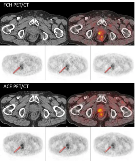
To compare the quantitative information provided by the two PET/CT scanners, we report the SUVmax of the positive lesions found in the two studies. In order to provide a measure of the contrast provided by the ACE and FCH PET images we also calculated target-to-background ratios (TBR). As a back-ground measure with a high reproducibility across studies we chose the blood-pool activity in the abdominal aorta. Fig. 1 Three-phase FCH and
three-phase ACE PET/CT showing concordant local recurrence (arrows)
Furthermore, a Jaszack phantom-based calibration was carried out to ensure compatibility between the two scanners. A radiologist interpreted the low-dose CT scans for distant sites. Standard of reference
All findings were evaluated on both PET and CT images acquired in the same session. Thus, CT images without con-trast enhancement were consistently available and allowed identification of lymph nodes and distant unrelated findings. As mentioned above, if findings were positive complementary imaging with MR and bone scintigraphy and biopsies of the suspected local recurrences were performed, when indicated and feasible. Planned patient follow-up was 2 years, and at the time of this report follow-up was ongoing with a current median of 11 months (range 1 to 20 months).
Statistical analysis
We measured the agreement between the two tracers consid-ering the results of each imaging test as positive, negative or equivocal for local recurrence, locoregional nodal status, bone metastasis and other distant findings (lung and other organs),
by using Cohen’s kappa coefficient [17]. We also used Cohen’s kappa to evaluate agreement between the two readers interpreting the ACE PET and FCH PET data. SUVmax and TBRs obtained for positive lesions in the two PET/CT studies were compared using a paired Student’s t test.
Results
Overall, we observed remarkable agreement between the two PET/CT imaging procedures, with a Cohen’s kappa of 0.929 (above the suggested cut-off of 0.8 [18]). The interrater agree-ment was slightly lower for ACE (0.657) than for FCH (0.771), but in both cases was above the conventionally accepted thresh-old of 0.6 for substantial interrater agreement [18].
For both PET/CT studies, positive, equivocal, or negative results on a per-patient basis were identical in 22 patients (Table2). In only one patient was an ACE PET/CT scan positive for nodal metastases while the corresponding FCH PET/CT scan was negative. In a second FCH PET/CT scan performed a few months later (once the PSA value had increased from 3.84 to 15 ng/ml), the previously negative lymph nodes this time showed tracer take-up reproducing the positive result of the previous ACE Table 2 PET results in 23 patients with relapse (on a per-patient basis) according to initial treatment and PSA value at the time of the PET scan PSA (ng/ml) Radical prostatectomy Primary radiotherapy
with or without androgen deprivation
Radical prostatectomy and salvage radiotherapy
All
Positive Equivocal Negative Positive Equivocal Positive Negative Discordant Positive Equivocal Negative Discordant
<1 2 1 2 1 3 (50 %) 1 2
1–3 2 2 4 1 6 (67 %) 3
>3–5 – 3 2 2 1a 5 (63 %) 2 0 1
All 2 1 4 5 2 7 1 1 14 (61 %) 3 5 1
a
Patient positive on ACE PET/CT and negative on FCH PET/CT
Table 3 PET results in 23 patients with relapse (on a per-patient basis) according to initial treatment and PSA doubling time PSA doubling
time (months)
Radical prostatectomy Primary
radiotherapy with or without androgen deprivation
Radical prostatectomy and salvage radiotherapy
All
Positive Equivocal Negative Positive Equivocal Positive Negative Discordant Positive Equivocal Negative Discordant
<5 1 2 4 1 4 (50 %) 1 2 1
5–10 1 3 3 1 6 (75 %) 2
>10 2a 1 2 2 4 (57 %) 2 1
All 2 1 4 5 2 7 1 1 14 (61 %) 3 5 1
a
Two patients in the surgery group had a positive PET scan (one local, one adenopathy) while presenting with particularly high doubling times of 86 and 35 months and low PSA of 0.96 and 0.4 ng/ml
PET/CT scan. PET double positivity was lowest in patients with PSA <1 ng/ml, with three of six patients being positive in this subgroup. A higher positivity rate was observed in those with a PSA value between 1 and 3 ng/ml (67 % positive) and a PSA value between 3 and 5 ng/ml (63 % positive). A correlation between double PET positivity rate and PSA doubling time was not observed (Table3); however, the subgroups were rather small. Table4summarizes all PET results according to site and concordance for ACE and FCH PET studies. Both PET/CTs were similarly positive for local recurrences in 6 patients.
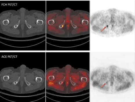
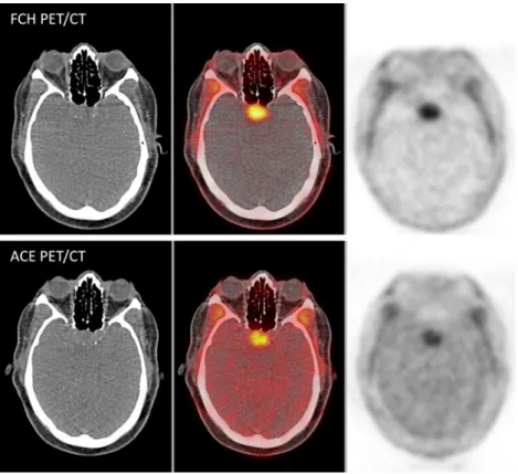
Four out of six local recurrences could be confirmed by the standard of reference investigations. In particular, three pa-tients underwent biopsies of the prostate and/or prostatic bed, which in all patients confirmed the local recurrence, and one patient had an MR scan which identified a local recurrence corresponding to the focal uptake visible on PET images. The two bone metastases identified in two patients were confirmed as isolated lesions on CT images and on positive bone scans. Two examples of concordantly positive imaging for local recurrence and for bone metastasis are shown in Figs.1and 2. Concordant imaging of incidental pathologies was observed in four patients with both radiotracers positive: a pituitary tumour (confirmed by MR and subsequently by histology; Fig. 3), a thyroid nodule (confirmed by ultrasound imaging and biological evaluation as a parathyroid adenoma), a lung tumour (confirmed by follow-up CT imaging as a stable nodule), and a supraclavicular lymph node (visible on CT images and probably unrelated to the baseline PC). Concor-dant information concerning lymph node status was obtained in 19 patients (9 positive, 8 negative, 2 equivocal). An exam-ple of concordantly positive findings for a pelvic lymph node is presented in Fig.4. For positive lymph nodes, the average SUVmax was 4.8±2.7 and 5.6±2.7 on ACE and FCH PET/ CT, respectively.
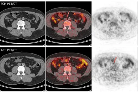
Differences between ACE and FCH PET/CT were ob-served in four patients. Among these, two patients had a clearly positive lymph node on ACE PET/CT but negative on FCH PET, as shown in Fig. 5. In these two patients, the SUVmax values found on ACE PET/CT were 3.3 and 2.9, that is in the range of lower values for positive lesions. In one patient a subsequent repeat FCH PET/CT scan confirmed the earlier ACE PET/CT positivity. In the third patient an external Table 4 PET results according to localization and concordance between
ACE and FCH PET expressed as the numbers of positive, double-equivocal, double-negative and discordant PET results per site Recurrence Concordant PET results Discordant
PET results Total per site Both positive Both equivocal Both negative Local 6 3 14 0 23 Lymph node 9 2 8 4 23 Bone metastasis 2 0 21 0 23 Total 17a 5 43 4 69 Unrelated patholo-gies 4b 0 19 0 23
aTwo patients were simultaneously double-positive for lymph nodes and
bone metastases, one patient simultaneously had a double-positive local recurrence and lymph nodes. With regard to Tables2and3, a total of 14 patients had double-positive PET results.
bThree concordant hyperactivities unrelated to PC were observed in one
hypophysis, one thyroid and one lung.
Fig. 2 Bone lesion concordantly positive (arrows) on both FCH and ACE PET/CT
iliac lymph node was equivocal on ACE PET and negative on FCH PET. In the fourth patient, retroperitoneal lymph nodes were equivocal on both FCH and ACE PET/CT, but some nodes were equivocal with one or the other of the radiotracers but not with both, although in this patient both PET scans were performed on the same day. Discordant lymph node PET/CT findings might occasionally reflect inflammatory situations, but in one patient an ACE-positive FCH-negative lymph node had progressed on imaging follow-up, suggesting a possible but small difference in sensitivity. No comparison with our
standard of reference was available for lymph node status, given that no biopsy could be performed and the follow-up at the time of this report was shorter than planned.
The SUVmax values of the concordantly positive lesions on ACE and FCH PET/CT studies were 4.4±1.7 (mean ± SD) and 5.3±2, respectively (Table 5). The values were signifi-cantly different between the two studies (p =0.017). The TBR values, however, did not differ significantly between the two studies (4.4±1.7 and 5.1±2.1 on ACE and FCH PET/CT studies, respectively; p =0.089).
Fig. 3 Pituitary gland concordantly positive on whole-body FCH and ACE PET/CT acquisitions corresponding to a macroadenoma
Fig. 4 Pelvic lymph node concordantly positive (arrows) on both FCH and ACE PET/CT
Discussion
11
C-Choline,18F-choline and11C-acetate are major agents for PET imaging of PC [1,2]. Only a few intraindividual com-parisons of PET have been performed with these radiotracers.
11
C-Acetate and 11C-choline PET have been directly com-pared in limited numbers of PC patients [19] and more recent-ly by PET/CT in bladder cancer patients [20]. Both studies concluded that the performance of both tracers is similar. It has been reported that11C-acetate PET fused retrospectively with CT and MR scans is essential for the final diagnosis of recurrent PC, notably for uptake in the prostate region [21]. Our findings are concordant with these results, given that we had a high rate of histological confirmation of positive local findings (all patients biopsied showed local recurrence). In addition, all our studies obtained on an integrated PET/CT scanner systematically included a whole-body unenhanced CT scan for localization and morphological analysis, discrim-inating between intestinal lymph node uptake. We had a higher rate of negative findings in our study (39 % vs. 10 %), that could be explained by the lower mean PSA at inclusion (2.3 vs. 6.3 ng/ml),
Acetate and choline are taken up by proliferating cells and can be transformed to phosphatidylcholine and phosphocholine, respectively [22,23]. Transmembrane transport of choline is dependent on the high-affinity choline transporter [24, 25], while transport of acetate is dependent on the monocarboxylate transporter-1 (MTC-1) or sodium-coupled MTC-1 (SMTC1) [26]. A relationship between 11C-choline and choline kinase expression has also been described [27], and in that study, this tracer was highly specific and sensitive for staging pelvic lymph nodes. Phosphatidylcholine can be incorporated into nascent cell membranes. In a direct comparison of the two tracers, uptake was correlated with the tumour cell proliferation rate measured with tritiated thymidine, with an uptake advantage
being found in the in vitro situation for ACE [22]. However, acetate can also be metabolized, notably in active musculature which allows cardiac [28] and striate muscle imaging [29]. Similarly, choline can be oxidized to betain aldehyde and is Fig. 5 Discordant interaortic
lymph node, negative on FCH PET/CT (above) and positive on ACE PET/CT (first whole-body acquisition, lower row)
Table 5 SUVmax values and TBR for positive findings on ACE or FCH PET/CT or both
ACE PET/CT FCH PET/CT
SUVmax TBR SUVmax TBR
Target Background Target Background Prostate 1 4.6 1.1 4.2 5.3 1.1 4.8 2 3.2 1.3 2.5 4.4 1.5 2.9 3 4.3 0.7 6.1 5.5 0.9 6.1 4 3.8 1.4 2.7 5.4 1.3 4.2 5 4 1 4.0 5.2 1.1 4.7 6 5.2 1 5.2 5 1.3 3.8 Lymph nodes 1 6.7 0.9 7.4 9.5 1 9.5 2 4.8 1.5 3.2 4.5 1 4.5 3 1.5 0.6 2.5 3.6 0.8 4.5 4 3.8 1.1 3.5 3.1 0.9 3.4 5 6.6 1 6.6 6.2 1.1 5.6 6 7.9 1.1 7.2 10.4 1 10.4 7 6.2 1.2 5.2 5.6 1 5.6 8 3 1 3.0 4.8 1.3 3.7 9 2.8 0.8 3.5 2.9 1.1 2.6 10 3.3 1.2 2.8 Negative Negative – 11 2.9 1 2.9 Negative Negative – Bone lesions 1 4.8 0.9 5.3 3.5 1 3.5 2 1.8 0.6 3.0 5.3 0.8 6.6
therefore taken up particularly in the liver and kidneys [22]. The uptake of18F-choline by cells is similar to that of11C-labelled choline, but after phosphorylation via choline kinase, the phospho-18F-choline remains trapped in the cytoplasm without further metabolism [30]. In the present study, we compared FCH and ACE PET/CT intraindividually in patients with bio-chemical recurrence and low PSA values. We sought to deter-mine whether an indication of higher sensitivity of one or the other agent would be observed in patients with low PSA levels or whether both tracers could be complementary. The localiza-tion of tumour recurrences in patients with low PSA levels is of clinical interest, since it could allow detection of tumours of restricted dissemination and therefore amenable to curative salvage therapy [7].
In contrast to ACE and11C-choline, FCH is a PET tracer with a half-life of 110 min and can be prepared on a commer-cial basis for distribution to PET centres without access to a cyclotron unit. FCH has become widely available and appears to be valuable for evaluation of PC patients with biochemical recurrence [13, 14, 31, 32]. With the use of early dynamic imaging centred on the prostate bed, the disadvantage of urinary excretion of18F can be partially compensated for since tumour uptake of FCH occurs earlier than urinary excretion [13,33]. Furthermore, late imaging with FCH might allow discrimination between inflammatory lymph nodes and tumour-infiltrated lymph nodes [13,33,34].
ACE has the advantage of a low to only moderate urinary excretion of11C, catabolized tracer being exhaled as11C-CO2.
On the other hand, its short half-life of 20 min does not allow the wider distribution of this tracer and requires a demanding and strict organization of tracer production and injection of patients. ACE PET/CT positivity in early recurrence appears to be correlated with blood PSA levels [35]. For PSA <1 ng/ ml, different groups have reported detection rates between 18 % and 36 % [36–40]. Positivity rates of FCH PET/CT in patients with similarly low PSA values are in the same range as for ACE PET/CT [12,13,32], but occasionally a higher percentage positivity (49 %) has also been reported [41], as was the case in this study with, however, only a small number of patients in this group. Nevertheless, only small numbers of patients have been studied at very low PSA values by both ACE and FCH PET/CT, and thus comparison of the results from the literature would not be reliable. In addition, the above studies underscore the need for a careful intraindividual as-sessment of FCH versus11C-choline PET/CT.
The current results show excellent agreement between FCH and ACE PET/CT. This suggests that both tracers visu-alize similar features of PC cells either on tracer integration in lipid synthesis or catabolic energy provision. These concor-dant data are in line with previous reports on ACE and11 C-choline [19,20], implying that FCH would be a valid alterna-tive to 11C-choline, although this point still needs to be addressed by a direct comparison.
This study has a main limitation, namely the absence of adequate follow-up to provide a“standard of reference” for all PET/CT findings. While the two positive bone findings were confirmed as bone metastases on CT and bone scintigraphy, and four of six positive findings for local recurrence could be confirmed, for other localizations, notably for lymph nodes, a conclusive interpretation is currently not possible. The limita-tion of these tracers, however, for lymph node disease is known [8], as also shown recently for 11C-choline [42] and reviewed with regard to PET/MR [43]. Thus neither tracer is the optimal tracer for recurrent PC, particularly for lymph node evaluation, as uptake is also observed in reactive medi-astinal and inguinal nodes. It is thus not surprising that the few discordant data in 4 of 23 patients observed with ACE and FCH concern lymph node status. A longer follow-up is cur-rently ongoing and is necessary to address this question. New tracers targeting specific molecules are currently under development for the diagnostic evaluation of PC, and some promising results have been shown in preliminary studies [1, 44,45].
Conclusion
These findings demonstrated that three-phase FCH PET/CT and an analogous ACE PET/CT protocol performed equally for early recurrent PC staging show an overall excellent con-cordance on a per-patient and a per-lesion basis.
Acknowledgments We are grateful to Fundació Privada Cellex for financing the study and to the staff of the Nuclear Medicine Division of the University Hospital of Geneva for their technical assistance and commitment. We thank Dr. A. Poncet, Unit of Methodological Support, University Hospital of Geneva, for advice and suggestions on the statis-tical evaluation of our data.
Conflicts of interest None.
References
1. Fox JJ, Schoder H, Larson SM. Molecular imaging of prostate cancer. Curr Opin Urol. 2012;22:320–7.
2. Jadvar H. Prostate cancer: PET with 18F-FDG, 18F- or 11C-acetate, and 18F- or 11C-choline. J Nucl Med. 2011;52:81–9.
3. Ward JF, Moul JW. Biochemical recurrence after definitive prostate cancer therapy. Part I: defining and localizing biochemical recurrence of prostate cancer. Curr Opin Urol. 2005;15:181–6.
4. Ward JF, Sebo TJ, Blute ML, Zincke H. Salvage surgery for radiorecurrent prostate cancer: contemporary outcomes. J Urol. 2005;173:1156–60.
5. Freedland SJ, Sutter ME, Dorey F, Aronson WJ. Defining the ideal cutpoint for determining PSA recurrence after radical prostatectomy. Prostate-specific antigen. Urology. 2003;61:365–9.
6. Abramowitz MC, Li T, Buyyounouski MK, Ross E, Uzzo RG, Pollack A, et al. The Phoenix definition of biochemical failure predicts for overall survival in patients with prostate cancer. Cancer. 2008;112:55–60.
7. Schick U, Jorcano S, Nouet P, Rouzaud M, Vees H, Zilli T, et al. Androgen deprivation and high-dose radiotherapy for oligometastatic prostate cancer patients with less than five regional and/or distant metastases. Acta Oncol. 2013. doi:10.3109/0284186X.2013.764010
8. Brogsitter C, Zophel K, Kotzerke J. F-Choline, choline and C-acetate PET/CT: comparative analysis for imaging prostate cancer patients. Eur J Nucl Med Mol Imaging. 2013;40 Suppl 1:18–27. 9. Giovacchini G, Picchio M, Garcia-Parra R, Mapelli P, Briganti A,
Montorsi F, et al. [11C]choline positron emission tomography/ computerized tomography for early detection of prostate cancer recurrence in patients with low increasing prostate specific antigen. J Urol. 2013;189:105–10.
10. Grassi I, Nanni C, Allegri V, Morigi JJ, Montini GC, Castellucci P, et al. The clinical use of PET with (11)C-acetate. Am J Nucl Med Mol Imaging. 2012;2:33–47.
11. Albrecht S, Buchegger F, Soloviev D, Zaidi H, Vees H, Khan HG, et al. (11)C-acetate PET in the early evaluation of prostate cancer recurrence. Eur J Nucl Med Mol Imaging. 2007;34:185–96. 12. Vees H, Buchegger F, Albrecht S, Khan H, Husarik D, Zaidi H, et al.
18F-choline and/or 11C-acetate positron emission tomography: de-tection of residual or progressive subclinical disease at very low prostate-specific antigen values (<1 ng/mL) after radical prostatecto-my. BJU Int. 2007;99:1415–20.
13. Steiner C, Vees H, Zaidi H, Berrebi O, Kossovsky MP, Khan GH, et al. Three-phase (18)F-fluorocholine PET/CT in the evaluation of prostate cancer recurrence. Nucl Med. 2009;48:1–9.
14. Soyka JD, Muster MA, Schmid DT, Seifert B, Schick U, Miralbell R, et al. Clinical impact of 18F-choline PET/CT in patients with recur-rent prostate cancer. Eur J Nucl Med Mol Imaging. 2012;39:936–43. 15. Michel C, Sibomana M, Boi A, Bernard X, Lonneux M, Defrise M, et al. Preserving Poisson characteristics of PET data with weighted OSEM reconstruction. Proceedings IEEE Nuclear Science Sympo-sium and Medical Imaging Conference, vol 2; 1998. p 1323–29. 16. Allal AS, Slosman DO, Kebdani T, Allaoua M, Lehmann W,
Dulguerov P. Prediction of outcome in head-and-neck cancer patients using the standardized uptake value of 2-[18F]fluoro-2-deoxy-D-glucose. Int J Radiat Oncol Biol Phys. 2004;59: 1295–300.
17. Cohen J. Weighted kappa: nominal scale agreement with provision for scaled disagreement or partial credit. Psychol Bull. 1968;70:213–20. 18. Landis JR, Koch GG. The measurement of observer agreement for
categorical data. Biometrics. 1977;33:159–74.
19. Kotzerke J, Volkmer BG, Glatting G, van den HJ, Gschwend JE, Messer P, et al. Intraindividual comparison of [11C]acetate and [11C]choline PET for detection of metastases of prostate cancer. Nucl Med. 2003;42:25–30.
20. Orevi M, Klein M, Mishani E, Chisin R, Freedman N, Gofrit ON. 11C-acetate PET/CT in bladder urothelial carcinoma: intraindividual comparison with 11C-choline. Clin Nucl Med. 2012;37:e67–72. 21. Wachter S, Tomek S, Kurtaran A, Wachter-Gerstner N, Djavan B,
Becherer A, et al. 11C-acetate positron emission tomography imag-ing and image fusion with computed tomography and magnetic resonance imaging in patients with recurrent prostate cancer. J Clin Oncol. 2006;24:2513–9.
22. Yoshimoto M, Waki A, Obata A, Furukawa T, Yonekura Y, Fujibayashi Y. Radiolabeled choline as a proliferation marker: com-parison with radiolabeled acetate. Nucl Med Biol. 2004;31:859–65. 23. Yoshimoto M, Waki A, Yonekura Y, Sadato N, Murata T, Omata N, et al. Characterization of acetate metabolism in tumor cells in relation to cell proliferation: acetate metabolism in tumor cells. Nucl Med Biol. 2001;28:117–22.
24. Okuda T, Osawa C, Yamada H, Hayashi K, Nishikawa S, Ushio T, et al. Transmembrane topology and oligomeric structure of the high-affinity choline transporter. J Biol Chem. 2012;287:42826–34. 25 . H ara T, K os aka N , K is hi H. Dev e lopment of ( 18 )F
-fluoroethylcholine for cancer imaging with PET: synthesis,
biochemistry, and prostate cancer imaging. J Nucl Med. 2002;43: 187–99.
26. Herrmann J, Hermes R, Breves G. Transepithelial transport and intraepithelial metabolism of short-chain fatty acids (SCFA) in the porcine proximal colon are influenced by SCFA concentra-tion and luminal pH. Comp Biochem Physiol Part A. 2011;158: 169–76.
27. Contractor K, Challapalli A, Barwick T, Winkler M, Hellawell G, Hazell S, et al. Use of [11C]choline PET-CT as a noninvasive method for detecting pelvic lymph node status from prostate cancer and relationship with choline kinase expression. Clin Cancer Res. 2011;17:7673–83.
28. Henes CG, Bergmann SR, Walsh MN, Sobel BE, Geltman EM. Assessment of myocardial oxidative metabolic reserve with positron emission tomography and carbon-11 acetate. J Nucl Med. 1989;30: 1489–99.
29. Buchegger F, Ratib O, Willi J-P, Steiner C, Seimbille Y, Zaidi H, et al. [11C]acetate PET/CT visualizes skeletal muscle exercise participa-tion, impaired function and recovery after hip arthroplasty; first results. Mol Imaging Biol. 2011;13:793–9.
30. DeGrado TR, Baldwin SW, Wang S, Orr MD, Liao RP, Friedman HS, et al. Synthesis and evaluation of (18)F-labeled choline analogs as oncologic PET tracers. J Nucl Med. 2001;42:1805–14.
31. Marzola MC, Chondrogiannis S, Ferretti A, Grassetto G, Rampin L, Massaro A, et al. Role of 18F-choline PET/CT in biochemically relapsed prostate cancer after radical prostatectomy: correlation with trigger PSA, PSA velocity, PSA doubling time, and metastatic distri-bution. Clin Nucl Med. 2013;38:e26–32
32. Pelosi E, Arena V, Skanjeti A, Pirro V, Douroukas A, Pupi A, et al. Role of whole-body 18F-choline PET/CT in disease detection in patients with biochemical relapse after radical treatment for prostate cancer. Radiol Med. 2008;113:895–904.
33. Massaro A, Ferretti A, Secchiero C, Cittadin S, Milan E, Tamiso L, et al. Optimising (18)F-choline PET/CT acquisition protocol in pros-tate cancer patients. N Am J Med Sci. 2012;4:416–20.
34. Oprea-Lager DE, Vincent AD, van Moorselaar RJ, Gerritsen WR, van den Eertwegh AJ, Eriksson J, et al. Dual-phase PET-CT to differentiate [18F]fluoromethylcholine uptake in reactive and malignant lymph nodes in patients with prostate cancer. PLoS One. 2012;7:e48430. 35. Picchio M, Briganti A, Fanti S, Heidenreich A, Krause BJ, Messa C,
et al. The role of choline positron emission tomography/computed tomography in the management of patients with prostate-specific antigen progression after radical treatment of prostate cancer. Eur Urol. 2011;59:51–60.
36. Souvatzoglou M, Krause BJ, Purschel A, Thamm R, Schuster T, Buck AK, et al. Influence of (11)C-choline PET/CT on the treatment planning for salvage radiation therapy in patients with biochemical recurrence of prostate cancer. Radiother Oncol. 2011;99:193–200. 37. Rybalov M, Breeuwsma AJ, Leliveld AM, Pruim J, Dierckx RA, de
Jong IJ. Impact of total PSA, PSA doubling time and PSA velocity on detection rates of (11)C-choline positron emission tomography in recurrent prostate cancer. World J Urol. 2012;31:319–23.
38. Krause BJ, Souvatzoglou M, Tuncel M, Herrmann K, Buck AK, Praus C, et al. The detection rate of [11C]choline-PET/CT depends on the serum PSA-value in patients with biochemical recurrence of prostate cancer. Eur J Nucl Med Mol Imaging. 2008;35:18–23. 39. Giovacchini G, Picchio M, Coradeschi E, Bettinardi V, Gianolli L,
Scattoni V, et al. Predictive factors of [11C]choline PET/CT in patients with biochemical failure after radical prostatectomy. Eur J Nucl Med Mol Imaging. 2010;37:301–9.
40. Castellucci P, Fuccio C, Nanni C, Santi I, Rizzello A, Lodi F, et al. Influence of trigger PSA and PSA kinetics on 11C-choline PET/CT detection rate in patients with biochemical relapse after radical pros-tatectomy. J Nucl Med. 2009;50:1394–400.
41. Detti B, Scoccianti S, Franceschini D, Cipressi S, Cassani S, Villari D, et al. Predictive factors of [18F]-choline PET/CT in 170 patients
with increasing PSA after primary radical treatment. J Cancer Res Clin Oncol. 2013;139:521–8.
42. Passoni NM, Suardi N, Abdollah F, Picchio M, Giovacchini G, Messa C, et al. Utility of [11C]choline PET/CT in guiding lesion-targeted salvage therapies in patients with prostate cancer recur-rence localized to a single lymph node at imaging: results from a pathologically validated series. Urol Oncol. 2013. doi:10.1016/j. urolonc.2013.03.006
43. Souvatzoglou M, Eiber M, Martinez-Moeller A, Furst S, Holzapfel K, Maurer T, et al. PET/MR in prostate cancer: technical aspects and
potential diagnostic value. Eur J Nucl Med Mol Imaging. 2013;40 Suppl 1:79–88.
44. Zhang H, Abiraj K, Thorek DL, Waser B, Smith-Jones PM, Honer M, et al. Evolution of bombesin conjugates for targeted PET imaging of tumors. PLoS One. 2012;7:e44046.
45. Nanni C, Schiavina R, Boschi S, Ambrosini V, Pettinato C, Brunocilla E, et al. Comparison of (18)F-FACBC and (11)C-choline PET/CT in patients with radically treated prostate cancer and bio-chemical relapse: preliminary results. Eur J Nucl Med Mol Imaging. 2013;40 Suppl 1:11–7.