ORIGINAL ARTICLE
Phosphate homeostasis in Bartter syndrome: a case
–control study
Alberto Bettinelli&Cristina Viganò&Maria Cristina Provero&Francesco Barretta&
Alessandra Albisetti&Silvana Tedeschi&Barbara Scicchitano&Mario G. Bianchetti
Received: 13 October 2013 / Revised: 24 April 2014 / Accepted: 29 April 2014 / Published online: 6 June 2014 # IPNA 2014
Abstract
Background Bartter patients may be hypercalciuric. Addition-al abnormAddition-alities in the metabolism of cAddition-alcium, phosphate, and calciotropic hormones have occasionally been reported. Methods The metabolism of calcium, phosphate, and calciotropic hormones was investigated in 15 patients with Bartter syndrome and 15 healthy subjects.
Results Compared to the controls, Bartter patients had signif-icantly reduced plasma phosphate {mean [interquartile range]:1.29 [1.16–1.46] vs. 1.61 [1.54–1.67] mmol/L} and maximal tubular phosphate reabsorption (1.16 [1.00–1.35] vs. 1.41 [1.37–1.47] mmol/L) and significantly increased parathyroid hormone (PTH) level (6.1 [4.5–7.7] vs. 2.8 [2.2–4.4] pmol/L). However, patients and controls did not differ in blood calcium, 25-hydroxyvitamin D, alkaline phos-phatase, and osteocalcin levels. In patients, an inverse corre-lation (P<0.05) was noted between total plasma calcium or glomerular filtration rate and PTH concentration. A positive correlation was also noted between PTH and osteocalcin
concentrations (P<0.005), as well as between chloriduria or natriuria and phosphaturia (P<0.001). No correlation was noted between calciuria and PTH concentration or between urinary or circulating phosphate and PTH.
Conclusions The results of this study demonstrate a tendency towards renal phosphate wasting and elevated circulating PTH levels in Bartter patients.
Keywords Bartter syndrome . Calcium .
Hypophosphatemia . Maximal tubular reabsorption of phosphate . Parathyroid hormone
Introduction
Bartter syndrome is a group of very similar recessive tubulopathies with a unifying pathophysiology consisting of
postproximal salt loss [1]. Affected patients present with
A. Bettinelli
:
M. C. Provero:
B. ScicchitanoDivision of Pediatrics, San Leopoldo Mandic Hospital, Merate, Lecco, Italy
C. Viganò
Division of Orthopedics, San Leopoldo Mandic Hospital, Merate, Lecco, Italy
M. C. Provero
Department of Clinical and Experimental Medicine, Ospedale Pediatrico Filippo del Ponte, Varese, Italy
F. Barretta
Department of Clinical Sciences and Community Health, University of Milan and Epidemiology Unit, Milan, Italy
F. Barretta
Department of Preventive Medicine, Foundation IRCCS Cà Granda Ospedale Maggiore Policlinico, Milan, Italy
A. Albisetti
Department of Clinical Orthopedics and Rehabilitation, University of Milan, Milan, Italy
S. Tedeschi
Laboratory of Medical Genetics, Foundation IRCCS Ca’ Granda Ospedale Maggiore Policlinico, Milan, Italy
M. G. Bianchetti
Integrated Department of Pediatrics, Ente Ospedaliero Cantonale Ticinese, University of Berne, Berne, Switzerland
M. G. Bianchetti (*)
San Giovanni Hospital, 6500 Bellinzona, Switzerland e-mail: [email protected]
normal or low blood pressure, hypokalemia, hyperreninemia, and a tendency towards nephrocalcinosis and hypercalciuria in
the neonatal variant [1]. Further abnormalities in the
metabo-lism of calcium, inorganic phosphate, and calciotropic hor-mones have sometimes been noted in these disorders, although data characterizing the levels of classic calciotropic hormones
during the stable phases of the condition are sparse [2,3].
Here we present the results of our study in which we investigated the metabolism of calcium, inorganic phosphate, and classic calciotropic hormones in patients affected with Bartter syndrome.
Patients and methods
Participants eligible for entry into the study were all Bartter patients on regular follow-up at the renal unit of the Division of Pediatrics, San Leopoldo Mandic Hospital, Merate (Italy). Fif-teen patients from 14 different families (8 females and 7 males, median age 7.3 years, age range 3.2–18) who presented for a scheduled visit between September and November 2012 were ultimately enrolled in the study. These patients had a history of polyhydramnios (N=13), premature birth (N=13), or ultrasound
findings consistent with nephrocalcinosis (N=13) (Table1). A
control group of 15 healthy subjects (8 females and 7 males median age 11 years, age range 6.9–18 years) was also studied.
The study design was approved by the Human Subjects Re-search Committee of Merate-Lecco Hospital.
The diagnosis of Bartter syndrome was based on the pres-ence of normal or low blood pressure, hypokalemia, inappro-priately high urinary excretion of chloride, and hyperreninemia. A molecular evaluation of the SLC12A1, KCNJ1, CLCNKB, and BSND genes had been performed on the 15 patients,
according to standard procedure [4,5]. The Bartter patients,
who did not discontinue their respective medication with po-tassium chloride (N=15), the inhibitor of prostaglandin synthe-sis indomethacin (N=14), and the potassium-sparing diuretic spironolactone (N=3), and the control subjects attended the outpatient clinic after overnight fasting and received a tap water
load of 400 mL [6]. After voiding the bladder, an
approximate-ly 2-h urine specimen was collected and a mid-point blood
sample was collected [6]. Creatinine, sodium, chloride,
potas-sium, total calcium and magnepotas-sium, and inorganic phosphate were assessed in both the blood and urine; urea, bicarbonate, albumin, 25-hydroxyvitamin D, alkaline phosphatase, intact parathyroid hormone (PTH), and osteocalcin were assessed in the blood only. Creatinine, urea, sodium, bicarbonate, chloride, potassium, albumin, total calcium and magnesium, and inor-ganic phosphate were measured on an automated auto-analyzer using colorimetric assays or ion-selective electrodes. Intact PTH was measured by a two-site radioimmunometric assay, 25-hydroxyvitamin D was measured using a competitive
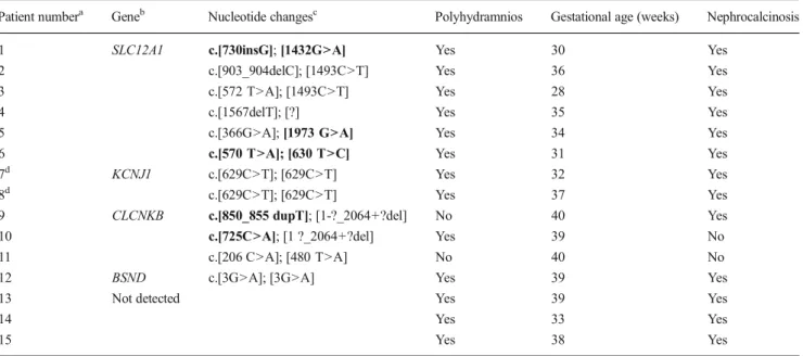
Table 1 Mutations and main clinical prenatal and neonatal data in 15 patients with Bartter syndromes
Patient numbera Geneb Nucleotide changesc Polyhydramnios Gestational age (weeks) Nephrocalcinosis 1 SLC12A1 c.[730insG]; [1432G>A] Yes 30 Yes
2 c.[903_904delC]; [1493C>T] Yes 36 Yes 3 c.[572 T>A]; [1493C>T] Yes 28 Yes
4 c.[1567delT]; [?] Yes 35 Yes
5 c.[366G>A]; [1973 G>A] Yes 34 Yes 6 c.[570 T>A]; [630 T>C] Yes 31 Yes 7d KCNJ1 c.[629C>T]; [629C>T] Yes 32 Yes 8d c.[629C>T]; [629C>T] Yes 37 Yes 9 CLCNKB c.[850_855 dupT]; [1-?_2064+?del] No 40 Yes 10 c.[725C>A]; [1 ?_2064+?del] Yes 39 No 11 c.[206 C>A]; [480 T>A] No 40 No 12 BSND c.[3G>A]; [3G>A] Yes 39 Yes
13 Not detected Yes 39 Yes
14 Yes 33 Yes
15 Yes 38 Yes
aPatients 1–8 and patients 14 and 15 were classified clinically as loop disorder Bartter syndrome; patients 9–11 and 13 were classified as distal
convoluted tubule disorder Bartter syndrome; patient 12 was classified as combined disorder Bartter syndrome
b
Mutations of the SLC12A1, KCNJ1, CLCNKB, and BSND genes were detected in 12 of the 15 patients. GenBank accession numbers as reference sequences: SLC12A3=NM_000339.2; SLC12A1=NM_000338.2; KCNJ1=NM_000220.3; CLCNKB=NM_000085.4. Nomenclature according to Human Genomic Variant Society
c
Novel mutations are given in bold font
d
protein-binding assay, and osteocalcin was measured by
radio-immunoassay, as previously reported [6].
The glomerular filtration rate (GFR) was estimated from
height measurements and plasma creatinine levels [7].
Stan-dard equations were used to calculate the fractional urinary excretion of sodium, chloride, potassium, calcium, magne-sium, and inorganic phosphate and the maximal tubular reab-sorption of inorganic phosphate, which is the best means of
defining the renal tubular handling of this ion [8,9].
Albumin-corrected plasma calcium was calculated as follows: for every
gram/liter by which plasma albumin exceeds 40 g/L, 0.025 is subtracted from the total calcium measurement, with the re-verse procedure for albumin values of <40 g/L. Blood and urinary levels of sodium, chloride, potassium, and magnesium
are age- and gender-independent [10], but blood levels of
inorganic phosphate and those of its maximal tubular
reab-sorption vary significantly with age and gender [8,9]. Hence,
the values obtained in our study were also compared with
those given in the literature [8, 9] and expressed as the
standard deviation score (SDS).
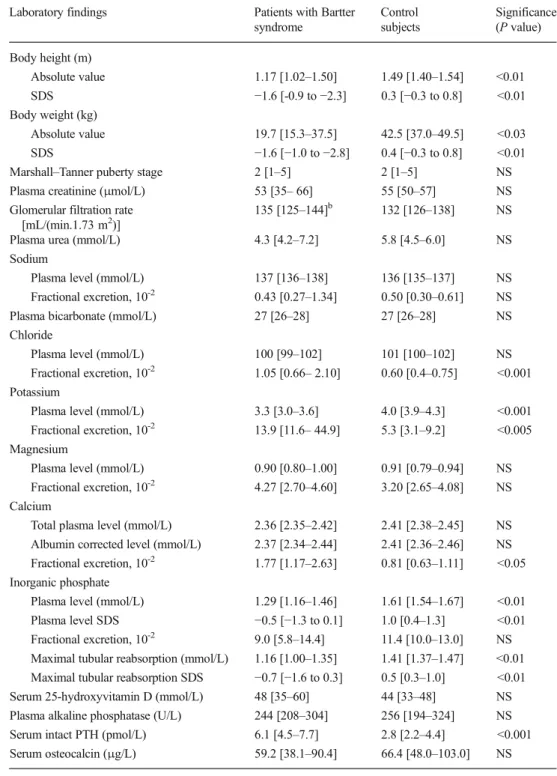
Table 2 Laboratory findings in the 15 patients affected with Bartter syndrome and in the 15 control subjectsa
Values are presented as the medi-an with the interquartile rmedi-ange (IQR) given in square brackets SDS, Standard deviation score; NS, not significant; PTH, para-thyroid hormone
a
Patients affected with Bartter syndrome: N = 15; 8 females, 7 males; median age 7.3 years; age range 3.2–18.0 years. Control subjects: N = 15; 7 females, 8 males; median age 11.1 years; age range 6.9-17.8 years
b
Glomerular filtration rate was ≥85 mL/(min.1.73 m2
) in 14 of the 15 patients
Laboratory findings Patients with Bartter syndrome Control subjects Significance (P value) Body height (m) Absolute value 1.17 [1.02–1.50] 1.49 [1.40–1.54] <0.01 SDS −1.6 [-0.9 to −2.3] 0.3 [−0.3 to 0.8] <0.01 Body weight (kg) Absolute value 19.7 [15.3–37.5] 42.5 [37.0–49.5] <0.03 SDS −1.6 [−1.0 to −2.8] 0.4 [−0.3 to 0.8] <0.01 Marshall–Tanner puberty stage 2 [1–5] 2 [1–5] NS Plasma creatinine (μmol/L) 53 [35– 66] 55 [50–57] NS Glomerular filtration rate
[mL/(min.1.73 m2)]
135 [125–144]b 132 [126–138] NS Plasma urea (mmol/L) 4.3 [4.2–7.2] 5.8 [4.5–6.0] NS Sodium
Plasma level (mmol/L) 137 [136–138] 136 [135–137] NS Fractional excretion, 10-2 0.43 [0.27–1.34] 0.50 [0.30–0.61] NS
Plasma bicarbonate (mmol/L) 27 [26–28] 27 [26–28] NS Chloride
Plasma level (mmol/L) 100 [99–102] 101 [100–102] NS Fractional excretion, 10-2 1.05 [0.66– 2.10] 0.60 [0.4–0.75] <0.001
Potassium
Plasma level (mmol/L) 3.3 [3.0–3.6] 4.0 [3.9–4.3] <0.001 Fractional excretion, 10-2 13.9 [11.6– 44.9] 5.3 [3.1–9.2] <0.005 Magnesium
Plasma level (mmol/L) 0.90 [0.80–1.00] 0.91 [0.79–0.94] NS Fractional excretion, 10-2 4.27 [2.70–4.60] 3.20 [2.65–4.08] NS Calcium
Total plasma level (mmol/L) 2.36 [2.35–2.42] 2.41 [2.38–2.45] NS Albumin corrected level (mmol/L) 2.37 [2.34–2.44] 2.41 [2.36–2.46] NS Fractional excretion, 10-2 1.77 [1.17–2.63] 0.81 [0.63–1.11] <0.05 Inorganic phosphate
Plasma level (mmol/L) 1.29 [1.16–1.46] 1.61 [1.54–1.67] <0.01 Plasma level SDS −0.5 [−1.3 to 0.1] 1.0 [0.4–1.3] <0.01 Fractional excretion, 10-2 9.0 [5.8–14.4] 11.4 [10.0–13.0] NS Maximal tubular reabsorption (mmol/L) 1.16 [1.00–1.35] 1.41 [1.37–1.47] <0.01 Maximal tubular reabsorption SDS −0.7 [−1.6 to 0.3] 0.5 [0.3–1.0] <0.01 Serum 25-hydroxyvitamin D (mmol/L) 48 [35–60] 44 [33–48] NS Plasma alkaline phosphatase (U/L) 244 [208–304] 256 [194–324] NS Serum intact PTH (pmol/L) 6.1 [4.5–7.7] 2.8 [2.2–4.4] <0.001 Serum osteocalcin (μg/L) 59.2 [38.1–90.4] 66.4 [48.0–103.0] NS
Descriptive statistics are presented as the median and in-terquartile range (25th to 75th percentile and includes half of the data points). Two-tailed Wilcoxon–Mann–Whitney tests for independent samples and linear regressions with the rank
correlation coefficient rs were performed for the analysis.
Significance was assumed when P<0.05.
Results
Molecular biology
Genetic analysis revealed mutations in 12 of the 15 patients enrolled in the study, with three patients having a homozygous
mutation, eight having a compound heterozygous mutation,
and one having a single heterozygous mutation (Table1). One
of these patients has been previously diagnosed as Bartter syndrome type IV based on the presence of a sensorineural
hearing loss [11]. Ten mutations had been previously
de-scribed, whereas seven mutations (4 missense and 1 frame-shift in the SLC12A1 gene and 1 missense and 1 frameframe-shift in the CLCNKB gene) are novel. All of the new variants were considered to be pathogenetic—the two frameshift mutations because they lead to truncated protein synthesis, and the missense mutations because all are on conserved residues and were not detected in 100 healthy chromosomes or found in the Exome Variant Server database. Molecular analysis did not detect any mutation in three patients.
Maximal T u bular Phosphate Re absorption (mm o l/L) 0.80 1.00 1.20 1.40 1.60 Serum 25-hy d roxy -v itamin D (nmol/L) 10 90 70 50 30 10 Fractional Calcium Excretion (10 -2) 1 0.1 0.01
P<0.05
P<0.01
NS
2.55 2.45 2.35 2.25 2.65 T o tal Plasma Ca lcium (mm o l/L)NS
P<0.01
1.60 1.40 1.20 1.00 1.80 Plasma Phosphate (mm o l/L)P<0.01
12.0 0.0 8.0 4.0 Serum Par a thy roid Hormone (pmol /L) Control subjects Bartter patients I-II (N = 8) III (N = 3) Barttin (N = 1) Unknown (N = 3)Fig. 1 Total plasma calcium and inorganic phosphate levels, fractional calcium excretion, maximal tubular reabsorption of inorganic phosphate, and serum intact parathyroid hormone and 25-hydroxyvitamin D levels in 15 Bartter syndrome patients and in 15 control subjects. Differently colored filled circles denote patients with mutations affecting either the SLC12A1 or the KCNJ1 gene (type I–II), patients with mutations affecting the CLCNK gene (type III) patients with mutations affecting the BSND (Barttin), and patients without any mutation, as well as the control subjects. NS Not significant. See text and Table2for a detailed description of the two study groups - 4.0 - 2.0 0.0 2.0 - 4.0 - 2.0 0.0 2.0
Maximal Tubular Reabsorption of Phosphate
SDS Plasma Phosphate Concentration
SDS Control subjects Bartter patients I-II (N = 8) III (N = 3) Barttin (N = 1) Unknown (N = 3)
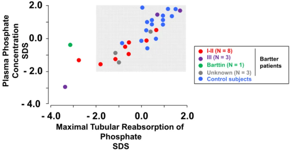
Fig. 2 Relationship between maximal tubular reabsorption of inorganic phosphate and corresponding plasma phosphate level in 15 Bartter syn-drome patients and 15 control subjects. The results are expressed as standard deviation score (SDS). Shaded area Reference values obtained from the literature. Differently colored filled circles denote patients with
mutations affecting either the SLC12A1 or the KCNJ1 gene (type I–II), patients with mutations affecting the CLCNK gene (type III) patients with mutations affecting the BSND (Barttin), and patients without any muta-tion, as well as the control subjects
Clinical and biochemical data
Growth and laboratory findings are shown in Table 2 and
Fig. 1. Body height, height SDS, weight and weight SDS
were significantly lower in the group of patients affected with Bartter syndrome than in the control group. Patients affected with Bartter syndrome and control subjects did not signifi-cantly differ with respect to puberty stage, plasma creatinine and urea, estimated GFR, plasma bicarbonate and chloride, and plasma and urinary sodium and magnesium. As expected, hypokalemia, hyperkaliuria, hyperchloriduria, and hypercal-ciuria were noted in Bartter patients. Plasma inorganic phos-phate level (P<0.01) and maximal tubular phosphos-phate reab-sorption were significantly reduced (P<0.01) and serum intact PTH level significantly increased (P<0.01) in Bartter patients as compared with control subjects. However, patients and controls did not significantly differ with respect to total calci-um blood level, sercalci-um 25-hydroxyvitamin D, plasma alkaline phosphatase and serum osteocalcin. PTH level was >10 pmol/L in three patients, with only one of these three patients
having a mildly reduced GFR of 58 mL/(min.1.73 m2).
Figure 2 shows the relationship between the maximal
tubular reabsorption of phosphate and its plasma level in Bartter patients and healthy controls, which are com-pared with age- and sex-specific reference values from
the literature [4, 5]. The maximal tubular reabsorption
of phosphate was low in three patients, and circulating phosphate level was low in one patient.
In Bartter patients we noted a significant inverse correlation (P<0.05) between total plasma calcium or GFR, taken as the
independent value, and PTH, taken as the dependent value
(Table3). Finally, a significant positive correlation was noted
between PTH and osteocalcin (P<0.005) and between urinary chloride or sodium and urinary phosphate (P < 0.001). On the contrary, no significant correlation was noted between urinary calcium and PTH, between urinary or circulating phosphate and PTH, and between maximal tubular reabsorption of phosphate and PTH.
Discussion
The clinical and laboratory characteristics of Bartter syndrome may be mimicked among others by treatment with loop
diuretics [1]. These natriuretic agents directly increase urinary
phosphate excretion in the presence of a normal extracellular
fluid volume [12]. On the other hand, there is no phosphaturia
in the presence of a contracted extracellular fluid volume, i.e.,
when urinary salt losses are not replaced [12]. Our case–
control study was performed in biochemically and genetically characterized Bartter patients. In these unselected patients, who were on long-term management with potassium chloride, indomethacin and, more rarely, spironolactone, we deter-mined hypercalciuria, renal phosphate wasting, and elevated circulating PTH levels.
The molecular biology studies performed in our patients also disclosed seven previously unreported mutations in either the SLC12A1 or CLCNKB genes.
To our knowledge, the metabolism of calcium, inorganic phosphate, and PTH in Bartter patients has only been
Table 3 Significant regressions in 15 patients with Bartter syndrome
Independent value Dependent value Slope Intercept Correlation coefficient (rs)
Significance (P)
Total calcium level Intact PTH −146.1 414 −0.529 <0.05 Glomerular filtration rate Intact PTH −0.277 98.9 −0.521 <0.05 Intact parathyroid hormone Osteocalcin 0.974 0.439 0.800 <0.005 Fraction chloride excretion Fractional phosphate excretion 2.90 5.76 0.822 <0.001 Fractional sodium excretion Fractional phosphate excretion 7.54 5.24 0.814 <0.001 PTH parathyroid hormone
Table 4 Data on blood calcium, inorganic phosphate, and parathyroid hormone levels and on urinary phosphate excretion in patients affected with Bartter syndromes
Reference Glomerular filtration rate [mL/(min.1.73 m2)]
Blood level (mmol/L) Urinary phosphate excretion PTH Calcium Phosphate
Leonhardt et al. [2] Reduced Elevated Low Elevated Normal Rodríguez-Soriano et al. [3] Normal Normal Normal–high Low Not assessed Present study Normal Elevated Normal Low Elevated PTH parathyroid hormone
addressed in our study and in two earlier studies [2, 3], although the latter two studies did not include a well-defined
pediatric control group (Table4). The tendency towards
ele-vated PTH levels and hyperphosphatemia noted in German
patients in the study of Leonhardt et al. [2] is likely the
consequence of a mildly reduced GFR. As in our patients, a tendency towards hypophosphatemia was noted in Span-ish pediatric patients, reported by Rodríguez-Soriano
et al. [3], with a normal GFR (phosphaturia was not
measured in these patients). However, contrary to our patients, in the Spanish patients, the PTH level was normal and the circulating calcium level was slightly elevated. As in our patients, hypophosphatemia and hy-perparathyroidism were also noted in an adolescent case
of Bartter syndrome reported 35 years ago [13].
The mechanisms underlying renal phosphate wasting and hypophosphatemia in Bartter patients remain elusive. Bartter patients tend to be alkalotic; it is recognized, however, that metabolic alkalosis, contrary to respiratory alkalosis, does not
noticeably modify phosphate metabolism [14–16]. There is
some phosphate wasting in primary hyperparathyroidism, but no correlation was noted between urinary or circulating
phos-phate and PTH in our Bartter patients [17]. Prostaglandin
overproduction, a constant finding in Bartter syndrome, might
underlie hyperphosphaturia in our patients [14–16]. This
as-sumption is supported by the observation that circulating phosphate levels increase in patients with Barter syndromes who are being treated with inhibitors of prostaglandin
synthe-sis [2]. When we take into account that our Bartter patients
were on treatment with indomethacin, an effective inhibitor of prostaglandin synthesis, we can assume that factors other than hyperprostaglandinism mainly account for renal phosphate wasting. We feel that the mild tendency to renal phosphate wasting observed in our Bartter patients results from a phosphaturic effect associated with the renal tubular dysfunc-tion, which is in part counterbalanced by existing extracellular
fluid volume contraction [12]. This impression is supported by
the significant correlation between urinary chloride or sodium and urinary phosphate.
There are a number of limitations to our study. First, we included Bartter patients with both prenatal and classic pre-sentation. Second, only a few patients (N = 15) were studied. Third, we did not measure urinary prostaglandin excretion and
phosphaturic hormone fibroblast growth factor level [15].
In conclusion, this case–control study demonstrates the existence of a tendency towards elevated circulating PTH levels and renal phosphate wasting in Bartter patients with normal GFR. A multicenter study with a large number of patients and the determination of prostaglandinuria,
reninemia, fibroblast growth factor 23, and 1,25-dihydroxyvitamin D should be helpful in establishing the mechanisms underlying these biochemical abnormalities.
Conflict of interest None.
References
1. Seyberth HW, Schlingmann KP (2011) Bartter- and Gitelman-like syndromes: salt-losing tubulopathies with loop or DCT defects. Pediatr Nephrol 26:1789–1802
2. Leonhardt A, Timmersmanns G, Roth B, Seyberth HV (1992) Calcium homeostasis and hypercalciuria in hyperprostaglandin E syndrome. J Pediatr 120:546–554
3. Rodríguez-Soriano J, Vallo A, Aguirre M (2005) Bone mineral density and bone turnover in patients with Bartter syndrome. Pediatr Nephrol 20:1120–1125
4. Bettinelli A, Borsa N, Bellantuono R, Syrèn ML, Calabrese R, Edefonti A, Komninos J, Santostefano M, Beccaria L, Pela I, Bianchetti MG, Tedeschi S (2007) Patients with biallelic mutations in the chloride channel gene CLCNKB: long-term management and outcome. Am J Kidney Dis 49:91–98
5. Puricelli E, Bettinelli A, Borsa N, Sironi F, Mattiello C, Tammaro F, Tedeschi S, Bianchetti MG, Italian Collaborative Group for Bartter Syndrome (2010) Long-term follow-up of patients with Bartter syn-drome type I and II. Nephrol Dial Transplant 25:2976–2981 6. Viganò C, Amoruso C, Barretta F, Minnici G, Albisetti W, Syrèn ML,
Bianchetti MG, Bettinelli A (2013) Renal phosphate handling in Gitelman syndrome - the results of a case-control study. Pediatr Nephrol 28:65–70
7. Schwartz GJ, Work DF (2009) Measurement and estimation of GFR in children and adolescents. Clin J Am Soc Nephrol 4:1832–1843 8. Brodehl J, Gellissen K, Weber HP (1982) Postnatal development of
tubular phosphate reabsorption. Clin Nephrol 17:163–171
9. Brodehl J (1994) Assessment and interpretation of the tubular thresh-old for phosphate in infants and children. Pediatr Nephrol 8:645 10. Burritt MF, Slockbower JM, Forsman RW, Offord KP, Bergstralh EJ,
Smithson WA (1990) Pediatric reference intervals for 19 biologic variables in healthy children. Mayo Clin Proc 65:329–336 11. Zaffanello M, Taranta A, Palma A, Bettinelli A, Marseglia GL,
Emma F (2006) Type IV Bartter syndrome: report of two new cases. Pediatr Nephrol 21:766–770
12. Greger R, Wangemann P (1987) Loop diuretics. Ren Physiol 10:174–183 13. Sann L, David L, Bernheim J, François R (1978) Hypophosphatemia and hyperparathyroidism in a case of Bartter's syndrome. Helv Paediatr Acta 33:299–310
14. Lang F, Greger R, Knox FG, Oberleithner H (1981) Factors modu-lating the renal handling of phosphate. Ren Physiol 4:1–16 15. Friedlander G (1996) Regulation of renal phosphate handling: recent
findings. Curr Opin Nephrol Hypertens 5:316–320
16. Hu MC, Shiizaki K, Kuro-o M, Moe OW (2013) Fibroblast growth factor 23 and Klotho: physiology and pathophysiology of an endo-crine network of mineral metabolism. Annu Rev Physiol 75:503–533 17. Bilezikian JP (2012) Primary hyperparathyroidism. Endocr Pract 18:
![Figure 2 shows the relationship between the maximal tubular reabsorption of phosphate and its plasma level in Bartter patients and healthy controls, which are com-pared with age- and sex-specific reference values from the literature [4, 5]](https://thumb-eu.123doks.com/thumbv2/123doknet/14813655.612491/5.892.76.815.106.256/relationship-reabsorption-phosphate-bartter-patients-controls-reference-literature.webp)
