*Corresponding author: Charaf Benarafa, Theodor Kocher Institute, University of Bern, Freiestrasse 1, CH-3012 Bern, Switzerland, e-mail: [email protected]. http://orcid.org/0000-0002-2049-7769
Sabrina S. Burgener, Mathias Baumann and Paola Basilico: Theodor Kocher Institute, University of Bern, Freiestrasse 1, CH-3012 Bern, Switzerland; and Graduate School for Cellular and Biomedical Sciences, University of Bern, CH-3012 Bern, Switzerland Eileen Remold-O’Donnell: Program in Cellular and Molecular Medicine and Division of Hematology/Oncology, Boston Children’s Hospital, Boston, MA 02115, USA; and Department of Pediatrics, Harvard Medical School, Boston, MA 02115, USA
Ivo P. Touw: Department of Hematology, Erasmus MC Cancer Institute, 3015 GE, Rotterdam, The Netherlands
Sabrina S. Burgener, Mathias Baumann, Paola Basilico, Eileen Remold-O’Donnell, Ivo P. Touw
and Charaf Benarafa*
Myeloid conditional deletion and transgenic
models reveal a threshold for the neutrophil
survival factor Serpinb1
DOI 10.1515/hsz-2016-0132
Received February 2, 2016; accepted April 20, 2016; previously published online April 22, 2016
Abstract: Serpinb1 is an inhibitor of neutrophil granule
ser-ine proteases cathepsin G, proteinase-3 and elastase. One
of its core physiological functions is to protect neutrophils
from granule protease-mediated cell death. Mice lacking
Serpinb1a (Sb1a
-/-), its mouse ortholog, have reduced bone
marrow neutrophil numbers due to cell death mediated
by cathepsin G and the mice show increased susceptibility
to lung infections. Here, we show that conditional deletion
of Serpinb1a using the Lyz2-cre and Cebpa-cre knock-in
mice effectively leads to recombination-mediated deletion
in neutrophils but protein-null neutrophils were only
obtained using the latter recombinase-expressing strain.
Absence of Serpinb1a protein in neutrophils caused
neu-tropenia and increased granule permeabilization-induced
cell death. We then generated transgenic mice
express-ing human Serpinb1 in neutrophils under the human
MRP8 (S100A8) promoter. Serpinb1a expression levels in
founder lines correlated positively with increased
neu-trophil survival when crossed with Sb1a
-/-mice, which
had their defective neutrophil phenotype rescued in the
higher expressing transgenic line. Using new conditional
and transgenic mouse models, our study demonstrates
the presence of a relatively low Serpinb1a protein
thresh-old in neutrophils that is required for sustained survival.
These models will also be helpful in delineating recently
described functions of Serpinb1 in metabolism and cancer.
Keywords: cell death; cre; serine protease; serpin.
Introduction
Neutrophils (PMNs) are granulocytes with central
func-tions in inflammatory disease and in innate immunity
against microbes (Kruger et al., 2015). Because isolated
PMNs are short-lived, they are poorly amenable to in vitro
manipulation such as RNA interference and transfection.
The use of genetic engineering in mice has thus been
helpful in delineating molecular pathways in PMN
home-ostasis and functions. With this approach, we and others
have shown that PMN survival in vivo depends in part
on inhibition of granule serine proteases by Serpinb1a
(Benarafa et al., 2011; Baumann et al., 2013; Loison et al.,
2014). Human SERPINB1 and its mouse homolog Serpinb1a
are intracellular inhibitors of neutrophil serine proteases
cathepsin G, proteinase-3 and elastase (Cooley et al., 2001;
Benarafa et al., 2002). Serpinb1a-deficient mice (Sb1a
-/-)
present a profound reduction in PMN survival, leading
to a neutropenia in the bone marrow (BM) in steady
state conditions as well as in the periphery during
infec-tion (Benarafa et al., 2007, 2011). Serpinb1a is expressed
broadly in all leukocytes including hematopoietic stem
cells (HSCs) but also at high levels in many organs such
as lungs, liver, pancreas and prostate (Benarafa et al.,
2002). There is an emerging literature correlating
varia-tions of Serpinb1 expression with progression of various
types of cancers and inflammatory diseases (Ashida et al.,
2004; Popova et al., 2006; Yasumatsu et al., 2006; Tseng
et al., 2009; Naito et al., 2010; Cui et al., 2014; Zhao et al.,
2014; Huasong et al., 2015; Sheng et al., 2015; El Ouaamari
et al., 2016). Therefore, additional models to investigate
specific functions of this serpin in vivo are needed. Here,
we developed and validated tools to conditionally delete
inversely, we generated a transgenic mouse expressing
human Serpinb1 (hSerpinb1) in PMNs that rescues the BM
neutropenia of Sb1a
-/-mice.
Results
Lyz2-driven cre recombination of Serpinb1a
floxed allele in neutrophils
We have previously targeted Serpinb1a in embryonic stem
(ES) cells using a 3-loxP strategy (Benarafa, 2011).
Tran-sient transfection of an ES cell clone with a cre
express-ing plasmid allowed removal of the selection cassette and
the generation of ES cell clones with a constitutive deleted
allele as well as clones with a conditional allele (Figure 1).
The characterization of constitutive Sb1a
-/-mice derived
from ES cells carrying a deleted allele was described
previ-ously (Benarafa et al., 2007, 2011). To investigate the
phe-notype of mice lacking Serpinb1a principally in myeloid
cells, conditional Sb1a
F/Fmice were interbred with the
commonly used knock-in mice expressing the cre
recom-binase from the endogenous Lyz2 (also known as LysM)
promoter (Lyz2
cre/cre)(Clausen et al., 1999). PCR analysis of
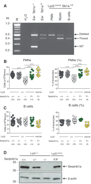
genomic DNA of sorted BM leukocyte subsets confirmed
almost complete cre-mediated recombination of the
Ser-pinb1a locus in PMNs, partial recombination in myelocytes
and, as expected, no recombination in B cells (Figure 2A).
However, absolute numbers and percentage of PMNs in
the BM of Lyz2
cre/creSb1a
F/-mice were normal and equivalent
to those of Sb1a
+/+, Sb1a
+/-and Lyz2
+/+Sb1a
F/-mice (Figure
2B), whereas Sb1a
-/-mice had reduced PMN numbers as we
Floxed (tm1.2)
4 56 7
3
2 SacI KpnI EcoRV 1 4 56 3 2 SacI EcoRV 1 Hyg-TK 7 KpnI Targeted 4 56 3
2 SacI KpnI EcoRV 1
Deleted (tm1.1)
Figure 1: Generation of Serpinb1aF/F mice.
Shown at the top is the targeted Serpinb1a locus with 3-loxP sites (black triangles) in ES cells. Cre recombinase in ES cells (indicated by white arrows) generated two new alleles: either a floxed (Serpinb1atm1.2(flox)Cben) allele or a deleted (Serpinb1atm1.1Cben) allele.
In vivo recombination (red arrow) of a floxed allele in Serpinb1aF/F
(Sb1aF/F) mice is expected to generate a deleted allele in cell
line-ages expressing cre. Deletion of exon 7, which encodes the reactive center loop of the serpin, was previously shown to be an effective null allele (Benarafa et al., 2007).
PMNs B cells Deleted Floxed WT 0.5 -B cell s My elo PMN kb
Lyz2cre/cre.Sb1aF/F
1.0 0.4 0.3 0.2 -M H2 O Ear Sb1a +/-Ear Sb1a F/-B cells (%) Serpinb1a Serpinb1a Lyz2+/+ +/+ +/- -/- F/F Lyz2cre/cre β-actin
D
A
C
B
55 35 35 -0 6 12 18 Cel ls (x10 6/fem ur ) +/+ **** +/- -/-Lyz2 Serpinb1a F/- F/-+/+ cre/cre ******** (20) (20) (20) (14) (8) 0 10 20 30 40 50 % of le ukocy te s +/+ **** +/- -/-Lyz2 Serpinb1a F/- F/-+/+ cre/cre ******** (20) (20) (20) (14) (8) PMNs (%) 0 3 6 9 12 15 Ce lls (x 10 6/fem ur ) +/+ +/- -/-Lyz2 Serpinb1a F/- F/-+/+ cre/cre (20) (20) (20) (14) (8) 0 10 20 30 40 50 % of le ukocyt es +/+ +/- -/-Lyz2 Serpinb1a F/- F/-+/+ cre/cre (20) (20) (20) (14) (8)Figure 2: Lyz2cre/cre deletion of Serpinb1aF/F.
(A) PCR analysis of genomic DNA isolated from flow cytometry sorted BM cells isolated from Lyz2cre/creSerpinb1aF/F mice. Arrows indicate
expected size of PCR products for Sb1a alleles (deleted 500bp; floxed 410bp; WT 250bp) as indicated by ear biopsy standard samples. Total number and percentage of BM PMN (B) and B cells (C). BM and blood cell numbers and percentages were analyzed by Mann-Whitney U-test (****p < 0.001). (D) Western blot analysis of Serpinb1a and β-actin of flow-sorted BM PMNs.
previously reported (Benarafa et al., 2011; Baumann et al.,
2013). As expected, B cell numbers were not significantly
different between all genotypes (Figure 2C). Western blot
analysis of sorted BM cells revealed that Serpinb1a protein
was still detectable in PMN lysates of Lyz2
cre/creSb1a
F/Fmice
These findings demonstrate that Lyz2
cre-driven deletion of
the Serpinb1a locus does not produce PMNs defective in
Serpinb1a protein despite efficient DNA recombination in
PMNs.
Cebpa-driven cre recombination of Serpinb1a
floxed allele in neutrophils
To evaluate an alternative model, Sb1a
F/Fmice were
inter-crossed with mice expressing the cre recombinase from
endogenous CCAAT/enhancer binding protein α (C/EBPα)
promoter, which drives expression at an earlier
develop-mental stage in myelopoiesis than Lyz2 (Wölfler et al.,
2010). Efficient deletion of the floxed allele was observed
in PMNs, myelocytes and monocytes but not in B cells
(Figure 3A). Western blot analysis also confirmed absence
of Serpinb1a protein in sorted PMN lysates of Cebpa
+/cre-Sb1a
F/-mice (Figure 3B). Accordingly, myeloid cell-specific
deletion of Serpinb1a in Cebpa
+/creSb1a
F/-mice reproduced
the phenotype of Sb1a
-/-mice characterized by reduced
absolute numbers and significantly lower percentage
of PMNs in the BM (Figure 4A). As in Sb1a
-/-mice, other
cell subsets in the BM were not altered in Cebpa
+/creSb1a
F/-mice (Figure 3B; Supplementary Figure S1A,B). Numbers
of PMNs and other leukocyte subsets in blood as well as
other blood parameters were not altered (Figure 4C;
Sup-plementary Figure S1C; Table 1), which is consistent with
the phenotype of Sb1a
-/-mice.
PMNs are highly susceptible to granule
permeabili-zation-induced cell death caused by L-leucyl-L- leucine
methyl ester (LLME). LLME treatment of PMNs of Cebpa
+/creSb1a
F/-mice showed reduced survival similar to those
of control Cebpa
+/creSb1a
-/-littermates and of Sb1a
-/-mice (Figure 4D). As shown previously for Sb1a
-/-PMNs
(Baumann et al., 2013), caspase inhibition with Q-VD-OPh
had no protective effect on cell death of Cebpa
+/creSb1a
F/-PMNs. Taken together, these data demonstrate that
mye-loid-specific deletion of Serpinb1a largely replicates the
PMN phenotype of constitutive Sb1a
-/-mice.
Transgenic rescue of Sb1a
-/-neutrophils with
human SERPINB1
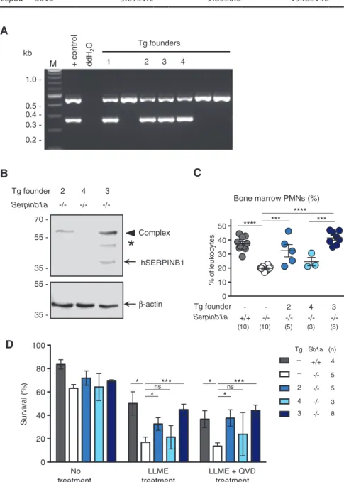
Human SERPINB1 cDNA was cloned downstream of the
human S100A8 (MRP8) promoter and was injected in the
pronucleus of C57BL/6J oocytes. Founder transgenic mice
were identified by PCR analysis (Figure 5A). Three of the
male founders were crossed with Sb1a
-/-mice. Western blot
analysis of PMN lysates revealed that the progeny of founder
55 35 70 -Serpinb1a β-actin 55 35 -Serpinb1a Cebpa+/+ +/+ +/- -/-
F/-A
B
M Ear Sb1 a +/ -Ear Sb1 a F/-Deleted Floxed WT B cells Myelo PMN Mono H2 OCebpa+/creSb1a
F/0.5 -kb 1.0 0.4 0.3 0.2 -Complex
*
Figure 3: Cebpa+/cre deletion of Serpinb1a.
(A) Genotyping PCR analysis of genomic DNA isolated from flow cytometry sorted BM cells of Cebpa+/creSb1aF/- mice. Arrows indicate
expected size of PCR products for Serpinb1a alleles (deleted 500 bp; floxed 410 bp; WT 250 bp) as indicated by ear biopsy stand-ard samples. (B) Western blot analysis for Serpinb1a and β-actin of flow-sorted BM PMNs. Serpinb1a-protease complex and partly degraded complex are indicated by an arrowhead and an asterisk, respectively.
3 (Tg3) typically expressed more Serpinb1 protein than
the progeny of Tg2 and that Serpinb1 expression was not
detectable in the progeny of Tg4 (Figure 5B). Percentage of
PMNs in the BM of the different Tg lines correlated with the
expression levels of each line (Figure 5C). Correspondingly,
protection from LLME-induced granule
permeabiliza-tion and cell death correlated with Serpinb1 expression
levels in the different Tg lines. Specifically, survival of high
expressors Tg3 PMNs was rescued to levels of WT PMNs.
Reduced survival was observed in PMNs of the Tg2 and
Tg4 lines and lowest survival was expectedly observed for
Sb1a
-/-PMNs (Figure 5D). Therefore, these data demonstrate
that Serpinb1 functions as a rheostat in protecting PMNs
from granule protease-mediated death in vivo and in vitro.
Discussion
We report that PMNs lacking Serpinb1a protein were
successfully generated in mice expressing the cre
models. Failure to generate Serpinb1a protein-null PMNs
despite efficient genomic recombination in Lyz2
cre/creSb1a
F/-mice is likely due to a combination of factors. First, the
floxed Serpinb1a locus was not fully recombined during
early stages of granulopoiesis. Second, low levels of
Ser-pinb1a protein may suffice to support PMN survival. Since
Serpinb1a transcription is active in HSCs and peaks early
in granulopoiesis at the promyelocyte/myelocytes stages
(Benarafa et al., 2011), Serpinb1a protein is already
accu-mulating in the cell before the floxed gene is recombined.
Thus, sufficient Serpinb1a protein levels may be sustained
throughout PMNs’ short life-span and maintain normal
PMN levels in the BM of Lyz2
cre/creSb1a
F/-mice.
PMNs develop in the BM from hematopoietic stem
cells via bipotent granulocyte/macrophage
progeni-tors (GMPs), which can develop into monocyte or PMN
lineages. Cebpa is expressed at high levels at the GMP
stage, whereas Lyz2 expression peaks later and
indepen-dently in both granulocyte and monocyte lineages.
Cre-mediated recombination using these two cre knock-in
models has been widely and successfully used to delete
genes in the myeloid compartment. Yet, as we have seen,
the timing of cre expression as well as the lineage may be
important in achieving effective gene recombination in
early stages of granulopoiesis. Recombination efficiency
at different stages of hematopoiesis also differs
depend-ing on the targeted locus and therefore makdepend-ing the right
choice of a cre deleter strain can be difficult and will
always require experimental confirmation. Our study
highlights that it remains crucial that gene deletion and
protein levels are effectively measured particularly when
recombination appears to have no effect on the studied
phenotype as shown by the absence of BM neutropenia
in Lyz2
cre/creSb1a
F/-mice. Far from a theoretical question, in
studies where protein levels were not verified and showing
no phenotype in mice with deleted myeloid cells (Rupec
et al., 2005; Kirkland et al., 2012), incomplete protein
dele-tion may have led to overlooking the contribudele-tion of the
studied proteins in myeloid cells.
We showed here that the extent of the rescue of PMN
survival by transgenic expression of human SERPINB1 in
Sb1a
-/-mice was dependent on transgene expression levels.
Evaluating the threshold levels of Serpinb1 necessary for
cytoprotection is challenging because Serpinb1-protease
complexes are processed rapidly into post-complex
cleav-age forms when proteases are in excess (Cooley et al., 2011).
Furthermore, the antibody used in Western blot analysis
may react differently with endogenous mouse and
trans-genic human Serpinb1 as well as with the different
com-plexes. In the Sb1a
-/-Tg2 progeny, the hSerpinb1 transgene
is relatively weakly expressed and only detectable in
0 20 40 60 80 100 Survival (%) No treatment LLME treatment LLME + QVD treatment Cebpa Serpinb1a +/+ -/- +/- -/- F/-+/+ +/+ +/cre +/cre +/cre (n) 3 9 5 7 11 * * * ns *** * ns **
D
A
B
Bone marrow PMNS Bone marrow PMNS (%)
C
Blood PMNs (%) Blood monocytes (%)Bone marrow monocytes Bone marrow monocytes (%) 0 20 40 60 % of le ukoc yt es Cebpa Serpinb1a +/+ F/-+/cre +/- -/- F/-** ** 0 5 10 15 20 Ce lls (x10 6 /fem ur ) ** Cebpa Serpinb1a +/+ F/-+/cre +/- -/- F/-(13) (8) (5) (11) (13) (8) (5) (11) (13) (8) (5) (11) (13) (8) (5) (11) 0 5 10 15 % of le ukocy te s Cebpa Serpinb1a +/+ F/-+/cre +/- -/- F/-0 2 4 6 8 Ce lls (x 10 6 /fem ur ) Cebpa Serpinb1a +/+ F/-+/cre +/- -/- F/-0 5 10 15 % of le uk oc yt es +/+ F/-+/cre +/- -/- F/-Cebpa Serpinb1a 0 5 10 15 % of le ukocyt es +/+ F/-+/cre +/- -/- F/-Cebpa Serpinb1a (12) (7) (5) (11) (11) (7) (5) (11)
Figure 4: Deletion of Serpinb1a in myeloid cells is sufficient to reduce PMN survival.
Total number and percentage of BM PMNs (A), BM monocytes (B). Percentage of blood PMNs and monocytes (C). BM cell numbers and percentages were analyzed by Mann-Whitney U-test (**p < 0.01). (D) Survival of PMNs treated with LLME (100 μM) in the presence or absence of the caspase inhibitor Q-VD-OPh (50 μM). Viability was assessed using Annexin V-FITC and 7- AAD staining of Ly-6G+
cells and analyzed by Student’s t-test relative to wild-type PMNs (*p < 0.05; **p < 0.01; ***p < 0.001).
recombinase driven by the Cebpa promoter but not by
the Lyz2 promoter. Accordingly, PMNs of Cebpa
+/creSb1a
F/-mice, but not PMNs of Lyz2
cre/creSb1a
F/-mice, recapitulated
the phenotype of Sb1a
-/-PMNs with reduced survival
in vitro and in vivo. We found that the floxed Serpinb1a
Table 1: Hematological analysis of whole blood.
Genotype WBC ( × 106 cells/ml) RBC ( × 109 cells/ml) PLT ( × 106 cells/ml) Hemoglobin (g/dl) Hematocrit (%) n
Sb1a+/+ 6.84±3.4 9.57±0.4 1295±248 15.8±0.7 52.7±2.6 8
Sb1a+/- 6.41±1.6 9.38±0.7 1309±160 15.6±1.2 48.2±5.4 7
Sb1a-/- 6.13±2.9 9.57±0.6 1656±129 16.3±0.4 51.2±3.0 4
Sb1aF/- 6.06±2.6 9.66±0.7 1479±182 16.2±1.0 50.4±4.3 13
Cepba+/creSb1a+/- 6.47±2.4 9.55±0.7 1361±258 15.9±1.2 49.9±5.1 8
Cepba+/creSb1a-/- 5.46±0.8 9.49±0.7 1532±187 15.7±1.0 50.3±2.9 5
Cepba+/creSb1aF/- 5.69±1.2 9.80±0.6 1348±142 16.1±0.9 51.0±4.4 9
M + control ddH 2 O
B
C
D
Bone marrow PMNs (%)A
0.5 -kb 1.0 0.4 0.3 0.2 -Tg founders 1 2 3 4 Serpinb1a Tg founder 35 - 55 - 70 - 2 -/- -/- -/-4 3 35 - 55 - hSERPINB1 β-actin Complex*
0 10 20 30 40 50 % of le uk oc yt es 2 4 3 Tg founder Serpinb1a +/+ -/- -/- -/- -/-- -*** **** *** **** (10) (10) (5) (3) (8) 0 20 40 60 80 100 Su rv iv al (% ) * *** * ns No treatment LLME treatment LLME + QVD treatment Tg Sb1a +/+ -/-_ _ 2 4 3 * *** *ns (n) 4 5 5 3 8Figure 5: Rescue of Serpinb1a-/- PMN survival by transgenic expression of human SERPINB1.
(A) PCR analysis of genomic DNA of transgenic founders using a single primer pair that recognizes both the human SERPINB1 transgene cDNA (266 bp) and mouse Serpinb1a gene (460 bp, which includes an intronic sequence). (B) Western blot analysis for SERPINB1 and β-actin of flow-sorted BM PMNs from offsprings of Tg founders 2, 3 and 4. hSerpinb1-protease complex and partly degraded complex are indicated by an arrowhead and an asterisk, respectively. (C) Percentage of PMN numbers in the BM of Sb1a-/-Tg2, Sb1a-/-Tg3 and Sb1a-/-Tg4 mice
(**p < 0.01). (D) Survival of PMNs treated with LLME (100 μM) in the presence or absence of the caspase inhibitor Q-VD-OPh (50 μm). Viability was assessed using Annexin V-FITC and 7- AAD staining of Ly-6G+ cells and analyzed by Student’s t-test relative to Sb1a-/- PMNs (*p < 0.05;
complex with proteases. With all the caveats in mind for
such a comparison, it suggests that the Tg2 line expresses
substantially lower levels than heterozygous Sb1a
+/-mice,
where active mouse Serpinb1a can be detected. Yet, in the
Sb1a
-/-Tg2 mice, we observed a partial rescue of the survival
phenotype in vivo and of LLME-induced death in vitro.
In the Sb1a
-/-Tg4 progeny, the transgene was not
detect-able by Western blot analysis, suggesting very low or no
expression and, accordingly, PMN numbers in the BM were
as low as in Sb1a
-/-mice. In the higher expressing
trans-genic line (Tg3), hSERPINB1 transgene was found in active
and complex forms and PMN survival was fully rescued
in vitro and in vivo. This new transgenic model will allow
us to evaluate the function of the ubiquitously expressed
Serpinb1a in non-hematopoietic tissues in the absence of
the PMN survival defect in various disease models.
More targeted deletions in tissue or cell subsets
could then follow using Sb1a
F/-mice crossed with other
cre-expressing mouse lines. Of note, reporter analysis of
Cebpa
+/cremice showed recombination in the liver and
lung airway epithelium (Wölfler et al., 2010). It is thus
likely that the Sb1a
F/-locus was recombined in some
non-hematopoietic cells of Cebpa
+/creSb1a
F/-mice. We previously
demonstrated that, in BM chimera, deletion of Sb1a in the
hematopoietic compartment was necessary and sufficient
to reproduce the phenotype of Sb1a
-/-mice (Baumann
et al., 2013). Therefore, effective deletion of Serpinb1a in
myeloid cells is undoubtedly the cause of BM
neutrope-nia in Cebpa
+/creSb1a
F/-mice. However, careful scrutiny of
Sb1a expression in various cell types and time points will
be required when using Cebpa
+/creSb1a
F/-mice in systemic
disease models. Finally, HSCs of Cebpa
+/creSb1a
F/-mice may
be most useful to reconstitute the immune system of
irra-diated mice to generate myeloid-specific deficiency.
Transgenic mice expressing cre under the hMRP-8
promoter (S100A8) and knock-in models such as
neu-trophil elastase knock-in mice (Elane
tm1(cre)Roes) or Ly6G
knock-in (Catchup) mice (Ly6g
tm2621(Cre-tdTomato)Arte) are
addi-tional models for PMN-specific gene deletion in PMNs
(Tkalcevic et al., 2000; Passegué et al., 2004; Hasenberg
et al., 2015). These models have reduced targeting of the
monocytic lineage than Lyz2 and Cebpa cre knock-in mice
used here. Whether Serpinb1a protein-null PMNs can be
obtained using these models remains to be tested but
is unlikely given the persistence of the protein in PMNs
of Lyz2
cre/creSb1
F/-mice and the small amount of protein
rescue needed for mitigating the Sb1a
-/-phenotype in
transgenic mice. For example, deletion of the Fcgr4 gene
was complete and specific for PMNs at the genomic level
in Catchup mice. While the mice showed a specific
pheno-type due to gene deletion, FcRIV expression on the PMN
surface measured by flow cytometry was only reduced by
50% (Hasenberg et al., 2015). Experimental approaches
using cre-mediated recombination in PMNs thus remain
challenging in choosing the right cre-expressing model(s)
and in the interpretation of the data. In addition to time
and resources to generate the mice, it requires specific
attention to the target gene expression pattern, protein
stability and functional protein threshold to choose the
right cre-expressing model(s) and to draw appropriate
conclusions.
In summary, we showed that the survival of PMNs
depends on a threshold level of Serpinb1 below which
serine protease activity is not controlled, leading to cell
death. These findings are consistent with the mode of
action of Serpinb1 as a stoichiometric inhibitor of
neutro-phil proteases. Our study further supports the notion that
intracellular serpins such as clade B serpins in vertebrates
and serpins of Caenorhabditis elegans have a
fundamen-tal cytoprotective function (Bird, 1999; Zhang et al., 2006;
Luke et al., 2007; Tan et al., 2013; Bird et al., 2014). We
have previously demonstrated that BM neutropenia of
Sb1a
-/-mice is dependent on cathepsin G in vivo (Baumann
et al., 2013). In addition, Serpinb1 prevents
spontane-ous PMN apoptosis by inhibiting proteinase-3-mediated
cleavage and activation of caspase-3 (Loison et al., 2014).
PMNs are exquisitely sensitive to death after granule
leakage induced by LLME treatment and cathepsin G is
required for this caspase-independent cell death pathway
that is critically regulated by Serpinb1 (Baumann et al.,
2013). Similarly, Serpinb9 protects cytotoxic lymphocytes
against granzyme B-mediated death following granule
leakage induced by LLME treatment or by T cell
activa-tion (Bird et al., 2014). Serpinb6a is another intracellular
serpin inhibitor of cathepsin G that is expressed in PMNs
and may contribute to the protease shield against granule
permeability-induced cell death. Ongoing studies using
multiple targeting of the large locus of clade B serpin
locus on mouse chromosome 13 will determine the
rela-tive contributions of Serpinb1a, Serpinb6a, Serpinb9 and
additional understudied serpin paralogs in cellular
home-ostasis. The models described here will provide important
tools for these studies.
Materials and methods
Ethics statement
All animal studies were approved by the Cantonal Veterinary Office of the canton of Bern and conducted in accordance with the Swiss federal legislation on animal welfare.
Mouse models for conditional deletion of Serpinb1a
Serpinb1a-/- (Serpinb1atm1.1Cben) mice were generated in 129S6/SvEvTac(129S6) background (Benarafa et al., 2007) and backcrossed in C57BL/6J background (Benarafa et al., 2011). The latter were used in this study. Serpinb1aF/F (Serpinb1atm1.2(flox)Cben) were generated in
parallel with Sb1a-/- mice. Briefly, 129S6/W4 ES cells (Taconic) were
targeted by homologous recombination with a linearized plasmid described previously (Benarafa, 2011). Homologous recombinant clones with 3-loxP sites were transiently transfected with Cre recom-binase to excise the floxed CMV-HYG/TK positive/negative selection cassette. Cells were further selected with gancyclovir to eliminate the clones where the deletion of the selection cassette did not occur. While most clones tested had recombined the first and third loxP sites to generate the deleted allele (serpinB1atm1.1Cben), we also found clones
that recombined the second and third loxP sites, leaving an allele (serpinB1atm1.2(flox)Cben) with a floxed exon 7 (Figure 1). ES cells (clone
2F7-F5) were injected into blastocysts at the transgenic core facility of the Brigham and Women Hospital (Boston). Sb1a+/F heterozygous
mice were generated from chimeric mice. During backcrossing into C57BL/6J for 10 generations, Sb1a+/F breeders were also selected
based on the polymorphic PCR markers D13Mit117 and D13Mit16 to reduce the portion of chromosome 13 belonging to the 129S6 strain to < 3–4 cM on each side of the Serpinb1a locus. Cre recombinase knock-in mice Lyz2cre/cre mice (B6.129P2-Lyz2tm1(cre)Ifo) (Clausen et al.,
1999) were obtained from the Jackson Laboratories at backcross gen-eration N6 and were further backcrossed to N10 with C57BL6/J mice before intercrossing with Sb1aF/F mice to generate the desired
geno-types. Cre recombinase knock-in mice Cebpa+/cre mice (Cebpatm1(cre)Touw)
were described previously (Wölfler et al., 2010).
Generation of human SERPINB1 transgenic mice
A 3.8 kb fragment upstream of the S100A8 start codon was amplified by PCR from a human BAC clone obtained from imaGenes (I.M.A.G.E. clone number RPCIB753O1168Q) and directionally cloned using
NheI and SalI restriction digest upstream of human SERPINB1 cDNA
flanked downstream by the SV40 small T intron and poly-adenyla-tion signal. The final DNA construct was verified by sequencing and the plasmid backbone was excised by restriction digest with ClaI and NotI. The transgene band was separated by agarose electropho-resis and gel purified. The transgene was microinjected in C57BL/6J oocytes at the Cryoconservation and Transgenic unit of the Theodor Kocher Institute, University of Bern. Four male and four female mice out of 21 total mice born were positive for the transgene by PCR. Three of the four transgene positive founder males transmitted the transgene to their progeny and were then crossed with Sb1a-/- mice.
The transgene was always maintained as hemizygous. The progeny of each founder were handled as independent transgenic lines named Tgx (full nomenclature B6J-Tg(S100A8-SERPINB1)xCben), where ‘x’ represents the founder number.
Flow cytometry and cell sorting
Leukocyte counts and hematopoietic lineage differential analysis of BM was performed in mice aged 6–8-weeks old. BM cells were har-vested from femurs by flushing with PBS supplemented with 1% FCS,
red blood cells were lyzed with ammonium chloride for 10 min, fur-ther washed in PBS and counted in a Neubauer chamber. Isolated BM cells were stained with fluorescently labeled antibodies (Biolegend and BD Biosciences) and analyzed on a FACScalibur flow cytometer (BD Biosciences) as described previously. Briefly, leukocyte subsets percentages were determined within CD45+ cells as PMNs (CD11b+
Ly-6G+), monocytes (CD115+), myelocytes (CD11b+, CD115neg, Ly6Gneg,
SSChigh) and B cells (CD19+ or CD45RB220+). Blood was collected by
retro-orbital venous puncture using heparinized microcapillary tubes and leukocyte subsets analyzed by flow cytometry as above. Total leukocyte, erythrocyte and platelet counts as well as hemo-globin and hematocrit measurements of whole blood were performed using a Scil Vet ABC hematology analyzer (Horiba Medical, Montpel-lier, France). Flow cytometry data was analyzed using FlowJo (FlowJo LLC). Flow sorting of PMNs, B cells and myelocytes was performed on single-cell suspensions of BM leukocytes stained with antibodies mentioned above using a FACS Aria II sorter (BD Biosciences) at the flow cytometry core facility of the Department of Clinical Research of the University of Bern.
Western blotting
Sorted cells were washed and lysed (107/ml) in RIPA buffer with
protease inhibitor cocktail (Roche). Lysates were resolved by SDS-PAGE under reducing conditions and immunoblotted using rabbit antiserum to human SERPINB1 provided by ERO (Rees et al., 1999). Blots were stripped and restained with anti-β-actin antibody (Cell Signaling Technology).
Cell death induced by granule permeabilization
BM cells were cultured in DMEM (4 mm L-Glut, 25 mm D-Glucose, 1 mm sodium pyruvate) (Life Technologies) containing 1% FCS and 1% penicillin/streptomycin at 1.0 × 10⁶ cells/ml in the presence or
absence of the pan-caspase inhibitor Q-VD-OPh (SM Biochemicals LLC) or LLME (G-2550; Bachem). Cells were harvested and viability was assessed using annexin V–fluorescein isothiocyanate (FITC) and 7-aminoactinomycin D (7AAD) and measured at the FACScalibur flow cytometer.
Statistical analysis
Leukocyte subset analysis was performed using Mann-Whitney
U-test or Student’s t-test with GraphPad Prism Mac 4.0c software
(GraphPad, San Diego, CA, USA). A p Value < 0.05 was considered sta-tistically significant.
Acknowledgments: We thank Elisabeth Frei and Stephan
Hirschi for excellent technical assistance. We acknowledge
Albert Witt for pronucleus microinjection, Lina Du for ES
cell injection and Bernadette Nyfeler for cell sorting. We
thank the ZEMB and DKF animal caretaker teams for
dedi-cated attention and husbandry. This study was supported
by grants from the Swiss National Science Foundation
(127464 and 149790) (CB), the EU/FP7 Marie Curie
Interna-tional Reintegration Grant (CB) grant 249297 and the Bern
University Research Foundation (CB). Serpinb1a
+/Fmice
were generated with support from the National Institutes
of Health grant HL66548 (ERO).
References
Ashida, S., Nakagawa, H., Katagiri, T., Furihata, M., Iiizumi, M., Anazawa, Y., Tsunoda, T., Takata, R., Kasahara, K., Miki, T., et al. (2004). Molecular features of the transition from prostatic intraepithelial neoplasia (PIN) to prostate cancer: genome-wide gene-expression profiles of prostate cancers and PINs. Cancer Res. 64, 5963–5972.
Baumann, M., Pham, C.T.N., and Benarafa, C. (2013). SerpinB1 is critical for neutrophil survival through cell-autonomous inhibi-tion of cathepsin G. Blood 121, 3900–3907.
Benarafa, C. (2011). The SerpinB1 knockout mouse a model for studying neutrophil protease regulation in homeostasis and inflammation. Methods Enzymol. 499, 135–148.
Benarafa, C., Cooley, J., Zeng, W., Bird, P.I., and Remold-O’Donnell, E. (2002). Characterization of four murine homologs of the human ov-serpin monocyte neutrophil elastase inhibitor MNEI (SERPINB1). J. Biol. Chem. 277, 42028–42033.
Benarafa, C., Priebe, G.P., and Remold-O’Donnell, E. (2007). The neutrophil serine protease inhibitor serpinb1 preserves lung defense functions in Pseudomonas aeruginosa infection. J. Exp. Med. 204, 1901–1909.
Benarafa, C., LeCuyer, T.E., Baumann, M., Stolley, J.M., Cremona, T.P., and Remold-O’Donnell, E. (2011). SerpinB1 protects the mature neutrophil reserve in the bone marrow. J. Leukoc. Biol. 90, 21–29. Bird, P.I. (1999). Regulation of pro-apoptotic leucocyte granule
serine proteinases by intracellular serpins. Immunol. Cell Biol.
77, 47–57.
Bird, C.H., Christensen, M.E., Mangan, M.S.J., Prakash, M.D., Sedelies, K.A., Smyth, M.J., Harper, I., Waterhouse, N.J., and Bird, P.I. (2014). The granzyme B-Serpinb9 axis controls the fate of lymphocytes after lysosomal stress. Cell Death Differ.
21, 876–887.
Clausen, B.E., Burkhardt, C., Reith, W., Renkawitz, R., and Förster, I. (1999). Conditional gene targeting in macrophages and granu-locytes using LysMcre mice. Transgenic Res. 8, 265–277. Cooley, J., Takayama, T.K., Shapiro, S.D., Schechter, N.M., and
Remold-O’Donnell, E. (2001). The serpin MNEI inhibits elastase-like and chymotrypsin-like serine proteases through efficient reactions at two active sites. Biochemistry 40, 15762–15770.
Cooley, J., Sontag, M.K., Accurso, F.J., and Remold-O’Donnell, E. (2011). SerpinB1 in cystic fibrosis airway fluids: quantity, molecular form and mechanism of elastase inhibition. Eur. Respir. J. 37, 1083–1090.
Cui, X., Liu, Y., Wan, C., Lu, C., Cai, J., He, S., Ni, T., Zhu, J., Wei, L., Zhang, Y., et al. (2014). Decreased expression of SERPINB1 cor-relates with tumor invasion and poor prognosis in hepatocel-lular carcinoma. J. Mol. Histol. 45, 59–68.
El Ouaamari, A., Dirice, E., Gedeon, N., Hu, J., Zhou, J.-Y.,
Shirakawa, J., Hou, L., Goodman, J., Karampelias, C., Qiang, G.,
et al. (2016). SerpinB1 promotes pancreatic β cell proliferation. Cell Metab. 23, 194–205.
Hasenberg, A., Hasenberg, M., Männ, L., Neumann, F., Borkenstein, L., Stecher, M., Kraus, A., Engel, D.R., Klingberg, A., Seddigh, P., et al. (2015). Catchup: a mouse model for imaging-based tracking and modulation of neutrophil granulocytes. Nat. Methods 12, 445–452.
Huasong, G., Zongmei, D., Jianfeng, H., Xiaojun, Q., Jun, G., Sun, G., Donglin, W., and Jianhong, Z. (2015). Serine protease inhibitor (SERPIN) B1 suppresses cell migration and invasion in glioma cells. Brain Res. 1600, 59–69.
Kirkland, D., Benson, A., Mirpuri, J., Pifer, R., Hou, B., DeFranco, A.L., and Yarovinsky, F. (2012). B cell-intrinsic MyD88 signaling prevents the lethal dissemination of commensal bacteria dur-ing colonic damage. Immunity 36, 228–238.
Kruger, P., Saffarzadeh, M., Weber, A.N.R., Rieber, N., Radsak, M., von Bernuth, H., Benarafa, C., Roos, D., Skokowa, J., and Hartl, D. (2015). Neutrophils: between host defence, immune modula-tion, and tissue injury. PLoS Pathog. 11, e1004651.
Loison, F., Zhu, H., Karatepe, K., Kasorn, A., Liu, P., Ye, K., Zhou, J., Cao, S., Gong, H., Jenne, D.E., et al. (2014). Proteinase 3-dependent caspase-3 cleavage modulates neutrophil death and inflammation. J. Clin. Invest. 124, 4445–4458.
Luke, C.J., Pak, S.C., Askew, Y.S., Naviglia, T.L., Askew, D.J., Nobar, S.M., Vetica, A.C., Long, O.S., Watkins, S.C., Stolz, D.B., et al. (2007). An intracellular serpin regulates necrosis by inhibit-ing the induction and sequelae of lysosomal injury. Cell 130, 1108–1119.
Naito, Y., Takagi, T., Okada, H., Omatsu, T., Mizushima, K., Handa, O., Kokura, S., Ichikawa, H., Fujiwake, H., and Yoshikawa, T. (2010). Identification of inflammation-related proteins in a murine colitis model by 2D fluorescence differ-ence gel electrophoresis and mass spectrometry. J. Gastroen-terol. Hepatol. 25(Suppl 1), S144–S148.
Passegué, E., Wagner, E.F., and Weissman, I.L. (2004). JunB deficiency leads to a myeloproliferative disorder arising from hematopoietic stem cells. Cell 119, 431–443.
Popova, E.Y., Claxton, D.F., Lukasova, E., Bird, P.I., and
Grigoryev, S.A. (2006). Epigenetic heterochromatin markers distinguish terminally differentiated leukocytes from incom-pletely differentiated leukemia cells in human blood. Exp. Hematol. 34, 453–462.
Rees, D.D., Rogers, R.A., Cooley, J., Mandle, R.J., Kenney, D.M., and Remold-O’Donnell, E. (1999). Recombinant human monocyte/ neutrophil elastase inhibitor protects rat lungs against injury from cystic fibrosis airway secretions. Am. J. Respir. Cell Mol. Biol. 20, 69–78.
Rupec, R.A., Jundt, F., Rebholz, B., Eckelt, B., Weindl, G.,
Herzinger, T., Flaig, M.J., Moosmann, S., Plewig, G., Dörken, B., et al. (2005). Stroma-mediated dysregulation of myelopoiesis in mice lacking IκBα. Immunity 22, 479–491.
Sheng, L., Anderson, P.H., Turner, A.G., Pishas, K.I., Dhatrak, D.J., Gill, P.G., Morris, H.A., and Callen, D.F. (2015). Identification of vitamin D3 target genes in human breast cancer tissue. J. Steroid Biochem. Mol. Biol. http://dx.doi.org/10.1016/j. jsbmb.2015.10.012 (in press).
Tan, J., Prakash, M.D., Kaiserman, D., and Bird, P.I. (2013). Absence of SERPINB6A causes sensorineural hearing loss with multiple histopathologies in the mouse inner ear. Am. J. Pathol. 183, 49–59.
Tkalcevic, J., Novelli, M., Phylactides, M., Iredale, J.P., Segal, A.W., and Roes, J. (2000). Impaired immunity and enhanced resist-ance to endotoxin in the absence of neutrophil elastase and cathepsin G. Immunity 12, 201–210.
Tseng, M.-Y., Liu, S.-Y., Chen, H.-R., Wu, Y.-J., Chiu, C.-C., Chan, P.-T., Chiang, W.-F., Liu, Y.-C., Lu, C.-Y., Jou, Y.-S., et al. (2009). Serine protease inhibitor (SERPIN) B1 promotes oral cancer cell motility and is over-expressed in invasive oral squamous cell carcinoma. Oral Oncol. 45, 771–776.
Wölfler, A., Danen-van Oorschot, A.A., Haanstra, J.R., Valkhof, M., Bodner, C., Vroegindeweij, E., van Strien, P., Novak, A., Cupedo, T., and Touw, I.P. (2010). Lineage-instructive function of C/EBPα in multipotent hematopoietic cells and early thymic progeni-tors. Blood 116, 4116–4125.
Yasumatsu, R., Altiok, O., Benarafa, C., Yasumatsu, C., Bingol-Karakoc, G., Remold-O’Donnell, E., and Cataltepe, S. (2006).
SERPINB1 upregulation is associated with in vivo complex for-mation with neutrophil elastase and cathepsin G in a baboon model of bronchopulmonary dysplasia. Am. J. Physiol. Lung Cell Mol. Physiol. 291, L619–L627.
Zhang, M., Park, S.-M., Wang, Y., Shah, R., Liu, N., Murmann, A.E., Wang, C.-R., Peter, M.E., and Ashton-Rickardt, P.G. (2006). Serine protease inhibitor 6 protects cytotoxic T cells from self-inflicted injury by ensuring the integrity of cytotoxic granules. Immunity 24, 451–461.
Zhao, P., Hou, L., Farley, K., Sundrud, M.S., and Remold-O’Donnell, E. (2014). SerpinB1 regulates homeostatic expansion of IL-17+ γδ
and CD4+ Th17 cells. J. Leukoc. Biol. 95, 521–530.
Supplemental Material: The online version of this article (DOI: 10.1515/hsz-2016-0132) offers supplementary material, available to authorized users.