Carcinogenesis vol.16 no.7 pp. 1499-1503, 1995
In situ analysis of transforming growth factor-Ps (TGF-pl,
TGF-J32, TGF-(33), and TGF-(3 type II receptor expression in
malignant melanoma
Peter Schmid
1, Peter Itin and Theo Rufli
Department of Dermatology, University Basel, Petersgraben 4, CH-4031 Basel, Switzerland
'To whom requests for reprints should be sent
We have analysed, by in situ hybridization, mRNA
expres-sion of TGF-pi, TGF-(32, TGF-p3, and of TGF-P type II
receptor in benign melanocytic naevi, primary melanomas,
and in skin metastases of malignant melanomas. Our
results show that melanoma progression correlates with
overexpression of TGF-p. All skin metastases and most
primary melanomas invasive to Clark's level IV-V revealed
specific TGF-P2 mRNA and protein expression. However,
expression of this cytokine was not observed in benign
melanocytic lesions and was detected only in one of five
early primary melanomas investigated. Some primary
melanomas and skin metastases also revealed specific
TGF-pl mRNA signals although expression of this isoform
was not found in benign naevi. TGF-p3 expression, which
was only barely detectable in benign melanocytic lesions,
was enhanced in some skin metastases. Interestingly, the
epidermis overlaying melanomas revealed lower levels
of TGF-p3 mRNA expression than epidermis of healthy
skin or epidermis adjacent to benign naevi, thereby
suggesting that paracrine mechanisms between tumour
cells and keratinocytes may influence melanoma
develop-ment. In primary melanomas TGF-P type II receptor
mRNA signals were much more heterogeneously distributed
when compared to benign melanocytic naevi, suggesting
variable degrees of TGF-P resistance among melanoma cells
within individual lesions. However, melanoma progression
appeared not to be correlated with a complete loss of
TGF-P type II receptor gene expression, since all skin
metastases revealed clearly detectable although
hetero-geneous levels of TGF-P type II receptor mRNA expression.
Introducton
Transforming growth factor-beta (TGF-p) is the name given
to a family of structurally and functionally related cytokines
which consist of three highly homologous mammalian
iso-forms, designated TGF-P 1, T G F - p , and TGF-P3. Although
expression of TGF-P isoforms is differentially regulated, these
pleiotrophic peptides show similar biological effects in most
experimental systems. TGF-ps control cell, proliferation and
differentiation processes, promote cell motility and
angio-genesis, have potent immunosuppressive effects, stimulate
synthesis of specific matrix proteins and integrins, and are
mediators of epithelial-mesenchymal interactions during
embryogenesis and wound healing (reviewed in 1,2). TGF-Ps
exert their effects via heteromeric complexes of TGF-p type
I (TPR I) and type II receptors (TpR II). A recent study has
shown that binding of TGF-P to TpR II is required for the
recruitment and activation of TPR I (3). Since TpR II can
interact with several type I receptors (4,5,6), the specificity of
the biological response to ligand appears to be defined by
the particular type I receptor, thus providing a rationale for
the multifunctional nature of TGF-Ps.
Recently, it has been suggested that escape from paracrine
or autocrine growth control by TGF-P during carcinogenesis
could involve genetic changes in the TpR II gene itself or
altered expression of its mRNA (7). Moreover, TGF-p switches
from an inhibitor of tumour cell growth to a stimulator of
growth and invasion during human colon carcinoma
progres-sion (8). Interestingly, elevated TGF-P mRNA levels have
been described in cell lines derived from diverse malignancies
(9), and increased expression of TGF-P in situ has been
observed in glioblastomas (10) and breast carcinomas (11).
An involvement of TGF-P in tumour progression has been
further substantiated by the finding that highly immunogenic
fibrosarcoma cells transfected with TGF-P 1 escaped
immuno-surveillance (12). Moreover, TGF-p2 has been shown to
suppress T-cell mediated immunity associated with
glio-blastoma multiforme (13), and anti-TGF-P antibodies
inhibited breast cancer cell tumorigenicity and increased mouse
spleen natural killer cell activity (14).
In vitro studies have shown that normal melanocytes and
some melanoma cells express TGF-P (15). However, normal
melanocytes are growth inhibited by TGF-P, whereas
melan-oma cells show various degrees of TGF-p resistance (16,17).
To test the hypothesis, that an altered TGF-P expression or
response may be implicated in melanoma progression in vivo,
we have investigated, by in situ hybridization, expression of
the three TGF-p isoforms and of TPR II, and by
immunohisto-chemistry, the protein distribution of TGF-p2, in histological
sections of benign melanocytic naevi, primary melanomas,
and skin metastases of malignant melanoma.
Materials and methods
Preparation of [3s-S]-labeled riboprobes
All riboprobe templates used in this study were described previously (18). 'Sense' and 'antisense' RNA probes were labeled according to the instructions of the manufacturer (Boehringer Mannheim: RNA Transkription Kit; Kat.Nr. 999644) with a-35S-UTP (>400 Ci/mmol, Amersham. Nr. SJ 263) to a
specific activity of >109 dprn/jig using SP6, T3 or T7 RNA polymerase.
Labeled riboprobes were extracted with phenol/chloroform and free nucleotides were removed by using a Sephadex G50 columne. RNA was precipitated in I vol. 7 M ammonium acetate/3.5 vol. ethanol overnight at -20°C, and then resuspended to an approximate concentration of 250 000 cpm/nl (5X concentrated stock solution) in 50% deionized formamide containing 20 mM dithiothreithol and stored at -70°C.
In situ hybridization
Biopsies were fixed overnight at 4°C in a freshly prepared solution of 4% paraformaldehyde in phosphate buffered salt solution (PBS), and then embedded in paraffin. 8 nm paraffin sections were placed on 3-aminopropyl-triethoxysilane treated slides, which bind sections covalently on the glass surface and prevent loss of sections during the experimental procedures. Paraffin sections were deparaffinized in xylene and absolute ethanol and air dried. Following rehydration with decreasing ethanol solutions sections were postfixed with 4% paraformaldehyde in PBS for 5 min, rinsed in PBS and
P.Sihmid, I'.ltin and T.RuHi
Hi(). depuimaled tin 2(1 nun vvilh (I 2 N HC1 .11 room lemperaluie treated 101 30 nun with 2XSSC l(>3 M NaCl. 0 03 M Na-cilralc. pH 7 0) at 70°C. dehydrated with inueasing elhanol solutions and linally air tried Piehybridi/.a-tion was performed at 54°C lor 3 h in 50r/{ deioni/ed toimamide. 10%
de\tiansiillale. 0 3 M NaCl. 10 111M Tn.s. 10 111M sodium phosphate pH 6 8. 20 111M dilhiothreitol. 0 2XDenhardt's reagent. 0 I mg/ml /.'<<>/i RNA. and 'cold' 0 5 uM ra-S-UTP Hybridization was carried out overnight in the same mix supplemented with 5XIO4 cpm/nl n - ^ S - U T P labelled RNA piobe in a humihed chamber at 54°C Slides were washed in hybridization solution without dextransultate. RNA. and 'cold' UTP conlaining 50% deioni/.ed lonnamide and 10 111M dithiothreitol at 55°C two times lor 1 h and cquilibiuted I01 15 mm in a buffet solution consisting ol 0 5 M NaCl. 10 niM Tris. I 111M liDTA, 10 111M D'lT pH 7 5 Sections weie then treated with 50 |ig ml ' RNasc A in equilibration butler toi 30 mm at 37"C to remove non-specihc bound probe Sections were washed in 2X.SSC I01 1 h and then in 0 I X S S C I01 I h at 37"C Slides were then sequentially dehydrated in h.V/(. X5'/i and l*5'i ( \ A ) ethanol solutions containing 300 mM ammonium
acetate and in absolute ethanol before being air dried Sections were coaled with a I 2 dilution ol llford K5 photoemulsion. air dried, and exposed for 12 days in a light sale box containing silica gel at 4°C Slides were developed in 1)19 developer (Kodak), hxed in 30% sodium thiosullate and stained with hacmatoxylin and eosm The pattern of hyhndr/ation signals on autotadio-graphed sections was analysed using a photomicroscope and hnghtheldV darklield illuminations For a semiquantitative analysis of mRNA expression, the average number ol silver grains per cell was calculated within individual tissues mRNA expression was defined as 'not detectable' it the average numbei ol silvei grains was less than two per cell and theietore not clearly distinguishable Irom non-specific background
Immunohistochemistiy was performed on paratlin sections using a labbit polyclonal IgG. which iccognizes icsidues 352-377 ol human TGF-|32 (Santa C m / Biotech) Sections were de-waxed in xylene. rehydrated in decreasing ethanol solutions, and incubated lot I h in I'BS containing 1% BSA The primary antibody was diluted (I ug/ml) in PBS containing 0 29f BSA and applied overnight at 4°C Antibody staining was performed using the StrAviGen Super Sensitive detection kit using last red as substrate (HioCienex. San Ramon. CA) Sections were counterstained vuth haematoxylin
Results
S/x'i ificily of \if>nah
The specificities of the TGF-(J 1/2/3 and T(3R II probes were determined by Northern blot analysis By using riboprobes encoding the three mature TGF-(3 isoforms. we have previously shown that no cross-hybridization occurs during in situ hybridization (IS). The specificity ol" the hybridization was further controlled by using the corresponding "sense" ribo-probes as negative controls which gave very weak and uni-formly distributed non-specific background signals comparable to the non-specific signals shown in Figures la. 1b and 2b.
Specificity of immunostaining was controlled by using non-immune serum which revealed no specific tissue staining (not shown).
Benign nielanocxtic mien
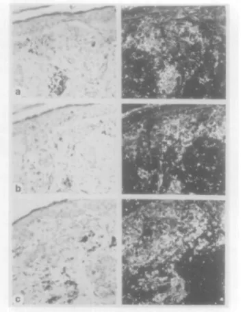
Four benign melanocytic naevi (1 compound. 1 junctional. 2 dermal) were investigated (Table I). In benign melanocytic naevi. specific expression of TGF-fjl and TGF-(32 mRNA was not detectable (Figures la.b). Only weak TGF-J33 hybridization signals (Figure Ic) were observed in three benign melanocytic lesions, although the epidermis (Figure lc) and hair follicle epithelia (not shown) revealed abundant TGF-fj3 mRNA expression. Expression of TpR 11 mRNA (Figure Id) was clearly detectable in all four naevi. the level of expression being comparable to that observed in epidermal keratinocytes (Figure Id) and vascular endotheha (not shown).
Primary melanomas
Five early melanomas according to Clark's level ll-III. and five advanced melanomas according to Clark's level IV-V were investigated (Table I) The ma|ority of early primary
v , ' * . • • • • • f ;
F i g . 1. In sun h y b i i d i / a t i o n analysis ol l a ) TGl-"-|M. ( b ) TGI-"-|32. (c) T G F - p 3 . a n d ( d ) Tf3R II m R N A e x p r e s s i o n in n a e x u s n a e v o e e l l u l a n s (left) B n g h t h c l d i l l u m i n a t i o n s , (iighl) d a r k l i e l d i l l u m i n a t i o n s ( a ) N o specific TGI--[3l h y b r i d i z a t i o n signals are visible ( b ) E x p r e s s i o n of T G l;- | i 2 m R N A is not visible ( c ) N a e v u s cells s h o w s onlv barely d e t e c t a b l e le\'els of TGF-J33 m R N A e x p r e s s i o n , a l t h o u g h e x p r e s s i o n of TGF-J33 m R N A is clearly visible m the e p i d e r m i s (d) A b u n d a n t T(3R II m R N A signals a i e visible both in n a e v u s cells a n d in the e p i d e i m i s
melanomas revealed no or only barely detectable expression of TGF-pi. TGF-P2 and TGF-P3 mRNA (Figure 2a-c). Abundant TGF-(3l mRNA expression was detected in two early melanomas and in one advanced melanoma. Specific signals forTGF-32 mRNA expression were found only in one early melanoma, although three advanced melanomas revealed abundant TGF-(32 expression. Two early melanomas and two advanced melanomas revealed levels of TGF-(33 mRNA expression which were stronger than those observed in benign naevi. Interestingly, the epidermis overlaying most primary melanomas (Figure 2c) revealed significantly lower levels of TGF-f53 mRNA expression (Figure 2c) when compared with epidermis of healthy skin (not shown) or epidermis overlaying
Transforming growth factor-(3s in malignant melanoma
Fig. 2. In situ hybridization analysis of (a) TGF-pi, (b) TGF-P2, and (c) TGF-P3 mRNA expression in an early primary melanoma, (left) Brightrield illuminations; (right) darkfield illuminations, (a) TGF-pl hybridization signals are only barely discernible within the tumour, (b) No specific TGF-P2 hybridization signals are visible, (c) TGF-P3 hybridization signals are only barely detectable both in the epidermis and within the tumour. Note, the bright spots in darkfield illuminations do not represent hybridization signals but light reflections from pigment granula.
Table 1. Traceability of TGF-pi, in melanocytic lesions by in situ
Benign naevi (n = 4) Early melanomas (n = 5) Advanced melanomas (n = 5) Skin metastases (n = 8) TGF-P2, and TGF-P3 mRNA hybridization TGF-PI (0/4) (2/5) (1/5) (3/8) TGF-P2 (0/4) (1/5) (3/5) (8/8) expression TGF-P3 (3/4) (2/5) (2/5) (4/8)
benign naevi (Figure lc). Expression of T(iR II was clearly
detectable in all primary melanomas. However, in primary
melanomas T0R II hybridization signals (Figure 3) were much
more heterogeneously distributed when compared with benign
naevi (Figure Id).
Skin metastases
Eight skin metastases from malignant melanomas were
investi-gated. All skin metastases (Table II) revealed clearly detectable
but various levels of TGF-(32 mRNA (Figure 4b) and protein
(Figure 5). Three metastases (Table II) showed also specific
TGF-P 1 hybridization signals (Figure 4a), and two (Table II)
revealed levels of TGF-(33 mRNA expression (Figure 4c)
which were markedly stronger than those observed in some
benign naevi and primary melanomas (Figures lc, 2c). The
level of TpR II mRNA expression varied among individual
metastases (Table II) and was stronger in endothelial cells than
in melanoma cells (Figure 4d).
Fig. 3. In situ hybridization analysis of TPR II mRNA in an advanced primary melanoma, (left) Brightfield illumination; (right) darkfield illumination. TpR II hybridization signals are very heterogeneously distributed within the tumour.
Table II. Semiquantitative analysis of TGF-PI, TGF-P2. TGF-P3, and TpR II mRNA expression in skin metastases of malignant melanomas
Biopsy no. TGF-PI TGF-P2 TGF-P3 TPRII
93.1983 93.2702 93.3766 93.3832 93.737 94.491 94.617 94.4092 + +•
Average number of silver grains per cell: - = < 2; + = 2-5; + + = 6-9; + + + = > 9.
Discussion
A previous study has shown that TGF-p3 is the predominantly
expressed isoform in human epidermis (18). Since TGF-P is
a potent inhibitor of melanocytic proliferation in vitro (16,17),
epidermal TGF-|J3 may have a physiological function to
control cell division and/or differentiation of melanocytes. The
finding that TGF-p3 mRNA expression is decreased in the
epidermis overlaying primary melanomas, when compared to
normal epidermis, suggests that melanoma cells may suppress
keratinocytic TGF-|33 expression via a paracrine mechanism.
Conversely, it is tempting to speculate that lack of
TGF-(33 expression in the epidermis could precede melanoma
development and promote clonal expansion of transformed
melanocytes. Melanomas may therefore arise predominantly
at sites of low epidermal TGF-p3 expression.
Furthermore, the heterogeneous pattern of T$R II expression
in primary melanomas may indicate variable degrees of
TGF-P resistance among melanoma cells, a finding which might
explain the decreased growth-inhibitory effect of TGF-p on
melanoma cells in vitro (16,17). However, melanoma
progres-sion does not imply a complete loss of TpR II mRNA
expression since all metastases revealed clearly detectable but
various levels of TpR II expression. Other mechanisms may
therefore contribute to the altered TGF-p reponsiveness of
1501
P.Schmid. P.ltin .mil T.Rufli Fin- 4 . In \iiu h y b i i d i / a t i o n a n a l y s i s ol l a ) T G F - [ M ( h i T G F - | 5 2 . (c) T G F - J 5 3 . a m i i d ) I[3R II i n R N A e x p i e s s i o n in a s k i n m e t a s t a s i s ( b i o p s y n o M3 2 7 0 2 1 ol m a l i g n a n t m e l a n o m a ( l e l t l t S n g h t l i e l d i l l u m i n a t i o n s , ( r i g h t ) d a i k h e k l i l l u n n n . i l i o i i s A h u n d . m l l a ) T G I - ( 5 l a n d ( b ) I G 1 - | 3 2 . a n d w e a k (<_) T G K | i 3 e\piL'sMon is v i s i b l e in m e l a n o m a e e l l s u l ) l - . \ p i e s s i o n ol T [ i R II is M r o n c e i in e n d o l h e l i a l e e l l s i h a n in m e l a n o m a e e l l s F i g . 5 . I m m u n o h i s l o e h e m i e a l a n a l y s i s ol T G F - J J 2 p r o t e i n d i s t r i b u t i o n in m a l i g n a n t m e l a n o m a ( l e l t i Iii.iily p r i m a r y m e l a n o m a N o s p e e i h e i m m u n o s i a i n i n g is w s i h l e ( l i g h t ) S k i n m e l a s i a s i s o f m a l i g n a n t m e l a n o m a TGF-[S2 i m m u n o i e a e l i v il\ is e l e a i l y \ isible i h i o u g h o u t t h e t u m o u r
melanoma colls, such as T(3R II gene mutations or processing
defects, missing TGF-p type I receptors, or alterations in the
intracellular TGF-P signal transduction pathways.
The present study contirms the recently puplished
observa-tion that TGF-(32 in malignant melanoma correlates with depth
of tumour invasion (19). In addition, we have shown that
overexprcssion of TGF-fil and/or TGF-(33 mRNA may also
occur in metastatic melanoma. However, a correlation of these
isoforms with melanoma progression appears less significant,
since not all metastases revealed elevated levels of TGF-|3l
and/or TGF-P3 transcripts. Expression of TGF-pi has been
found in cultured melanocytes (15) although we failed to
detect specitic TGF-(3l mRNA expression in benign
mclano-cytic lesions, suggesting that the level of TGF-fil expression
in benign melanocytic lesions is below the limit of detection
by //( situ hybridization. However, the growth medium for
melanocytes is supplemented with growth factors and phorbol
ester, the latter being a potent inducer of TGF-(3l transcription
(20.21). It remains therefore questionable if production of
TGF-pM by melanocytes m vitro has a physiological relevance.
A role of TGF-fJ3 to control melanocytic growth or
differenti-ation appears more likely, since normal epidermis and several
benign naevi showed detectable mRNA expression of this
isoform.
It appears that in melanomas a selective pressure favors
overproduction of TGF-fj which might promote tumour
pro-gression by diverse possible mechanisms. TGF-(i has potent
immunosuppressive effects, because it inhibits IL-1 stimulated
proliferation of T-lymphocytes (22). suppresses cytokine
stimu-lated activation of natural killer cells (23). and appears to be
an important negative regulator of MHC class I and class II
expression (24). A recent review of the literature (25) has
shown that HLA class I antigens are not detectable in about 40%
of metastatic melanoma lesions. Conversely, the cytotoxicity of
the V(3I6+ T cell line, which has been generated by ;/; vitro
expansion of tumour infiltrating lymphocytes of a patient with
regressing melanoma (26). is HLA class I restricted. Therefore,
secretion of bioactive TGF-p2 by melanoma cells may well
contribute to suppress local immunosurveillance. although this
mechanism appears not to be the only way which allows
melanoma cells to escape immune recognition, since Hubcr
et al. (27) have shown that some melanoma cell lines can
inhibit, by direct contact, the proliferation of tumour infiltrating
lymphocytes via a TGF-(32 independent pathway. The abilities
of TGF-|3s to induce angiogencsis and to promote the formation
of stroma (28) may be another role of these polypeptides in
the maintenance and progression of transformed cells in the
host (29). Interestingly, human melanoma cells have been
shown to require ligution of the intcgrin avf3, to sustain viability
and growth in three dimensional dermal collagen (30). Since
TGF-P up-regulates expression of integrin avP, (31).
over-expression of TGF-P may contribute to rescue melanoma
cells from apoptosis. Another possible puthomcchanism may
implicate the metastasis suppressor gene nm23 (32). In
well-differentiated human colon carcinoma cells nm23 functions in
the TGF-p signalling pathway leading to growth arrest and
differentiation, whereas colon carcinoma cells which have
progressed to a more aggressive phenotype have lost nm23
function and use TGF-P to stimulate growth and invasion (33).
Since expression of the nm.23 gene has also been inversely
correlated with tumour metastatic potential in human malignant
melanomas (34) a similar mechanism may contribute to
melan-oma metastasis.
Transforming growth factor-Ps in malignant melanoma
Acknowledgements
This work was supported by a grant from the Swiss National Fond (project no. 32-40488.94). We are very grateful to Dr G.K.McMaster (Ciba-Geigy Ltd) for the generous gift of TGF-p 1/2/3 and T0R II riboprobe templates.
References
l.Wahl.S.M. (1992) Transforming growth factor beta (TGF-P) in inflammation: a cause and a cure. J. Clin. Immunol., 12, 61-74. 2,Sporn,M.B. and Roberts.A.B. (1992) Transforming growth factor-P: recent
progress and new challenges. J. Cell Bioi, 119, 1017-1021.
3.Wrana,J.L, Attisano.L., Wieser.R., Ventura.F. and MassaqueJ. (1994) Mechanism of activation of the TGF-p receptor. Nature, 370, 341-347. 4.Attisano,L., Carcamo.J., Ventura.F.. Weis.M.B., MassaqueJ. and
WranaJ.L. (1993) Identification of human activin and TGFp type I receptors that form heteromeric kinase complexes with type II receptors.
Cell, 75,671-680.
5.Franzen,R, ten Dijke.P., Ichijo.H. Yamashita.H., Schulz.R, Heldin,C.-H. and Miyazono.K. (1993) Cloning of a TGF-p type I receptor that forms a heteromeric complex with the TGF-P type II receptor. Cell, 75, 681-692. 6.Bassing,C.H., YinglingJ.M., Howe.D.J., Wang,T., He.W.W., Gustafson, M.L., Shah.R, Danahoe,RK. and Wang,X-F. (1994) A transforming growth factor P type I receptor that signals to activate gene expression. Science, 263. 87-89.
7.Park,K., Kim.S.-J., Bang,Y-J., Park,J.-G., Kim.N.K., Roberts.A.B. and Sporn.M.B. (1994) Genetic changes in the transforming growth factor P (TGF-P) type II receptor gene in human gastric cancer cells: correlation with sensitivity to growth inhibition by TGF-p. Proc. Nad Acad. Sci. USA, 91, 8772-8776.
8,Hsu,S., Huang.F., Hafez.M., Winawer.S. and Friedman.E. (1994) Colon carcinoma cells switch their response to transforming growth factor pi with tumor progression. Cell Growth Differ., 5, 257-275.
9. Derynck.R., Goeddel.D.V, Ullnch.A.. Guttermann,J.U., Williams.R.D., Bringman.R.D. and Berger.W.H. (1987) Synthesis of messenger RNAs for transforming growth factors alpha and beta and the epidermal growth factor receptor by human tumors Cancer Res., 47, 707—712
10. Samuels,V., Barret.J.M., Bockman.S., Pantazis.C.G. and Allen.M.B. (1989) Immunocytochemical study of transforming growth factor expression in benign and malignant gliomas. Am. J. Pathol.. 134, 894—902.
ll.Gorsch.S.M., Memoli.V.A., Stukel.T.A.. Gold.L.I. and Arrick.B.A. (1992) Immunohistochemical staining for transforming growth factor beta 1 associates with disease progression in human breast cancer. Cancer Res., 52, 6949-6952.
12.Torre Aminone.G., Beaucham.R.D., Koeppen.H., Park,B.H., Schreiber.H., Moses.H.L. and Rowley.D.A. (1990) Highly immunogenic tumor transfected with a murine transforming growth factor pi cDNA escapes immune surveillance. Proc. Nad Acad. Sci. USA, 87, 1486-1490. 13.Bodmer,S., Stommer.K., Frei.K. Siepl.C, De Triboleit.N., Heid.I. and
Fontana.A. (1989) Immunosuppression and transforming growth factor-p in glioblastoma. Preferential production of transforming growth factor-P2.
J. Immunol., 143, 3222-3229.
14.Artega,C.L., Hurd.S.D., Winnier.A.R., Johnson.M.D.. Fendly.B.M. and Forbes.J.T. (1993) Anti-transforming growth factor (TGF)-P antibodies inhibit breast cancer cell tumongenicity and increase mouse spleen natural killer cell activity. J. Clin. Invest., 929-2576.
15.Albino,A.R, Davis.B.M. and Nanus.D.M. (1991) Induction of growth factor RNA expression in human malignant melanoma: markers of transformation. Cancer Res., 51, 4815-4820.
16,Rodeck,U., Bossler.A., Graeven.U., Fox.F.E., Nowell.P.C, Knabbe.C. and Kari.C. (1994) Transforming growth factor p production and responsiveness in normal human melanocytes and melanoma cells. Cancer
Res., 54,575-581.
17,MacDougall,J.R., Kobayashi.H. and Kerbel.R.S. (1993) Responsiveness of normal dysplastic melanocytes, and melanoma cells from different lesional stages of disease progression to the growth inhibitory effects of TGF-p.
Mol. Cell. Differ., 1, 21-40
l8.Schmid,P, Cox.D., BiIbe.G., McMaster.G.K., Morrison.Ch., Stahelin.H., Liischer.N. and Seller,W.O. (1994) TGF-Ps and TGF-p type II receptor in human epidermis: differential expression in acute and chronic skin wounds.
J. Pathol., 171, 191-197.
19.Reed,J.A., McNutt.S., Prieto.V.G. and Albino.P.A. (1994) Expression of transforming growth factor-P2 in malignant melanoma correlates with the depth of tumor invasion. Implications for tumor progression. Am. J.
Pathol., 145. 97-104.
2O.Akhurst,R.J., Fee.F. and Balmain.A. (1988) Localized production of TGF-p mRNA in tumor TGF-promotor-stimulated mouse eTGF-pidermis. Nature, 331,
363-365.
21.Sing,G.K., Ruscetti.F.W., Beckwith.M.. KellerJ.R.j Ellingsworth.L., Urba.W. and Longo.D.L. (1990) Growth inhibition of a human lymphoma cell line: induction of a transforming growth factor-p-tnediated autocrine negative loop by phorbol myristate acetate. Cell Growth Diff., 1, 549-557. 22.Ellingsworth,L.R., Nakayama.D.. Segarini.P., Daschll.. Carillo.P. and
Waegell.W. (1988) Transforming growth factor-Ps arejequipotent growth inhibitors of interleukin-1-induced thymocyte proliferation. Cell. Immunol., 114, 41-54.
23.Rook.A.H.. Kehrl.J.H., Wakefield.L.M.. Roberts.A.B., Sporn.M.B., Burhngton.D.B., Lane.H.C. and Fauci.A.S. (1986) Effects of transforming growth factor p on the functions of natural killer cells: depressed cytolytic activity and blunting of interferon responsiveness. /. Immunol., 136. 3916-3920.
24.Geiser,A.G., Letterio.J.J., Kulkarni.A.B., Karlson.S.. Roberts.A.B. and Sporn.M.B. (1993) Transforming growth factor pi (TGF-pi) controls expression of major histocompatibility genes in thei postnatal mouse: aberrant histocompatibility antigen expression in the pathogenesis of the TGF-PI null mouse phenotype. Proc. Nad Acad. Sci. USA. 90, 9944-9948. 25.Ruiter,D.J., Mattijessen.V., Broecker,E.-B. and Ferrone.S. (1991) MHC
antigens in human melanomas. Semin. Cancer Biol., 2,' 35-45.
26.Mackensen,A., Carcelain.G., Viel.S. Raynal.M.-C, Michalaki.H.. Triebel.F., Bosq.J. and Hercend.T. (1994) Direct evidence to support the immunosurveillance concept in a human regressive melanoma. J. Clin.
Invest., 93, 1397-1402. ',
27.Huber,D.. Philipp,J. and Fontana.A. (1992) Protease inhibitors interfere with the transforming growth factor-P-dependent but not the transforming growth factor-P-independent pathway of tumor cell-mediated immunosuppression. J. Immunol., 148, 277-284.
28. Roberts.A.B., Sporn.M.B., Assoian,R.K., SmithJ.M., Roche.N.S.. Wakefield.L.M., Heine.U.I., Liotta.L.A., Falanga.V, Kehrl.J.H. and Fauci, A.S. (1986) Transforming growth factor type P: rapid induction of fibrosis and angiogenesis in vivo and stimulation of collagen formation in vitro.
Proc. Nad Acad. Sci. USA, 83, 4167-4171.
29. Roberts.A.B., Thompson,N.L.. Heien.C, Flanders.C. and Sporn.M.B. (1988) Transforming growth factor-P: possible roles in carcinogenesis. Br.
J. Cancer, 57. 594-600.
30. Montgomery.A.M.P, Reisfeld.R.A. and Cheresh.D.A. (1994) Integrin avp3
rescues melanoma cells from apoptosis in three-dimensional dermal collagen. Proc. Nad Acad. Sci. USA, 91, 8856-8860.
31.Ignotz,R.A., HeinoJ. and MassaqueJ. (1989) Regulation of cell adhesion receptors by transforming growth factor-p. Regulation of vitronectin receptor and LFA-1. J. Biol Chem.. 264, 389-392. ',
32.Leone,A., Flatow.U., King.C.R., Sandeen.M.A., iMargulies.I.M.K., Liotta.L.A. and Steeg.P.S. (1991) Reduced tumor incidence, metastatic potential, and cytokine responsiveness of nm23-transfected melanoma cells. Cell, 65, 25-35.
33. Hsu,S., Huang.F.. Wang.L.. Banerjee.S., Winawer.S. and Friedman.E. (1994) The role of nm23 in transforming growth factor PI-mediated adherence and growth arrest. Cell Growth Diff., 5. 909-917.
34. Florenes.V.A., Aamadal.S., Myklebost.O., Maelandsmo.G.M.. Bruland.O.S. and Fodstad.O. (1992) Levels of nm23 mRNA in metastatic malignant melanomas: inverse correlation to disease progression. Cancer Res., 52. 6088-6091.
Received on February 14, 1995; revised on March 29, 1995; accepted on March 29, 1995