Restenosis and its determinants in first and repeat
coronary angioplasty
H. J. RAPOLD*, P. R. DAVID, P. GUITERAS VAL, A. L. MATA, P. A. CREAN AND M. G. BOURASSA
Department of Medicine, Montreal Heart Institute, 5000 East Belanger Street, Montreal, Quebec, Canada HI T1C8 and * Section of Cardiology, Department of Medicine, University of Berne Medical School,
Inselspital, 3010 Berne, Switzerland
KEY WORDS: PTCA, restenosis, repeat PTCA, PTCA outcome determinants.
Restenosis is the main problem limiting long-term success of percutaneous transluminal coronary angioplasty (PTCA) and is most accurately evaluated by follow-up angiography. We compared the primary and long-term results of angioplasty in 268 consecutive patients (293 segments) with first PTCA (PTCA 1, angiographic follow-up 98% ) and in 66patients (76 segments) with repeat PTCA after restenosis (PTCA 2, angiographic follow-up 92%). Forty clinical, angiographic and procedural factors were assessed in relation to outcome. Primary success rate was higher in PTCA 2 (91% vs 67-5%) and major complications were fewer (4-5% vs 16%). Higher inflation pressure (7-9±2-3 vs 6-8±l-8atm, P<0005) and larger balloons (3-5±0-5 vs 3-2 ± 0-5 mm, P < 0.005) were used for PTCA 2, resulting in lesser residual stenosis (33 ± 16% vs 40 ± 18%, P < 0-05). Restenosis rate (^70%) after PTCA I and after PTCA 2 (27% vs 36%, P = NS) and the mean time to recurrence (4- 7 vs 5-3 months, P = NS) were similar. Procedural factors were the main determinants of long-term success in primary PTCA. The restenosis risk was independently related to residual stenosis ^45% (P<0-001), variant angina (P<0-05) and multivessel disease (P<0-05) after PTCA 1 and to male sex (P<0001) and higher inflation pressure (P<0-05) after PTCA 2. Mild to moderate intimal tearing was associated with less restenosis after PTC A 1, but not after PTCA 2. Including 9 patients (10 segments) with a third PTCA, 70% of the 66 patients with repeat PTCA had a successful long-term outcome. Repeat angio-plasty should therefore be considered as an integral part of PTCA therapy. Restenosis however remains a major concern. An optimal primary result with a minimal residual stenosis is decisive for first PTCA, whereas avoidance of a dissection by using lower inflation pressure on a restenosis might improve the long-term outcome of repeat PTCA.
Since its introduction'1 2' successful percutaneous long-term success of PTCA. Reported restenosis
transluminal coronary angioplasty (PTCA) has rates ( 1 4 - 4 7 % ) vary, depending in part on the den-been shown to relieve angina and myocardial nition used, but according to the larger series'18"22',
ischaemia as assessed by exercise electrocardiog- one patient in three with successful PTCA must raphy13', thallium scintigraphy*3'4', a n d measure- expect recurrence, most of them within 6-8 m o n t h s
ment of coronary blood flow and myocardial after the intervention. Repeat P T C A has been metabolism'5'6'. T h e original indications'2' have been done with a higher primary success rate and fewer
expanded'7"171. But regardless of selection criteria, complications than the initial procedure.
Neverthe-restenosis with recurrence of myocardial ischaemia less, there remains a substantial rate of second and symptoms remains the major problem limiting recurrences'23"251.
The presence of angina as an indicator for restenosis has a predictive accuracy of only 5 6 % Submitted for publication on 23 June 1986 and in revised form 21 after first a n d 6 7 % after Second P T C A according tO
O c t o b C T l 9 8 6 the N H L B I report"8 2 3 1. One patient in four with
For the period that this work was carried out, Dr Rapold was restenosis has no chest pain. In a population at high supported by a grant oftheSwisi National Science Foundation. risk of restenosis, Such as in patients undergoing . . . , , „ „ , „ .. . . repeat PTCA, a higher percentage of angiographic
Address for correspondence: Dr Ham J. Rapold, Section of r ,. , _r , , . „ „ , , , , „ ,
Cardiology, Department of Medicine. University of Berne, Medical follow-up than that SO far reported ( 4 8 % and 6 1 % , School, inseispitai, CH-3010 Berne, Switzerland. respectively)123-24' might therefore be m a n d a t o r y , in 0195-<>68X/87/O6O575 + 1 2 $02.00/0 © 1987 The European Society of Cardiology
576 H. J. Rapoldet al.
order to evaluate the value of a repeat procedure, as stated in the NHLBI report"8-31 and by others'261.
This study, based on an angiographic follow-up of 98% and 92%, respectively, of the patients, reports on our long-term results of first and of repeat PTCA. Factors related to restenosis after both conditions and their impact on procedural recommendations are discussed.
Methods
STUDY POPULATION (REPEAT PTCA)
Between February 1980 and February 1984, 609 consecutive patients underwent PTCA at the Montreal Heart Institute. Among them 66 patients, 52 men and 14 women, had a second PTCA (PTCA 2) on the same coronary segment because of restenosis after an initially successful procedure.
Ten patients had a repeat PTCA on two segments. Nine patients underwent a third PTCA (PTCA 3) because of restenosis on an initially successful second PTCA, one of them on 2 segments. Figure 1 summarizes results and follow-up of the patients with repeat angioplasty.
REFERENCE POPULATION (FIRST P T C A )
Among our first consecutive 268 angioplasties (February 1980 until March 1983) 181 were pri-marily successful and 178 of them (98%) had an angiographic follow-up. Since that time, we have not performed control angiography systematically on patients with a first successful and uncompli-cated PTCA of a single coronary vessel, that are asymptomatic and had a negative stress test in their follow-up. To avoid a bias by the selection of
PTCA 2 primary results
PTCA 2 long-term results
PTCA 3 primary results
PTCA 3 . long-term results
I | Success with angiogrophic control tijjjj Success without angiographic control • • Failure or restenosis with
^ * angiographic control inn j Failure or restenosis without •=21 angiographic control
CABG I
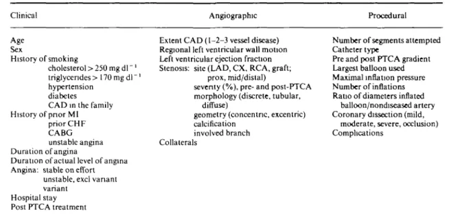
Table 1 Variables entering univariale analysis
Clinical Angiographic Procedural
Age Sex History of smoking cholesterol > 250 m g d l "1 triglycendes > 170 mg dl" hypertension diabetes
CAD in the family History of prior MI
prior CHF CABG unstable angina Duration of angina
Duration of actual level of angina Angina: stable on effort
unstable, excl variant variant
Hospital stay Post PTCA treatment
Extent CAD (1-2-3 vessel disease) Regional left ventricular wall motion Left ventricular ejection fraction Stenosis: site (LAD, CX, RCA, graft;
prox, mid/distal)
seventy (%), pre- and post-PTCA morphology (discrete, tubular,
diffuse)
geometry (concentric, excentric) calcification
involved branch Collaterals
Number of segments attempted Catheter type
Pre and post PTCA gradient Largest balloon used Maximal inflation pressure Number of inflations Ratio of diameters inflated
balloon/nondiseased artery Coronary dissection (mild,
moderate, severe, occlusion) Complications
angiographically controlled patients, we compare therefore our repeat PTCA with the first consecutive 268 initial procedures.
Table 1 shows the forty clinical, angiographical and procedural variables assessed for their discrimi-nant and predictive value of primary and long-term success in PTCA 1 and PTCA 2.
PTCA PROCEDURE AND ADJUNCTIVE THERAPY
Angioplasty was performed by femoral approach following a previously described protocol'271 and using steerable catheters since April 1982. The percent severity of the stenosis pre- and post-PTCA was evaluated angiographically in several angu-lations. In addition, the absolute luminal diameter of the adjacent healthy coronary segment was measured pre-PTCA, corrected for X-ray magnifi-cation by comparing it with the shadow of the 8F catheter (2-7 mm). Since the real balloon size exceeds the one given by manufacturers with press-ures above 6 atmospheres'281, the balloons were measured immediately after the procedure, inflated at the maximal pressure used. As a consequence of previous analyses'291 the inflation pressure used tended to be higher (8-12 atm) and balloons larger during 1984 with the goal of achieving a ratio of 11 to 1-3 in the diameters of inflated balloon/adjacent healthy coronary segment, which we believe to be optimal'281.
All patients were treated with platelet-inhibitors (either sulfinpyrazone 200mgq.i.d. or aspirin 650 mg plus dipyridamole 75mgt.i.d.) for six months after successful PTCA. Diltiazem, 90mgt.i.d. in addition to anti-platelet treatment was given systematically for 3 months during a randomized trial in 1982/19831301 but, based on physicians' preference, only intermittently since.
FOLLOW-UP
The mean clinical follow-up after PTCA 1 was 14-2±11 months, and after PTCA 2 l l - 6 ± 7 months. It included exercise stress tests, usually at 3 and 6 months after the intervention. No patient was lost to follow-up. 178 of the 181 patients with a successful PTCA 1 (98%) had a control angio-graphy 10-6 ± 6-8 months after the intervention, and so did 55 of the 60 patients (92%) 9 + 7 months after a successful PTCA 2. Six patients refused control angiography. In 2 patients it was not done after PTCA 2 because of complications previously.
The time of angiographic control was chosen to compare the time interval to recurrence after and repeat PTCA, since the information on reappear-ance of symptoms, especially in the presence of persisting atypical pains, was not precise in a number of cases. Control angiography, however, was done early (within 1-4 weeks) after reappearance of symptoms.
578 H.J.Rapoldeta\.
CRITERIA FOR SUCCESS AND RESTENOSIS
Primary success of PTCA was defined as ^20% stenosis reduction without a major inhospital complication.
Restenosis was defined as reappearance of a successfully dilated stenosis to ^ 70% in the angu-lation showing maximum severity of the lesion. Each restenosis definition, including those proposed by the NHLBI, have shortcomings as shown by comparing them to each other1'8'. Our restenosis definition is based on a percent severity that we believe to be of clinical relevance. With an angiographic follow-up of 98% for the PTCA 1 population and of 92% for the redilated patients, assessment of short and long term success is based exclusively on angiographic data, unless otherwise mentioned.
STATISTICAL ANALYSIS
To compare baseline and follow-up character-istics, univariate analysis was performed using the chi-square test for categorical data and the Student /-test for continuous data. A stepwise logistic regression analysis identified among the univariately significant variables those with an independent discriminant or predictive value, using primary suc-cess and restenosis (definitions above) as outcome variables. BMDP software was used for statistical testing.
Results
PATIENT CHARACTERISTICS
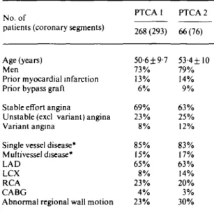
The baseline characteristics of both PTCA 1 and PTCA 2 patients are shown in Table 2. Sixty-six patients underwent a second intervention for restenosis 4-7 ± 4 months after the first one. Their mean age was 53-4 years. Seventy-nine percent were men. Fourteen percent had suffered a prior myo-cardial infarction and 9% had undergone previous coronary artery bypass grafting (CABG). Stable angina on effort was present in 63%, whereas 37% of the patients were in an unstable condition, including 12% with variant angina'3'1. One vessel disease was present in 83% and abnormal regional left ventricular wall motion in 30%.
Although the mean age was higher in the popu-lation that underwent repeat PTCA compared to the PTCA 1 population (53-4± 10 vs 50-8±10 years, P = 005), the percentage of patients above 60 years in both groups was not significantly different. There was a trend for more men and more variant
angina in the PTCA 2 group, but no other signifi-cant difference in the baseline characteristics of the patients.
REPEAT PTCA PROCEDURE
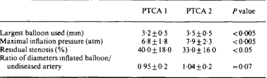
Compared to the first PTCA in the same patients several factors of the second procedure changed significantly (Table 3): larger balloons (mean 3-5 mm vs 3-2 mm, P< 0-005) with a higher ratio of diameters inflated balloon/nondiseased artery (mean 104 vs 0-95, P = 007) and higher inflation pressure (mean 7-9 vs 6-8 atmospheres P < 0-005) were used. The resulting residual stenosis was less (mean 33% vi40%, P<005).
PRIMARY RESULTS OF PTCA 1/PTCA 2 AND THEIR PREDICTORS
The immediate outcome of both PTCA 1 and PTCA 2 is summarized in Table 4. The primary success rate of PTCA 2 was 91% (60/66) of the patients and 92% (70/76) of the segments which was significantly higher than in the PTCA 1 population (181 /268 patients = 67-5% and 205/293 segments = 70%, respectively). Failures were due to inability to
Table 2 Baseline characteristics of the patients with first and repeat PTCA
No. of
patients (coronary segments)
Age (years) Men
Prior myocardial infarction Prior bypass graft Stable effort angina
Unstable (excl variant) angina Variant angina
Single vessel disease* Multivessel disease* LAD
LCX RCA CABG
Abnormal regional wall motion
PTCA 1 268(293) 5O-6±9-7 73% 13% 6% 69% 23% 8% 85% 15% 65% 8% 23% 4% 23% PTCA 2 66(76) 53-4±10 79% 14% 9% 63% 25% 12% 83% 17% 63% 14% 20% 3% 30%
* ? 70% stenosis on major coronary artery or > 50% on left main stem.
LAD — left anterior descending, LCX — circumflex, RCA — right coronary artery, CABG — coronary artery bypass graft.
Table 3 Differences between first and second procedures in patients with repeat PTC A (N = 66 patients or 76 coronary segments)
PTCA1 PTCA2 Pvalue
Largest balloon used (mm) Maximal inflation pressure (atm) Residual stenosis (%)
Ratio of diameters inflated balloon/ undiseased artery 3-2±0-5 6-8 ±1-8 4 0 0 ± 1 8 0 0 95 ±0-2 3-5±O-5 7-9±2-3 330±16 0 l-04±0-2 < 0-005 < 0-005 <0.05 = 007
Table 4 Results of first and repeal PTC A
All patients (coronary segments) primary success
failure: lesion non traversed non dilatable occluded complications: in hospital death
infarction emergency CABG one of the above Patients (coronary segments) with
angiographic control long term success restenosis N 268 (293) 181(205) ( 58) ( 7) ( 23) 19 36 43 178(202) 130(154) 48 ( 48) PTCA 1 % 100 67-5 (70) (20) ( 2) ( 8) 7 13 16 100 73(76) 27(24) N 66(76) 60(70) ( 3) ( 3) -2 1 3 55(65) 35(41) 20 (24) PTCA 2 % 100 91 (92) ( 4-5) ( 4-5) -3 1-5 4-5 100 64(63) 36(37)
cross the stenosis (4-5% in PTCA 2, 20% in PTCA 1), early occlusion (4-5% in PTCA 2, 8% in PTCA l)and nondilatable lesions (none in PTCA 2,2% in PTCA 1).
Significantly fewer major complications occurred due to PTCA 2 compared to PTCA 1 (myocardial infarction 3% vs 7%, emergency coronary artery bypass grafting 1-5% vs 13%, no in hospital death; overall4-5% vs 16%, P<005).
Due to considerable improvement in PTCA materials since 1982 (steerable catheters, low pro-file balloons) and growing experience (e.g. in the decision to perform emergency bypass grafting) the primary results in the PTCA 1 group do not reflect the present standard and should therefore only cautiously be compared with the PTCA 2 group. Nevertheless, the primary results in the PTCA 2 subgroup treated during the same period as the
PTCA 1 group (Feb 1980-March 1983: J V = 3 5 ,
success rate 32/35 = 91%, major complications 1/35 = 2-9%) had already reached that excellent standard.
Factors that were independently related to primary success in our PTCA 1 group are not men-tioned here, since they might no longer be relevant with the new materials available. The number of failures in the PTCA 2 group is too small to evaluate factors influencing the immediate outcome.
LONG-TERM RESULTS OF PTCA 1/PTCA 2 AND THEIR PREDICTORS
Restenosis >70% was angiographically docu-mented in 48/178 (27%) patients or 48/205 (24%) coronary segments at a mean of 4-7 ± 4 months after successful PTCA 1. The use of the NHLBI defin-ition of restenosis'181 would have added two more
580 H.J.Rapoldeta).
Table 5 Factors significantly related to restenosis after first PTCA
N Restenosis Lasting success
rates, except* rates, except* P value
Overall results
Patients (coronary segments) with angiographic control after successful PTCA 1 Univariate analysis of subgroups
Residual stenosis
Coronary lesion: one vessel disease, fixed one vessel d. + variant angina multivessel disease
Coronary dissection by PTCA. no mild moderate severe Calcification artery: yes
no
Ratio diameter inflated balloon/artery Number of inflations with largest balloon
178 (202) (202) 134 20 24 (110) ( 19) ( 64) ( 9) ( 50) (152) (202) (202) 27% (24%) *48±14% 20% 45% 46% (27%) ( 5%) (17%) (55%) (34%) (20%) *0-98±0-2 •5-1 ±2-9 Multivariate analysis Residual stenosis ^ 4 5 %
Coronary lesion: one vessel disease with variant angina or multivessel disease Coronary dissection by PTCA: none or severe
Ratio of diameters inflated balloon/artery < 1
73% (76%) •36 ± 1 3 % 80% 55% 54% (73%) (95%) (83%) (45%) (66%) (80%) *l-06±0-2 *4-2±2-4 < 0-001 <0 005 <001 <005 <005 <005 /"-value < 0-001 <005 $ 0 0 7 = 007
patients. 84% of the restenoses occurred within 6 months, 96% within 8 months. The restenosis rate after PTCA 2 tended to be higher: 20/55 (36%) patients or 24/65 (37%) segments (/> = NS for patients, = 005 for segments). The mean interval to restenosis after PTCA 2 was 5-3 + 5 months, with 76% of the recurrences within 6 months and 88% within 8 months after the intervention (/>=NS vs PTCA 1).
Factors that favoured restenosis after PTCA 1 according to uni- and multivariate analysis of 40 variables (Table 1) are listed in Table 5. The resteno-sis group was characterized by a higher degree of residual stenosis immediately after PTCA (mean 48 ± 14% with 29/48 segments between 50 and 60%
vs mean 36 ± 1 3 % with only 28/154 segments
between 50 and 60% in the group with lasting success, />< 0-001), a smaller ratio in the diameters of inflated balloon/adjacent healthy coronary seg-ment (0-98±0-2 vs 1 06±0-2, P<005) and more inflations (5-1 ± 2 9 vs 4-2±2-4 P<005). Other
factors predicting a high restenosis rate were: variant angina (45%), multivessel disease (46%), a calcined artery (34%) and a severe intimal dissection (55%). Interestingly, a mild dissection led to significant less restenosis than no visible dissection (5% vs 27%,
P < 001). Among variables not related to restenosis
were the coronary risk factors, the clinical ante-cedents, gender and age. The multivariate analysis selected three independent predictors of restenosis after PTCA I: a residual stenosis of 45% or more (/"< 0-001), the presence of variant angina (/><0-05) and multivessel disease (/><005). The degree of dissection produced by PTCA and the ratio in diameters of inflated balloon/artery almost reached statistical significance (/> = 007 each).
The same forty variables (Table 1) were assessed for their predictive value of restenosis after PTCA 2 (Table 6). The restenosis group was characterized by the use of higher inflation pressure (9 ± 2 vs 7±2atm, /><0-005) and a high initial gradient (57±15 v* 45±15mmHg, P<0Q5). Only men
Table 6 Factors significantly related to restenosis after repeat PTC A
N rates, except*Restenosis Lasting successrates, except' /•value
Overall results
Patients (coronary segments) with angiographic control after successful PTCA 2
Univariate analysis of subgroups Sex: male
female Inflation pressure
Coronary dissection by PTCA: no mild moderate Extent of coronary disease' one vessel
multivessel Initial gradient before PTCA
Multivariate analysis Male sex
Inflation pressure > 8 atm
55(65) 43 12 (62) (35) (26) ( 4) 35 20 49 36% (37%) 47% 0% *8-9±2-4atm ( 26%) ( 42%) (100%) 23% 60% •57±15mmHg 64% (63%) 53% 100% •7 ±1-9 atm (74%) (58%) ( 0%) 77% 40% •45±15mmHg <0001 <0005 <0-01 <0 05 <0-05 lvalue < 0-001 < 0 0 5
suffered a second recurrence (restenosis rate 47% vs 0% for female, />< 0-001). Other factors related to a high restenosis risk were: a mild or moderate intimal dissection (restenosis rate 42% and 100%, respectively, vs 26% with no dissection, P<005) and multivessel disease (60% vs 23% in one vessel disease, P<005). The trend of a higher recurrence rate in patients with prior unstable (57%) or variant angina (50%) was not significant, probably because of the small size of the subgroups (N= 14 and 6, respectively).
Multivariate analysis retained male sex
(P < 0001) and a higher inflation pressure {P < 005)
as indepedent predictors of restenosis after PTCA 2.
When the clinical results of patients without angiographic control (3 asymptomatic, 2 atypically symptomatic but with positive exercise stress test) are taken into account, the recurrence rate after PTCA 2 does not change (22/60 = 36-6%).
THIRD PTCA
Of the 20 patients with a second recurrence after PTCA 2, 9 underwent a third PTCA (PTCA 3), 6 had elective coronary artery bypass grafting and 5 were controlled with medical treatment (see Fig. 1). PTCA 3 was primarily successful in all patients. Follow-up based on angiograms (6 patients),
exer-cise stress tests (1 patient) and interviews (2 patients) demonstrated sustained success and absence of symptoms in 8 of 9 patients. One patient, with 3 consecutive PTCAs and restenoses on the proximal segment of a venous bypass graft had a reoperation. Thus, analyzing PTCA 2 and 3 together, repeat angioplasty in 66 patients with restenosis after the first intervention resulted in lasting success in 70% of them (41 of 58 patients with angiographic follow-up or 46 of all 66 patients with clinical follow-follow-up).
Discussion
PATIENTS
The patients undergoing repeat PTCA had base-line characteristics similar to the patients with single or initial PTCA. The trend of more men having a second angioplasty is in line with the other two series published so far123-241. The high incidence of variant angina in PTCA 2 patients might be due to our research interest in that area'1731' and the high restenosis rate among those patients'171.
PROCEDURE
The fact that larger balloons with higher inflation pressure were used for the second PTCA, leading to a higher ratio of diameters inflated balloon/ nondiseased artery and lesser residual stenosis,
582 H. J.Rapoldetal.
reflects our1281 and others'32-331 experience, that these procedural factors are more important determi-nants of immediate PTCA success than the clinical or angiographic pattern.
PRIMARY RESULTS
A second PTCA, performed because of restenosis had a higher primary success rate than a first pro-cedure (91 vs 67-5%, much like that in the other series (85% vs 61 %(231,97 vs 85%'24), 96%|26]. Major complications were fewer, also in agreement with published reports123"241. The reasons for both include the improvement in PTCA material since 1982, the increasing experience of the operators and the selec-tion of a PTCA 2 populaselec-tion with lesions suitable for a successful intervention. The primary results of our PTCA 1 group 1980-1983 certainly do not reflect the current standard. Furthermore the number of patients (especially of failures) in PTCA 2 group is small. Factors influencing the immediate outcome of both PTCA 1 and PTCA 2 are therefore not discussed here.
LONG-TERM RESULTS
Restenosis remains a major concern limiting long-term success of both first and repeat PTCA in spite of improved primary results. The lowest pub-lished restenosis rate in a larger series with an angio-graphical follow-up comparable to ours was 17% after initial and 33% after repeat PTCA1201. In most other series of first PTCA a higher recurrence rate between 25% and 35%1'8'"-21-221 was documented with a less complete angiographic follow-up, but in agreement with the 27% of our PTCA 1 group.
The restenosis rate tends to be even higher after repeat PTCA (36%), as shown by others as wel]P3-25i T h i s i s a l s o t m e f o r t h e p j c A 2 per-formed since March 1983, and therefore indepen-dent of technical improvement, but not surprising for a selected population at high risk of restenosis. In the NHLBI study1231 24% of the patients had clinical events and 52% were symptomatic after a second PTCA. In the Atlanta experience124', 26% of the patients were not improved clinically after a mean of 8-5 months. The Mid America Heart Insti-tute reported a higher restenosis rate (37%) after repeat angioplasty*251. With an angiographic follow-up of 48%(23) and 61%(24) (not detailed in1251), the restenosis rates (34%(23>, 41 %1241, 37%'25]) in these respective series might not be representative of the total PTCA 2 population. If, as suggested by the Atlanta study1241, the mean time interval between intervention and recurrence lengthens after repeat
PTCA, a more complete angiographical follow-up might have revealed restenoses not yet clinically apparent. In our study, however, the time to recur-rence was similar after PTCA 1 (4-7 ± 4 months) and PTCA 2 (5-3 ± 5 months) with 96% and 85% of the restenoses being demonstrated within 8 months after the intervention.
RESTENOSIS RISK FACTORS
Non procedural factors
Unstable and especially variant angina indepen-dently increased the restenosis risk after primary PTCA, in agreement with other reports'18-2'1.
Multivessel disease was related to restenosis after
both first and repeat PTCA (cf ref.1221). Male sex was a strong risk factor of recurrence after repeat PTCA. This contrasts with the higher bypass patency reported in men'34351, but confirms the data of the NHLBI registry"81. Other published associations with a high restenosis risk, such as dia-betes"8-221, PTCA of occluded vessels"4-2'1 or PTCA of a proximal vein graft stenosis"3-361 could not be verified due to small subgroups in our series. Dilated stenoses on the left anterior descending artery did not recur more often than those on the circumflex or the right coronary artery, as reported by others'2'-221; the use of an adequate balloon size after measuring the luminal diameter of the adjacent healthy coronary segment (see Methods) might have prevented this.
Procedural factors
Procedural factors of PTCA seem to be the main determinants of its long term outcome.
The residual stenosis, especially when >45%, was the main predictor of restenosis after first PTCA. The importance of the immediate result has been emphasized by other groups as well121'221. A severe intimal dissection increased the recurrence risk, but no visible dissection as well. We believe like others'21*221 that mild intimal tearing is an angio-graphic marker that predicts a good long term result in primary PTCA. The choice of an adequate balloon
size is thereby important. A ratio of diameters
inflated balloon/undiseased artery < 1 increased the restenosis risk, 1-1-1-3 being optimal'281. The un-predictable resistance and histology of the plaque to be treated certainly also determines the inflation pressure needed and the extent of the resulting dissection.
For repeat PTCA higher inflation pressure and even mild intimal dissection led to more restenosis
(c)
(d)(e)
(f)
(g)
Figure 2 Angiographic pattern of restenosis post-PTC A and repeal PTCA
Sixty-three year old man with single vessel disease. The angulation for all frames is RAO 15°/25° caudal, except for e and f (RAO 30')- (a) April 1983 — excentric short stenosis of 75% on proximal LAD; (b) immediate result of PTCA 1 (balloon 3-7 mm, 4 atm): residual stenosis of 20%, no visible dissection; (c) October 1983 — restenosis of 60%, longer and smoother than the original stenosis; (d) immediate result of PTCA 2 (balloon 4-3 mm, 12 atm): residual stenosis of 25%, long dissection without impairment of flow; (e) February 1984 —restenosis of 60%, long and irregular; (f) immediate result of PTCA 3 (balloon 3-5 mm, 8 atm): residual stenosis of 30%, no visible dissection; (g) September 1984 — control, no significant restenosis.
584 H.J.Rapoldeta\.
in spite of a good primary result. This point deserves special attention. If we are dealing with a different, more compilable histological substrate in repeat PTCA, the use of lower inflation pressure might indeed be optimal. In PTCA on an original hard atheromatous plaque, disruption of the internal elastic membrane and overstretching of the muscu-lar media with some cellumuscu-lar damage under high pressure is probably essential for success'37!. ]n repeat PTCA, however, it might be sufficient to distend and perhaps compress to some degree the proliferating fibrocellular neointima with lower pressure.
No drug regimen was associated with a signifi-cantly lower recurrence rate after primary or repeat PTCA. That was also true for other retrospective studies'18"21'. According to prospective trials, aspirin is at least as effective as coumadin in preventing restenosis after PTCA'381 and diltiazem does not decrease the recurrence rate in patients without variant angina'30'. Calcium antagonist therapy how-ever, seems to reduce the high restenosis risk of
PTCA in patients with variant angina'171.
PATHOLOGY OF RESTENOSIS
The difference in procedural risk factors for restenosis after primary and repeat PTCA seems rational if the histology of restenosis differs from the original lesion. Although a few histological case reports of restenosis 3—6 months after PTCA have documented recurrent lesions indistinguishable from de novo atherosclerosis'39' others have shown restenosis by fibrocellular proliferation only*40-41' resembling more the experimental model'42'. Restenosis by intimal fibrous hyperplasia only has also been described after venous bypass angioplasty performed early (2 months) after graft insertion143' whereas vein graft stenoses occurring beyond one year after insertion are known to consist of typical atherosclerotic plaque144'. It is a common experience that restenoses are more symmetrical and smoother in their angiographic appearance (see Fig. 2) and less resistant when being dilated, suggesting that the consistency and histology indeed differ from the original lesions. The role of platelets in coating a damaged surface immediately after PTCA'45"47] and that of thrombus organization'42' has been demon-strated for restenosis in the animal model. Intimal proliferation of smooth muscle cells seems to be the main mechanism of recurrence in man'40"41"43'. It can be triggered by platelet-derived growth factors, but might also be provoked by the balloon injury itself independently of serum factors'481 and thus even
under antiplatelet or anticoagulant drugs. Spasm is probably not the major stimulus of restenosis in patients without variant angina'30', but occurs early after PTCA[49>.
THIRD PTCA
Our number of third angioplasties performed on the same restenosing segment is too small to draw firm conclusions. However, a majority of this highly selected population (8/9 in our experience, 6/7 in the Atlanta report'24' 13/23 in the series of the Mid America Heart Institute'251 seems to have a favour-able long term outcome. The only restenosis after PTCA 3 we experienced was on the proximal part of a venous bypass graft.
Conclusions
Restenosis after PTCA remains a major problem. Besides some inherent risk factors such as male sex, variant angina and multivessel disease, procedural factors are the main determinants of long-term success in PTCA. An optimal primary result with a minimal residual stenosis, that might well include mild intimal tearing, is important for first PTCA, whereas avoidance of any visible dissection by using lower inflation pressure on a restenosis of probably different histology could improve the long-terrn outcome of repeat PTCA. Repeat PTCA in patients with restenosis after a first procedure has a high primary success rate, and complications are rare. Although the restenosis rate after a second angio-plasty is at least as high as after the first inter-vention, 70% of the patients with repeat PTCA have a successful long-term outcome, if one includes third interventions. Women seem to do better than men. Thus, repeat angioplasty should be considered as an integral part of PTCA therapy.
Addendum
Up to 15 November 1984, 136 coronary segments in 117 patients were redilated because of a restenosis at the Montreal Heart Institute. The overall primary success rate was 91 %. Of the 95 segments successfully dilated before 15 May 1984, 82 (86%) were angiographically controlled within 6 months (by 15 November). Restenosis > 70% was present on 27 (33%) of the segments. No recurrence occurred after a successful second PTCA in women
{N=\iy
The above conclusions, therefore remain valid for our entire experience.
We acknowledge the excellent secretarial assistance of Heidi Schuppbach in the preparation of this manuscript.
References
[1] Gruentzig AR. Transluminal dilatation of coronary-artery stenosis (letter to the editor). Lancet 1978; 1: 263. [2] Gruentzig AR, Senning A, Siegenthaler WE. Non-operative dilatation of coronary artery stenosis. N Engl ^ JMed 1979; 301: 61.
[3] Scholl JM, Chaitman BR, David PR et al. Exercise electrocardiography and myocardial scintigraphy in the serial evaluation of results of PTCA. Circulation 1982; 66: 380.
[4] Hirzel HO, Nuesch K, Gruentzig AR, Horst W, Krayenbuehl HP. Thallium 201 exercise scintigraphy after percutaneous transluminal coronary angioplasty •"' of coronary artery stenosis. Med Clinics of North
America 1980; 64: 163.
[5] Williams DO, Riley RS, Singh AK, Most AS. Coronary circulatory dynamics before and after successful angio-plasty. J Am Coll Cardiol 1983; 1: 1268.
[6] O'Neill WW, Wolton JA, Bates ER et al Criteria for successful coronary angioplasty as assessed by alter-ations in coronary vasodilatory reserve J Am Coll •N Cardiol 1984; 3: 1382.
[7] Bentivoglio LG, Van Raden MJ, Kelsey SF, Detre KM. PTCA in patients with relative contraindications: Results of the NHLBI PTCA Registry. Am J Cardiol 1984; 53: 82C.
[8] Mock MB and other participants in NHLBI PTCA registry: PTCA in the elderly patient. Experience in the NHLBI PTCA registry. Am J Cardiol 1984; 53: 89C. ( [9] Dorros G, Stertzer SH, Cowley MJ, Myler RK.
Complex coronary angioplasty: multiple coronary dilatations. Am J Cardiol 1984; 53: 126C.
[10] Vhetstra RE, Holmes DR, Reeder GS et al. Balloon angioplasty in multivessel coronary artery disease. Mayo ClinProc 1983; 58: 563.
[11] Mata LA, Bosch X, David PR, Rapold HJ, Corcos T, Bourassa MG. Clinical and angiographic assessment 6
J months after double perculaneous coronary
angio-plasty. J Am Coll Cardiol 1985; 6: 1239.
•; [12] Faxon DP, Detre KM, M c C a b e C H ^ al. Role of PTCA in the treatment of unstable angina. Am J Cardiol 1984; 53: 131C.
[13] Block PC, Cowley MJ, Kaltenbach M, Kent FM, Simpson J. Percutaneous angioplasty of stenoses of bypass grafts or of bypass graft anastomotic sites. Am J N Cardiol 1984, 53:666.
[14] Holmes DR, Vlietstra RE, Reeder GS, Bresnalan JF, < Smith HC, Bove AA. Angioplasty in total coronary
artery occlusion. J Am Coll Cardiol 1984; 3: 845. [15] HartzlerGO, Rutherford BD, McConahay DR. PTCA:
Application for acute myocardial infarction. Am J Cardiol 1984,53: 117C
[16] Hitchcok JF, Robes dc Medina E. Angioplasty of the main left coronary artery for isolated left main coronary artery disease. Am J Cardiol 1982; 49: 956 (Abstr). v [17] Corcos T, David PR, Bourassa MG etal. PTCA for the
treatment of variant angina. J Am Coll Cardiol 1985; 5: 1046.
[ 18] Holmes DR and other participants in the NHLBI PTCA registry. Restenosis after PTCA: a report from the PTCA registry of the NHLBI. Am J Cardiol 1984; 53: 77C.
[19] Gruentzig AR, Meier B. PTCA. The first five years and the future. IntJ Cardiol 1983; 12:3.
[20] Kaltenbach M, Kober G, Scherer D, Vollbracht C. Recurrence rate after successful coronary angioplasty Eur Heart 1985; 6: 276.
[21] Leimgruber PP, Roubin GS, Hollman J e / a / Restenosis after successful coronary angioplasty in patients with single vessel disease. Circulation 1986; 73: 710. [22] Hollman J, Galan K, Franco I et al: Recurrent stenosis
after coronary angioplasty. J Am Coll Cardiol 1986; 7: 20A (Abstr).
[23] Williams DO, Gruentzig AR, Kent KM, Detre KM, Kelsey SF, To T. Efficacy of repeat PTCA for coronary restenosis. Am J Cardiol 1984; 53: 32C.
[24] Meier B, King SB, Gruentzig AR el al. Repeat coronary angioplasty. J Am Coll Cardiol 1984; 3: 463.
[25] Giorgi LV, Hartzler GO, Rutherford BD, McConahay DR. Should repeat PTCA be performed on patients with multiple recurring restenoses? Circulation 1984; 70 (Suppl II) 11-177 (Abstr).
[26] Fleck E, Dacian S, Dirschinger J, Hall D, Rudolph W. Quantitative changes in stenotic coronary artery lesions during follow-up after PTCA. Circulation 1984; 70 (Suppl II) 11-176 (Abstr).
[27] Bourassa MG, David PR, Guiteras Val P. Percutaneous transluminal coronary angioplasty. In: Rowlands DJ, ed. Recent Advances in Cardiology 9. Edinburgh: Churchill Livingstone, 1984; 193-212.
[28] Duprat G, David PR, Lesperance J et al. An optimal size of balloon catheter is critical to angiographic success early after PTCA. Circulation 1984; 70 (Suppl II) 11-295 (Abstr).
[29] David PR, Renkin J, Moise A, Dangoisse V, Guiteras PV, Bourassa MG. Can patient selection and optimiz-ation of technique reduce the rate of restenosis after PTCA. J Am Coll Cardiol 1984; 3: 470 (Abstr). [30] Corcos T, David PR, Renkin J et al. A randomized trial
of Diltiazem for prevention of restenosis after PTCA. Am Heart J 1985; 109:926.
[31] Waters DD, Miller DD, Szlachcic J et al. Factors influencing the long term prognosis of treated patients with variant angina. Circulation 1983; 68: 258. [32] Faxon DP, Kelsey SF, Ryan TJ, McCabe CH, Detre K.
Determinants of successful PTCA. Report from the NHLBI registry. Am Heart J 1984, 108: 1019. [33] Meier B, Gruentzig AR, King SB et al. Higher balloon
dilatation pressure in coronary angioplasty. Am Heart J 1984; 107:619.
[34] Douglas JS, King SB, Jones E et al. Reduced efficacy of coronary bypass surgery in women. Circulation (Suppl
I I ) 1 9 8 1 ; I I : 1 1 .
[35] Loop FD, Golding LR, MacMillian JP et al. Coronary artery surgery in women compared with men: analysis of risks and long term results. J Am Coll Cardiol 1983; 1: 383.
[36] Douglas JS, Gruntzig AR, King SB et al. PTCA in patients with prior coronary bypass surgery. J Am Coll Cardiol 1983; 2: 745.
[37] Block PC. Mechanism of transluminal angioplasty. Am J Cardiol 1984;53:69C.
[38] Thornton MA, Gruentzig AR, Hollman J, King SP, Douglas JS. Coumadin and Aspirin in prevention of recurrence after transluminal coronary angioplasty: a randomized study. Circulation 1984; 69:721.
[39] Waller BF, McManus BM, Kishel JC. Sudden death 90 to 180 days after PTCA: Severe narrowing by
athero-586 H.J. Rapoldt\a\.
sclerotic plaque at necropsy at the site of previous angioplasty. Circulation 1982; 66(Suppl II) II: 4. [40] Essed CE, Van den Brand M, Becker AE. PTCA and
early restenosis: Fibrocellular occlusion after wall laceration. Br Heart J 1983; 49: 393.
[41] Austin GE, Ratliff NB, Hollman J ei at. Intimal pro-liferation of smooth muscle cells as an explanation for recurrent coronary artery stenosis after PTCA. J Am CollCardioll985;6:369.
[42] Faxon DP, Sanborn TA, Weber VJ et at. Restenosis following transluminal angioplasty in experimental atherosclerosis. Atherosclerosis 1984; 4: 189.
[43] Waller BF, Rothbaum DA, Garfinkel HJ, Ulbnght TM, Linnemeier TJ, Berger SM. Morphologic observations after percutaneous transluminal balloon angioplasty of early and late aortocoronary saphenous vein bypass grafts. J Am Coll Cardiol 1984; 4: 784.
[44] Lie JT, Lawrie GM, Morris G O Aortocoronary bypass saphenous vein graft atherosclerosis. Anatomic study of 99 vein grafts from normal and hyperlipoproteinemic patients up to 75 months postoperatively. Am J Cardiol
1977; 40: 906.
[45] Pastemac RC, Baughman KL, Fallon JT, Block PL. Scanning electron microscopy after coronary trans-luminal angioplasty in experimental atherosclerosis. Arteriosclerosis 1984; 4- 189.
[46] Steele PM, Chesebro JH, Stanson AW. Balloon angio-plasty: natural history of the pathophysiologica] response to injury in a pig model. Circulation Res 1985; 57: 105.
[47] Lam J, Chesebro JH, Steele PM, Badimon L, Fuster V. Production of tears into media during arterial angio-plasty predisposes to platelet thrombus deposition. J Am Coll Cardiol 1985; 5- 520(Abstr).
[48] Grunwald J, Haudenschild CC. Intimal injury in vivo activates vascular muscle cell migration and explant out-growth in vivo. Arteriosclerosis 1984; 4: 183.
[49] Hollman J, Austin GE, Gruntag AR el al. Coronary artery spasm at the site of angioplasty in the first two months after successful PTCA. J Am Coll Cardiol 1983; 2: 1039.