Developing staining protocols for visualization of tissue-engineering scaffolds
using micro computed tomography in native wet state
Kuo W1, Nimeskern L1, Martínez Ávila H2, Hofmann S1, Freedman, J3,Grinstaff MW3, Müller R1, Stok KS1 1Institute for Biomechanics, ETH Zürich, Switzerland
2Biopolymer Technology, Department of Chemical and Biological Engineering, Chalmers University of Technology, Sweden
3Departments of Biomedical Engineering and Chemistry, Boston University, United States [email protected]
Abstract: BNC-alginate and silk fibroin tissue-engineering
scaffolds were stained with X-ray contrast agents in order to visualize internal microstructure in the native wet state with microcomputed tomography. A successful protocol employ-ing amphiphilic contrast agents (CAs) dissolved in a water-based staining solution was used. The CAs were then fixed to the scaffold by neutralizing their charged functional groups, increasing their hydrophobicity and retention on the scaffold surface in water. While some unresolved issues concerning homogeneous staining and strength of contrast remain, these first successes constitute an important milestone by identify-ing good contrast agent candidates and stainidentify-ing protocols for longitudinal monitoring of tissue-engineering studies.
Keywords: contrast agent, silk fibroin, bacterial
nanocellu-lose, alginate
Introduction
Controlling scaffold porosity, pore size and interconnectivity is necessary to facilitate cell migration in tissue-engineering (TE). However quantification of 3D morphometric parame-ters is not possible with routine histology, and advanced quantification methods are required, such as micro computed tomography (µCT). While 3D internal microstructure is visible with µCT when scaffolds are scanned in dry state, imaging scaffolds in dry state is of limited interest, as scaf-fold morphology may change when immersed in a physio-logical solution. In water-based solution however, the scaf-fold structure is no longer visible with µCT due to insuffi-cient contrast above the background water.
Contrast agents (CA) are therefore required for imaging scaffolds in a physiological environment. An adequate CA should have a higher affinity for the scaffold material than for water. For that reason, a trade-off should be identified between a highly hydrophilic CA which would distribute evenly through the scaffold but have poorer contrast due to affinity to the background water, and a hydrophobic CA which could adsorb to a scaffold surface but may not diffuse evenly in the scaffold.
The goal of this project was to develop staining protocols applicable to various scaffold materials used in cartilage TE to allow assessment of their 3D internal microstructure in their physiological state using µCT. A focus was set on protocols requiring limited deviation from physiological conditions and on minimizing chemical modifications of the original scaffold.
Methods
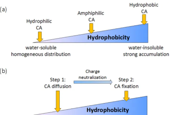
Two different scaffold materials used in cartilage TE were investigated in this study: bacterial nanocellulose-alginate (BNC-alginate) and silk fibroin (SF) scaffolds. BNC-alginate is a composite material made of cellulose fibres [1] coated with an alginate gel. Silk fibroin scaffolds were produced from B. mori according to methods published elsewhere [2]. CA were selected in order to take advantage of either the anionic (BNC-alginate) or the hydrophobic (SF scaffolds) nature of the scaffold material. Three initial strategies were identified: (A) hydrophilic, cationic CA with anionic scaf-fold, (B) hydrophobic CA with hydrophobic scafscaf-fold, (C) amphiphilic CA with hydrophobic scaffolds. Additionally two-step strategies were investigated: (D) scaffolds were immersed with amphiphilic, anionic CA at alkaline pH, keeping it in its more hydrophilic form to improve CA dis-tribution, then pH was lowered to fix the CA on the scaffold structures. A similar strategy was developed where the hy-drophilicity of the amphiphilic CA was lowered by bringing it in contact with opposite charges on the scaffold surface (E). These different strategies are illustrated in Figure 1. Table 1 summarizes which strategy was investigated on the scaffold materials. The protocols are detailed below:
(A) Hydrophilic, cationic CA with anionic scaffold
BNC-alginate with CA4+
The BNC-alginate scaffold was kept in an 80 mg I/ml solu-tion of CA4+ [3] overnight, rinsed shortly with water and
scanned wet in air. All scans were performed with a µCT50 scanner (Scanco Medical AG, Brüttisellen, Switzerland).
(B) Hydrophobic CA with hydrophobic scaffold
Silk with Lipiodol® and SDS (sodium dodecyl sulfate)
0.62 ml Lipiodol® [480 mg I/ml] (Guerbet, Villepinte, France) was shaken together with 9 ml PBS (phosphate buffered saline), 0.25 ml acetic acid and 200 mg SDS to receive a 30 mg I/ml emulsion. The emulsion was homoge-nized by pressing it quickly through a 0.2 m syringe filter with a 10 ml syringe. A scaffold was stained for two days, rinsed for 45 min in a solution of 0.5 g SDS in 65 ml water and scanned submerged in a SDS / water solution.
(C) Amphiphilic CA with hydrophobic scaffold
SF scaffolds with erythrosine
A silk scaffold was stained in a 15 mg I/ml solution of eryth-rosine in water for 15 days. It was stirred in water overnight and scanned submerged in water.
(D) Amphiphilic CA combined with pH shift
SF scaffolds with 2,3,5-triiodobenzoic acid (TIBA)
197 mg TIBA was added to 10 ml water and adjusted with 4 ml 1 M NaOH to pH 8. Approximately half the TIBA dis-solved, the rest stayed as solid powder. One scaffold was stained for 5 days, stirred in 0.05 M HCl overnight and scanned submerged in 0.05 M HCl.
(E) Amphiphilic CA combined with opposite charge
BNC-alginate with polylysine coating and erythrosine
BNC-alginate scaffold was kept in a 0.5 ml 1% polylysine solution mixed with 0.5 ml PBS for three days to give to anionic scaffold material a cationic surface. It was then left to dry in air for one day, stained overnight in 15 mg I/ml solution of erythrosine in water, rinsed thoroughly in water and then scanned submerged in 0.1 M CaCl2.
Figure 1: (a) One-step A-C, and (b) two-step strategies D-E.
BNC-alginate SF
A Hydrophilic, cationic CA
B Hydrophobic CA
C Amphiphilic CA
D Amphiphilic CA with pH shift
E Amph. CA with opposite charge
Table 1: Summary of the different scaffolds and strategies investigated. Checks indicate promising protocols.
Results
(A) Hydrophilic, cationic CA with anionic scaffold
BNC-alginate and with CA4: A fraction of the CA adsorbs on
the scaffold surface, yet the resulting contrast is weak.
(B) Hydrophobic CA with hydrophobic scaffold
Silk with Lipiodol® and SDS: Bright spots are observed on
the µCT scan suggesting an accumulation of contrast in pores rather than on the scaffold.
(C) Amphiphilic CA with hydrophobic scaffold
Silk with erythrosine: A fraction of the CA adsorbs on the
scaffold surface, yet the resulting contrast is weak.
(D) Amphiphilic CA combined with pH shift
Silk with 2,3,5-Triiodobenzoic acid (TIBA): Accumulation of
CA on the scaffold surface is observed, although its distribu-tion is slightly inhomogeneous; see figure 2.
(E) Amphiphilic CA combined with opposite charge
BNC-alginate with polylysine coating and erythrosine: The
internal structure is clearly visible in the scaffold, however, the staining is inhomogeneous and should be improved.
Figure 2:Amphiphilic CA combined with pH shift: i.e. silk and TIBA, provides contrast on the scaffold surface.
Discussion
The weakness of the staining of the BNC-alginate scaffold with CA4+ is in agreement with result reported in literature
using dyes [4], and is likely due to its high hydrophilicity and low charge number [4]. Staining silk with Lipiodol® emulsified with SDS yielded poor contrast distribution which was not improved through homogenization. Amphiphilic erythrosine was expected to perform better, given its similarity to eosin, a common histology dye for silk fibroin scaffolds. Although it provides good visual coloration, its X-ray absorption is not sufficient for µCT contrast. The greater success of the TIBA staining of silk scaffolds can therefore be explained by greater affinity of contrast agent as a result of the increased hydrophobicity at low pH and the larger iodine density. Similarly, erythrosine performed better when its charges were neutralized through contact with the polycationic polylysine, which increased its hydrophobicity. There are still some issues remaining concerning homogeneous staining and strength of the contrast that have to be resolved before being able to quantify 3D morphometric parameters of TE scaffolds. The results of this study provide insights into the important chemical interactions and properties required for a TE scaffold contrast agent, and a working, reliable solution is now only a matter of optimization.
Acknowledgement
Funding from the Swiss National Science Foundation and ERA-NET/EuroNanoMed (EAREG-406340-131009/1).
Bibliography
[1] Svensson, A., et al., Biomater, 2005, 26, 419. [2] Hofmann, S., et al, Biomater, 2007, 28, 1152.
[3] Joshi, N.S., et al., J. Am. Chem. Soc., 2009, 131, 13234. [4] Prentø, P., Biotechnic & Histochem, 2001, 76 (3), 137.