Video-assisted Thoracoscopic Resection
of a Small Pulmonary Nodule after Computed
Tomography–guided Localization
with a Hook-wire System
Experience in 45 Consecutive Patients
Olivier Pittet, MD,
1Michel Christodoulou, MD,
1Edgardo Pezzetta, MD,
1Sabine Schmidt, MD,
2Pierre Schnyder, MD,
2Hans-Beat Ris, MD
11
Division of Thoracic Surgery, Centre Hospitalier Universitaire Vaudois, 1011 Lausanne, Switzerland
2
Department of Radiology, Centre Hospitalier Universitaire Vaudois, 1011 Lausanne, Switzerland
Abstract
Background: This study is a single-institution validation of video-assisted thoracoscopic (VATS) resection of a small solitary pulmonary nodule (SPN) previously localized by a CT-guided hook-wire system in a consecutive series of 45 patients.
Methods: The records of all patients undergoing VATS resection for SPN preoperatively localized by CT-guided a hook-wire system from January 2002 to December 2004 were assessed with respect to failure to localize the lesion by the hook-wire system, conversion thoracotomy rate, duration of operation, postoperative complications, and histology of SPN.
Results: Forty-five patients underwent 49 VATS resections, with simultaneous bilateral SPN resection performed in 4. Preoperative CT-guided hook-wire localization failed in two patients (4%). Conversion thoracotomy was necessary in two patients (4%) because it was not possible to resect the lesion by a VATS approach. The average operative time was 50 min. Postoperative complications occurred in 3 patients (6%), one hemothorax and two pneumonia. The mean hospital stay was 5 days (range: 2–18 days). Histological assessment revealed inflammatory disease in 17 patients (38%), metastasis in 17 (38%), non-small-cell lung cancer (NSCLC) in 4 (9%), lymphoma in 3 (6%), interstitial fibrosis in 2 (4%), histiocytoma in one (2%), and hamartoma in one (2%).
Conclusions: Histological analysis of resected SPN revealed unexpected malignant disease in more than 50% of the patients indicating that histological clarification of SPN seems warranted. Video-assisted thoracoscopic resection of SPN previously localized by a CT-guided hook-wire system is related to a low conversion thoracotomy rate, a short operation time, and few postop-erative complications, and it is well suited for the clarification of SPN.
S
mall pulmonary nodules (SPN) are defined as intraparenchymal lung lesions smaller than 3cm in diameter without association with atelectasis or lymph node enlargement.1Small pulmonary nodules have been Presented at the 41st World Congress of Surgery of ISS/SIC 2005,Durban, South Africa.
Correspondence to: Hans-Beat Ris, MD, e-mail: [email protected]
Ó 2007 by the Socie´te´ Internationale de Chirurgie World J Surg (2007) 31: 575–578 Published Online: 1 February 2007 DOI: 10.1007/s00268-006-0343-7
diagnosed with increasing frequency since the advent of computed tomography.2,3 Because about 50% of SPN are related to malignancy, the necessity for rapid and precise histological diagnosis of SPN has been stres-sed.3–5Transthoracic or transbronchial fine-needle biop-sy may be considered in selected cases; however, the diagnostic yield for SPN has been reported to be rather low in general. Excisional biopsy of SPN may be con-sidered but requires invasive access, such as thoracot-omy. With the advent of video-assisted thoracoscopic surgery (VATS), a minimally invasive procedure has emerged that allows complete resection of SPN with minimal morbidity.6However VATS is limited because the surgeon is unable to palpate the lung tissue during operation, which may render the intraoperative identifi-cation of SPN difficult.7 Thus, failure to visualize or palpate SPN has resulted in conversion thoracotomy rates of up to 46%.7 Various techniques designed to localize SPN precisely during VATS resection have been reported, among them, preoperative CT-guided marking of the lesion by methylene blue,8,9 Lipidol, or radionuc-lides,10,11or intraoperative VATS-guided ultrasound.12,13 Recently, a hook-wire marking system has been pro-posed that involves preoperative CT-guided introduction under local anesthesia and anchorage in the lesion. In this report, we present our experience with VATS resec-tion of SPN previously marked by a CT-guided hook-wire system in a consecutive series of 45 patients.
PATIENTS AND METHOD
All patients undergoing VATS resection of SPN previ-ously marked by use of the hook-wire system at our insti-tution from 2002 to 2004 are the subject of this report. Patients with infracentimetric SPN underwent preopera-tive CT-guided localization of their lesion by use of the hook-wire system. Small pulmonary nodules in direct contact with the pleura or larger than 1cm and at a distance of less than 1 cm beneath the pleural surface were not considered for hook-wire implantation. Smokers with soli-tary spiculated lesions on CT-scan compatible with primary non-metastatic lung cancer underwent lobectomy and formal mediastinal lymph node dissection by thoracotomy. A commercially available hook-wire system (Ariadne’s Thread; Biosphere Medical, Louvres, France) was used. This system consists of a circular hook-wire of 8 mm diameter connected to a 50-cm-long suture thread. The hook is contained within a 20-gauge, 10-cm-long needle and regains its circular configuration after deployment.
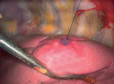
The hook-wire was deployed within the nodule under CT-guided control14 (Fig. 1). A control CT-scan after hook-wire implantation was performed to verify correct positioning. Video-assisted thoracoscopic resection of the lesion was performed using 3 thoraco-ports and endo-scopic stapler devices (EndoGIAÒ Autosuture; Endo-pathÒ Ethicon). The surgical specimen was extracted by use of an EndobagÒ (Autosuture) to prevent chest wall implantation of malignant disease (Fig. 2; Video 1). The specimen was processed by frozen examination during the operation, and lobectomy and mediastinal lymph node dissection were carried out when it was established that the lesion was non-small-cell lung cancer (NSCLC).
Figure 1. The hook-wire, attached to a long suture thread, is deployed under computed tomographic (CT) control into the nodule and anchored to the adjacent lung parenchyma. CT-scan documentation is performed after deployment.
Figure 2. Intraoperative photograph of SPN previously marked by CT-guided deployment of the hook-wire device. Light traction on the suture thread during video-assisted thoracoscopic (VATS) resection renders deep-seated lesions more superficial and facilitates excision by endo-staplers.
The files of the patients were analyzed with respect to patient demographics, underlying diseases, histology of SPN, and procedure-related characteristics such as fail-ure to localize the lesion by the hook-wire system, con-version thoracotomy rate, duration of operation, and postoperative complications.
RESULTS
Forty-five patients underwent 49 VATS resections of SPN previously marked by the hook-wire system at our institution from 2002 to 2004. Four patients underwent simultaneous bilateral procedures for bilateral SPN. There were 23 men and 22 women with an average age at operation of 58 years (range: 22–79 years). Small pulmonary nodules were localized in the left and right lung in 45% and 55% of the patients, respectively. During the same time period, we performed a total of 320 VATS procedures and 20 VATS resections for SPN without using a hook-wire device.
Computed tomography (CT)-guided hook-wire implan-tation was performed without complications in all but 2 patients (4%). One patient developed an important pneumothorax requiring immediate preoperative chest tube drainage. One patient underwent misplaced hook-wire implantation requiring repositioning of the hook-hook-wire before operation.
Video-assisted thoractomy resection of SPN was fea-sible in all but 2 patients (4%) who required conversion thoracotomy. Both patients revealed a too centrally localized lesion with respect to accessibility and resection properties of the endoscopic stapler device. Four addi-tional patients had conversion thoracotomy for lobectomy and mediastinal lymph node dissection for NSCLC. A hook-wire system was noted to be in place during VATS exploration in all patients. The operation times were less than 60 min, 60–90 min, and 90–120 min in 56%, 28%, and 16% of the patients, respectively. Longer operation times were mainly recorded in patients under-going the bilateral procedure for SPN. Postoperative complications were recorded in 3 patients (6%), pneu-monia in two (4%) and hemothorax requiring reoperation and hemostasis in one (2%). Retrospectively, this patient was found to have hemophilia.
Table 1 summarizes results of the histological exami-nation of the excised SPN. The size of SPN varied between 2 and 20 mm and measured < 10 mm in 68% of the patients. Malignant disease was found in 53% of the patients, and primary NSCLC in 9%.
Comment
The recent advent of VATS resection for SPN has al-lowed rapid and precise diagnosis of SPN by use of a minimally invasive procedure while avoiding the draw-backs of formal thoracotomy. Video-assisted thoraco-scopic resection of SPN has become widely accepted, but conversion thoracotomy was frequently necessary because preoperative SPN localization was inadequate. This holds true especially for small subpleural lesions seated deeply in the parenchyma. Several techniques have been developed to facilitate intraoperative localiza-tion of SPN during VATS reseclocaliza-tion such as preoperative CT-guided injection of methylene blue or intraoperative ultrasound localization of the lesion.8,9,12,13 Methylene blue injection carries the risk of spreading the colorant on the pleural surface and the chest cavity during application, which renders subsequent intraoperative VATS-guided localization of SPN difficult. Intraoperative VATS-guided ultrasound localization of SPN is operator dependent and requires complete collapse of the assessed lung, which is difficult to obtain in emphysematous lungs.
The CT-guided hook-wire system allows precise and quick preoperative localization of SPN. It is also well-suited for the localization of small and deep-seated lesions. Light traction on the suture thread during VATS resection renders deep-seated lesions more superficial and facilitates complete excision by endo-staplers. Because the hook-wire is placed before the operation with local anaesthesia, the duration of general anaes-thesia and operation is considerably reduced, and this may translate into reduced procedure-related costs.
Our results confirm low morbidity associated with the procedure and demonstrated the feasibility of preopera-tive CT-guided hook-wire implantation in all but one patient. One patient with marked emphysema developed a pneumothorax after hook-wire implantation requiring chest tube drainage. In fact, caution is indicated in patients with marked emphysema undergoing preopera-tive hook-wire implantation for SPN localization, and
Table 1.
Histological findings of the pulmonary nodules Histological finding Number of patients Percent Inflammatory diseases 17 38 Metastatic disease 17 38 Non-small-cell lung cancer 4 9
Lymphoma 3 7
Lung fibrosis 2 4
Histiocytoma 1 2
Hamartoma 1 2
preoperative insertion of chest tubes should be liberally performed in these situations. In a few other patients, a small pneumothorax was noticed on control CT after hook-wire implantation without clinical impact. However, prompt surgical intervention is advocated after hook-wire implantation in order to reduce the risk of a symptomatic pneumothorax.
The CT-guided hook-wire localization of SPN yields an excellent accuracy, with all SPN in our patients being correctly marked. In contrast to other reports, no dis-lodgement of the hook-wire was noted, even if traction was exerted during VATS resection. We speculate that this was due to the flexibility of this hook-wire system because of the long suture thread. However, two patients (4%) required a conversion thoracotomy because it was impossible to completely resect their deep-seated lesions by use of endo-staplers.
The histological analysis of SPN confirms the high percentage of malignant disease and endorses the previously reported results in this respect. Video-as-sisted thoracoscopic resection of SPN offers an elegant minimally invasive alternative to thoracotomy for this purpose. Preoperative CT-guided hook-wire marking of SPN is a rapid and efficient technique that facilitates intraoperative localization of small, deep-seated lesions. However, because of the additional costs, we consid-ered hook-wire implantation for deep-seated lesions (> 10 mm from pleural surface) and small infra-centri-metric lesions not in contact with the pleural surface, as suggested by Suzuki et al.7
REFERENCES
1. Fraser RG, Sanders C, Barnes GT, et al. Digital imaging of the chest. Radiology 1989;171:297–307.
2. Kaneko M, Eguchi K, Ohmatsu H, et al. Peripheral lung cancer: screening and detection with low-dose spiral CT versus radiography. Radiology 1996;201:798–802.
3. Lillington GA. Management of solitary pulmonary nodules. Dis Mon 1991;37:271–318.
4. Lillington GA, Caskey CI. Evaluation and management of solitary and multiple pulmonary nodules. Clin Chest Med 1993;14:111–119.
5. Viggiano RW, Swensen SJ, Rosenow EC III. Evaluation and management of solitary and multiple pulmonary nod-ules. Clin Chest Med 1992;13:83–95.
6. Allen MS, Deschamps C, Lee RE, et al. Video-assisted thoracoscopic stapled wedge excision for indeterminate pulmonary nodules. J Thorac Cardiovasc Surg 1993;106: 1048–1052.
7. Suzuki K, Nagai K, Yoshida J, et al. Video-assisted tho-racoscopic surgery for small indeterminate pulmonary nodules: indications for preoperative marking. Chest 1999;115:563–568.
8. Lenglinger FX, Schwarz CD, Artmann W. Localization of pulmonary nodules before thoracoscopic surgery: value of percutaneous staining with methylene blue. AJR Am J Roentgenol 1994;163:297–300.
9. Wicky S, Mayor B, Cuttat JF, et al. CT-guided localizations of pulmonary nodules with methylene blue injections for thoracoscopic resections. Chest 1994;106:1326–1328. 10. Chella A, Lucchi M, Ambrogi MC, et al. A pilot study of the
role of TC-99 radionuclide in localization of pulmonary nodular lesions for thoracoscopic resection. Eur J Cardio-thorac Surg 2000;18:17–21.
11. Nomori H, Horio H, Naruke T, et al. Fluoroscopy-assisted thoracoscopic resection of lung nodules marked with lipiodol. Ann Thorac Surg 2002;74:170–173.
12. Gruppioni F, Piolanti M, Coppola F, et al. [Intraoperative echography in the localization of pulmonary nodules during video-assisted thoracic surgery]. Radiol Med (Torino) 2000;100:223–228.
13. Sortini D, Feo CV, Carcoforo P, et al. Thoracoscopic localization techniques for patients with solitary pulmonary nodule and history of malignancy. Ann Thorac Surg 2005;79:258–262.
14. Wicky S, Dusmet M, Doenz F, et al. Computed tomography-guided localization of small lung nodules before video-assisted resection: experience with an efficient hook-wire system. J Thorac Cardiovasc Surg 2002;124:401–403. 578 Pittet et al.: Video-assisted Thoracoscopic Resection of a Small Pulmonary Nodule