HAL Id: inserm-00583476
https://www.hal.inserm.fr/inserm-00583476
Submitted on 7 Mar 2012
HAL is a multi-disciplinary open access
archive for the deposit and dissemination of
sci-entific research documents, whether they are
pub-lished or not. The documents may come from
teaching and research institutions in France or
abroad, or from public or private research centers.
L’archive ouverte pluridisciplinaire HAL, est
destinée au dépôt et à la diffusion de documents
scientifiques de niveau recherche, publiés ou non,
émanant des établissements d’enseignement et de
recherche français ou étrangers, des laboratoires
publics ou privés.
MRI predictors of cognitive outcome in early multiple
sclerosis.
Mathilde Deloire, Aurélie Ruet, Delphine Hamel, Melissa Bonnet, Vincent
Dousset, Bruno Brochet
To cite this version:
Mathilde Deloire, Aurélie Ruet, Delphine Hamel, Melissa Bonnet, Vincent Dousset, et al.. MRI
pre-dictors of cognitive outcome in early multiple sclerosis.. Neurology, American Academy of Neurology,
2011, 76 (13), pp.1161-7. �10.1212/WNL.0b013e318212a8be�. �inserm-00583476�
MRI predictors of cognitive outcome in early multiple sclerosis
Mathilde S. A. Deloire 1 2 , Aur lie Ruet é 1 2 , Delphine Hamel 1 , Melissa C. Bonnet 1 , Vincent Dousset 1 3 , Bruno Brochet 1 *
Neuroinflammation: imagerie et th rapie de la scl rose en plaques
1 é é INSERM : U1049 , Universit Victor Segalen - Bordeaux II : EA2966 é , 146 rue L o Saignat BP78 33076 Bordeaux Cedex, FRé
Service de neurologie
2 CHU Bordeaux , Groupe hospitalier Pellegrin , 33076 Bordeaux Cedex, FR Service de neuroimagerie
3 CHU Bordeaux , Groupe hospitalier Pellegrin , 33076 Bordeaux Cedex, FR
* Correspondence should be adressed to: Bruno Brochet <bruno.brochet@chu-bordeaux.fr >
Abstract
Objective
To determine MRI predictors for cognitive outcome in early relapsing remitting multiple sclerosis (MS) patients. Methods
Inception Cohort. Forty-four patients recently diagnosed with clinically definite MS, were followed-up with clinical and cognitive evaluations at 1, 2, 5 and 7 years and underwent brain magnetic resonance imaging (MRI) including magnetization transfer (MT) imaging at baseline and two years. Cognitive evaluation was also performed in 56 matched healthy subjects at baseline. Cognitive testing included the Brief Repeatable Battery (BRB). Imaging parameters included lesion load, brain parenchyma fraction, ventricular fraction (VF), mean MT ratio (MTR) of lesion and normal appearing brain tissue (NABT) masks.
Results
At baseline, patients presented deficits of memory, attention and information processing speed (IPS). Over 2 years all MR parameters deteriorated significantly. Over 7 years, EDSS deteriorated significantly. Fifty percent of patients deteriorated on memory cognitive domain and 22.7 of patients on IPS domain. Seven-year change of memory scores was significantly associated with baseline diffuse% brain damage (NABT MTR). IPS z score change over 7 years was correlated with baseline global atrophy (BPF), baseline diffuse brain damage and central brain atrophy (VF) change over 2 years.
Conclusion
The main predictors of cognitive changes over 7 years are baseline diffuse brain damage and progressive central brain atrophy over the 2 years after MS diagnosis.
MESH Keywords Adult ; Biological Markers ; Cognition ; physiology ; Cognition Disorders ; etiology ; pathology ; physiopathology ; Female ; Humans ; Longitudinal Studies ; Magnetic Resonance Imaging ; methods ; Male ; Middle Aged ; Multiple Sclerosis ; complications ; pathology ; physiopathology ; Neuropsychological Tests ; Predictive Value of Tests ; Regression Analysis
Author Keywords Multiple sclerosis ; Brain atrophy ; Ventricular fraction ; Cognition ; Magnetic resonance imaging.
INTRODUCTION
Cognitive impairment is an important feature of Multiple Sclerosis (MS) and may affect everyday activities. Deficits in memory, information processing speed (IPS), attention, working memory, and executive functions are frequently seen . Cognitive dysfunction has1 long been considered to be confined to patients at later stages of the disease but such impairments have been described in clinically isolated syndromes (CIS) and in early relapsing-remitting MS (RRMS) .2 3
The association between cognitive deficiencies and several magnetic resonance imaging (MRI) markers, including lesion load (LL), diffuse brain abnormalities and brain atrophy, has been investigated . However, little is known about the value of these parameters at the1 early stages of MS to predict cognitive decline over time and the value of magnetization transfer (MT) imaging parameters. We designed a longitudinal study in newly diagnosed clinically definite MS (CDMS) patients to investigate MRI markers predictive for deteriorating cognitive function. Cross-sectional data at baseline showed that cognitive impairment was associated with MR parameters evaluating diffuse brain damage . We hypothesized that diffuse brain damage outside lesions in early RRMS detected at baseline and their change3 over 2 years predict the deterioration of cognitive performance over 7 years. Baseline values of various MR parameters, including LL, MT ratio (MTR) metrics, and global and central atrophy, and their change over 2 years were investigated for predicting the deterioration of cognitive performance over 7 years.
METHODS
MR and cognitive decline in RRMS
Fifty-eight patients, diagnosed with RRMS within the previous six months without other selection criteria, were included by the coordinating centre between November 2000 and November 2001. Patients were consecutively referred by practicing neurologists from a neurological network in south western France for the purpose of that study. None of these patients received disease-modifying therapies before the second exacerbation.
One patient was excluded from the study due to reconsideration of the MS diagnosis. Forty-four patients (77.2 ) completed all% evaluations during the 7-year study period and were used for the analysis. All patients underwent clinical and cognitive assessment at baseline (year 0) and after years (y) 1, 2, 5 and 7, and underwent brain MRI at y0 and y2. A sample of 56 healthy subjects matched for sex, age, and educational level was also studied at baseline. Baseline clinical, imaging, and cognitive characteristics of patients and controls have been published previously .3
Standards Protocol Approvals and Patient consents
The study was approved by the institutional review board (CPP Bordeaux 2000/28) and patients gave written informed consent.#
Clinical evaluation
Clinical evaluation included Expanded Disability Status Scale (EDSS), Multiple Sclerosis Functional Composite (MSFC), and Montgomery and Asberg Depression Rating Scale (MADRS).
Neuropsychological assessment
The neuropsychological assessment was previously described in detail . It included the similarities test and the Brief Repeatable3 Battery (BRB-N) with the Selective Reminding Test (SRT, and 3 subscores: SRT-LTS Long term storage; SRT-CLTR Consistent= = Long term retrieval ; SRT-DR delay recall; verbal memory), the 10/36 spatial recall test testing short- (SPART) and long-term= visuo-spatial memory (SPART DR), the Symbol Digit Modalities Test (SDMT, attention and IPS), the PASAT 3 and 2 second versions (PASAT 3s and PASAT 2s; working memory and IPS) and the Word List Generation test (WLG; verbal fluency). At y7, we applied alternate forms for all these neuropsychological tests to limit practice effect. Data concerning other tests used at baseline (Go/No-Go; Stroop, similarities, Boston and RFF tests) and for all tests at y1, 2 and 5 follow-ups were not used in this report, because no alternate forms were used.
At baseline, patients were classified as patient cognitively impaired (PCI) if they performed less than the 5th percentile of matched controls on at least two tests of the battery (n 22) and patient cognitively unimpaired (PCU) if they did not (n 22).= =
At each time point, the raw scores of each cognitive score were transformed to z scores using the mean and standard deviation (SD) baseline scores of the 44 patients as the reference data. Z scores were used to study longitudinal changes of cognitive tests and correlations. Additionally, two domain-specific z-scores were created for memory (SRT-LTS, SRT-CLTR, SRT-DR, SPART and SPART-DR) and IPS (SDMT, PASAT 3s and 2s).
MRI evaluation
Image acquisition
Methods for image acquisition and analysis have been detailed previously . Brain MRI scans were obtained on a Philips Gyroscan3 ACS-NT 1.5 T scanner, including Fast Fluid-Attenuated Inversion-Recovery (FLAIR) images (TR/TE/TI: 11000/140/2725), MT images using a proton density sequence (TR/TE: 37/2.3 and flip angle 8) both with and without an MT saturation pulse, and T1-weighted images= (TR/TE: 450/12) before and after administration of gadolinium-DTPA (0.1 mmol/Kg). For all sequences, 26 contiguous interleaved axial slices were acquired with 5 mm slices, 256x256 matrices and 230 230 fields of view. The slices were positioned to run parallel to a line× joining the most inferoanterior and inferoposterior parts of the corpus callosum.
Native and pre-processed data were displayed on workstations running software developed by Bio-Clinica, Inc. (Lyon, France). Following parameters were considered in the analysis: LL, Brain Parenchymal Fraction (BPF), which is defined as the ratio of whole brain parenchyma volume to the intracranial volume , Ventricular Fraction (VF), defined as the ratio of ventricular volume to the intracranial4 volume , and means of lesion and normal-appearing brain tissue (NABT) MTR masks.5
Statistical analysis
Statview version 5.0 (Windows) was used for statistical analysis. Matching between MS patients (n 44) and healthy controls (n 56)= = was verified using the Chi square test for gender and the t test for age and years of education. For each score, the percentages of patients deteriorating or improving more than 1.0 SD of controls scores or stable over 7 years were calculated in whole group, PCI and PCU groups. For IPS and memory domains, patients were classified in the deterioration group if they have more than one SD decrease based on baseline scores of healthy controls. Additionally, at the follow-up evaluations, the patients were considered cognitively deteriorated
according to their deterioration score modified from Kujala . For each domain (IPS, memory), if the subject scored below 1.0 SD6 − compared with the norms, he/she received one deterioration point; if below 1.5 SD, two; if below 2.0 SD, three. All comparisons were− − performed using the t test when distribution of values was normal or by non parametric Wilcoxon test when it was not.
Participants who fulfilled all follow-ups were compared to the original sample with regard to demographic data (age, gender, and years of education), baseline neuropsychological scores, EDSS, MSFC, MADRS, disease duration; LL, MTR means, BPF, and VF.
Differences were considered significant, for all analyses, when p values were less than 5 . Correlations between change in cognitive% scores and MADRS scores were studied by Pearson s correlation coefficient.’
Univariate linear regressions were performed to assess correlation between individual baseline MR variables or changes over the first two years (LL, mean lesion MTR, BPF, VF and mean NABT MTR) and changes over 7 years of the domain specific cognitive z scores (memory and IPS) in the whole group. To determine which MRI parameters were the best predictors of cognitive change over 7 years in the whole group, we performed two stepwise multiple linear regression models for each cognitive change over 7 years (memory and IPS domains) as dependent variable. Independent variables were baseline MRI parameters (LL, mean lesion MTR, BPF, VF and mean NABT MTR) in the two first model and MRI parameters changes over 2 years (LL, mean lesion MTR, BPF, VF and mean NABT MTR) in the other models. In all models, we entered age, education level, gender and MADRS scores as independent variables and baseline EDSS was forced in all models as a covariate. Only independent variables with a conservative significance level of p<0.25 at the univariate analysis were entered simultaneously in the linear regression model. All parameters with p>0.05 were removed from the model by stepwise elimination.
Logistic regression analyses were also performed (see Table e-1 at www.neurology.org ).
RESULTS
Demographics, clinical and MRI data
summarizes demographics and disease characteristics of patients and controls at baseline. MS patients were matched to Table 1
healthy controls according to gender (p 0.16,= χ2 = 1.98), age (p 0.67, t 0.42), and mean number of years of education (p 0.72, t = = = =−0.36 ). No statistical difference was observed for baseline demographics, clinical, and cognitive scores and MRI parameters between the 44 patients who completed all follow-ups over 7 years and the entire original cohort of 56 patients (all p values >0.05).
The percentage of patients receiving disease-modifying therapies at any time during the study was 95.6 . During the follow-up% period, 9 of the 44 patients followed over 7 years converted from RRMS to secondary progressive MS.
None of the patients reached the MADRS threshold for severe depression (MADRS>34) at baseline and only one patient at 7 years7 (MADRS 46). Mild depression (MADRS score between 7 and 19) was diagnosed in 9 patients at baseline and in 16 patients at y7.= MADRS median range increased over the 7 years (3.0 0 21 at baseline and 10.5 0.0 46 at y7, p <0.001, z 3.712), and deteriorated[ ] [ – ] [ – ] = − in both groups (in CI : 3.0 0 18 at baseline and 11 0 46 at y7, p 0.017, z 2.39, in CU : 4.0 0 21 at baseline and 10.5 0 31 at y7, p[ – ] [ – ] = = − [ – ] [ – ] = 0.004, z 2.80).= −
Median EDSS deteriorated between y0 (2.0 0.0 5.5 ) and y7 (2.5 0.0 8.0 ) (p<0.001, z 3.586). but the MSFC did not change[ – ] [ – ] = − during seven years (p 0.20, t 1.31 ). The number of patients with EDSS <3 was 77.3 (34/44) at baseline and 59.1 (26/44) at y7. The= = % % number of patients with EDSS 6 was 0 at baseline and 3 at y7 (6.8 ). Median EDSS increase is 1.0 2.0 5.0 . During follow-up,≥ % [− – + ] disability progression was noted in 21 (48 ) patients.%
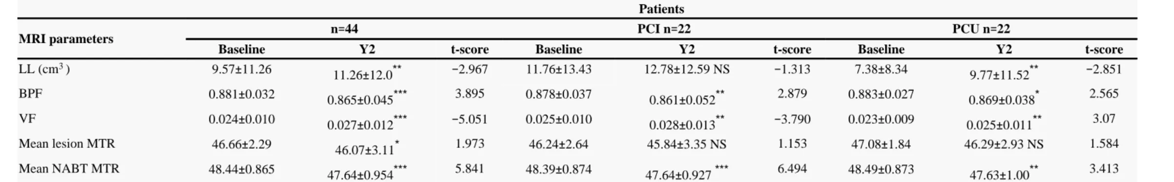
Over 2 years, all MR parameters deteriorated (p<0.05) in the whole group (Table 2 ). Cognitive evolution during follow-up
The baseline results have been published . Differences were observed between RRMS patients and matched controls for memory,3 attention, IPS, inhibition, and conceptualization.
Nineteen out of 44 patients (43.18 ) had a global deterioration score >2 (sum of memory and IPS domains deterioration scores) and% 9/44 (20.45 ) a score >4. The proportions of patients deteriorating by cognitive domains more than 1.0 SD of controls scores, improving% more than 1.0 SD of controls scores and stable over 7 years are presented in Table 3 for the three groups (all patients, PCI and PCU) without any difference (Fisher test) between the PCI and PCU groups, for memory (p 0.37, OR 2.05; 95 CI 0.57 8.2) and IPS (p= = % = – = 1.0, OR 1.0, 95 CI 0.19 5.2). The proportion of patients deteriorating was 50 for memory (9 patients deteriorated for one score, 8= % = – % patients for two score and 5 patients for more than 2 scores), 22.7 for IPS (5 for one score and 5 for at least 2 scores) and 13.6 for the% % WLG at y7.
MR and cognitive decline in RRMS
Changes over 7 years in cognitive scores were not correlated with baseline MADRS scores or MADRS change over 7 years (whole, PCI, and PCU groups) (Pearson s correlation coefficient |r| <0.30, p >0.05).’
Regression analyses (Tables 4 )
For memory or IPS z scores change over 7 years as dependent variable, two multiple regression models were performed with (i) baseline MRI parameters (LL, mean lesion MTR, BPF, VF and mean NABT MTR) as independent variables and (ii) MRI changes over 2 years (LL, mean lesion MTR, BPF, VF and mean NABT MTR). In univariate linear regression analyses, memory z score change over 7 years was associated with baseline LL and mean NABT MTR and with VF changes over 2 years. IPS z score change over 7 years was correlated with baseline MRI parameters (LL, BPF, VF and mean NABT MTR) and with VF and BPF changes over 2 years (all p value <0.05, see Table 4 ). Parameters with p<0.25 in univariate analyses and the baseline EDSS were entered in the two multivariate models ( ). After stepwise elimination, the only independent predictor of memory decline was the baseline NABT MTR. After stepwise Table 4
elimination, baseline NABT MTR and BPF and 2 years change of VF remained in the models for prediction of IPS decline. No MR parameter predicted WLG z score change in multivariate analysis.
DISCUSSION
This study emphasizes the frequency of cognitive deterioration in the early stages of MS. This deterioration, which may have many impacts in the daily life of patients, including working and social activities, needs to be detected in order to adapt therapeutic strategies. The present study was designed to determine which MR parameter, measured early in MS course, could help predict subjects who will deteriorate. The results showed that MRI parameters reflecting diffuse brain damage (BPF, VF and mean NABT MTR) predict more strongly cognitive deterioration than lesions.
The mechanisms of cognitive dysfunction in MS are not fully understood, but convergent evidence suggests that disconnection between cortical areas might be the basis of this dysfunction . Although focal lesions might contribute to this disconnection, several9 cross-sectional studies using diffusion and MT imaging suggested that axonal damage largely contributes to cognitive alterations by interrupting critical networks , 3 9 –15 . In the present longitudinal study in early MS we observed that baseline NABT MTR, which reflects diffuse brain injury accumulated so far, but no MR parameters studying focal lesions, predicts cognitive changes over 7 years in the two main domains memory and IPS, in multivariate analyses. Although, LL was associated with cognitive outcome in univariate analyses, it did not remain in multivariate models. These results confirmed the importance of diffuse brain damage in cognition alteration in RRMS. Such disconnections could interfere to the allocation of brain resources for a specific task and may explain that the most common cognitive impairment in MS concerned IPS16 .
Recent reports have shown correlations between cognitive disabilities and central atrophy and are in agreement with the hypothesis that disconnection between the sub-cortical and cortical structures involved in these networks may contribute to cognitive dysfunction 17 – . Although atrophy measures did not correlate with cognitive functions at baseline , baseline BPF, a measure of global atrophy and
23 3
two-year VF change, a measure of central brain atrophy predict decline in IPS performance over 7 years. Although periventricular white-matter atrophy could contribute to VF change, and in particular corpus callosum atrophy, which has been implicated in cognitive impairment in MS24 , it is likely that damage of the deep gray-matter and the surrounding white matter is determinant in cognitive deterioration affecting IPS in early RRMS. These results are in agreement with a recent study that analyzed the rates of atrophy in different parts of the brain and correlated them to various clinical indexes including the PASAT as part of the MSFC25 . The authors found that a worse performance on the PASAT at follow-up was predominantly related to atrophy development around the ventricles and, to a lesser extent, brainstem atrophy.
Several studies investigated the association between MR parameters and cognition evolution in RRMS but none used MT imaging. One study showed that one-year global atrophy rate was associated with cognitive deficiencies measured several years later26 . In that study, prediction of cognitive deterioration could not be evaluated since cognitive data were only measured at the last follow-up. Another study that examined the relation between changes of LL and brain parenchymal volumes over 2 years together with change in cognition during the same time-frame and found that the best predictor was brain parenchymal volumes27 . These two studies did not specifically evaluate central atrophy. Another study found that only neocortical volume changes, but not deep gray matter volume changes, were significantly associated with deteriorating cognitive performance over 2.5 years28 . It is possible that atrophy of the cerebral cortex accounts for more of the cognitive deterioration when the disease progresses, since the mean disease duration was longer in that later study than in ours (4.0 2.8 vs. 2.0 / 2.2 years). It has been suggested that deep central atrophy may peak at earlier stages compared to whole± + − brain atrophy29 and that central atrophy progresses preferentially in the early stages of the disease30 . These data can explain that, although baseline BPF which reflects brain whole atrophy developed so far is predictive of cognitive deterioration, it is only the central atrophy change which account for cognitive deterioration in attention/IPS domain. We hypothesized that disruption of deep central brain cognitive networks involved in attentional occurs in our population. It is worth mentioning that cortical atrophy also occurs in early MS31 ,32 but appears to progress less rapidly than central atrophy29 . In our study, we did not specifically study neocortical volume.
Interestingly, it was recently shown that memory performance and mesial temporal volumes, in particular hippocampal atrophy, were correlated in MS33 34 , This suggests regional specificity of correlations between atrophy and alterations of a given cognitive domain. In our study, we did not find correlation between memory change and atrophy measures although a trend was observed with VF change (data not shown). However baseline NABT MTR predicted memory changes over 7 years suggesting that diffuse alterations contribute also to the memory deficits by interrupting the relevant networks.
There are some limitations to this study. When performing imaging, it would have been informative to study regional changes in brain volume and MTR, for example, using voxel-based morphometry. Unfortunately those techniques were not available at the beginning of this study. Concerning cognition, the main limitation of longitudinal studies in MS is that relatively few patients deteriorate for cognitive scores and that a relatively large number of them remain stable. One possible reason is practice effect. To limit practice effect we used alternate forms at the 7 year evaluation, never used in the previous evaluations, but we cannot exclude that some practice effect remains. Another explanation for the limited changes in cognitive performances is that cognitive skills deteriorate slowly at the early stages of MS because of the presence of compensatory mechanisms35 .
Further studies are needed to determine which therapeutics strategies could limit diffuse brain damage at the early stage of MS and therefore ongoing cognitive deterioration.
Acknowledgements:
The study received support from ARSEP (Association pour la Recherche contre la Scl rose en Plaques) and Schering France. Work was madeé possible by the neurologists of the AQUISEP network.
Supported in part by research grants from Association pour la Recherche contre la Scl rose en Plaques, (ARSEP, France) and Bayeré Healthcare France SA.
Sponsors did not participate in design and conduct of the study; collection, management, analysis, and interpretation of the data; and preparation, review, or approval of the manuscript.
Dr. Melissa C. Bonnet has participated as speaker to a symposium organized by Bayer.
Footnotes:
Statistical analysis were performed by Mathilde Deloire, PhD and Jeremy Jove, statistician, INSERM U657, University de Bordeaux, Bordeaux, France.
Dr Ruet reports no disclosure in relation with this study. She has been member of advisory boards or participate as speaker to symposia organized by Biogen-Idec.
She is or has been investigator for studies promoted by Novartis, Bayer-Schering, Roche, Lilly, Peptimmune and Merck-Serono and has received subventions for this activity.
Dr Dousset reports no disclosure in relation with this study. He has been member of advisory boards or participate as speeker or chairman to symposia organized by Biogen-Idec. He is or has been investigator for studies promoted by Biogen-Idec, Novartis, Bayer-Schering, Teva, Peptimmune, Lilly and AB sciences and his institution received subventions for this activity
Dr Deloire and Ms Hamel reports no disclosure.
Dr Brochet reports no disclosure in relation with this study. He has been member of advisory boards or participates as speaker or chairman to symposia organized by Merck-Serono, Bayer-Schering, Novartis, Biogen-Idec, Teva and Sanofi/Aventis. He is or has been investigator for studies promoted by Biogen-Idec, Novartis, Roche, Sanofi-Aventis, Bayer-Schering, Teva, Peptimmune, Lilly and AB sciences and his institution received subventions for this activity..
References:
1 .Hoffmann S , Tittgemeyer M , von Cramon DY . Cognitive impairment in multiple sclerosis . Curr Opin Neurol . 2007 ; 20 : 275 - 280
2 .Feuillet L , Reuter F , Audoin B . Early cognitive impairment in patients with clinically isolated syndrome suggestive of multiple sclerosis . Mult Scler . 2007 ; 13 : 124 -127
3 .Deloire MS , Salort E , Bonnet M . Cognitive impairment as marker of diffuse brain abnormalities in early relapsing-remitting multiple sclerosis . J Neurol Neurosurg Psychiatry . 2005 ; 76 : 519 - 526
4 .Rudick RA , Fisher E , Lee JC , Simon J , Jacobs L . Use of the brain parenchymal fraction to measure whole brain atrophy in relapsing-remitting MS. Multiple Sclerosis Collaborative Research Group . Neurology . 1999 ; 53 : 1698 - 1704
5 .Kalkers NF , Bergers E , Castelijns JA . Optimizing the association between disability and biological markers in MS . Neurology . 2001 ; 57 : 1253 - 1258 6 .Kujala P , Portin R , Ruutiainen J . The progress of cognitive decline in multiple sclerosis A controlled 3-year follow-up . Brain . 1997 ; 120 : 289 - 297
7 .Snaith RP , Harrop FM , Newby DA , Teale C . Grade scores of the Montgomery Asberg depression and the clinical anxiety scales – . Br J Psychiatry . 1986 ; 148 : 599 -601
8 .Calabrese P , Penner IK . Cognitive dysfunctions in multiple sclerosis-a multiple disconnection syndrome ? “ ” . J Neurol . 2007 ; 254 : (Suppl 2 ) 18 - 21
9 .Rovaris M , Iannucci G , Falautano M . Cognitive dysfunction in patients with mildly disabling relapsing-remitting multiple sclerosis: an exploratory study with diffusion tensor MR imaging . J Neurol Sci . 2002 ; 195 : 103 - 109
10 .Cox D , Pelletier D , Genain C . The unique impact of changes in normal appearing brain tissue on cognitive dysfunction in secondary progressive multiple sclerosis patients . Mult Scler . 2004 ; 10 : 626 - 629
MR and cognitive decline in RRMS
11 .Ranjeva JP , Audoin B , Au Duong MV . Structural and functional surrogates of cognitive impairment at the very early stage of multiple sclerosis . J Neurol Sci . 2006 ; 245 : 161 - 167
12 .Benedict RH , Bruce J , Dwyer MG . Diffusion-weighted imaging predicts cognitive impairment in multiple sclerosis . Mult Scler . 2007 ; 13 : 722 - 30 13 .Roca M , Torralva T , Meli F . Cognitive deficits in multiple sclerosis correlate with changes in fronto-subcortical tracts . Mult Scler . 2008 ; 14 : 364 - 369
14 .Lin X , Tench CR , Morgan PS , Constantinescu CS . Use of combined conventional and quantitative MRI to quantify pathology related to cognitive impairment in multiple sclerosis . J Neurol Neurosurg Psychiatry . 2008 ; 79 : 437 - 441
15 .Rovaris M , Riccitelli G , Judica E . Cognitive impairment and structural brain damage in benign multiple sclerosis . Neurology . 2008 ; 71 : 1521 - 6
16 .Forn C , Belenguer A , Parcet-Ibars MA , Avila C . Information-processing speed is the primary deficit underlying the poor performance of multiple sclerosis patients in the Paced Auditory Serial Addition Test (PASAT) . J Clin Exp Neuropsychol . 2008 ; 13 : 1 - 8
17 .Bermel RA , Bakshi R , Tjoa C , Puli SR , Jacobs L . Bicaudate ratio as a magnetic resonance imaging marker of brain atrophy in multiple sclerosis . Arch Neurol . 2002 ; 59 : 275 - 280
18 .Christodoulou C , Krupp LB , Liang Z . Cognitive performance and MR markers of cerebral injury in cognitively impaired MS patients . Neurology . 2003 ; 60 : 1793 -1798
19 .Lazeron RH , Boringa JB , Schouten M . Brain atrophy and lesion load as explaining parameters for cognitive impairment in multiple sclerosis . Mult Scler . 2005 ; 11 : 524 - 531
20 .Hildebrandt H , Hahn HK , Kraus JA , Schulte-Herbr ggen ü A , Schwarze B , Schwendemann G . Memory performance in multiple sclerosis patients correlates with central brain atrophy . Mult Scler . 2006 ; 12 : 428 - 436
21 .Benedict RH , Bruce JM , Dwyer MG . Neocortical atrophy, third ventricular width, and cognitive dysfunction in multiple sclerosis . Arch Neurol . 2006 ; 63 : 1301 -1306
22 .Houtchens MK , Benedict RH , Killiany R . Thalamic atrophy and cognition in multiple sclerosis . Neurology . 2007 ; 69 : 1213 - 1223
23 . Tekok-Kilic A , Benedict RH , Weinstock-Guttman B . Independent contributions of cortical gray matter atrophy and ventricle enlargement for predicting neuropsychological impairment in multiple sclerosis . Neuroimage . 2007 ; 36 : 1294 - 1300
24 .Pelletier J , Suchet L , Witjas T . A longitudinal study of callosal atrophy and interhemispheric dysfunction in relapsing-remitting multiple sclerosis . Arch Neurol . 2001 ; 58 : 105 - 11
25 .Jasperse B , Vrenken H , Sanz-Arigita E . Regional brain atrophy development is related to specific aspects of clinical dysfunction in multiple sclerosis . Neuroimage . 2007 ; 38 : 529 - 537
26 .Summers M , Fisniku L , Anderson V , Miller D , Cipolotti L , Ron M . Cognitive impairment in relapsing-remitting multiple sclerosis can be predicted by imaging performed several years earlier . Mult Scler . 2008 ; 14 : 197 - 204
27 .Zivadinov R , Sepcic J , Nasuelli D . A longitudinal study of brain atrophy and cognitive disturbances in the early phase of relapsing-remitting multiple sclerosis . J Neurol Neurosurg Psychiatry . 2001 ; 70 : 773 - 780
28 .Amato MP , Portaccio E , Goretti B . Association of neocortical volume changes with cognitive deterioration in relapsing-remitting multiple sclerosis . Arch Neurol . 2007 ; 64 : 1157 - 1161
29 .Simon JH . Brain atrophy in multiple sclerosis: what we know and would like to know . Mult Scler . 2006 ; 12 : 679 - 687
30 .Audoin B , Davies GR , Finisku L , Chard DT , Thompson AJ , Miller DH . Localization of grey matter atrophy in early RRMS : A longitudinal study . J Neurol . 2006 ; 253 : 1495 - 1501
31 .Morgen K , Sammer G , Courtney SM . Evidence for a direct association between cortical atrophy and cognitive impairment in relapsing-remitting MS . Neuroimage . 2006 ; 30 : 891 - 898
32 .Calabrese M , Agosta F , Rinaldi F . Cortical lesions and atrophy associated with cognitive impairment in relapsing-remitting multiple sclerosis . Arch Neurol . 2009 ; 66 : 1144 - 50
33 .Sicotte NL , Kern KC , Giesser BS . Regional hippocampal atrophy in multiple sclerosis . Brain . 2008 ; 131 : (Pt 4 ) 1134 - 41
34 .Benedict RH , Ramasamy D , Munschauer F , Weinstock-Guttman B , Zivadinov R . Memory Impairment in Multiple Sclerosis: Correlation with Deep Gray Matter and Mesial Temporal Atrophy . J Neurol Neurosurg Psychiatry . 2009 ; 80 : 201 - 6
Table 1
Demographic and clinical baseline characteristics of RRMS patients as well as separately for CPU and CPU groups included in the follow-up
Patients n 44= PCI n 22= PCU n 22= Controls ( n 56)= Gender (male/female) 10/34 5/17 5/17 20/36 Age (years) 39.0 (8.7) 40.4 (9.9) 37.7 (7.2) 38.2 (10.5)
Educational level (years of schooling) 12.2 (2.5) 11.3 (2.3) 13.2 (2.3) 13.05 (2.6)
Disease duration (months) 24.3 (27.4) 22.5(21.7) 26.1(32.6)
EDSS 2.0 0.0 5.5[ – ] 2.0 0.0 3.5[ – ] 1.5 0.0 5.5[ – ]
MSFC −0.053 (0.701) −0.409(0.681) 0.339 (0.490)
MADRS 3.0 0 21[ – ] 2.0 0 18[ – ] 4.0 0 21[ – ]
PCI patients cognitively impaired; PCU: patients cognitively unimpaired. For all clinical data, scores are expressed as mean (SD) except for EDSS and MADRS which are median (range). EDSS:= expanded disability status scale; MSFC: multiple sclerosis functional composite; MADRS: Montgomery and Asberg depression rating scale.
Table 2
MRI parameters
Patients
MRI parameters n 44= PCI n 22= PCU n 22=
Baseline Y2 t-score Baseline Y2 t-score Baseline Y2 t-score
LL (cm )3 9.57 11.26± 11.26 12.0± ** −2.967 11.76 13.43± 12.78 12.59 NS± −1.313 7.38 8.34± 9.77 11.52± ** −2.851 BPF 0.881 0.032± 0.865 0.045± *** 3.895 0.878 0.037± 0.861 0.052± ** 2.879 0.883 0.027± 0.869 0.038± * 2.565 VF 0.024 0.010± 0.027 0.012± *** −5.051 0.025 0.010± 0.028 0.013± ** −3.790 0.023 0.009± 0.025 0.011± ** 3.07 Mean lesion MTR 46.66 2.29± 46.07 3.11± * 1.973 46.24 2.64± 45.84 3.35 NS± 1.153 47.08 1.84± 46.29 2.93 NS± 1.584 Mean NABT MTR 48.44 0.865± 47.64 0.954± *** 5.841 48.39 0.874± 47.64 0.927 ± *** 6.494 48.49 0.873± 47.63 1.00± ** 3.413 PCI patients cognitively impaired; PCU: patients cognitively unimpaired. MRI parameters are expressed as mean (SD). LL : lesion load; BPF : Brain Parenchymal Fraction; VF : Ventricular Fraction;= NABT : Normal-appearing brain tissue. MTR: Magnetization Transfer Ratio. P values and t-scores concerned comparison between baseline and y2.
NS : p > 0.05;
*p 0.05;≤ **p< 0.01; ***p < 0.001.
Table 3
Number of patients with deterioration, no change or improvement for the two main cognitive domains over 7 years.
All Patients (n 44)= PCI (n 22)= PCU (n 22)=
Improvement No change Deterioration Improvement No change Deterioration Improvement No change Deterioration
Memory domain (SRT-LTS, SRT-CLTR, SRT-DR, SPART, SPART-DR) 1 21 22 1 12 9 0 9 13
IPS domain (SDMT, PASAT 3s & 2s) 0 34 10 0 17 5 0 17 5
PCI patients cognitively impaired at baseline; PCU patients cognitively unimpaired at baseline (see methods).= =
SRT, Selective Reminding Test; SRT-LTS, long-term storage; SRT-CLTR, consistent long-term retrieval; SRT-DR, delayed recall; SPART, Spatial Recall Test; SPART-DR, Delayed recall; SDMT, symbol digit modalities test; PASAT, paced auditory serial addition test.
MR and cognitive decline in RRMS
TABLE 4
Linear regression models predicting change in cognitive z scores over 7 years from MR parameters at baseline or changes over 2 years.
Cognitive z scores change (y7-baseline) MR parameters Included in models R Univariate analyses P values Univariate analyses Adjusted R model2 P values multivariate analyses
Memory domain Baseline EDSS −0.095 0.54 0.144 <0.05
Baseline LL −0.333 <0.05
Baseline BPF 0.126 0.42
Baseline VF −0.175 0.26
Baseline mean lesion MTR 0.253 0.10
Baseline mean NABT MTR 0.377 <0.05
Baseline EDSS −0.095 0.54 0.009 0.54
LL change over 2 years −0.085 0.59
BPF change over 2 years 0.200 0.19 VF change over 2 years −0.290 <0.05
Mean lesion MTR change over 2 years − 0.090 0.57 Mean NABT MTR change over 2 years −0.057 0.72
IPS domain Baseline EDSS −0.273 0.07 0.416 <0.0001
Baseline LL −0.425 <0.01
Baseline BPF 0.551 <0.0001
Baseline VF −0.414 <0.01
Baseline mean lesion MTR 0.148 0.34
Baseline mean NABT MTR 0.511 <0.001
Baseline EDSS −0.273 0.07 0.326 <0.001
LL change over 2 years 0.117 0.45
BPF change over 2 years 0.435 <0.01
VF change over 2 years −0.545 <0.0001
Mean lesion MTR change over 2 years 0.163 0.29 Mean NABT MTR change over 2 years −0.016 0.92
Memory domain includes the Selective Reminding Test (long-term storage; consistent long-term retrieval; delayed recall) and the Spatial Recall Test (recall score and Delayed recall). IPS domain includes the Symbol Digit Modalities Test, the Paced Auditory Serial Addition test (3 and 2 sec versions).
LL : Lesion Load; MTR: Magnetization Transfer Ratio; NABT: normal appearing brain tissue; BPF; Brain Parenchymal Fraction; VF: Ventricular Fraction. Univariate analyses for age, MADRS scores, gender and educational levels showed not significant results (p>0.05). Variables with a p value <0.25 in univariate analyses were included in multivariate analyses. Variables included in multivariate Variables significantly correlated to cognitive change in multivariate analyses are in bold
analyses but not significantly correlated with cognitive Z scores changes in final models were in italic police.