REVIEW ARTICLE
Patent foramen ovale and neurosurgery in sitting position: a
systematic review
A.-R. Fathi
1†, P. Eshtehardi
2 †and B. Meier
2*
‡1
Department of Neurosurgery and Intensive Care Medicine and
2Department of Cardiology, Bern University
Hospital, 3010 Bern, Switzerland
*Corresponding author. E-mail: [email protected]
We have conducted a systematic review of air embolism complications of neurosurgery in the
sitting position and patent foramen ovale (PFO) closure. It assesses the risk and benefit of
PFO closure before neurosurgery in the sitting position. The databases Medline, Embase, and
Cochrane Controlled Trial Register were systematically searched from inception to November
2007 for keywords in both topics separately. In total, 4806 patients were considered for
neuro-surgery in sitting position and 5416 patients underwent percutaneous PFO closure. The overall
rate of venous air embolism during neurosurgery in sitting position was 39% for posterior
fossa surgery and 12% for cervical surgery. The rate of clinical and transoesophageal
echocar-diography detected paradoxical air embolism was reported between 0% and 14%. The overall
success rate for PFO closure using new and the most common closure devices was reported
99%, whereas the average risk of major complications is ,1%. On the basis of our systematic
review, we recommend screening for PFO and considering closure in cases in which the sitting
position is the preferred neurosurgical approach. Our proposed management including the
time of PFO closure according to available data is presented. However, the conclusions from
our systematic review may be limited due to the lack of level A evidence and from using data
from observational cohort studies. Thus, definite evidence-based recommendations require
prospective evaluation of the issue in well-designed studies.
Br J Anaesth 2009; 102: 588–96
Keywords: anaesthesia, neurosurgical; complications, embolism, air; closure device, foramen
ovale, patent/complications; heart, congenital defects, patent foramen ovale; position, sitting
Fatal air embolism in a patient operated on in the sitting
position was first reported by Barlow
9in 1830, and the
sitting position in brain and cervical spine surgery has been
a subject of interest since then.
30 37 85 90During this time,
expert opinion has been revised repeatedly. The sitting
position has advantages but also has intrinsic
compli-cations.
73 85The most severe risks are venous air embolism
(VAE) and its potentially fatal complication, paradoxical
air embolism (PAE).
32 52 79 90VAE is defined as the
entrainment of air (or exogenously delivered gas) into the
venous system from the operative site or another
communi-cation with the environment. It may produce a broad array
of systemic effects and outcomes. PAE occurs when VAE
passes into the systemic arterial circulation, for example,
through a patent foramen ovale (PFO). A negative pressure
in cranial veins in the sitting position leads to air aspiration.
However, a lower incidence of VAE during neurosurgery
has also been reported in the horizontal position.
2 19 42 92 99VAE may have catastrophic cardiovascular, pulmonary,
and neurological sequelae regardless of the presence of a
PFO. A PFO is the major mechanism for cerebral
arterial air embolism during neurosurgery in that
situ-ation.
37 38 59 69 85 87 89Its prevalence in the normal
popu-lation is about 25%,
56and .40% in adults with
cryptogenic stroke.
72The presence of a PFO represents a significant
contraindi-cation to neurosurgery in the sitting position, and many
sur-geons avoid the sitting position in order to minimize the risk
of PAE.
19 37 45 49 52 74 76 90 96Therefore, transoesophageal
echocardiography (TOE) or transcranial Doppler ultrasound
has become a routine preoperative evaluation in many
†These authors contributed equally to the manuscript.
‡Declaration of interest. B.M. is proctor for and recipient of research
grants and speaker fees from AGA Medical, manufacturer of PFO closure devices.
centres to rule out a PFO in such patients.
52 70 74 85 90 95However, neurosurgery in the sitting position cannot always
be avoided in this population, as some neurosurgeons feel
uncomfortable with the horizontal position for certain
oper-ations and are prepared to accept the increased risk for
PAE.
19 94Meanwhile, percutaneous PFO closure using
dedi-cated devices has become a routine procedure with a low
risk and high success rate.
116The indications of preoperative PFO closure in order to
minimize the risk for neurosurgery in the sitting position
remain to be defined.
The aim of this systematic review is to assess the risk
and benefit of PFO closure before neurosurgery in the
sitting position.
Literature search
Two groups were defined for the literature review: Group
1, studies of air embolism during neurosurgical procedures
in sitting position, and Group 2, percutaneous PFO closure
studies. The databases Medline, Embase, and Cochrane
Controlled Trial Register were systematically searched
from inception to November 2007 by various
combi-nations of the keyword groups: (i) neurosurgery, sitting
position, air embolism, complications, patent foramen
ovale and (ii) patent foramen ovale, persistent foramen
ovale, PFO, and closure. All abstracts were screened
according to the research question. Bibliographies of
ident-ified articles and reviews in this field were also searched.
Additionally, hand searching of pertinent journals for
issues in the last 6 months was undertaken. Articles in
multiple languages were searched.
Inclusion criteria for Group 1 were cohort studies with 10
or more patients undergoing neurosurgical procedures in the
sitting position with reporting of episodes of VAE or PAE.
Inclusion criteria for Group 2 were cohort studies including
10 or more patients undergoing percutaneous PFO closure
for any reason with follow-up of at least 3 months. The
reports including both PFO and atrial septal defect (ASD)
closures were included when the data were stratified for PFO
and ASD closures. Exclusion criteria for both groups were
studies of exclusively paediatric patients, experimental
studies, animal studies, case reports, expert opinions,
repeated reports from individual centres (the last
all-inclus-ive report was used), and studies with unclear methods.
Data extraction and analysis
Database search and data extraction were performed
inde-pendently by two authors and resulted in 127 abstracts
meeting the inclusion and exclusion criteria for Group 1
and 89 for Group 2. After full-text analysis, 28 remained
in Group 1 and 33 in Group 2. All were in English, except
for two papers in French. Quality was independently
assessed by two reviewers (A.-R.F. and P.E.) and
differ-ences were reconciled by mutual agreement of the senior
author (B.M.). The following data were tabulated from
included studies and inserted into a standardized excel
sheet (Microsoft Office 2000, Microsoft, Redmond, WA,
USA). Groups 1 and 2: author, publication year, journal,
language, study design, number of cases, mean age of
patients. Group 1: type of neurosurgical procedure, rates of
VAE and PAE, method of air embolism detection,
screen-ing for PFO, PFO prevalence, author recommendations
regarding PFO, and neurosurgery in the sitting position.
Group 2: PFO closure reasons, PFO closure devices,
pro-cedural success rates, major and minor peri-propro-cedural
complication rates, and residual shunt rates. Comparison
of clinical outcome of air embolism was not feasible due
to clinical heterogeneity between studies with regard to
populations, interventions, form of outcome assessment, or
study method. The data were pooled using Comprehensive
Meta-analysis Version 2, Biostat, Englewood, NJ, USA
(2005), when it was feasible, and there was no
heterogen-eity in definitions or methodologies.
Definitions
The definitions of PAE and VAE were given above. The
most frequently used methods were TOE, precordial
Doppler study, and end-tidal CO
2(
ECO20). PFO closure
success rates were defined as successful device
implan-tation. Minor procedural complications were haematoma or
bleeding not requiring transfusion, transient atrial
arrhyth-mia or atrioventricular node block, device embolization, air
embolization, transient ST-segment elevation, or femoral
arteriovenous fistula. Major complications were death,
stroke, cardiac tamponade, emergency surgery, haematoma,
or bleeding requiring blood transfusion or surgery, transient
ischaemic
attack,
significant
( persistent)
arrhythmia,
cardiac perforation, device malposition, septicaemia,
myo-cardial infarction, and massive pulmonary embolism.
Results
No randomized controlled trial or studies with level A
evi-dence were found. Therefore, we included both
retrospec-tive and prospecretrospec-tive clinical cohort studies (evidence level
B of American Academy of Family Physicians).
104VAE and PAE in sitting position
Twenty-eight studies published between 1972 and 2007
were included for data analysis.
2 3 15 18 19 25 33 38 42 47 5253 70 71 75 77 79 81 84 87 92 97 99 103 107 109 115 120
In total,
4806 patients were included (Supplementary Table S1 in
online version). Fifty-four per cent of the studies were
prospective, six reported on VAE rates during posterior
fossa surgery alone, two during cervical procedures
alone, and 21 reported on both procedures. No study
evaluated the occurrence of PAE for either procedure
sep-arately. The overall occurrence of VAE ranged from 0%
to 76% irrespective of the method of detection and
pos-ition. The incidence of VAE derived from pooled data
was 1 – 76% in the sitting position and 0 – 12% in the
horizontal position. The incidence of VAE from the
studies comparing two positions was 28.4% (15 – 45%)
(95% CI, 20.3 – 38.0) sitting and 5.5% (0 – 12%) (95% CI,
2.6 – 11.3) horizontal (Table 1). The rate of VAE was
38.6% (7 – 76%) (95% CI, 30.5 – 47.4) in posterior fossa
surgery and 11.8% (2 – 35%) (95% CI, 6.7 – 19.9) in
cer-vical procedures (Table 2). In the studies included,
patients were screened before operation for the presence
of PFO in 10 (36%) studies, and seven of them published
their prevalence. A PFO was detected in 5 – 33% of the
neurosurgical patients. Overall, 10 studies considered the
presence of PFO as an absolute contraindication for the
sitting position and patients were operated in the
horizon-tal position. Two of the 28 studies did not consider the
presence of PFO, if known, as a contraindication for
neu-rosurgery in the sitting position. Of note, none of the
studies mentioned the possibility of preoperative PFO
closure. The rate of clinical and TOE detected PAE was
reported in 20 of 28 studies (0214%, 0% in 14 studies).
Of the 28 studies included, TOE was used as a detection
method in nine. In three studies, the presence of a PFO
was not regarded as a contraindication to neurosurgery in
the sitting position (or at least not mentioned as a
contraindication). In these three studies, VAE was found
in 38%, 43%, and 60% and PAE in 14%, 0%, and 6.6%
of the study population, respectively (the one with 0%
PAE had partly excluded patients with PFO).
Patent foramen ovale
Thirty-three non-randomized studies published between
1992 and 2007, 19 prospective and 14 retrospective, were
included for data analysis (Supplementary Table S2 in
online version).
1 6 – 8 11 – 13 21 – 24 27 28 31 40 41 44 46 60 61 63 – 6680 86 91 93 101 105 106 111 116
In total, 5416 patients were
included. The cohorts ranged from 10 to 1006 patients and
the mean age of the patients from 30 to 57 yr. More than
80% of PFO closures were performed as a secondary
pre-vention for paradoxical embolism in patients with at least
one documented thromboembolic event. The remaining
indications were mostly migraine and diving. PFO closure
devices were ASDOS, Rashkind, Sideris, Buttoned Device,
Double-Umbrella, Angel Wing, PFO-Star, Amplatzer ASD
occluder, Amplatzer PFO occluder, CardioSEAL family
(including Clamshell and STAR-Flex), Cardia PFO
occlu-der (including IntraSept), Premere, Cierra, and Helex.
Amplatzer
PFO
occluder,
CardioSEAL
family,
and
PFO-Star family were the most common devices. The
prin-ciple of percutaneous PFO closure is shown in Figure 1.
Table 1 Comparison of the rate of VAE between sitting position and horizontal position
Authors Year of publication VAE in SP VAE in HP Method of detection
Albin and colleagues 1976 25% 11% PD
Black and colleagues 1988 45% 12% PD
Duke and colleagues 1998 28% 5% E0CO2and PD
Rath and colleagues 2007 15% 1% E0CO2
Schwarz and colleagues 1994 27% 0% E0CO2and PD
Overall 28.4% (95% CI, 20.3 – 38.0) 5.5% (95% CI, 2.6 – 11.3)
Table 2 Comparison of the rate of VAE between posterior fossa surgery and cervical surgery in sitting position
Authors Year of publication VAE in posterior fossa surgery VAE in cervical surgery
Michenfelder and colleagues 1972 42% 24%
Albin and colleagues 1976 25%
Buckland and Manners 1976 33%
Cucchiara and colleagues 1984 60% TOE
Standefer and colleagues 1984 7%
Matjasko and colleagues 1985 41% 9%
Young and colleagues 1986 43% 13%
Black and colleagues 1988 45%
Losasso and colleagues 1992 43% 7%
Papadopoulos and colleagues 1994 76% 25% TOE
Simo Moyo and colleagues 1995 31%
Duke and colleagues 1998 28%
Stendel and colleagues 2000 75% 35% TOE
Schmitt and Hemmerling 2002 72% TOE
Girard and colleagues 2003 2%
Bithal and colleagues 2004 28%
Leslie and colleagues 2006 15% 6%
Overall 38.6% (95% CI, 30.5 – 47.4) 11.8% (95% CI, 6.7 – 19.9)
The pooled procedural success rate (reported in all
included studies) was 99.2% (95% CI, 98.5 – 99.6). Minor
peri-procedural complication rates were reported in 29
studies with a mean of 3.5% (95% CI, 2.7 – 4.5). Moreover,
pooled data of 30 studies showed the rate of 1.4% (95%
CI, 1.1 – 1.9) for major peri-procedural complications
mostly seen using the first generation devices and in early
experiences. Residual shunt rates evaluated by TOE were
reported at different follow-up time points and are
summar-ized in Supplementary Table S2 (online version).
As this initial review included a number of PFO closure
devices which are not used any more, and also some
studies of early experiences with some devices, we
per-formed a subanalysis of PFO closure studies which used
only the most common and current PFO closure devices
(Amplatzer PFO occluder, CardioSEAL family, PFO-Star
family, and Helex), and not early clinical experience.
There were 15 studies (Supplementary Table S2 in online
version)
10 – 15 18 – 22 24 26 28 29meeting these criteria. The
success rate was reported in all of them and was 99%
(95% CI, 99.0 – 99.7). Minor and major peri-procedural
complication rates were reported in 13 studies with the
mean of 4% (95% CI, 2.4 – 5.6) and 1% (95% CI, 0.7 –
1.8), respectively, with seven studies reporting no major
complication.
Discussion
Since the sitting position for neurosurgery was advocated
by De Martel
35in 1931, the debate about its value and
risk has not abated (Table 3). Recently, there has been
a worldwide decline in its use.
19 45 74 76 95A few studies
have shown better neurological outcome and less blood loss
in the sitting position compared with the horizontal
position.
2 19 42 92 99The main reason for not performing
neurosurgery in the sitting position is the risk of PAE
through a PFO. Many authors consider the presence of a
PFO as absolute contraindication for neurosurgical
pro-cedures in the sitting position (Supplementary Table S1 in
online version). However, some neurosurgeons prefer the
sitting position according to their personal results and
posi-tive experience and still perform neurosurgery in the sitting
position even in the presence of a PFO.
30 42 47 76 94 107Our pooled data analysis revealed an overall incidence of
VAE in the sitting position of about 39% in posterior fossa
surgery and 11% in cervical procedures. The reported
inci-dence of PAE during neurosurgical procedures is quite low
in most studies (between 0% and 14%, Table 2).
25 33 79 87Possible reasons for this are avoidance of surgery in the
sitting position in patients with PFO, non-standard and
inaccurate methods of detection (most of the studies did not
search for and detect PAE using intraoperative TOE, and
the report of PAE was only limited to clinically
sympto-matic PAEs), and incomplete data registration. However
rare, PAE can result in severe brain ischaemia and
poten-tially other organ damage.
34 48 50 55 78 88 112 118Relationship between VAE and PAE
In general, the morbidity and mortality of VAE are directly
related to the volume of air entrainment and rate of
accumulation.
85In several animal models, a close
relation-ship between VAE and PAE has been described.
29 114 119However, not every VAE results in PAE, and the clinical
consequences depend on the quantity of air that crosses
over into the arterial circulation.
33Accordingly, great
attention should be paid to minimizing the frequency and
volume of air in any position during surgery, as VAE also
occurs in the horizontal position and in other types of
surgery regardless of the presence of PFO.
85One
expla-nation of the pathophysiology of PAE could be that the
distribution of blood volume between the intra- and
extra-thoracic compartment occurs after a change from the
supine to the sitting position.
26With the number of
moni-toring modalities available, most episodes of VAE are
pre-ventable.
39 47 58As demonstrated in the study of Duke and colleagues,
42the percentage of patients who were monitored
intraopera-tively with TOE or Doppler in the horizontal position was
IVC RA SVC LA PV SS SP PV PV PV
Fig 1Schematic diagram of an Amplatzer PFO occluder placed in a PFO. The grey area represents endocardium embedding the device within a few months after the implantation. IVC, inferior vena cava; LA, left atrium; PV, pulmonary vein; RA, right atrium; SVC, superior vena cava.
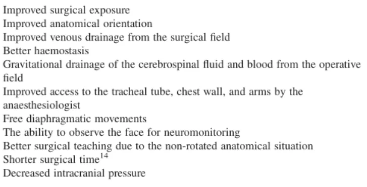
Table 3Advantages of neurosurgery in sitting position5 9 42 92 Improved surgical exposure
Improved anatomical orientation
Improved venous drainage from the surgical field Better haemostasis
Gravitational drainage of the cerebrospinal fluid and blood from the operative field
Improved access to the tracheal tube, chest wall, and arms by the anaesthesiologist
Free diaphragmatic movements
The ability to observe the face for neuromonitoring
Better surgical teaching due to the non-rotated anatomical situation Shorter surgical time14
much lower than that in the sitting patients. This
underesti-mates the effective rate of VAE in the horizontal position.
Other factors raising the hazard of VAE are the early stage
of surgery, including muscle preparation and craniotomy,
the use of nitrous oxide (N
2O), and the use of PEEP over
5 cm H
2O.
2 3 15Both N
2O and PEEP should be used
cau-tiously to minimize further harm by entrained air.
17 43 77Furthermore, a large prospective study found that PEEP
(10 cm H
2O) is associated with adverse cardiopulmonary
effects without altering the incidence of VAE.
51This
con-firms an earlier study conducted in paediatric
neurosurgi-cal procedures.
83Methods of detection
Early detection of left-sided cardiac air is vital to prevent
poor neurological outcome and the use of TOE or
precor-dial Doppler is essential in this way.
38 77 79Depending on
the method of detection, the frequency of VAE detection
is up to 70% (Table 1). The most commonly used methods
are TOE, precordial Doppler,
E0CO2, right heart
catheteriza-tion, and oesophageal stethoscope in decreasing order of
sensitivity for air detection.
16The potential hazard of PFO
PFO has been increasingly recognized as a source of
para-doxical embolism. The mechanism for this phenomenon is
thought to involve the passage of air, thrombus, or fat from
the right atrium to the left atrium through the PFO and then
on to the systemic circulation.
20 36 72 117As the prevalence of
PFO decreases with age from 35% during the first decade to
20% during the ninth decade of life, a selective mortality
(increased risk of early death in patients with PFO) has to be
considered in addition to spontaneous closure.
56An
additional proof of the prognostic importance is the doubled
mortality risk and the tripled stroke risk of patients with
pul-monary embolism in the presence of a PFO.
67Percutaneous PFO closure with modern closure devices
is the most simple of all catheter-based cardiac
interven-tions. The procedure can be performed as an outpatient
procedure in ,30 min with a fluoroscopy time of ,5 min
and good long-term safety.
116The currently accepted
indi-cations are limited to patients with (recurrent) cryptogenic
stroke, but are likely to expand soon to those with
migraine and divers.
The most commonly used devices so far for PFO
closure are Amplatzer PFO occluder, CardioSEAL family,
and PFO-Star family. Partially bioabsorbable devices
(BioSTAR
device),
self-expanding
stent
(Coherex
FlatStent PFO Closure System), and suturing devices
(HeartStich Suturing device) are under clinical
investi-gation. Finally, a radiofrequency energy source for closure
of PFO (PFx Closure System) has been clinically tested
but has not yet reached the market.
The review of percutaneous PFO closure in 5416
patients showed the safety and efficacy of this procedure
in the short and long term with a high procedural success
rate and low morbidity. Consequently, based on the best
available scientific evidence, it is appropriate to consider
percutaneous PFO closure in the preoperative management
of patients requiring neurosurgery in the sitting position.
This is particularly important when PFO is the only
concern, and the sitting position confers a significantly
lower surgical risk (Fig. 2).
Complications
Cerebrovascular ischaemia as a complication during or
after PFO closure is a rare event. Peri-interventional
com-plications were mostly minor, reversible, and decrease
further with experience and device improvement. Some
studies reported no complications with new devices.
100 116Our subanalysis in 15 studies using newer and more
common closure devices showed an overall 3.7% minor
complication rate, and 1.2% major complication rate with
no major complications in half of the studies. Mid- and
long-term complications are also rare and mostly without
clinical significance.
82Residual shunt
Many studies indicate that small PFOs are a very rare
source of paradoxical embolism in patients with
crypto-genic stroke and not clinically relevant.
5 62 98 108 110 113Moreover, Braun and colleagues
22showed that the
pre-sence of a minimal residual shunt after percutaneous PFO
closure cannot be considered as a risk factor for recurrent
Indication for neurosurgery in sitting position Preoperative TOE Sitting position No PFO detected PFO detected
Low surgical risk High surgical risk
Successful Declined or not
successful
Consider PFO closure (2–4 weeks before surgery) Horizontal position
Horizontal position Sitting position
Fig 2 Flow chart for PFO closure as a preoperative management in candidates of neurosurgery in the sitting position.
thromboembolic events. Hence, the small residual shunt
rate at 1 month in our systematic review of the literature
may not be a matter of concern as these shunts were
mostly trivial, and considered as clinically effective
occlu-sion. Neurosurgery in the sitting position could be safely
performed at that time.
Antithrombotic therapy
Initial antithrombotic therapy after the percutaneous PFO
closure is recommended to prevent device thrombus
for-mation. Although there is no established antithrombotic
therapy regimen and duration after percutaneous PFO
closure, a common regimen is acetylsalicylic acid for 6
months, occasionally accompanied by clopidogrel for 1 – 3
months. No study has verified the efficacy and necessity of
such antiplatelet therapy. One study
10has shown no
increase in the platelet activation markers after
percuta-neous PFO closure and questioned the necessity of
antipla-telet therapy. Moreover, acetylsalicylic acid has been
prescribed in different dosages in different studies. In a
report of 63 cases with percutaneous PFO closure,
64only
46% of patients were on acetylsalicylic acid and 17% on
anticoagulation therapy after implantation. No device
thrombosis during the mean follow-up period of 2.6 yr
was found. Krumsdorf and colleagues
68did not find any
influence of anticoagulation regimens on device thrombus
formation. Overall, the reported incidence of thrombus
for-mation is low, and in most cases without clinical sequela.
However, there are some differences which favour
Amplatzer and Helex occluders.
4 68 116Endothelialization of different devices for closure of
patent ductus arteriosus in animal models was reported
to start as early as 13 days and to be completed at
5 weeks.
54 102An animal study
57showed partial and
com-plete endothelialization at 1 and 3 months, respectively,
after the implantation of Amplatzer occluders. Early
endothelialization was observed 2 weeks after implantation
of a PFO Star in a patient who underwent surgical
explan-tation of the device due to dislocation.
22For most intra-dural surgical procedures, antithrombotic
therapy should be stopped 7 – 10 days before surgery and not
recommenced until 1 week after surgery, provided no
peri-operative major bleeding occurs. A significant proportion of
patients who undergo neurosurgical procedures are already
on treatment with aspirin or anticoagulants, and their
peri-operative management is well established. In view of this, it
seems safe to stop acetylsalicylic acid and clopidogrel for
neurosurgery for 2 weeks even early after PFO device
implantation. Ideally, PFO closure should take place at least
2 – 4 weeks before surgery. In an emergency operation or
when it cannot be delayed, we suggest closure of the PFO
immediately before surgery or to use the horizontal position.
Even the PFO has been closed before operation, techniques
for prevention of air embolism should still be used.
85Conclusions
On the basis of our systematic review, we recommend
screening for PFO and considering PFO closure in cases
where using the sitting position for neurosurgery has
major advantages for the outcome. However, the result of
our systematic review may be limited due to the lack of
data of level A evidence and from using data from cohort
studies with observational nature. Therefore, our proposal
is based on available evidence, and we have proposed a
policy to reduce the risk of PAE and consequently the risk
of neurosurgery in the sitting position in patients with
PFO. The evidence-based recommendations require
pro-spective evaluation in well-designed studies.
Supplementary material
Supplementary material is available at British Journal of
Anaesthesia online.
References
1 Alameddine F, Block PC. Transcatheter patent foramen ovale closure for secondary prevention of paradoxical embolic events: acute results from the FORECAST registry. Catheter Cardiovasc Interv 2004; 62: 512 – 6
2 Albin MS, Babinski M, Maroon JC, Jannetta PJ. Anesthetic man-agement of posterior fossa surgery in the sitting position. Acta Anaesthesiol Scand 1976; 20: 117 – 28
3 Albin MS, Carroll RG, Maroon JC. Clinical considerations con-cerning detection of venous air embolism. Neurosurgery 1978; 3: 380 – 4
4 Anzai H, Child J, Natterson B, et al. Incidence of thrombus for-mation on the CardioSEAL and the Amplatzer interatrial closure devices. Am J Cardiol 2004; 93: 426 – 31
5 Anzola GP, Zavarize P, Morandi E, Rozzini L, Parrinello G. Transcranial Doppler and risk of recurrence in patients with stroke and patent foramen ovale. Eur J Neurol 2003; 10: 129 – 35 6 Aslam F, Iliadis AE, Blankenship JC. Percutaneous closure of patent foramen ovale: success and outcomes of a low-volume procedure at a rural medical center. J Invasive Cardiol 2007; 19: 20 – 4
7 Azarbal B, Tobis J, Suh W, Chan V, Dao C, Gaster R. Association of interatrial shunts and migraine headaches: impact of transcatheter closure. J Am Coll Cardiol 2005; 45: 489 – 92 8 Bailey CE, Allaqaband S, Bajwa TK. Current management of
patients with patent foramen ovale and cryptogenic stroke: our experience and review of the literature. WMJ 2004; 103: 32 – 6 9 Barlow J. An account of the removal of a tumor situated on the
cheek. RocMedChirSoc 1830; 16: 19
10 Bedard E, Rodes-Cabau J, Houde C, et al. Enhanced thrombo-genesis but not platelet activation is associated with transcath-eter closure of patent foramen ovale in patients with cryptogenic stroke. Stroke 2007; 38: 100 – 4
11 Beitzke A, Schuchlenz H, Gamillscheg A, Stein JI, Wendelin G. Catheter closure of the persistent foramen ovale: mid-term results in 162 patients. J Interv Cardiol 2001; 14: 223 – 9
12 Berger F, Ewert P, Bjornstad PG, et al. Transcatheter closure as standard treatment for most interatrial defects: experience in
200 patients treated with the Amplatzer Septal Occluder. Cardiol Young 1999; 9: 468 – 73
13 Bijl JM, Ruygrok PN, Hornung TS, Wilson NJ, West T. Percutaneous closure of patent foramen ovale. Intern Med J 2005; 35: 706 – 10
14 Bithal PK, Pandia MP, Chouhan RS, et al. Hemodynamic and bispec-tral index changes following skull pin attachment with and without local anesthetic infiltration of the scalp. J Anesth 2007; 21: 442–4 15 Bithal PK, Pandia MP, Dash HH, Chouhan RS, Mohanty B, Padhy
N. Comparative incidence of venous air embolism and associ-ated hypotension in adults and children operassoci-ated for neurosur-gery in the sitting position. Eur J Anaesthesiol 2004; 21: 517 – 22 16 Black S, Cucchiara RF. Tumor surgery. In: Cucchiara RF, Black S,
Michenfelder JD, eds. Clinical Neuroanesthesia. New York: Churchill-Livingstone, 1998; 340 – 65
17 Black S, Cucchiara RF, Nishimura RA, Michenfelder JD. Parameters affecting occurrence of paradoxical air embolism. Anesthesiology 1989; 71: 235 – 41
18 Black S, Muzzi DA, Nishimura RA, Cucchiara RF. Preoperative and intraoperative echocardiography to detect right-to-left shunt in patients undergoing neurosurgical procedures in the sitting position. Anesthesiology 1990; 72: 436 – 8
19 Black S, Ockert DB, Oliver WC, Jr, Cucchiara RF. Outcome fol-lowing posterior fossa craniectomy in patients in the sitting or horizontal positions. Anesthesiology 1988; 69: 49 – 56
20 Bogousslavsky J, Garazi S, Jeanrenaud X, Aebischer N, Van Melle G. Stroke recurrence in patients with patent foramen ovale: the Lausanne Study. Lausanne Stroke with Paradoxal Embolism Study Group. Neurology 1996; 46: 1301 – 5
21 Braun M, Gliech V, Boscheri A, et al. Transcatheter closure of patent foramen ovale (PFO) in patients with paradoxical embo-lism. Periprocedural safety and mid-term follow-up results of three different device occluder systems. Eur Heart J 2004; 25: 424 – 30
22 Braun MU, Fassbender D, Schoen SP, et al. Transcatheter closure of patent foramen ovale in patients with cerebral ischemia. J Am Coll Cardiol 2002; 39: 2019 – 25
23 Bridges ND, Hellenbrand W, Latson L, Filiano J, Newburger JW, Lock JE. Transcatheter closure of patent foramen ovale after presumed paradoxical embolism. Circulation 1992; 86: 1902 – 8 24 Bruch L, Parsi A, Grad MO, et al. Transcatheter closure of
interatrial communications for secondary prevention of para-doxical embolism: single-center experience. Circulation 2002; 105: 2845 – 8
25 Buckland RW, Manners JM. Venous air embolism during neuro-surgery. A comparison of various methods of detection in man. Anaesthesia 1976; 31: 633 – 43
26 Buhre W, Weyland A, Buhre K, et al. Effects of the sitting pos-ition on the distribution of blood volume in patients undergoing neurosurgical procedures. Br J Anaesth 2000; 84: 354 – 7 27 Buscheck F, Sievert H, Kleber F, et al. Patent foramen ovale
using the Premere device: the results of the CLOSEUP trial. J Interv Cardiol 2006; 19: 328 – 33
28 Butera G, Bini MR, Chessa M, Bedogni F, Onofri M, Carminati M. Transcatheter closure of patent foramen ovale in patients with cryptogenic stroke. Ital Heart J 2001; 2: 115 – 8
29 Butler BD, Leiman BC, Katz J. Arterial air embolism of venous origin in dogs: effect of nitrous oxide in combination with halothane and pentobarbitone. Can J Anaesth 1987; 34: 570 – 5 30 Charbel F, Kehrli P, Pain L. The sitting position in neurosurgery: the
viewpoint of the surgeon. Ann Fr Anesth Reanim 1998; 17: 160– 3 31 Chatterjee T, Petzsch M, Ince H, et al. Interventional closure
with Amplatzer PFO occluder of patent foramen ovale in
patients with paradoxical cerebral embolism. J Interv Cardiol 2005; 18: 173 – 9
32 Clayton DG, Evans P, Williams C, Thurlow AC. Paradoxical air embolism during neurosurgery. Anaesthesia 1985; 40: 981 – 9 33 Cucchiara RF, Nugent M, Seward JB, Messick JM. Air embolism
in upright neurosurgical patients: detection and localization by two-dimensional transesophageal echocardiography. Anesthesiology 1984; 60: 353 – 5
34 Cucchiara RF, Seward JB, Nishimura RA, Nugent M, Faust RJ. Identification of patent foramen ovale during sitting position craniotomy by transesophageal echocardiography with positive airway pressure. Anesthesiology 1985; 63: 107 – 9
35 De Martel T. Surgical treatment of cerebral tumors: technical considerations. Surg Gynecol Obstet 1931; 52: 381 – 5
36 Di Tullio M, Sacco RL, Venketasubramanian N, Sherman D, Mohr JP, Homma S. Comparison of diagnostic techniques for the detection of a patent foramen ovale in stroke patients. Stroke 1993; 24: 1020 – 4
37 Domaingue CM. Anaesthesia for neurosurgery in the sitting position: a practical approach. Anaesth Intensive Care 2005; 33: 323 – 31
38 Domaingue CM. Neurosurgery in the sitting position: a case series. Anaesth Intensive Care 2005; 33: 332 – 5
39 Draganov J, Scheeren TW. Incidental detection of paradoxical air embolism with a transoesophageal Doppler probe inserted for measuring descending aortic blood flow. Br J Anaesth 2003; 90: 520 – 2
40 Du ZD, Hijazi ZM, Kleinman CS, Silverman NH, Larntz K. Comparison between transcatheter and surgical closure of secundum atrial septal defect in children and adults: results of a multicenter nonrandomized trial. J Am Coll Cardiol 2002; 39: 1836 – 44
41 Dubiel M, Bruch L, Liebner M, et al. Exclusion of patients with arteriosclerosis reduces long-term recurrence rate of presumed arterial embolism after PFO closure. J Interv Cardiol 2007; 20: 275 – 81
42 Duke DA, Lynch JJ, Harner SG, Faust RJ, Ebersold MJ. Venous air embolism in sitting and supine patients undergoing vestibular schwannoma resection. Neurosurgery 1998; 42: 1282 – 6, discus-sion 6 – 7
43 Eger EI, 2nd, Saidman LJ. Hazards of nitrous oxide anesthesia in bowel obstruction and pneumothorax. Anesthesiology 1965; 26: 61 – 6
44 Egred M, Andron M, Albouaini K, Alahmar A, Grainger R, Morrison WL. Percutaneous closure of patent foramen ovale and atrial septal defect: procedure outcome and medium-term follow-up. J Interv Cardiol 2007; 20: 395 – 401
45 Elton RJ, Howell RS. The sitting position in neurosurgical anaes-thesia: a survey of British practice in 1991. Br J Anaesth 1994; 73: 247 – 8
46 Ende DJ, Chopra PS, Rao PS. Transcatheter closure of atrial septal defect or patent foramen ovale with the buttoned device for prevention of recurrence of paradoxic embolism. Am J Cardiol 1996; 78: 233 – 6
47 Engelhardt M, Folkers W, Brenke C, et al. Neurosurgical oper-ations with the patient in sitting position: analysis of risk factors using transcranial Doppler sonography. Br J Anaesth 2006; 96: 467 – 72
48 Furuya H, Suzuki T, Okumura F, Kishi Y, Uefuji T. Detection of air embolism by transesophageal echocardiography. Anesthesiology 1983; 58: 124 – 9
49 Gale T, Leslie K. Anaesthesia for neurosurgery in the sitting position. J Clin Neurosci 2004; 11: 693 – 6
50 Garachemani A, Eshtehardi P, Meier B. Paradoxical emboli through the patent foramen ovale as the suspected cause of myocardial and renal infarction in a 48-year-old woman. Catheter Cardiovasc Interv 2007; 70: 1010 – 2
51 Giebler R, Kollenberg B, Pohlen G, Peters J. Effect of positive end-expiratory pressure on the incidence of venous air embo-lism and on the cardiovascular response to the sitting position during neurosurgery. Br J Anaesth 1998; 80: 30 – 5
52 Girard F, Ruel M, McKenty S, et al. Incidences of venous air embolism and patent foramen ovale among patients undergoing selective peripheral denervation in the sitting position. Neurosurgery 2003; 53: 316 – 9, discussion 9 – 20
53 Glenski JA, Cucchiara RF. Transcutaneous O2and CO2
monitor-ing of neurosurgical patients: detection of air embolism. Anesthesiology 1986; 64: 546 – 50
54 Grabitz RG, Schrader R, Sigler M, et al. Retrievable patent ductus arteriosus plug for interventional, transvenous occlusion of the patent ductus arteriosus. Evaluation in lambs and prelimi-nary clinical results. Invest Radiol 1997; 32: 523 – 8
55 Gronert GA, Messick JM, Jr, Cucchiara RF, Michenfelder JD. Paradoxical air embolism from a patent foramen ovale. Anesthesiology 1979; 50: 548 – 9
56 Hagen PT, Scholz DG, Edwards WD. Incidence and size of patent foramen ovale during the first 10 decades of life: an autopsy study of 965 normal hearts. Mayo Clin Proc 1984; 59: 17– 20 57 Han YM, Gu X, Titus JL, et al. New self-expanding patent
foramen ovale occlusion device. Catheter Cardiovasc Interv 1999; 47: 370 – 6
58 Harrison EA, Mackersie A, McEwan A, Facer E. The sitting pos-ition for neurosurgery in children: a review of 16 years’ experi-ence. Br J Anaesth 2002; 88: 12 – 7
59 Heckmann JG, Lang CJ, Kindler K, Huk W, Erbguth FJ, Neundorfer B. Neurologic manifestations of cerebral air embo-lism as a complication of central venous catheterization. Crit Care Med 2000; 28: 1621 – 5
60 Hein R, Buscheck F, Fischer E, et al. Atrial and ventricular septal defects can safely be closed by percutaneous intervention. J Interv Cardiol 2005; 18: 515 – 22
61 Herrmann HC, Silvestry FE, Glaser R, et al. Percutaneous patent foramen ovale and atrial septal defect closure in adults: results and device comparison in 100 consecutive implants at a single center. Catheter Cardiovasc Interv 2005; 64: 197 – 203
62 Homma S, Di Tullio MR, Sacco RL, Mihalatos D, Li Mandri G, Mohr JP. Characteristics of patent foramen ovale associated with cryptogenic stroke. A biplane transesophageal echocardio-graphic study. Stroke 1994; 25: 582 – 6
63 Hong TE, Thaler D, Brorson J, Heitschmidt M, Hijazi ZM. Transcatheter closure of patent foramen ovale associated with paradoxical embolism using the amplatzer PFO occluder: initial and intermediate-term results of the US multicenter clinical trial. Catheter Cardiovasc Interv 2003; 60: 524 – 8
64 Hung J, Landzberg MJ, Jenkins KJ, et al. Closure of patent foramen ovale for paradoxical emboli: intermediate-term risk of recurrent neurological events following transcatheter device pla-cement. J Am Coll Cardiol 2000; 35: 1311 – 6
65 Khositseth A, Cabalka AK, Sweeney JP, et al. Transcatheter Amplatzer device closure of atrial septal defect and patent foramen ovale in patients with presumed paradoxical embolism. Mayo Clin Proc 2004; 79: 35 – 41
66 Kiblawi FM, Sommer RJ, Levchuck SG. Transcatheter closure of patent foramen ovale in older adults. Catheter Cardiovasc Interv 2006; 68: 136 – 42, discussion 43 – 4
67 Konstantinides S, Geibel A, Kasper W, Olschewski M, Blumel L, Just H. Patent foramen ovale is an important predictor of adverse outcome in patients with major pulmonary embolism. Circulation 1998; 97: 1946 – 51
68 Krumsdorf U, Ostermayer S, Billinger K, et al. Incidence and clinical course of thrombus formation on atrial septal defect and patient foramen ovale closure devices in 1,000 consecutive patients. J Am Coll Cardiol 2004; 43: 302 – 9
69 Kubo S, Nakata H. Air embolism due to a patent foramen ovale visualised by harmonic contrast echocardiography. J Neurol Neurosurg Psychiatry 2001; 71: 555
70 Kwapisz MM, Deinsberger W, Muller M, et al. Transesophageal echocardiography as a guide for patient positioning before neu-rosurgical procedures in semi-sitting position. J Neurosurg Anesthesiol 2004; 16: 277 – 81
71 Lechat P, Guggiari M, Lascault G, et al. Detection by contrast ultrasonography of patent foramen ovale before neurosurgery. Presse Med 1986; 15: 1409 – 10
72 Lechat P, Mas JL, Lascault G, et al. Prevalence of patent foramen ovale in patients with stroke. N Engl J Med 1988; 318: 1148 – 52 73 Leivers D, Spilsbury RA, Young JV. Air embolism during
neuro-surgery in the sitting position. Two case reports. Br J Anaesth 1971; 43: 84 – 90
74 Leonard IE, Cunningham AJ. The sitting position in neurosur-gery—not yet obsolete! Br J Anaesth 2002; 88: 1 – 3
75 Leslie K, Hui R, Kaye AH. Venous air embolism and the sitting position: a case series. J Clin Neurosci 2006; 13: 419 – 22 76 Liutkus D, Gouraud JP, Blanloeil Y. The sitting position in
neuro-surgical anaesthesia: a survey of French practice. Ann Fr Anesth Reanim 2003; 22: 296 – 300
77 Losasso TJ, Muzzi DA, Dietz NM, Cucchiara RF. Fifty percent nitrous oxide does not increase the risk of venous air embolism in neurosurgical patients operated upon in the sitting position. Anesthesiology 1992; 77: 21 – 30
78 Loscalzo J. Paradoxical embolism: clinical presentation, diagnos-tic strategies, and therapeudiagnos-tic options. Am Heart J 1986; 112: 141 – 5
79 Mammoto T, Hayashi Y, Ohnishi Y, Kuro M. Incidence of venous and paradoxical air embolism in neurosurgical patients in the sitting position: detection by transesophageal echocardiography. Acta Anaesthesiol Scand 1998; 42: 643 – 7
80 Martin F, Sanchez PL, Doherty E, et al. Percutaneous transcath-eter closure of patent foramen ovale in patients with paradoxi-cal embolism. Circulation 2002; 106: 1121 – 6
81 Matjasko J, Petrozza P, Cohen M, Steinberg P. Anesthesia and surgery in the seated position: analysis of 554 cases. Neurosurgery 1985; 17: 695 – 702
82 Meier B. Closure of patent foramen ovale: technique, pitfalls, complications, and follow up. Heart 2005; 91: 444 – 8
83 Meyer PG, Cuttaree H, Charron B, Jarreau MM, Perie AC, Sainte-Rose C. Prevention of venous air embolism in paediatric neurosurgical procedures performed in the sitting position by combined use of MAST suit and PEEP. Br J Anaesth 1994; 73: 795 – 800
84 Michenfelder JD, Miller RH, Gronert GA. Evaluation of an ultra-sonic device (Doppler) for the diagnosis of venous air embo-lism. Anesthesiology 1972; 36: 164 – 7
85 Mirski MA, Lele AV, Fitzsimmons L, Toung TJ. Diagnosis and treatment of vascular air embolism. Anesthesiology 2007; 106: 164 – 77
86 Onorato E, Melzi G, Casilli F, et al. Patent foramen ovale with paradoxical embolism: mid-term results of transcatheter closure in 256 patients. J Interv Cardiol 2003; 16: 43 – 50
87 Papadopoulos G, Kuhly P, Brock M, Rudolph KH, Link J, Eyrich K. Venous and paradoxical air embolism in the sitting position. A prospective study with transoesophageal echocardiography. Acta Neurochir (Wien) 1994; 126: 140 – 3
88 Perkins-Pearson NA, Marshall WK, Bedford RF. Atrial pressures in the seated position: implication for paradoxical air embolism. Anesthesiology 1982; 57: 493 – 7
89 Pham Dang C, Pereon Y, Champin P, Delecrin J, Passuti N. Paradoxical air embolism from patent foramen ovale in scoliosis surgery. Spine 2002; 27: E291 – 5
90 Porter JM, Pidgeon C, Cunningham AJ. The sitting position in neurosurgery: a critical appraisal. Br J Anaesth 1999; 82: 117 – 28 91 Post MC, Van Deyk K, Budts W. Percutaneous closure of a patent foramen ovale: single-centre experience using different types of devices and mid-term outcome. Acta Cardiol 2005; 60: 515– 9
92 Rath GP, Bithal PK, Chaturvedi A, Dash HH. Complications related to positioning in posterior fossa craniectomy. J Clin Neurosci 2007; 14: 520 – 5
93 Reisman M, Christofferson RD, Jesurum J, et al. Migraine head-ache relief after transcatheter closure of patent foramen ovale. J Am Coll Cardiol 2005; 45: 493 – 5
94 Samii M, Matthies C. Management of 1000 vestibular schwanno-mas (acoustic neuroschwanno-mas): the facial nerve—preservation and restitution of function. Neurosurgery 1997; 40: 684 – 94
95 Schaffranietz L, Grothe A, Olthoff D. Use of the sitting position in neurosurgery. Results of a 1998 survey in Germany. Anaesthesist 2000; 49: 269 – 74
96 Schaffranietz L, Gunther L. The sitting position in neurosurgical operations. Results of a survey. Anaesthesist 1997; 46: 91 – 5 97 Schmitt HJ, Hemmerling TM. Venous air emboli occur during
release of positive end-expiratory pressure and repositioning after sitting position surgery. Anesth Analg 2002; 94: 400 – 3, table of contents
98 Schuchlenz HW, Weihs W, Horner S, Quehenberger F. The association between the diameter of a patent foramen ovale and the risk of embolic cerebrovascular events. Am J Med 2000; 109: 456 – 62
99 Schwarz G, Fuchs G, Weihs W, Tritthart H, Schalk HV, Kaltenbock F. Sitting position for neurosurgery: experience with preoperative contrast echocardiography in 301 patients. J Neurosurg Anesthesiol 1994; 6: 83 – 8
100 Sievert H, Fischer E, Heinisch C, Majunke N, Roemer A, Wunderlich N. Transcatheter closure of patent foramen ovale without an implant: initial clinical experience. Circulation 2007; 116: 1701 – 6
101 Sievert H, Horvath K, Zadan E, et al. Patent foramen ovale closure in patients with transient ischemia attack/stroke. J Interv Cardiol 2001; 14: 261 – 6
102 Sigler M, Handt S, Seghaye MC, Von Bernuth G, Grabitz RG. Evaluation of in vivo biocompatibility of different devices for interventional closure of the patent ductus arteriosus in an animal model. Heart 2000; 83: 570 – 3
103 Simo Moyo J, Adnet P, Wambo M. Detection of gas embolism in neurosurgery by capnography. Apropos of 32 patients surgically treated in seated position. Cah Anesthesiol 1995; 43: 77 – 9
104 Siwek J, Gourlay ML, Slawson DC, Shaughnessy AF. How to write an evidence-based clinical review article. Am Fam Physician 2002; 65: 251 – 8
105 Slavin L, Tobis JM, Rangarajan K, Dao C, Krivokapich J, Liebeskind DS. Five-year experience with percutaneous closure of patent foramen ovale. Am J Cardiol 2007; 99: 1316 – 20 106 Spies C, Strasheim R, Timmermanns I, Schraeder R. Patent
foramen ovale closure in patients with cryptogenic thrombo-embolic events using the Cardia PFO occluder. Eur Heart J 2006; 27: 365 – 71
107 Standefer M, Bay JW, Trusso R. The sitting position in neurosur-gery: a retrospective analysis of 488 cases. Neurosurgery 1984; 14: 649 – 58
108 Steiner MM, Di Tullio MR, Rundek T, et al. Patent foramen ovale size and embolic brain imaging findings among patients with ischemic stroke. Stroke 1998; 29: 944 – 8
109 Stendel R, Gramm HJ, Schroder K, Lober C, Brock M. Transcranial Doppler ultrasonography as a screening technique for detection of a patent foramen ovale before surgery in the sitting position. Anesthesiology 2000; 93: 971 – 5
110 Stone DA, Godard J, Corretti MC, et al. Patent foramen ovale: association between the degree of shunt by contrast transeso-phageal echocardiography and the risk of future ischemic neuro-logic events. Am Heart J 1996; 131: 158 – 61
111 Thanopoulos BV, Dardas PD, Karanasios E, Mezilis N. Transcatheter closure versus medical therapy of patent foramen ovale and cryptogenic stroke. Catheter Cardiovasc Interv 2006; 68: 741 – 6
112 Thompson T, Evans W. Paradoxical embolism. Q J Med 1930; 23: 135 – 50
113 Van Camp G, Schulze D, Cosyns B, Vandenbossche JL. Relation between patent foramen ovale and unexplained stroke. Am J Cardiol 1993; 71: 596 – 8
114 Vik A, Brubakk AO, Hennessy TR, Jenssen BM, Ekker M, Slordahl SA. Venous air embolism in swine: transport of gas bubbles through the pulmonary circulation. J Appl Physiol 1990; 69: 237 – 44
115 Voorhies RM, Fraser RA, Van Poznak A. Prevention of air embo-lism with positive end expiratory pressure. Neurosurgery 1983; 12: 503 – 6
116 Wahl A, Kunz M, Moschovitis A, et al. Long-term results after fluoroscopy-guided closure of patent foramen ovale for second-ary prevention of paradoxical embolism. Heart 2008; 94: 336– 41 117 Webster MW, Chancellor AM, Smith HJ, et al. Patent foramen
ovale in young stroke patients. Lancet 1988; 2: 11 – 2
118 Windecker S, Wahl A, Chatterjee T, et al. Percutaneous closure of patent foramen ovale in patients with paradoxical embolism: long-term risk of recurrent thromboembolic events. Circulation 2000; 101: 893 – 8
119 Yahagi N, Furuya H. The effects of halothane and pentobarbital on the threshold of transpulmonary passage of venous air emboli in dogs. Anesthesiology 1987; 67: 905 – 9
120 Young ML, Smith DS, Murtagh F, Vasquez A, Levitt J. Comparison of surgical and anesthetic complications in neuro-surgical patients experiencing venous air embolism in the sitting position. Neurosurgery 1986; 18: 157 – 61